Gold nanoparticles from Celastrus hindsii and HAuCl4: Green synthesis, characteristics, and their cytotoxic effects on HeLa cells
-
Xuan-Truong Mai
 ,
Minh-Chien Tran
,
Minh-Chien Tran

Abstract
The extract from Celastrus hindsii (C. hindsii), a plant that naturally grows in the forests of several provinces of Vietnam, has been traditionally used as an alternative medicine for the treatment of inflammation because of its anticancer and antitumor properties. This study reported the green synthesis of stable gold nanoparticles (Au-NPs) derived from HAuCl4 using the extract of C. hindsii as reducing and capping agents. Their particle size could be controlled by adjusting the ratio of the extract to HAuCl4 solution used (1.25%, 2.5%, 3.75%, 5.0%, and 6.25%). The optimal ratio of the extract was 3.75% (Au-NPs-3.75%). The X-ray powder diffraction analysis demonstrated that the Au-NPs was successfully synthesized. Fourier-transform infrared spectroscopy result indicated the possible presence of phenolic acids and flavonoids (acting as reducing agents and potential natural antioxidants). Transmission electron microscopy images showed that the particle diameter of Au-NPs-3.75% varied between 13 and 53 nm (average: ∼30 nm) in its spherical shape. The biosynthesized Au-NPs-3.75% exhibited dose-dependent cytotoxicity against HeLa cells, and the inhibitory concentration (IC50) was 12.5 µg/mL at 48 h. Therefore, Au-NPs that were synthesized from environmentally friendly method without the presence of potentially toxic chemicals were highly possible in biomedical applications.
Graphical abstract
Green synthesis of gold nanoparticles from the Celastrus hindsii extract and its application for anticancer activity.

1 Introduction
Nanoscience and technology has attracted more concerns from scientific communities because of its wide application. Among current nanomaterials, gold nanoparticles (Au-NPs) have been widely applied (especially in bio-medication [1,2]) because of their unique and advantageous properties. The multiple surface functionality of Au-NPs makes it easier to attach with drugs [3], oligonucleotides [4], and antibodies [5]. The unique surface plasmon resonance (SPR) of Au-NPs triggers their photostability and extremely efficient absorptivity of light [6]. Therefore, Au-NPs offer multiple modalities for biological and medical applications [7] such as cancer nanotechnology [7,8], tissue engineering, and drug and gene delivery [6].
Green nanotechnology is a thrust area of research with a significant focus on developing eco-friendly methods for the production of high-quality nanostructures with sustainable commercial viability. Among existing Au-NPs synthesis, the method based on biological systems (i.e., microbes, fungi, and extracts) is the most common and appropriate because of its simplicity and low preparation cost. Besides, this method is eco-friendly and green, resulting from the absence of toxic chemicals during the synthesis process. In general, various plant species (the extracts from them) can serve as potential reductant and capping agents for synthesizing Au-NPs. In the literature, the extracts of many plant species have been applied for the successful biosynthesis of Au-NPs. They included Chenopodium album [9], Sorbus aucuparia [10], Sugar beet pulp [11], Terminalia catappa [12], Hibiscus rosa sinensis [13], Mangifera indica [14], Syzygium aromaticum [15], Anacardium occidentale [16], Macrotyloma uniflorum [17], and Murraya koenigii [18]. The gold nanomaterials biosynthesized from those extracts often exhibit their excellent compatibility with biomolecules [19]. Different methods that were used for the plant extract might lead to different properties of resultant Au-NPs. In addition, the application of the extract method is strongly dependent on the unique nature and property of each plant. Therefore, the extract method often plays an important role in controlling the properties and sizes of resultant Au-NPs [9].
Celastrus hindsii (C. hindsii; its family: Celastraceae) that is abundantly found in America, China, and Vietnam [20,21] can be used as herbal and traditional medicines for treating cancers [20,22]. In Vietnam, C. hindsii plants naturally thrive in forests and have been chemically studied because of their biological activities. For example, Duyen et al. [23] used ethanol and then fractionated with different solvents (n-hexane, n-butanol, and ethyl acetate) to extract certain chemicals (i.e., vitamin C, vitamin E, and polyphenols) from the leaves of C. hindsii. They found that the highest cytoxicity effects on the cells of lung cancer and liver cancer were obtained by using the ethyl acetate fraction, with an inhibitory concentration (IC50) value being 13 and 33.7 mg/mL, respectively. They concluded that the extract of the leaves of C. hindsii has strong antioxidant and cytotoxicity activities. An identical conclusion was reported by Pham et al. [21]. The authors used ethanol assisted with ultrasound to extract flavonoids from the leaves of C. hindsii. The IC50 values for cancer cell lines were 88 µg/mL (MCF7), 120 µg/mL (A549), and 118 µg/mL (HeLa).
In this study, the extract of C. hindsii leaves and stems was used as a natural reducing and capping agent for biosynthesizing well-dispersed Au-NPs. A simple method of extraction without any chemicals or solvents was developed. The extract and Au-NPs were well-characterized by various techniques. Au-NPs were then applied for exploring their cytotoxic effects on HeLa cells.
2 Materials and methods
2.1 Materials and reagents
The dried leaves and stems of C. hindsii were procured from local herbal markets. Gold(iii) chloride trihydrate (99.9%; HAuCl4‧3H2O) is acquired from Sigma Aldrich. Other chemicals were of analytical grade, such as Ammoniac, Dulbecco’s modified Eagles medium, phosphate-buffered saline, 3-(4,5 dimethylthiazol-2-yl)-2,5-diphenyl tetrazolium bromide (MTT), fetal bovine serum (FBS), and dimethyl sulfoxide. Those chemicals were purchased from Sigma-Aldrich and ACROS Organics and directly used.
2.2 Preparation of the plan extract from C. hindsii
The leaves and stems of C. hindsii were thoroughly washed with de-ionized water, dried in the air, and then ground into powder particles. A mixture of C. hindsii powder (50 g) and de-ionized water (500 mL) was heated up to 60°C for 30 min, and cellulose was then removed by filtrations to obtain the aqueous black extract. After 100 mL of the extract had been evaporated under reduced pressures, 1.55 g of C. hindsii powder was obtained. Finally, the extract of C. hindsii powder was dispersed in 100 mL of de-ionized water.
2.3 Green synthesis of Au-NPs
The synthesis method of Au-NPs was adapted from the literature [24,25]. Briefly, the extract of C. hindsii (at different volumes: 0.25, 0.5, 0.75, 1.0, and 1.25 mL) was added to a HAuCl4‧3H2O solution (1.0 mM and pH 2.75) in a flask (under stirring 250 rpm) at 30°C for 1.0 min. The total volume of the mixture of the extract and the HAuCl4 was 20 mL. This means that the ratio of the extract (v/v) used was 1.25%, 2.5%, 3.75%, 5.0%, and 6.25%. For convenience, the final samples were named according to the ratio of the extract used, such as Au-NPs-1.25%, Au-NPs-2.5%, Au-NPs-3.75%, Au-NPs-5.0%, Au-NPs-6.25%, respectively. After the above reaction time (∼1.0 min), the mixture containing Au-NPs was centrifuged at 10,000 rpm for 30 min, repeatedly washed with de-ionized water, and then stored in de-ionized water (20 mL) at 4°C for further use.
2.4 Characterization of the prepared Au-NPs sample
The prepared Au-NPs samples were distinguished by UV-Visible spectrometer (Shimadzu UV-1800) for evaluating their fascination potentials. The UV-Visible spectra of the Au-NPs samples were observed at a wavelength range between 450 and 650 nm. In addition, their morphological properties (including their size and shape) were analyzed by transmission electron microscopy (TEM) using a JEM1010-JEOL microscope. The aqueous re-dispersed suspension of a drop of Au-NPs was located on a carbon-coated copper grid and air-dried before the TEM assessment. Moreover, the crystallographic structure of the synthesized Au-NPs was detected by X-ray diffraction (XRD; EQUINOX 5000 diffractometer) using Cu K radiation (λ = 0.1542 nm). Meanwhile, the Fourier-transform infrared spectroscopy (FTIR) spectra of C. hindsii and Au-NPs were used to determine the presence of main functional groups on the materials’ surface. The FTIR absorption spectra were conducted at room temperature at the level of 4,000–500 cm−1 using the IR Prestige-21 Shimadzu spectrometer. Finally, the concentration of Au in Au-NPs and solution was determined by inductively coupled plasma mass spectrometry (ICP-MS; PerkinElmer NexION 2000).
2.5 Anticancer activity of Au-NPs
The prepared Au-NPs sample anticancer activity was analyzed by calculating the inhibitory effect (IC50) in HeLa cell line using MTT assay [26]. HeLa cells were cultured in a composite of DMEM/F12 with 10% FBS and 1% penicillin/streptomycin. The cells were maintained at 37°C with a 5% CO2 atmosphere and detached by trypsin/ethylenediaminetetraacetic acid (0.25%; Gibco) when confluence reached around 80–90%. The cell suspension was seeded in a 96-well plate at density 5 × 10³ cells in 100 µL per well. Subsequently, they were cultured stably in media (DMEM/F12, 10% FBS, and 1% PS) in 24 h. After removing media, the cells were treated with various concentrations of Au-NPs (i.e., 1.5, 3, 5. 12.5, 25, 50, and 73 µg/mL) to each well for 48 h. The whole cultured cells were incubated at 37°C in a 5% CO2 atmosphere. After 48 h of incubation, the images of cells cultured were captured using inverted microscopy (Eclipse Ti-U, Nikon) before adding dye steps.
After the incubation period, the medium was removed, and 100 µL of 7-hydroxy-3H-phenox azin-3-one 10-oxide dye (resazurin; 0.02 mg/mL) was added to each well. Those mixtures were incubated within avoiding-light requirements for 3 h at 37°C and 5% CO2. The samples were read by a microplate reader (Varioskan™, ThermoScientific, USA) at a wavelength of 530 nm, and fluorescent signals were measured at 590 nm. Notably, the data were presented and reported as average ± standard deviation (SD).
3 Results and discussion
3.1 Characterization of Au-NPs
In essence, UV-visible absorption spectroscopy (an efficient technique) is often used to monitor the optical properties of quantum-sized particles [27]. Figure 1a shows the maximum band corresponding to the unique SPR at around 540 nm. The results confirmed the formation of Au-NPs because the presence of a maxima band of absorbance at around 540 nm is a specific signal to identify gold nanoparticles [28,29]. In addition, the color change of solution (Figure 1c) after reaction from yellow (pristine HAuCl4) to ruby-red suggested the successful formation of Au-NPs [30]. Moreover, the SPR band indicated a slight shift towards a higher maximal wavelength number with an increase in the extract ratios from C. hindsii used (Figure 1a), consistent with the change of color observed in Figure 1c. The result suggested an increase in particle sizes of the resultant Au-NPs when the ratios of the extract used increased from 2.5% to 6.25% (Figure A1), except that of 1.25%. In fact, the sample of Au-NPs-1.25% was not reliable because some Au-NPs were not observed by TEM images (data not shown). This is consistent with its visible color in Figure 1c, so this sample was not used for further discussion. The average particle diameters of each gold nanoparticle increased as the following order: 14 nm (Au-NPs-2.5%) < 30 nm (Au-NPs-3.75%) < 36 nm (Au-NPs-5%) < 54 nm (Au-NPs-6.25%). A similar phenomenon was reported by Singh and Srivastava [31]. Notably, the SPR band became broader when the ratio of the extract was higher than 3.75% (i.e., 5% and 6.25%) because of the formation of large anisotropic particles [30]. Therefore, the sample (Au-NPs-3.75%) was used for further studies of its characteristics and anticancer activity.

(a) UV-Visible spectra of the synthesized gold colloids, (b) its stability over times, and (c) manifestation of different colors of Au-NPs samples prepared from different ratios of the C. hindsii extract.
The analysis of UV-Visible absorption spectroscopy was repeated for a period from 0, 5, 10, 20, and 30 days to evaluate the stability of Au-NPs-3.75%. Figure 1b indicates that the band of the water solution containing Au-NPs-3.75% was almost stable after 30 days. The result confirmed that Au-NPs-3.75% exhibited too high stability and good dispersion in the water solution without any aggregation. A similar conclusion was reported by other researchers who synthesized Au-NPs from the extract of Anacardium occidentale leaves [16] or marine algae Padina gymnospora [30].
Secondly, FTIR was performed to investigate the biomolecules of C. hindsii extract involved in the Au-NPs formation and identify the molecules responsible for capping and efficient stabilization of synthesized Au-NPs. The FTIR spectrum of C. hindsii extract (Figure 2a) was compared with that of synthesized Au-NPs (Figure 2b). As shown in Figure 2a, an intensive band at around 1,067 cm−1 can be assigned to the stretching vibration of the C–O bond derived from proteins in the plant extract [30]. Distinct bands in the regions of 1,238 and 1,385 cm−1 may be attributed to the presence of stretching vibrations of alcohols, ethers, esters, carboxylic acids, and amino groups. A band at the 1,385 cm−1 is related to the carbon double bonds (C═C) in aromatic of common flavonoids in C. hindsii [16]. Notably, the presence of the amides (–C(═O)N═) overlapping the carbonyl group (C═O) in proteins is identified at approximately 1,610 cm−1. Meanwhile, a band at 1,730 cm−1 is also related to carboxylic groups. The stretching vibrations of the secondary amines in the N–H and O–H bonds are observed at 3,420 cm−1 [25].
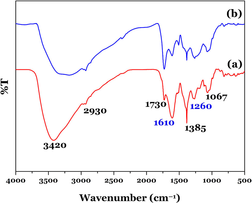
FTIR spectra of (a) C. hindsii extract and (b) Au-NPs-3.75%.
The FTIR spectrum of Au-NPs-3.75% (Figure 2b) is similar to that of the C. hindsii extract (Figure 2a). However, the intensity of some important bands remarkably decreased, suggesting certain reactions exist between some compounds in the C. hindsii extract and HAuCl4 to form Au-NPs. For example, the intensity of two bands located at around 1,385 cm−1 (identified the presence of the flavonoids) and 3,420 cm−1 (phenols) in the spectrum of Au-NPs-3.75% decreased compared to that in the C. hindsii extract. The result suggested that the phytochemical compounds (i.e., flavonoids and phenols) available in the C. hindsii extract simulated generating Au-NPs through the bio-reduction of Au3+ ions [16,31]. Meanwhile, a remarkable decrease in the intensity of a band at 1,610 cm−1 (amides) confirmed that the amide groups had a strong capability to capture Au-NPs.
Thirdly, the XRD result suggested that Au-NPs-3.75% was well-crystalline (Figure 3). The XRD peaks located at 2θ = 38.18°, 44.38°, 64.57°, and 77.56° correspond to the (111), (200), (220), and (311) planes of the Au-NPs, respectively [12,17,32]. The standard diffraction bands showed the face-centered cubic phase of Au based on JCPDS 65-2870 and COD LINK 9008463 [33]. The result suggested that the Au-NPs were successfully synthesized by the C. hindsii extract and HAuCl4.
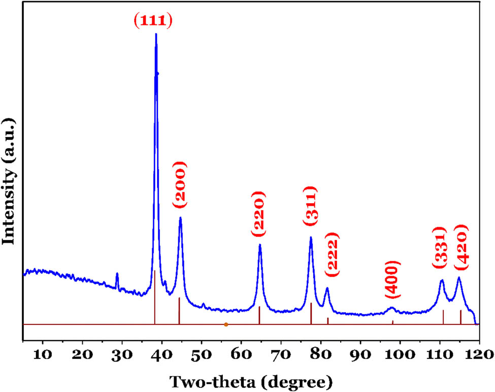
XRD pattern of the biogenic Au-NPs-3.75%.
Furthermore, the properties of morphology, size, and shape of the prepared Au-NPs are explored by the TEM technique. Figure 4a shows that Au-NPs-3.75% exhibited two distinguishing morphologies, such as spherical (accounting for >95%) and triangular. An analogous observation was obtained by Singh and Srivastava [31] for synthesizing Au-NPs using black cardamom. As expected, each sphere (or triangle) indicated a high mono-dispersed particle and non-agglomerated phenomenon (Figure 4b). This might result from the effective stabilization of several bio-organic components or proteins (Figure 2a) in the C. hindsii extract [30]. Those compounds can act as an important ligand to encapsulate Au-NPs (Figure 4a). Notably, some previous studies demonstrated that the spherical shape of common nanoparticles could be simulating to increasing cytotoxic antibacterial effects such as silver nanoparticles [34] and Au-NPs [16]. Furthermore, the TEM images of the other samples (Au-NPs-1.25%, Au-NPs-2.5%, Au-NPs-5.0%, and Au-NPs-6.25%) are provided in Figure A2 for comparison.
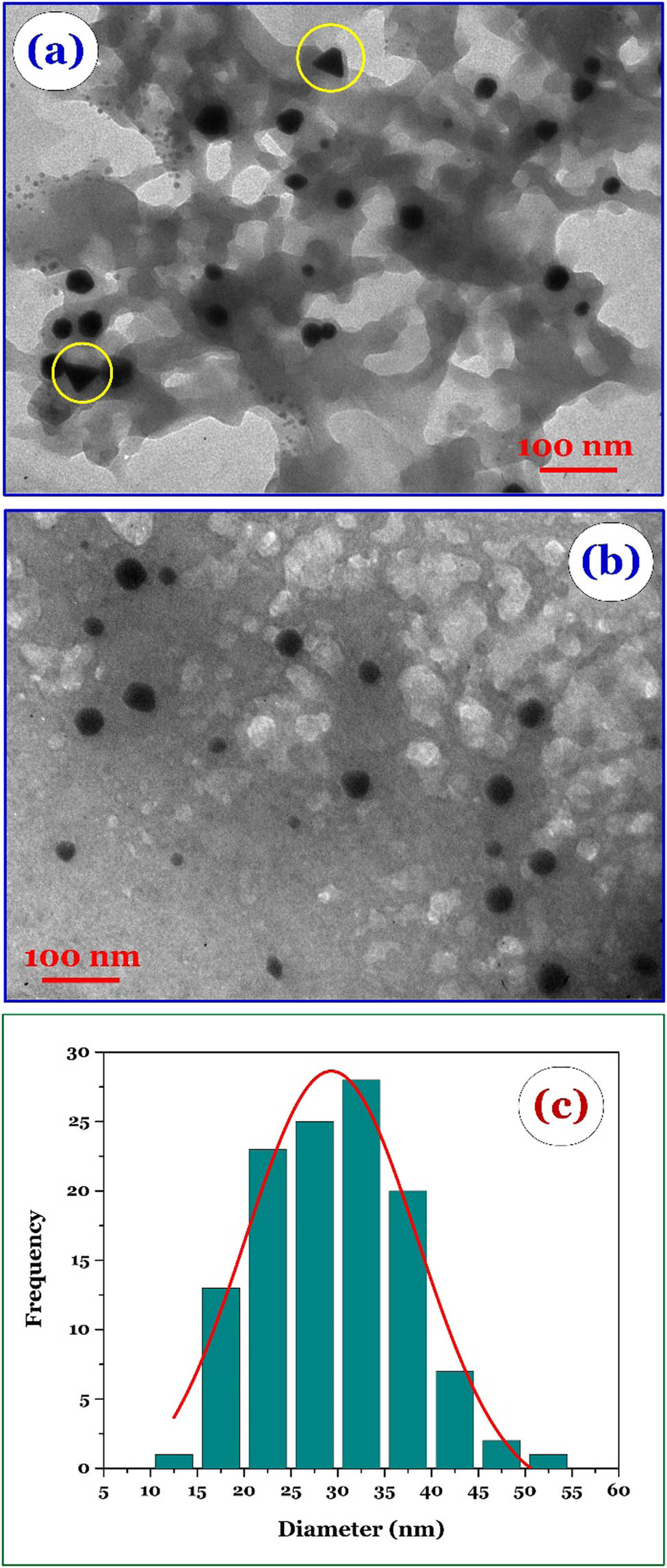
(a and b) TEM images of Au-NPs-3.75% and (c) distribution of its particle diameter.
The particle size of Au-NPs-3.75% (obtained from the TEM data) ranged from 13 to 53 nm in diameter, with the most diameter being in the range of 23–37 nm (centered at 30 nm) (Figure 4c). Some scholars also reported the similar diameters of Au-NPs such as 15–25 nm for Au-NPs performed from Cordyceps militaris [25], 25–35 nm from black cardamom [31], and 30–50 nm from Marsdenia tenacissima [24].
Finally, the total concentration of Au in Au-NPs-3.75% in 20 mL was determined by the ICP-MS technique. The result of three repeated experiments indicated that the Au concentration was 72.9 ± 0.3 µg/mL (average ± SD). This concentration was used as a stock solution to prepare running solutions in the investigation of anticancer activity.
3.2 Anticancer activity of Au-NPs
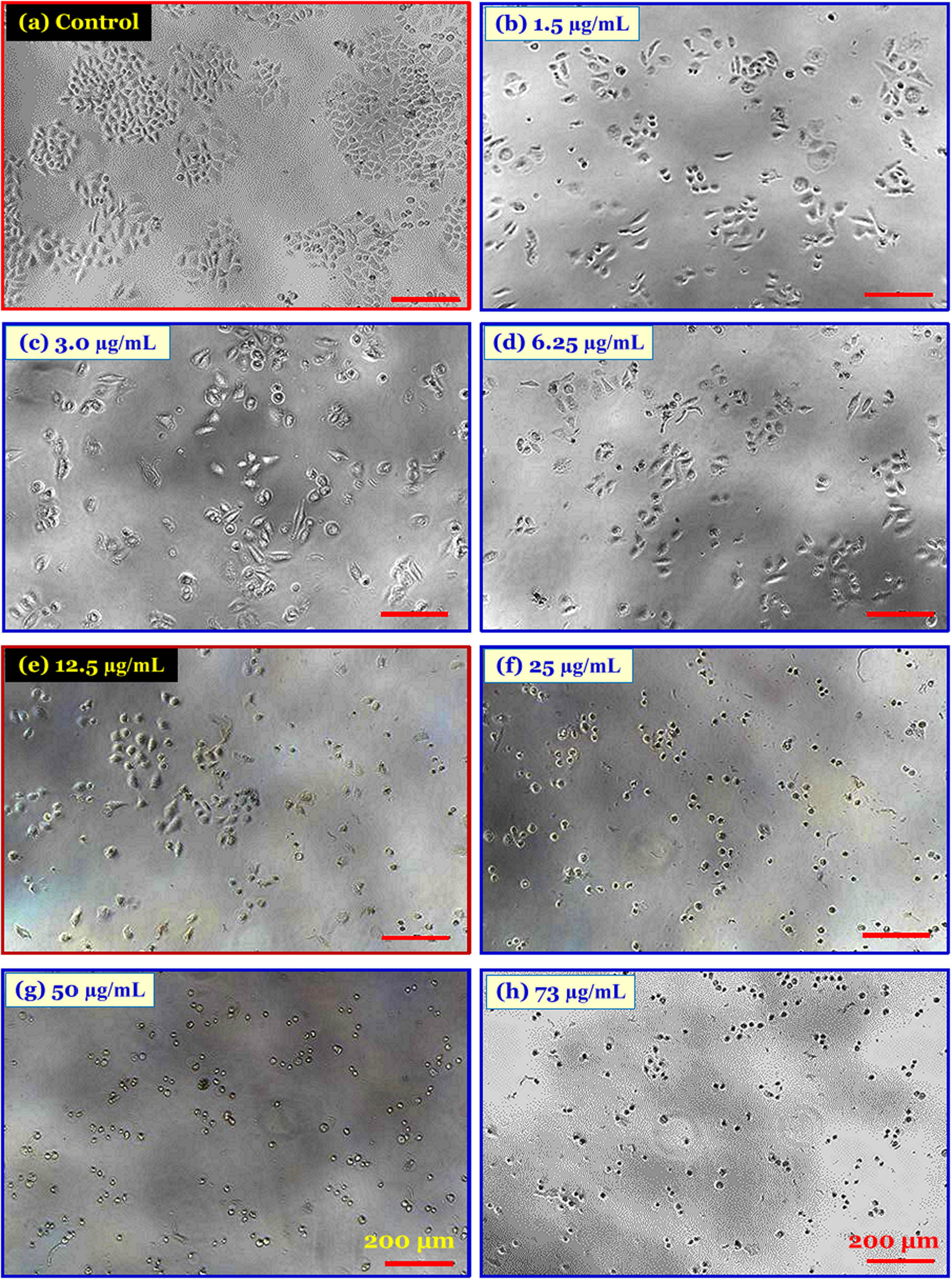
Figure 5 shows the cytotoxic effect of C. hindsii Au-NPs on HeLa cells. The cell shape changed and became unstable when increasing the concentrations of Au-NPs (µg/mL). In the control group (0 µg/mL), the population of HeLa cells proliferate, have lance-shaped, and grow in clusters (Figure A3). In the presence of Au-NPs, the change of the shape and stretching due to chemical stress indicated the increase in membrane rupture cells when increasing the concentrations of Au-NPs [35,36]. Clearly, when high concentrations of Au-NPs (i.e., 25, 50, and 73 µg/mL) were used, the state of HeLa cells was unstable: they lose lance-shaped and their membranes are entirely ruptured. In addition, there was no “connecting” in adjacent HeLa cells because of the sparsed population. Those were some manifestations of cellular stress.
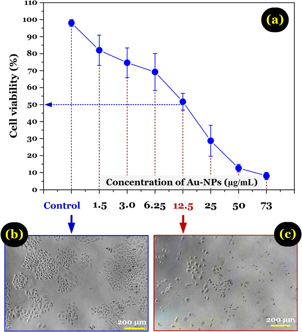
(a) Effect of the concentrations of Au-NPs-3.75% on the viability of HeLa cells (average ± SD; raw data presented in Table A1); microscopic images of HeLa cells without treated with Au-NPs-3.75% (b) and after treated with Au-NPs-3.75% (12.5 μg/mL) for 48 h (c).
Au-NPs, as novel agents for cancer therapy, are gaining substantial demand in medical applications. However, studies on the cytotoxic activity of biosynthesized Au-NPs against cancer cell lines are limited. The MTT assay was used to assess the effect of Au-NPs on the proliferation of HeLa cells. The present study found that the induction of cell death modalities could be the possible mechanism for the antiproliferative activity of biosynthesized Au-NPs.
The dose-dependent cytotoxicity was also observed in Au-NP-treated HeLa cells. The results demonstrated that living cells decreased when increasing the concentration of Au-NPs, especially in the 73 µg/mL group, nearly all death cells (only approximately 10% cell viability). Considering 50% of HeLa cell death, the inhibitory concentration (IC50) value of biosynthesized Au-NPs is 12.5 µg/mL in 48 h (Figure 5) [24]. The previous research showed that the amphiphilic nature of the Au-NPs helps them easily penetrate the cell membrane and increases metabolic activity; these factors increase reactive oxygen species (ROS), as a result creating hypoxia condition in the cancerous cells, which has an inhibitory effect on metabolic activities and endothelial balance, resulting in inhibiting these cells and leading to a decrease or a stop in growth at different concentrations [19,37]. In addition, the ingredients of C. hindsii extract are also believed to contribute to cancer prevention [20,21,23], and the surface of Au-NPs, after covered with biomolecules, can be delivered drug [38]. Briefly, the experimental results proved the excellent anticancer activity of Au-NPs against the HeLa cell line [35,36].
4 Conclusion
The green synthesis of stable and spherical-shaped Au-NPs using the extract of C. hindsii as a simple, economical, nontoxic, and efficient green approach was successfully developed. The ratio of the extract used played a critical role in the size dispersity of NPs. The reduction of Au3+ occurred very fast at room temperature, and the Au-NPs remained stable over time. The crystalline nature of NPs was evident from clear lattice fringes in the TEM images and bands in the XRD pattern. FTIR spectra revealed that biomolecules responsible for capping and stabilizing Au-NPs were phenolic and polyphenolic compounds found in the extract. The high phenolic content in C. hindsii with a robust antioxidant activity reduced Au cations to Au-NPs.
Moreover, the cytotoxicity effects of Au-NPs on the HeLa cell line were assessed using cell viability and staining techniques, thus proving that Au-NPs exert cytotoxicity effects on HeLa cell lines. Further studies are required to elucidate the precise molecular mechanism involved in cell growth inhibition, thereby permitting the biosynthesized Au-NPs as chemo-preventive and therapeutic cancer agents.
Appendix
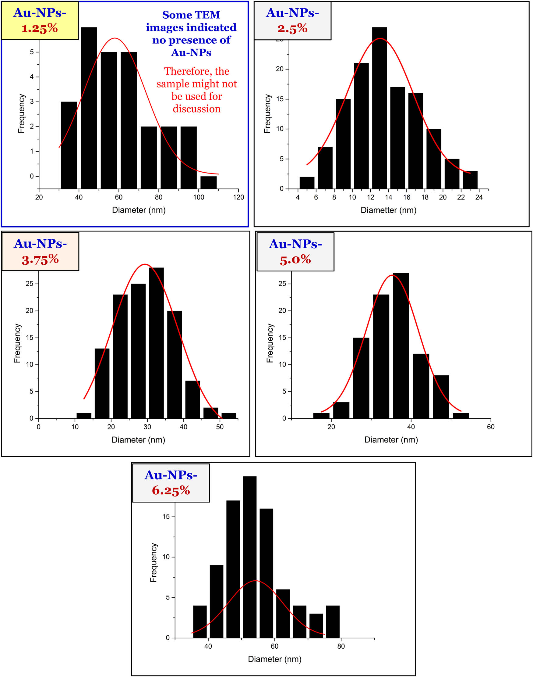
Particles size distribution of Au-NPs prepared at different ratios of the extract used.
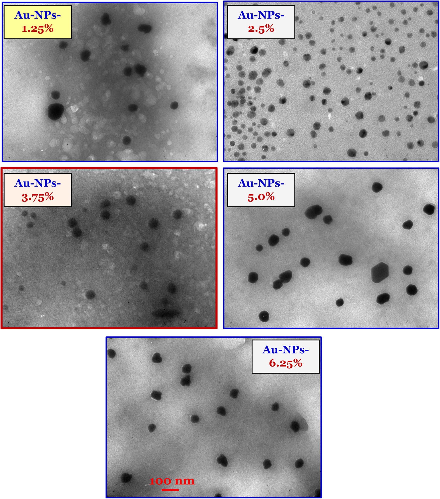
TEM images of Au-NPs prepared at different ratios of the extract used.
Cell growth inhibition assays of C. Hindsii-AuNPs on HeLa cells.
The inhibition of cell growth assays of C. Hindsii-AuNPs on HeLa cells
| Control | Concentration of Au-NPs (ppm) | |||||||
|---|---|---|---|---|---|---|---|---|
| 1.5 ppm | 3 ppm | 6.25 ppm | 12.5 ppm | 25 ppm | 50 ppm | 73 ppm | ||
| Repeat 1 | 577 | 550 | 501 | 484 | 312 | 241 | 89 | 57 |
| Repeat 2 | 656 | 500 | 479 | 457 | 311 | 134 | 71 | 44 |
| Repeat 3 | 634 | 491 | 418 | 368 | 365 | 169 | 70 | 38 |
| Repeat 3 | 693 | 548 | 505 | 452 | 331 | 180 | 92 | 69 |
| Average | 640 | 522 | 476 | 440 | 330 | 181 | 81 | 52 |
| SD | 48.5 | 31.1 | 40.2 | 50.2 | 25.2 | 44.5 | 11.6 | 13.8 |
| SE | 24.3 | 15.6 | 20.1 | 25.1 | 12.6 | 22.3 | 5.8 | 6.9 |
-
Research funding: The study was supported by The Youth Incubator for Science and Technology Programe, managed by Youth Development Science and Technology Center-Ho Chi Minh Communist Youth Union and Department of Science and Technology of Ho Chi Minh City, the contract number is “23/2019/HĐ-KHCN-VƯ”.
-
Author contributions: Xuan-Truong Mai: conceptualization, writing – original draft, methodology, investigation, funding acquisition; Minh-Chien Tran: methodology, investigation, software; Anh-Quan Hoang: methodology, investigation; Phuc Dang-Ngoc Nguyen: investigation; Thi-Hiep Nguyen: methodology, validation, formal analysis; Hai Nguyen Tran: writing – review and editing, validation, formal analysis, visualization; Phuong-Tung Nguyen: conceptualization, writing – review and editing, methodology, validation, supervision, project administration.
-
Conflict of interest: One of the authors (Hai Nguyen Tran) is a member of the Editorial Board of Green Processing and Synthesis.
References
[1] Bagheri S, Yasemi M, Safaie-Qamsari E, Rashidiani J, Abkar M, Hassani M, et al. Using gold nanoparticles in diagnosis and treatment of melanoma cancer. Artif Cell Nanomed Biotechnol. 2018;46:462–71. 10.1080/21691401.2018.1430585.Search in Google Scholar PubMed
[2] Lévy R, Thanh NTK, Doty RC, Hussain I, Nichols RJ, Schiffrin DJ, et al. Rational and combinatorial design of peptide capping ligands for gold nanoparticles. J Am Chem Soc. 2004;126:10076–84. 10.1021/ja0487269.Search in Google Scholar PubMed
[3] Han G, Ghosh P, Rotello VM. Functionalized gold nanoparticles for drug delivery. Nanobiotechnology. 2007;3:40–5. 10.2217/17435889.2.1.113.Search in Google Scholar PubMed
[4] Kim J-H, Yeom J-H, Ko J-J, Han MS, Lee K, Na SY, et al. Effective delivery of anti-miRNA DNA oligonucleotides by functionalized gold nanoparticles. J Biotechnol. 2011;155:287–92. 10.1016/j.jbiotec.2011.07.014.Search in Google Scholar PubMed
[5] Li S, Bouchy S, Penninckx S, Marega R, Fichera O, Gallez B, et al. Antibody-functionalized gold nanoparticles as tumor-targeting radiosensitizers for proton therapy. Nanomedicine. 2019;14:317–33. 10.2217/nnm-2018-0161.Search in Google Scholar PubMed
[6] Sharifi M, Attar F, Saboury AA, Akhtari K, Hooshmand N, Hasan A, et al. Plasmonic gold nanoparticles: optical manipulation, imaging, drug delivery and therapy. J Control Rel. 2019;311–2:170–89. 10.1016/j.jconrel.2019.08.032.Search in Google Scholar PubMed
[7] Amendoeira A, García LR, Fernandes AR, Baptista PV. Light irradiation of gold nanoparticles toward advanced cancer therapeutics. Adv Ther. 2020;3:1900153. 10.1002/adtp.201900153.Search in Google Scholar
[8] Taghizadeh S, Alimardani V, Roudbali PL, Ghasemi Y, Kaviani E. Gold nanoparticles application in liver cancer. Photodiagnosis Photodyn Ther. 2019;25:389–400. 10.1016/j.pdpdt.2019.01.027.Search in Google Scholar PubMed
[9] Abdelghany A, Abdelrazek E, Badr S, Abdel-Aziz M, Morsi M. Effect of gamma-irradiation on biosynthesized gold nanoparticles using Chenopodium murale leaf extract. J Saudi Chem Soc. 2017;21:528–37. 10.1016/j.jscs.2015.10.002.Search in Google Scholar
[10] Dubey S, Lahtinen M, Särkkä H, Sillanpää M. Bioprospective of Sorbus aucuparia leaf extract in development of silver and gold nanocolloids. Colloids Surf B. 2010;80:26–33. 10.1016/j.colsurfb.2010.05.024.Search in Google Scholar PubMed
[11] Castro L, Blázquez ML, Muñoz JA, González F, García-Balboa C, Ballester A. Biosynthesis of gold nanowires using sugar beet pulp. Process Biochem. 2011;46:1076–82. 10.1016/j.procbio.2011.01.025.Search in Google Scholar
[12] Ankamwar B. Biosynthesis of gold nanoparticles (green-gold) using leaf extract of terminalia catappa. E-J Chem. 2010;7:745120–1339. 10.1155/2010/745120.Search in Google Scholar
[13] Philip D. Green synthesis of gold and silver nanoparticles using Hibiscus rosa sinensis. Phys E Low Dimens Syst Nanostruct. 2010;42:1417–24. 10.1016/j.physe.2009.11.081.Search in Google Scholar
[14] Philip D, Spectroscopy B. Rapid green synthesis of spherical gold nanoparticles using Mangifera indica leaf. Acta A Mol Biomol Spectrosc. 2010;77:807–10. 10.1016/j.saa.2010.08.008.Search in Google Scholar PubMed
[15] Kaur B, Markan M, Singh M. Green synthesis of gold nanoparticles from Syzygium aromaticum extract and its use in enhancing the response of a colorimetric urea biosensor. BioNanoScience. 2012;2:251–8. 10.1007/s12668-012-0062-5.Search in Google Scholar
[16] Sunderam V, Thiyagarajan D, Lawrence AV, Mohammed SSS, Selvaraj A. In vitro antimicrobial and anticancer properties of green synthesized gold nanoparticles using Anacardium occidentale leaves extract. Saudi J Biol Sci. 2019;26:455–9. 10.1016/j.sjbs.2018.12.001.Search in Google Scholar PubMed PubMed Central
[17] Manivasagan P, Oh J. Production of a novel fucoidanase for the green synthesis of gold nanoparticles by streptomyces sp. and its cytotoxic effect on HeLa cells. Mar Drugs. 2015;13:6818–37. 10.3390/md13116818.Search in Google Scholar PubMed PubMed Central
[18] Philip D, Unni C, Aromal SA, Vidhu V, Spectroscopy B. Murraya koenigii leaf-assisted rapid green synthesis of silver and gold nanoparticles. Spectrochim Acta A Mol Biomol. 2011;78:899–904. 10.1016/j.saa.2010.12.060.Search in Google Scholar PubMed
[19] Ahmad B, Hafeez N, Bashir S, Rauf A, Mujeeb ur R. Phytofabricated gold nanoparticles and their biomedical applications. Biomed Pharmacother. 2017;89:414–25. 10.1016/j.biopha.2017.02.058.Search in Google Scholar PubMed
[20] Ly TN, Shimoyamada M, Yamauchi R. Isolation and characterization of rosmarinic acid oligomers in Celastrus hindsii benth leaves and their antioxidative activity. J Agric Food Chem. 2006;54:3786–93. 10.1021/jf052743f.Search in Google Scholar PubMed
[21] Pham D-C, Nguyen H-C, Nguyen T-HL, Ho HL, Trinh TK, Riyaphan J, et al. Optimization of ultrasound-assisted extraction of flavonoids from Celastrus hindsii leaves using response surface methodology and evaluation of their antioxidant and antitumor activities. Biomed Res Int. 2020;2020:1–9. 10.1155/2020/3497107.Search in Google Scholar PubMed PubMed Central
[22] Viet TD, Xuan TD, Van TM, Andriana Y, Rayee R, Tran HD. Comprehensive fractionation of antioxidants and GC-MS and ESI-MS fingerprints of Celastrus hindsii Leaves. Medicines. 2019;6:64. 10.3390/medicines6020064.Search in Google Scholar PubMed PubMed Central
[23] Duyen BTT, Hung VM, Tung BT. Cytotoxicity and antioxidant effects of Celastrus hindsii benth. Leaf extract. VNU J Sci Med Pharm Sci. 2020;36:36. 10.25073/2588-1132/vnumps.4203.Search in Google Scholar
[24] Li L, Zhang W, Desikan Seshadri VD, Cao G. Synthesis and characterization of gold nanoparticles from Marsdenia tenacissima and its anticancer activity of liver cancer HepG2 cells. Artif Cell Nanomed Biotechnol. 2019;47:3029–36. 10.1080/21691401.2019.1642902.Search in Google Scholar PubMed
[25] Ji Y, Cao Y, Song Y. Green synthesis of gold nanoparticles using a Cordyceps militaris extract and their antiproliferative effect in liver cancer cells (HepG2). Artif Cell Nanomed Biotechnol. 2019;47:2737–45. 10.1080/21691401.2019.1629952.Search in Google Scholar PubMed
[26] Xi D, Dong S, Meng X, Lu Q, Meng L, Ye J. Gold nanoparticles as computerized tomography (CT) contrast agents. RSC Adv. 2012;2:12515–24. 10.1039/C2RA21263C.Search in Google Scholar
[27] Haiss W, Thanh NT, Aveyard J, Fernig DG. Determination of size and concentration of gold nanoparticles from UV−Vis spectra. Anal Chem. 2007;79:4215–21. 10.1021/ac0702084.Search in Google Scholar PubMed
[28] Morsi M, Abdelghany A. UV-irradiation assisted control of the structural, optical and thermal properties of PEO/PVP blended gold nanoparticles. Mater Chem Phys. 2017;201:100–12. 10.1016/j.matchemphys.2017.08.022.Search in Google Scholar
[29] Ghosh S, Patil S, Ahire M, Kitture R, Gurav DD, Jabgunde AM, et al. Gnidia glauca flower extract mediated synthesis of gold nanoparticles and evaluation of its chemocatalytic potential. J Nanobiotechnol. 2012;10:17. 10.1186/1477-3155-10-17.Search in Google Scholar PubMed PubMed Central
[30] Singh M, Kalaivani R, Manikandan S, Sangeetha N, Kumaraguru AK. Facile green synthesis of variable metallic gold nanoparticle using Padina gymnospora, a brown marine macroalga. Appl Nanosci. 2013;3:145–51. 10.1007/s13204-012-0115-7.Search in Google Scholar
[31] Singh AK, Srivastava ON. One-step green synthesis of gold nanoparticles using black cardamom and effect of pH on its synthesis. Nanoscale Res Lett. 2015;10:353. 10.1186/s11671-015-1055-4.Search in Google Scholar PubMed PubMed Central
[32] Chen M, He Y, Liu X, Zhu J, Liu R. Synthesis and optical properties of size-controlled gold nanoparticles. Powder Technol. 2017;311:25–33. 10.1016/j.powtec.2017.01.087.Search in Google Scholar
[33] Wyckoff RWG. Crystal structures (vol 1). New York: Interscience Publishers; 1963. p. 7–83. http://www.crystallography.net/cod/9008463.htmlSearch in Google Scholar
[34] Raza M, Kanwal Z, Rauf A, Sabri A, Riaz S, Naseem S. Size- and shape-dependent antibacterial studies of silver nanoparticles synthesized by wet chemical routes. Nanomaterials. 2016;6:74. 10.3390/nano6040074.Search in Google Scholar PubMed PubMed Central
[35] Wang P, Wang X, Wang L, Hou X, Liu W, Chen C. Interaction of gold nanoparticles with proteins and cells. Sci Technol Adv Mater. 2015;16:034610. 10.1088/1468-6996/16/3/034610.Search in Google Scholar PubMed PubMed Central
[36] Cui L, Zahedi P, Saraceno J, Bristow R, Jaffray D, Allen C. Neoplastic cell response to tiopronin-coated gold nanoparticles. Nanomedicine. 2013;9:264–73. 10.1016/j.nano.2012.05.016.Search in Google Scholar PubMed
[37] Tran N, Le A, Ho M, Dang N, Thi Thanh HH, Truong L, et al. Polyurethane/polycaprolactone membrane grafted with conjugated linoleic acid for artificial vascular graft application. Sci Technol Adv Mater. 2020;21:56–66. 10.1080/14686996.2020.1718549.Search in Google Scholar PubMed PubMed Central
[38] Khan T, Ullah N, Khan MA, Nadhman A. Plant-based gold nanoparticles; a comprehensive review of the decade-long research on synthesis, mechanistic aspects and diverse applications. Adv Colloid Interface Sci. 2019;272:102017. 10.1016/j.cis.2019.102017.Search in Google Scholar PubMed
© 2021 Xuan-Truong Mai et al., published by De Gruyter
This work is licensed under the Creative Commons Attribution 4.0 International License.
Articles in the same Issue
- Research Articles
- MW irradiation and ionic liquids as green tools in hydrolyses and alcoholyses
- Effect of CaO on catalytic combustion of semi-coke
- Studies of Penicillium species associated with blue mold disease of grapes and management through plant essential oils as non-hazardous botanical fungicides
- Development of leftover rice/gelatin interpenetrating polymer network films for food packaging
- Potent antibacterial action of phycosynthesized selenium nanoparticles using Spirulina platensis extract
- Green synthesized silver and copper nanoparticles induced changes in biomass parameters, secondary metabolites production, and antioxidant activity in callus cultures of Artemisia absinthium L.
- Gold nanoparticles from Celastrus hindsii and HAuCl4: Green synthesis, characteristics, and their cytotoxic effects on HeLa cells
- Green synthesis of silver nanoparticles using Tropaeolum majus: Phytochemical screening and antibacterial studies
- One-step preparation of metal-free phthalocyanine with controllable crystal form
- In vitro and in vivo applications of Euphorbia wallichii shoot extract-mediated gold nanospheres
- Fabrication of green ZnO nanoparticles using walnut leaf extract to develop an antibacterial film based on polyethylene–starch–ZnO NPs
- Preparation of Zn-MOFs by microwave-assisted ball milling for removal of tetracycline hydrochloride and Congo red from wastewater
- Feasibility of fly ash as fluxing agent in mid- and low-grade phosphate rock carbothermal reduction and its reaction kinetics
- Three combined pretreatments for reactive gasification feedstock from wet coffee grounds waste
- Biosynthesis and antioxidation of nano-selenium using lemon juice as a reducing agent
- Combustion and gasification characteristics of low-temperature pyrolytic semi-coke prepared through atmosphere rich in CH4 and H2
- Microwave-assisted reactions: Efficient and versatile one-step synthesis of 8-substituted xanthines and substituted pyrimidopteridine-2,4,6,8-tetraones under controlled microwave heating
- New approach in process intensification based on subcritical water, as green solvent, in propolis oil in water nanoemulsion preparation
- Continuous sulfonation of hexadecylbenzene in a microreactor
- Synthesis, characterization, biological activities, and catalytic applications of alcoholic extract of saffron (Crocus sativus) flower stigma-based gold nanoparticles
- Foliar applications of plant-based titanium dioxide nanoparticles to improve agronomic and physiological attributes of wheat (Triticum aestivum L.) plants under salinity stress
- Simultaneous leaching of rare earth elements and phosphorus from a Chinese phosphate ore using H3PO4
- Silica extraction from bauxite reaction residue and synthesis water glass
- Metal–organic framework-derived nanoporous titanium dioxide–heteropoly acid composites and its application in esterification
- Highly Cr(vi)-tolerant Staphylococcus simulans assisting chromate evacuation from tannery effluent
- A green method for the preparation of phoxim based on high-boiling nitrite
- Silver nanoparticles elicited physiological, biochemical, and antioxidant modifications in rice plants to control Aspergillus flavus
- Mixed gel electrolytes: Synthesis, characterization, and gas release on PbSb electrode
- Supported on mesoporous silica nanospheres, molecularly imprinted polymer for selective adsorption of dichlorophen
- Synthesis of zeolite from fly ash and its adsorption of phosphorus in wastewater
- Development of a continuous PET depolymerization process as a basis for a back-to-monomer recycling method
- Green synthesis of ZnS nanoparticles and fabrication of ZnS–chitosan nanocomposites for the removal of Cr(vi) ion from wastewater
- Synthesis, surface modification, and characterization of Fe3O4@SiO2 core@shell nanostructure
- Antioxidant potential of bulk and nanoparticles of naringenin against cadmium-induced oxidative stress in Nile tilapia, Oreochromis niloticus
- Variability and improvement of optical and antimicrobial performances for CQDs/mesoporous SiO2/Ag NPs composites via in situ synthesis
- Green synthesis of silver nanoparticles: Characterization and its potential biomedical applications
- Green synthesis, characterization, and antimicrobial activity of silver nanoparticles prepared using Trigonella foenum-graecum L. leaves grown in Saudi Arabia
- Intensification process in thyme essential oil nanoemulsion preparation based on subcritical water as green solvent and six different emulsifiers
- Synthesis and biological activities of alcohol extract of black cumin seeds (Bunium persicum)-based gold nanoparticles and their catalytic applications
- Digera muricata (L.) Mart. mediated synthesis of antimicrobial and enzymatic inhibitory zinc oxide bionanoparticles
- Aqueous synthesis of Nb-modified SnO2 quantum dots for efficient photocatalytic degradation of polyethylene for in situ agricultural waste treatment
- Study on the effect of microwave roasting pretreatment on nickel extraction from nickel-containing residue using sulfuric acid
- Green nanotechnology synthesized silver nanoparticles: Characterization and testing its antibacterial activity
- Phyto-fabrication of selenium nanorods using extract of pomegranate rind wastes and their potentialities for inhibiting fish-borne pathogens
- Hydrophilic modification of PVDF membranes by in situ synthesis of nano-Ag with nano-ZrO2
- Paracrine study of adipose tissue-derived mesenchymal stem cells (ADMSCs) in a self-assembling nano-polypeptide hydrogel environment
- Study of the corrosion-inhibiting activity of the green materials of the Posidonia oceanica leaves’ ethanolic extract based on PVP in corrosive media (1 M of HCl)
- Callus-mediated biosynthesis of Ag and ZnO nanoparticles using aqueous callus extract of Cannabis sativa: Their cytotoxic potential and clinical potential against human pathogenic bacteria and fungi
- Ionic liquids as capping agents of silver nanoparticles. Part II: Antimicrobial and cytotoxic study
- CO2 hydrogenation to dimethyl ether over In2O3 catalysts supported on aluminosilicate halloysite nanotubes
- Corylus avellana leaf extract-mediated green synthesis of antifungal silver nanoparticles using microwave irradiation and assessment of their properties
- Novel design and combination strategy of minocycline and OECs-loaded CeO2 nanoparticles with SF for the treatment of spinal cord injury: In vitro and in vivo evaluations
- Fe3+ and Ce3+ modified nano-TiO2 for degradation of exhaust gas in tunnels
- Analysis of enzyme activity and microbial community structure changes in the anaerobic digestion process of cattle manure at sub-mesophilic temperatures
- Synthesis of greener silver nanoparticle-based chitosan nanocomposites and their potential antimicrobial activity against oral pathogens
- Baeyer–Villiger co-oxidation of cyclohexanone with Fe–Sn–O catalysts in an O2/benzaldehyde system
- Increased flexibility to improve the catalytic performance of carbon-based solid acid catalysts
- Study on titanium dioxide nanoparticles as MALDI MS matrix for the determination of lipids in the brain
- Green-synthesized silver nanoparticles with aqueous extract of green algae Chaetomorpha ligustica and its anticancer potential
- Curcumin-removed turmeric oleoresin nano-emulsion as a novel botanical fungicide to control anthracnose (Colletotrichum gloeosporioides) in litchi
- Antibacterial greener silver nanoparticles synthesized using Marsilea quadrifolia extract and their eco-friendly evaluation against Zika virus vector, Aedes aegypti
- Optimization for simultaneous removal of NH3-N and COD from coking wastewater via a three-dimensional electrode system with coal-based electrode materials by RSM method
- Effect of Cu doping on the optical property of green synthesised l-cystein-capped CdSe quantum dots
- Anticandidal potentiality of biosynthesized and decorated nanometals with fucoidan
- Biosynthesis of silver nanoparticles using leaves of Mentha pulegium, their characterization, and antifungal properties
- A study on the coordination of cyclohexanocucurbit[6]uril with copper, zinc, and magnesium ions
- Ultrasound-assisted l-cysteine whole-cell bioconversion by recombinant Escherichia coli with tryptophan synthase
- Green synthesis of silver nanoparticles using aqueous extract of Citrus sinensis peels and evaluation of their antibacterial efficacy
- Preparation and characterization of sodium alginate/acrylic acid composite hydrogels conjugated to silver nanoparticles as an antibiotic delivery system
- Synthesis of tert-amylbenzene for side-chain alkylation of cumene catalyzed by a solid superbase
- Punica granatum peel extracts mediated the green synthesis of gold nanoparticles and their detailed in vivo biological activities
- Simulation and improvement of the separation process of synthesizing vinyl acetate by acetylene gas-phase method
- Review Articles
- Carbon dots: Discovery, structure, fluorescent properties, and applications
- Potential applications of biogenic selenium nanoparticles in alleviating biotic and abiotic stresses in plants: A comprehensive insight on the mechanistic approach and future perspectives
- Review on functionalized magnetic nanoparticles for the pretreatment of organophosphorus pesticides
- Extraction and modification of hemicellulose from lignocellulosic biomass: A review
- Topical Issue: Recent advances in deep eutectic solvents: Fundamentals and applications (Guest Editors: Santiago Aparicio and Mert Atilhan)
- Delignification of unbleached pulp by ternary deep eutectic solvents
- Removal of thiophene from model oil by polyethylene glycol via forming deep eutectic solvents
- Valorization of birch bark using a low transition temperature mixture composed of choline chloride and lactic acid
- Topical Issue: Flow chemistry and microreaction technologies for circular processes (Guest Editor: Gianvito Vilé)
- Stille, Heck, and Sonogashira coupling and hydrogenation catalyzed by porous-silica-gel-supported palladium in batch and flow
- In-flow enantioselective homogeneous organic synthesis