Preparation and characterization of a novel composite membrane of natural silk fiber/nano-hydroxyapatite/chitosan for guided bone tissue regeneration
-
 and
and

Abstract
Natural silk fiber (SF) was introduced into the chitosan/nano-hydroxyapatite (CS/n-HA) system to fabricate a novel guided bone tissue regeneration (GBR) membrane. The effect of different treatment methods (degummed, un-degummed, or dissolved SF) and different contents of SF on the properties of the CS/n-HA composite membrane was investigated. Results demonstrated that the degummed SF/CS/n-HA composite membrane with a weight ratio of 2:6:2 possessed the highest mechanical strength, where SF supported the composite membrane as a skeleton frame in the form of primeval state, while the un-degummed SF and dissolved SF had weaker reinforce effect due to the poor interface or poor interaction between SF and CS, and the dissolved SF/CS/n-HA composite membrane displayed the fastest degradation. However, the three SF could all improve the cell biocompatibility of the CS/n-HA composite membrane. Conclusively, the study revealed that degummed SF could in situ reinforce the CS/n-HA composite membrane with a simple and green processing method, which would provide an important guidance significant to develop a novel GBR membrane.
1 Introduction
Guided bone tissue regeneration (GBR) is an effective method for regeneration of many bony defects, where an ideal membrane was placed on the defect acting as a mechanical barrier membrane, aimed to prevent epithelial cells and fibroblasts from growing into the defect; thus, a singular space was created under the membrane, so as to permit the essential time for the proliferation of osteoblasts and new bone formation. Generally, the GBR membranes should have high mechanical strength, suitable biodegradation, and excellent biocompatibility (1,2,3).
Among the selection of membrane, the nano-hydroxyapatite/chitosan (n-HA/CS) composite membrane has been usually regarded as a candidate for the GBR membrane (4,5). However, it displayed relatively poor mechanical strength and fast degradation. To solve these problems, glutaraldehyde was added acting as chemical crosslinking. Unluckily, the remaining glutaraldehyde would inevitably bring toxicity, which was not conducive to its biocompatibility of the n-HA/CS composite (6). Therefore, it was expected to introduce other polymers to improve the mechanical strength and slow down the degradation of the n-HA/CS composite membrane.
With the research of natural fibers, natural hemp, kenaf and silk were known to reinforce polymers (7,8,9,10). Especially, silk fiber (SF), a natural macromolecule polypeptide produced by Bombyx mori, is mainly composed of silk fibroin and sericin (11). SF has good mechanical properties, blood compatibility, water permeability, and antibacterial property, which is known as the “queen of fiber”, owing to its good arrangement structure and component. Therefore, SF was used to reinforce polymers (12,13,14,15). However, most of researches focused on the addition of silk protein, not natural SF, which required a variety of complicated steps, such as degumming, dissolution, dialysis, and filtration, then the silk protein solution was combined with other polymers to be manufactured into membrane or other scaffolds (16,17,18,19). Inevitably, the tedious procession would be adverse for production, and the fiber’s strengthening effect might be significantly reduced after the silk was dissolved.
Additionally, based on its excellent biological and mechanical properties, many studies have demonstrated the importance of the development of SF-based bone biomaterials, in particular GBR composite membranes by addition of SF (20,21,22). However, SF has not been used for reinforcing the n-HA/CS membrane. Therefore, it is worth exploring whether natural SF would in situ reinforce the CS/n-HA composite membrane by a simple and green processing method.
Based on the above considerations, in this work, natural SF was used to prepare the SF/CS/n-HA composite membrane, in which natural SF would support the composite as a skeleton frame in the form of primeval state, whose weight ratios of the SF/CS/n-HA were 1:6:2, 2:6:2, and 6:6:2, respectively, compared with the CS/n-HA, un-degummed SF/CS/n-HA, and dissolved SF/CS/n-HA composite membranes. Moreover, Fourier transformation infrared (FTIR), scanning electron microscope (SEM), and mechanical analyses were carried out. Furthermore, in vitro degradation behavior of the SF/CS/n-HA composite membranes soaked in simulated body fluid (SBF) was investigated by weight loss, water absorption rate, tensile strength reduction, and SEM morphology observation. Finally, the preliminary cell culture experiment was carried out, including fluorescence microscope observation and cell proliferation. The main aim of the study is to explore a simple and green processing method by adding the primeval state natural SF into the CS/n-HA composite membrane as a skeleton frame, so as to obtain an SF/CS/n-HA composite membrane for the GBR membrane.
2 Experimental
2.1 Materials
Chitosan (CS) was purchased from Jinan haidebei marine Bioengineering Co. Ltd., whose degree of deacetylation was 90% and molecular weight was 3 × 10⁵. n-HA was prepared in our laboratory by the solution precipitation method according to the relative literature (23), whose size was 100–200 nm in length and 30–70 nm in width. Silkworm silks were obtained by home feeding in Xian Zi Jiao Zhen, Dao County, Yongzhou City, Hunan Province. All other agents were of analytical grade.
2.2 Preparation of SF/CS/n-HA composite membranes
A total of 5.0 g silkworm silks were boiled in 300 mL of 0.02 mol/L Na2CO3 aqueous solution for 30 min, rinsed, and dried, noted as degummed SF. A 10 mg degummed SF was dissolved in 25 mL of mixture solution composed of 11.1 g CaCl2, 9.2 g ethanol, and 14.4 g deionized water, named as dissolved SF. Some of the silkworm silks were rinsed with distilled water and dried, noted as un-degummed SF.
CS solution (3 wt%) was obtained with acetic acid 2%, and n-HA slurry was added with the weight ratio of CS/n-HA of 6:2. Half of the CS/n-HA mixture solution was poured into a clean and dry glass plate, degummed SF was spread out on it, and then the remaining half of the CS/n-HA mixture solution was casted on the degummed SF, whose weight ratio was 1:6:2, 2:6:2, and 6:6:2, respectively. Similarly, the un-degummed SF or dissolved SF was introduced according to the same procedure with a weight ratio of 1:6:2, and the CS/n-HA composite membrane (6:2) was also prepared by the solution cast method as controls.
2.3 Material characterization
2.3.1 Fourier transformation infrared (FTIR)
The FTIR analysis of samples was performed with a Thermo Niclet 670 spectrometer, and the spectra were collected between 600 and 4,000 cm−1.
2.3.2 Surface morphological analysis
The surface morphology of samples was observed by SEM (S-520, Hitachi, Japan) after being sprayed with gold for 60 s.
2.3.3 Tensile strength measurement
The tensile strength of dry samples was measured using a universal testing machine (CMT6000, Sans, China) with a speed of 50 mm/min at 25°C. Three parallel samples of each specimen were tested and the mean values were given.
2.3.4 In vitro degradation investigation
The samples were immersed in the SBF for 8 weeks according to related reference (24). After soaking for 2, 4, and 8 weeks, the samples were taken out from SBF, washed with deionized water, and the surface water was adsorbed with filter paper for about 5 s. The weight loss and water absorption rate were given as follows:
where W 1 was the original weight of the sample, and W 2 and W 3 were the wet weight and the dry weight after being entirely dried, respectively.
Moreover, the tensile strength reduction was measured after soaking. The surface morphologies of samples after being soaked for 2 and 8 weeks were observed with SEM according to the previous procedure.
2.3.5 In vitro cell experiment
To primarily evaluate in vitro cell biocompatibility, bone mesenchymal stem cells (BMSCs) were isolated from a week-old SD (Sprague Dawley) rat neonatal by the sequential enzymatic digestive process (25,26). Briefly, the femur and tibia of the hind legs of SD rats were taken out and cut off, DMEM/F-12 medium containing 10% fetal bovine serum (FBS) was added with a 1 mL syringe, and washed until the bone wall turns white. The mixture was centrifuged at 800 rpm for 3 min, and the supernatant was resuspended with 1 mL of DMEM/F-12 culture solution containing 10% FBS, then 9 mL of culture medium was added into the resulting mixture, gently shook to fully mix with the cells, finally, the supernatant was placed in the incubator at 37°C, 5% CO2, and 90% relative humidity. After five passages, hematopoietic stem cells, red blood cells, and other hybrid cells were removed to obtain pure BMSC. The samples with the size of Φ 6.1 mm × 0.2 mm were soaked with 75% ethanol solution and sterilized with an ultraviolet lamp. The treated samples were placed in a 96-well plate with the density of 8,000 cells per well, and incubated without being disturbed for 3 h, followed by an additional 1 mL culture medium into each well.
Fluorescent pictures were used to intuitively evaluate the adhesion and proliferation of BMSCs on the surface of composite membranes. After incubation for 1, 2, and 3 days, the samples were taken out and rinsed with phosphate buffer solution (PBS) twice, then dyed for 10 min with 1 mL of acridine orange (AO) solution (0.1 mg/mL), and rinsed with PBS twice again. Finally, the fluorescence microscope (Axioskop 2, ZEISS, Germany) was used to observe the morphology and growth of cells.
MTT (Cell Proliferation and Cytotoxicity Assay Kit) assay was performed to evaluate the cell proliferation (27,28). After 1, 2, and 3 days, the medium in the cell-seeded materials was discarded, and 1 mL of MTT solution of 3 mg/mL was added, followed by incubation for over 4 h at 37°C in an air atmosphere containing 5% CO2. Then, the formazan crystals were dissolved with 1 mL of DMSO, and 100 µL of purple solution was transferred into a new 96-well plate, whose optical density values were measured with a microplate reader (Synergy HTX, BIOTEK, USA) at 492 nm.
3 Results and discussion
3.1 Physical–chemical properties of SF/CS/n-HA composite membrane
3.1.1 FT-IR analysis
Figure 1 shows FT-IR spectra of samples. It can be found that there was an obvious wide peak of 3,500 cm−1 attributed to –OH or –NH2, when different contents of degummed SF were introduced, which showed that there was no chemical bonding between degummed SF and CS/n-HA matrix, meaning they were physical adsorption among various components, that is to say, degummed SF only acted as a skeleton frame to support the CS/n-HA composite. However, the wide peak disappeared when dissolved or un-degummed SF was added. Especially, when the peaks of 800–1,800 cm−1 were partially enlarged, it could be seen that the peak of 1,645 cm−1 (Amid I and II of CS) also disappeared, suggesting that there existed chemical bonding, which was formed between amino acid of sericin and some functional groups of CS (29). The reason was that the dissolved SF contained amino acids, and un-degummed SF remained sericin, as we know, sericin was composed of i, ii, iii, and iv proteins from outside to inside, and it accounted for 20–30% of silk.

FT-IR spectra of the composite membranes: (a) CS/n-HA (6:2), (b) SF/CS/n-HA (1:6:2), (c) SF/CS/n-HA (2:6:2), (d) SF/CS/n-HA (6:6:2), (e) un-degummed SF/CS/n-HA (1:6:2), and (f) dissolved SF/CS/n-HA (1:6:2).
3.1.2 Surface morphology observation
Figure 2 gives the surface morphology of samples. The un-degummed SF displayed some jelly on the SF surface (noted in red arrow in Figure 2g), while the degummed SF seemed smoother (shown in Figure 2h). For the CS/n-HA composite membrane, some agglomerate n-HA nanoparticles were present in the CS matrix. For the un-degummed SF or dissolved SF, there were still a few agglomerated n-HA nanoparticles. While the degummed SF membrane was embedded between the two CS/n-HA layers, SF was covered with CS and n-HA nanoparticles without agglomeration, and even the SF amount was increased to 43 wt% (namely a weight ratio of SF/CS/n-HA of 6:6:2). The reason was that n-HA nanoparticles were adhered on the SF, showing that the degummed SF could prevent the agglomeration of n-HA nanoparticles.

SEM micrographs of the composite membranes: (a) CS/n-HA (6:2), (b) SF/CS/n-HA (1:6:2), (c) SF/CS/n-HA (2:6:2), (d) SF/CS/n-HA (6:6:2), (e) un-degummed SF/CS/n-HA (1:6:2), (f) dissolved SF/CS/n-HA (1:6:2), (g) un-degummed SF, and (h) degummed SF.
3.1.3 Tensile strength analysis
Figure 3 shows the tensile strength of the membranes, when the degummed SF was introduced to the CS/n-HA composite membrane, whose tensile strength was enhanced with the increasing of degummed SF, and the reinforce effect was by far better than that of the un-degummed SF and dissolved SF. The reason was that sericin was removed for the degummed SF, which was more propitious to uniformly attach n-HA nanoparticles, and SF would act as a membrane skeleton, so as to endow the composite membrane with excellent mechanical property, which was consistent with the SEM observation. While the dissolved SF displayed worst reinforce effect, which was caused by the poor interaction between SF and CS.

Tensile strengths of the composite membranes: (a) CS/n-HA (6:2), (b) SF/CS/n-HA (1:6:2), (c) SF/CS/n-HA (2:6:2), (d) SF/CS/n-HA (6:6:2), (e) un-degummed SF/CS/n-HA (1:6:2), and (f) dissolved SF/CS/n-HA (1:6:2).
3.2 Degradation of SF/CS/n-HA composite membrane
3.2.1 Weight loss and water absorption rate analysis
The weight loss and water absorption rate of samples are shown in Figures 4 and 5, respectively. The data indicated that the weight loss rate of all composite membranes gradually decreased, and the dissolved SF/CS/n-HA composite membrane reflected the greatest weight loss rate among all the samples, and other samples showed little difference. Meanwhile, it could be seen that the water absorption of the dissolved SF/CS/n-HA composite membrane was the highest, further suggesting that the dissolved SF/CS/n-HA composite membrane displayed the fastest degradation rate, which were all originated from the better hydrophilicity of the dissolved SF (30).

Weight loss of the composite membranes during the soaking.

Water absorption of the composite membranes during the soaking.
3.2.2 Tensile strength reduction
Figure 6 shows the mechanical strength reduction during the soaking period. It could be found that the tensile strength of samples gradually reduced with the prolonging degradation time. The SF/CS/n-HA composite membranes possessed higher tensile strength than the un-degummed SF and dissolved SF composite membrane at 8 weeks, which could keep over 17 MPa, while the dissolved SF/CS/n-HA composite membrane and CS/n-HA composite membrane only maintained 5 MPa or so. The reason was contributed to the faster degradation of dissolved SF. Obviously, the degummed SF/CS/n-HA composite membranes would keep mechanical support longer, and the mechanical support strength was higher than the relative literature (31).

Tensile strength reduction of the composite membranes during the soaking.
3.2.3 Surface morphology change
To investigate the bioactivity, Figure 7 shows the SEM photograph of samples after being soaked for 2 and 8 weeks. As expected, it could be clearly found that the surface of all SF/CS/n-HA composite membranes got whiter, compared with the CS/n-HA composite membrane, which showed a large amount of apatite deposition on the surface of the SF/CS/n-HA composite membrane, and there was no difference for different treated SF, suggesting that SF was adverse for apatite deposition (32).
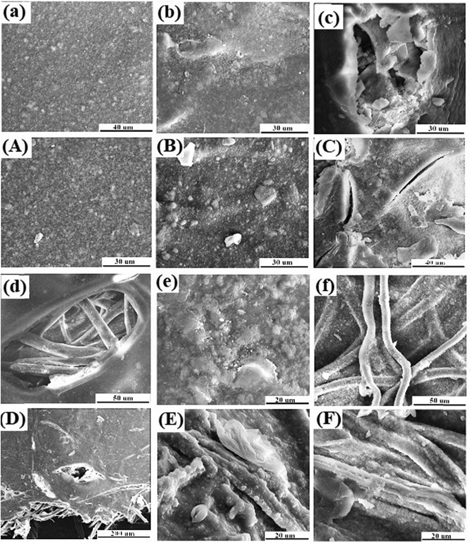
SEM micrographs of composite scaffolds with different weight ratios after soaking in SBF: (a and A) CS/n-HA (6:2), (b and B) SF/CS/n-HA (1:6:2), (c and C) SF/CS/n-HA (2:6:2), (d and D) SF/CS/n-HA (6:6:2), (e and E) dissolved SF/CS/n-HA (1:6:2), and (f and F) un-degummed SF/CS/n-HA (1:6:2). (a–f) Soaked for 2 weeks, (A–F) soaked for 8 weeks.
3.3 Cell biocompatibility of SF/CS/n-HA composite membrane
3.3.1 Fluorescence photograph observation
Figure 8 presents the cell fluorescence photographs of the samples. It could be seen that all of the SF/CS/n-HA composite membranes exhibited better cell attachment, compared with the CS/n-HA composite membrane, which demonstrated that the introduction of SF was profitable for cell biocompatibility. As we know, both chemical and topographical features of a surface could influence cell adhesion. Many relative literature indicated that SF possessed good biocompatibility (33), and the agglomerated n-HA nanoparticles in the matrix were not conducive to cell adhesion. So, we think that the reason was contributed to the good biocompatibility of SF and the better dispersion of n-HA in the matrix of SF/CS/n-HA composite membranes, compared with the CS/n-HA composite membrane.

Fluorescence staining micrographs on sample surface: (a) CS/n-HA (6:2), (b) SF/CS/n-HA (1:6:2), (c) SF/CS/n-HA (2:6:2), (d) SF/CS/n-HA (6:6:2), (e) dissolved SF/CS/n-HA (1:6:2), and (f) un-degummed SF/CS/n-HA (1:6:2). The magnification was 40 times.
3.3.2 Cell proliferation analysis
Figure 9 shows the cell proliferation, and it was expected to find the cells had distinct proliferation tendency after being cultivated for 1, 2, and 3 days, indicating that all samples were nontoxic. Comparing the subtle difference, the SF/CS/n-HA composite membranes possessed more remarkable proliferation than the CS/n-HA composite membrane, and the SF/CS/n-HA composite membrane with higher SF content displayed better cell proliferation. However, there was no significant difference for different SFs, which was consistent with the results of apatite deposition and fluorescence photographs.

MTT assays of cell culture on sample surface.
4 Conclusion
In this study, SF was introduced into the CS/n-HA composite membrane by different treatment methods and different contents. Results showed that the degummed SF supported the composite membrane as a skeleton frame in the form of primeval state, which produced the highest mechanical strength and appropriate degradation of the composite membrane, compared with the membrane with un-degummed SF or dissolved SF, although the three different SFs were all profitable for cell biocompatibility without significant difference. The study revealed that the degummed SF could be used to in situ reinforce a CS/n-HA composite membrane by a simple and green processing method, which would have a great potential for the GBR membrane.
Acknowledgments
The authors would like to thank Prof. Jiang Liuyun for guidance.
-
Funding information: The authors would like to thank the support of the Natural Science Foundation of Hunan province (2020JJ4430).
-
Author contributions: Shuo Tang: writing – original draft, writing – review and editing, methodology, formal analysis; Weijia Wang: formal analysis, visualization.
-
Conflict of interest: Authors declare no potential conflicts of interest with respect to the research, authorship, and/or publication of this article.
-
Data availability statement: The raw/processed data required to reproduce these findings cannot be shared at this time as the data also form part of an ongoing study.
References
(1) Li PY, Li YF, Kwok T, Yang T, Liu C, Li WC, et al. A bi-layered membrane with micro-nano bioactive glass for guided bone regeneration. Colloids Surf B. 2021;205:111886.10.1016/j.colsurfb.2021.111886Search in Google Scholar PubMed
(2) Niu XL, Wang LF, Xu MJ, Qin M, Zhao LQ, Wei Y, et al. Electrospun polyamide-6/chitosan nanofibers reinforced nano-hydroxyapatite/polyamide-6 composite bilayered membranes for guided bone regeneration. Carbohyd Polym. 2021;260:117769.10.1016/j.carbpol.2021.117769Search in Google Scholar PubMed
(3) Brum IS, Elias CN, de Carvalho JJ, Pires JLS, Pereira MJS, de Biasi RS. Properties of a bovine collagen type I membrane for guided bone regeneration applications. e-Polymers. 2021;21:210–21.10.1515/epoly-2021-0021Search in Google Scholar
(4) Tu Y, Chen C, Li YB, Hou Y, Huang M, Zhang L. Fabrication of nano-hydroxyapatite/chitosan membrane with asymmetric structure and its applications in guided bone regeneration. Bio-Med Mater Eng. 2017;28:223–33.10.3233/BME-171669Search in Google Scholar PubMed
(5) Cheng XM, Li YB, Zuo Y, Zhang L, Li JD, Wang HN. Properties and in vitro biological evaluation of nano-hydroxyapatite/chitosan membranes for bone guided regeneration. Mat Sci Eng C-Mater. 2009;29:29–35.10.1016/j.msec.2008.05.008Search in Google Scholar
(6) Oliveira MZFD, Fernandes TSM, Carvalho TV. Synthesis and characterization of commercial chitosan beads cross-linked with glutaraldehyde. Materia (Brazil). 2021;26:2.Search in Google Scholar
(7) Ude AU, Ariffin AK, Zhari CHA. Impact damage characteristics in reinforced woven natural silk/epoxy composite face-sheet and sandwich foam, core, mat and honeycomb materials. Int J Impact Eng. 2013;58:31–8.10.1016/j.ijimpeng.2013.03.003Search in Google Scholar
(8) Tamrakar S, Kiziltas A, Mielewski D, Zander R. Characterization of kenaf and glass fiber reinforced hybrid composites for underbody shield applications. Compos Part B-Eng. 2021;216:108805.10.1016/j.compositesb.2021.108805Search in Google Scholar
(9) Manaia JP, Manaia A. Interface modification, water absorption behaviour and mechanical properties of injection moulded short hemp fiber-reinforced thermoplastic composites. Polymers (Basel). 2021;13:1638.10.3390/polym13101638Search in Google Scholar PubMed PubMed Central
(10) Gu YQ, Yu LZ, Mou JG, Wu DH, Zhou PJ, Xu MS. Mechanical properties and application analysis of spider silk bionic material. e-Polymers. 2020;20:443–57.10.1515/epoly-2020-0049Search in Google Scholar
(11) Cheerarot O, Baimark Y. Biodegradable silk fibroin/chitosan blend microparticles prepared by emulsification-diffusion method. e-Polymers. 2015;15:67–74.10.1515/epoly-2014-0134Search in Google Scholar
(12) Yang K, Guan J, Shao ZZ, Ritchie RO. Mechanical properties and toughening mechanisms of natural silkworm silks and their composites. J Mech Behav Biomed. 2020;110:103942.10.1016/j.jmbbm.2020.103942Search in Google Scholar PubMed
(13) Vyas C, Zhang J, Ovrebo O, Huang BY, Roberts I, Setty M, et al. 3D printing of silk microparticle reinforced polycaprolactone scaffolds for tissue engineering applications. Mat Sci Eng C-Mater. 2021;118:111433.10.1016/j.msec.2020.111433Search in Google Scholar PubMed
(14) Piri N, Mottaghitalab V, Arbab S. Conductive regenerated silk fibroin composite fiber containing MWNTs. e-Polymers. 2013;13:7.10.1515/epoly-2013-0107Search in Google Scholar
(15) Kale RD, Alemayehu TG, Gorade VG. Development and characterization study of silk filament reinforced chitosan biocomposite. J Nat Fibers. 2020;17:66–74.10.1080/15440478.2018.1465878Search in Google Scholar
(16) Zhao SH, Pan HL, Liu YL, Zeng YR, Liu HL, Yu WD. Silk fabric protection obtained via chemical conjugation of transglutaminase and silk fibroin reinforcement. Text Res J. 2019;89:4581–94.10.1177/0040517519837727Search in Google Scholar
(17) Narita C, Okahisa Y, Yamada K. A novel technique in the preparation of environmentally friendly cellulose nanofiber/silk fibroin fiber composite films with improved thermal and mechanical properties. J Clean Prod. 2019;234:200–7.10.1016/j.jclepro.2019.06.215Search in Google Scholar
(18) Zhang R, Han Q, Li Y, Cai YF, Zhu XY, Zhang TQ, et al. High antibacterial performance of electrospinning silk fibroin/gelatin film modified with graphene oxide-sliver nanoparticles. J Appl Polym Sci. 2019;136:47904.10.1002/app.47904Search in Google Scholar
(19) Yin YL, Zhao XF, Xiong J. Modeling analysis of silk fibroin/poly(epsilon-caprolactone) nanofibrous membrane under uniaxial tension. Nanomaterials (Basel). 2019;9:1149.10.3390/nano9081149Search in Google Scholar PubMed PubMed Central
(20) Wu M, Han ZY, Liu W, Yao JR, Zhao BJ, Shao ZZ, et al. Silk-based hybrid microfibrous mats as guided bone regeneration membranes. J Mater Chem B. 2021;9:2025–32.10.1039/D0TB02687ESearch in Google Scholar
(21) Imsombut T, Srisa-ard M, Srihanam P, Baimark Y. Preparation of silk fibroin microspheres by emulsification-diffusion method for controlled release drug delivery applications. e-Polymers. 2011;088.10.1515/epoly.2011.11.1.936Search in Google Scholar
(22) Karatepe UY, Ozdemir T. Improving mechanical and antibacterial properties of PMMA via polyblend electrospinning with silk fibroin and polyethyleneimine towards dental applications. Bioact Mater. 2020;5:510–5.10.1016/j.bioactmat.2020.04.005Search in Google Scholar PubMed PubMed Central
(23) Xu LJ, Jiang LY, Xiong CD, Jiang LX. Effect of different synthesis conditions on the microstructure, crystallinity and solubility of Mg-substituted hydroxyapatite nanopowder. Adv Powder Technol. 2014;25:1142–6.10.1016/j.apt.2014.02.019Search in Google Scholar
(24) Tang S, Jiang LY, Ma BL, Tang CY, Wen Y, Zhang N, et al. Preparation and characterization of bamboo fiber/chitosan/nano-hydroxyapatite composite membrane by ionic crosslinking. Cellulose. 2020;27:5089–100.10.1007/s10570-020-03145-2Search in Google Scholar
(25) Zhang BJ, He L, Han ZW, Li XG, Zhi W, Zheng W, et al. Enhanced osteogenesis of multilayered pore-closed microsphere-immobilized hydroxyapatite scaffold via sequential delivery of osteogenic growth peptide and BMP-2. J Mater Chem B. 2017;5:8238–53.10.1039/C7TB01970JSearch in Google Scholar PubMed
(26) Zhou JK, Zhang K, Ma SS, Liu TF, Yao MH, Li JG, Wang XF, Guan FX. Preparing an injectable hydrogel with sodium alginate and Type I collagen to create better MSCs growth microenvironment. e-Polymers. 2019;19:87–91.10.1515/epoly-2019-0011Search in Google Scholar
(27) Chang GX, Ren KF, Zhao YX, Sun YX, Ji J. Modulation of cell behaviors by electrochemically active polyelectrolyte multilayers. e-Polymers. 2014;14:297–304.10.1515/epoly-2014-0075Search in Google Scholar
(28) Sharma P, Mathur G, Goswami N, Sharma SK, Dhakate SR, Chand S, Mathur A. Evaluating the potential of chitosan/poly (vinyl alcohol) membranes as alternative carrier material for proliferation of Vero cells. e-Polymers. 2015;15:237–43.10.1515/epoly-2015-0021Search in Google Scholar
(29) Min L, Liu M, Liu LL, Rao ZQ, Zhu C, Fan LH. Enzymatic synthesis of quaternary ammonium chitosan-silk fibroin peptide copolymer and its characterization. Int J Biol Macromol. 2018;109:1125–31.10.1016/j.ijbiomac.2017.11.108Search in Google Scholar PubMed
(30) Wang C, Du YL, Chen BY, Chen SJ, Wang YP. A novel highly stretchable, adhesive and self-healing silk fibroin powder-based hydrogel containing dual-network structure. Mater Lett. 2019;252:126–9.10.1016/j.matlet.2019.05.129Search in Google Scholar
(31) Xu LJ, Jiang LY, Xiong CD, Jiang LX, Li Y. Study on a novel double-layered composite membrane of Mg-substituted nano-hydroxyapatite/poly(l-lactide-co-ε-caprolactone): effect of different L-lactide/ε-caprolactone ratios. Mat Sci Eng A-Struct. 2014;615:361–6.10.1016/j.msea.2014.07.044Search in Google Scholar
(32) Zhou BG, Zhou Q, Wang P, Yuan JG, Yu YY, Deng C. HRP-mediated graft polymerization of acrylic acid onto silk fibroins and in situ biomimetic mineralization. J Mater Sci Mater M. 2018;29:72.10.1007/s10856-018-6084-ySearch in Google Scholar PubMed
(33) Li Y, Zhao LH, Yao Y, Guo XF. Single-molecule nanotechnologies: an evolution in biological dynamics detection. ACS Appl Bio Mater. 2020;4:1369–80.10.1021/acsabm.9b00840Search in Google Scholar PubMed
© 2021 Shuo Tang and Weijia Wang, published by De Gruyter
This work is licensed under the Creative Commons Attribution 4.0 International License.
Articles in the same Issue
- Research Articles
- Research on the mechanism of gel accelerator on gel transition of PAN solution by rheology and dynamic light scattering
- Gel point determination of gellan biopolymer gel from DC electrical conductivity
- Composite of polylactic acid and microcellulose from kombucha membranes
- Synthesis of highly branched water-soluble polyester and its surface sizing agent strengthening mechanism
- Fabrication and characterization of poly(3-hydroxybutyrate-co-3-hydroxyhexanoate) modified with nano-montmorillonite biocomposite
- Fabrication of N-halamine polyurethane films with excellent antibacterial properties
- Formulation and optimization of gastroretentive bilayer tablets of calcium carbonate using D-optimal mixture design
- Sustainable nanocomposite films based on SiO2 and biodegradable poly(3-hydroxybutyrate-co-3-hydroxyhexanoate) (PHBH) for food packaging
- Evaluation of physicochemical properties of film-based alginate for food packing applications
- Electrically conductive and light-weight branched polylactic acid-based carbon nanotube foams
- Structuring of hydroxy-terminated polydimethylsiloxane filled by fumed silica
- Surface functionalization of nanostructured Cu/Ag-deposited polypropylene fiber by magnetron sputtering
- Influence of composite structure design on the ablation performance of ethylene propylene diene monomer composites
- MOFs/PVA hybrid membranes with enhanced mechanical and ion-conductive properties
- Improvement of the electromechanical properties of thermoplastic polyurethane composite by ionic liquid modified multiwall carbon nanotubes
- Natural rubber latex/MXene foam with robust and multifunctional properties
- Rheological properties of two high polymers suspended in an abrasive slurry jet
- Two-step polyaniline loading in polyelectrolyte complex membranes for improved pseudo-capacitor electrodes
- Preparation and application of carbon and hollow TiO2 microspheres by microwave heating at a low temperature
- Properties of a bovine collagen type I membrane for guided bone regeneration applications
- Fabrication and characterization of thermoresponsive composite carriers: PNIPAAm-grafted glass spheres
- Effect of talc and diatomite on compatible, morphological, and mechanical behavior of PLA/PBAT blends
- Multifunctional graphene nanofiller in flame retarded polybutadiene/chloroprene/carbon black composites
- Strain-dependent wicking behavior of cotton/lycra elastic woven fabric for sportswear
- Enhanced dielectric properties and breakdown strength of polymer/carbon nanotube composites by coating an SrTiO3 layer
- Analysis of effect of modification of silica and carbon black co-filled rubber composite on mechanical properties
- Polytriazole resins toughened by an azide-terminated polyhedral oligomeric silsesquioxane (OADTP)
- Phosphine oxide for reducing flammability of ethylene-vinyl-acetate copolymer
- Study on preparation and properties of bentonite-modified epoxy sheet molding compound
- Polyhedral oligomeric silsesquioxane (POSS)-modified phenolic resin: Synthesis and anti-oxidation properties
- Study on structure and properties of natural indigo spun-dyed viscose fiber
- Biodegradable thermoplastic copolyester elastomers: Methyl branched PBAmT
- Investigations of polyethylene of raised temperature resistance service performance using autoclave test under sour medium conditions
- Investigation of corrosion and thermal behavior of PU–PDMS-coated AISI 316L
- Modification of sodium bicarbonate and its effect on foaming behavior of polypropylene
- Effect of coupling agents on the olive pomace-filled polypropylene composite
- High strength and conductive hydrogel with fully interpenetrated structure from alginate and acrylamide
- Removal of methylene blue in water by electrospun PAN/β-CD nanofibre membrane
- Theoretical and experimental studies on the fabrication of cylindrical-electrode-assisted solution blowing spinning nanofibers
- Influence of l-quebrachitol on the properties of centrifuged natural rubber
- Ultrasonic-modified montmorillonite uniting ethylene glycol diglycidyl ether to reinforce protein-based composite films
- Experimental study on the dissolution of supercritical CO2 in PS under different agitators
- Experimental research on the performance of the thermal-reflective coatings with liquid silicone rubber for pavement applications
- Study on controlling nicotine release from snus by the SIPN membranes
- Catalase biosensor based on the PAni/cMWCNT support for peroxide sensing
- Synthesis and characterization of different soybean oil-based polyols with fatty alcohol and aromatic alcohol
- Molecularly imprinted electrospun fiber membrane for colorimetric detection of hexanoic acid
- Poly(propylene carbonate) networks with excellent properties: Terpolymerization of carbon dioxide, propylene oxide, and 4,4ʹ-(hexafluoroisopropylidene) diphthalic anhydride
- Polypropylene/graphene nanoplatelets nanocomposites with high conductivity via solid-state shear mixing
- Mechanical properties of fiber-reinforced asphalt concrete: Finite element simulation and experimental study
- Applying design of experiments (DoE) on the properties of buccal film for nicotine delivery
- Preparation and characterizations of antibacterial–antioxidant film from soy protein isolate incorporated with mangosteen peel extract
- Preparation and adsorption properties of Ni(ii) ion-imprinted polymers based on synthesized novel functional monomer
- Rare-earth doped radioluminescent hydrogel as a potential phantom material for 3D gel dosimeter
- Effects of cryogenic treatment and interface modifications of basalt fibre on the mechanical properties of hybrid fibre-reinforced composites
- Stable super-hydrophobic and comfort PDMS-coated polyester fabric
- Impact of a nanomixture of carbon black and clay on the mechanical properties of a series of irradiated natural rubber/butyl rubber blend
- Preparation and characterization of a novel composite membrane of natural silk fiber/nano-hydroxyapatite/chitosan for guided bone tissue regeneration
- Study on the thermal properties and insulation resistance of epoxy resin modified by hexagonal boron nitride
- A new method for plugging the dominant seepage channel after polymer flooding and its mechanism: Fracturing–seepage–plugging
- Analysis of the rheological property and crystallization behavior of polylactic acid (Ingeo™ Biopolymer 4032D) at different process temperatures
- Hybrid green organic/inorganic filler polypropylene composites: Morphological study and mechanical performance investigations
- In situ polymerization of PEDOT:PSS films based on EMI-TFSI and the analysis of electrochromic performance
- Effect of laser irradiation on morphology and dielectric properties of quartz fiber reinforced epoxy resin composite
- The optimization of Carreau model and rheological behavior of alumina/linear low-density polyethylene composites with different alumina content and diameter
- Properties of polyurethane foam with fourth-generation blowing agent
- Hydrophobicity and corrosion resistance of waterborne fluorinated acrylate/silica nanocomposite coatings
- Investigation on in situ silica dispersed in natural rubber latex matrix combined with spray sputtering technology
- The degradable time evaluation of degradable polymer film in agriculture based on polyethylene film experiments
- Improving mechanical and water vapor barrier properties of the parylene C film by UV-curable polyurethane acrylate coating
- Thermal conductivity of silicone elastomer with a porous alumina continuum
- Copolymerization of CO2, propylene oxide, and itaconic anhydride with double metal cyanide complex catalyst to form crosslinked polypropylene carbonate
- Combining good dispersion with tailored charge trapping in nanodielectrics by hybrid functionalization of silica
- Thermosensitive hydrogel for in situ-controlled methotrexate delivery
- Analysis of the aging mechanism and life evaluation of elastomers in simulated proton exchange membrane fuel cell environments
- The crystallization and mechanical properties of poly(4-methyl-1-pentene) hard elastic film with different melt draw ratios
- Review Articles
- Aromatic polyamide nonporous membranes for gas separation application
- Optical elements from 3D printed polymers
- Evidence for bicomponent fibers: A review
- Mapping the scientific research on the ionizing radiation impacts on polymers (1975–2019)
- Recent advances in compatibility and toughness of poly(lactic acid)/poly(butylene succinate) blends
- Topical Issue: (Micro)plastics pollution - Knowns and unknows (Guest Editor: João Pinto da Costa)
- Simple pyrolysis of polystyrene into valuable chemicals
- Topical Issue: Recent advances of chitosan- and cellulose-based materials: From production to application (Guest Editor: Marc Delgado-Aguilar)
- In situ photo-crosslinking hydrogel with rapid healing, antibacterial, and hemostatic activities
- A novel CT contrast agent for intestinal-targeted imaging through rectal administration
- Properties and applications of cellulose regenerated from cellulose/imidazolium-based ionic liquid/co-solvent solutions: A short review
- Towards the use of acrylic acid graft-copolymerized plant biofiber in sustainable fortified composites: Manufacturing and characterization