BRCA1 subcellular localization regulated by PI3K signaling pathway in triple-negative breast cancer MDA-MB-231 cells and hormone-sensitive T47D cells
-
Bin Ma


Abstract
This study is to investigate the effect of the PI3K/Akt signaling pathway on the regulation of BRCA1 subcellular localization in triple-negative breast cancer (TNBC) MDA-MB-231 cells and hormone-sensitive T47D cells. We found that heregulin-activated T47D cells showed more nuclear localization of BRCA1, but BRCA1 nuclear localization decreased after the inhibition of the PI3K signaling pathway. In MDA-MB-231 cells, activation or inhibition of the PI3K signaling pathway did not significantly affect cell apoptosis and BRCA1 nuclear translocation (P > 0.05). However, in T47D cells, the activation of the PI3K pathway significantly increased cell apoptosis (P < 0.05). In the heregulin-activated MDA-MB-231 and T47D cells, the phosphorylation of Akt and BRCA1 was significantly increased (P < 0.05), while that was significantly reduced after PI3K pathway inhibition (P < 0.05). The changing trends of the mRNA levels of Akt and BRCA1 in MDA-MB-231 and T47D cells after PI3K pathway activation or inhibition were consistent with the trends of their proteins. In both MDA-MB-231 and T47D cells, BRCA1 phosphorylation is regulated by the PI3K signaling pathway, but the nuclear localization of BRCA1 is different in these two cell lines. Moreover, the apoptosis rates of these two cell lines are different.
1 Introduction
Breast cancer is one of the most common malignant tumors among women worldwide, which seriously threatens women’s health [1]. However, the etiology and pathogenesis of breast cancer have not yet been fully elucidated. Breast cancer susceptibility gene-1 (BRCA1) has been recognized as one of the risk factors for breast cancer occurrence and development [2]. BRCA1 is an essential protein at the G2/M checkpoint, which can inhibit the cell cycle progression, regulate the DNA damage repair [3], inhibit cell proliferation, and induce apoptosis [4,5,6]. The BRCA1 gene mutations can lead to reduced BRCA1 protein expression and loss of function [7]. Meanwhile, the subcellular localization of BRCA1 also has important effects on its function. The decreased BRCA1 expression and the subcellular localization abnormalities have been observed in triple-negative breast cancer (TNBC) [8,9]. BRCA1 is mainly located in the nucleus of normal breast tissue [10]. A large number of studies have shown that after being regulated by different signals, BRCA1 can be translocated from the nucleus to the cytoplasm, which can affect its function [11,12].
The pathogenesis of breast cancer is regulated by multiple signaling pathways. Many studies have shown that the Notch signaling pathway [13,14], Wnt signaling pathway [15,16], NF-κb pathway [17], JAK-STAT signaling pathway [18], and PI3K/AKT signaling pathway [19] are all related to the occurrence and development of breast cancer. Among these signaling pathways, the PI3K/AKT signaling pathway plays a leading role. More than 70% of breast cancer cases have PIK3CA gene mutations, and the mutated PIK3CA gene can cause the activation of the PI3K/AKT pathway, which subsequently leads to rapid proliferation and invasion of breast cancer cells, probably ending up with metastasis [20]. Meanwhile, heregulin (HRG) can promote the BRCA1 phosphorylation [21] and nuclear localization [22] through activating AKT.
Our previous study [23] found that the BRCA1 expression was decreased in breast cancer tissues, and its subcellular localization changed from the nucleus to cytoplasm. The BRCA1-associated RING domain protein 1 gene, which is closely related to BRCA1, also showed subcellular localization changes. Previous studies [21,22] mainly focused on hormone-sensitive T47D breast cancer cell lines (ER and PR positive). TNBC has no receptor markers like other types of breast cancer [24] and has a poor prognosis [25,26]. There are no controlled studies concerning the changes in BRCA1 subcellular localization and function in different breast cancer types. Additionally, whether BRCA1 subcellular localization and function can be regulated by the PI3K/PTEN/AKT signaling pathway in TNBC is still unknown. This study investigated the effect of PI3K/PTEN/AKT signaling pathway on the subcellular localization of BRCA1 in the TNBC MDA-MB-231 and hormone-sensitive T47D cell lines.
2 Materials and methods
2.1 Cell lines
T47D (hormone-sensitive) and MDA-MB-231 (TNBC) cell lines were purchased from Shanghai Genechem Co., Ltd (Shanghai, China). Both cell lines were cultured in DMEM supplemented with 10% fetal bovine serum and 1% penicillin–streptomycin at 37°C under a humidified atmosphere containing 5% CO2.
2.2 Lentiviral transfection
According to the target gene sequence of AKT1 (accession number NM_001014432), three RNA interference target sequences were designed and synthesized by Shanghai Genechem Co., Ltd (Shanghai, China), namely, ATCGCTTCTTTGCCGGTAT, ACAAGGACGGGCACATTAA, and TCCTCAAGAAGGAAGTCAT. The lentiviral particles were constructed using the GV112 lentiviral vector, while the sequence of TTCTCCGAACGTGTCACGT was used to construct an unrelated sequence vector.
Cells were seeded onto 96-well plates at a concentration of 4 × 10³ cells/well. After 24 h of incubation, the culture medium was replaced with fresh medium containing 5 μg/mL polybrene. Lentivirus suspension at a different viral multiplicity of infection (MOI = 0, 5, 10, 20, 40) was added, and fresh medium was changed after 16 h. Puromycin was used for screening. The optimal concentration of puromycin for MDA-MB-231 cells and T47D cells was determined at 2 μg/mL and 4 μg/mL, respectively. The optimal MOI of MDA-MB-231 cells and T47D cells was 5 and 10, respectively. The successful transfection of shRNA was detected by Western blot.
2.3 Cell treatment and grouping
After seeding into 96-well plates, the cells were divided into different groups and treated as follows: Control group, cells were incubated with complete DMEM; PI3K activation group (HRG group), cells were treated with 30 ng/mL HRG for 24 h; PI3K inhibition group (HRG + LY294002 group), cells were treated with 30 ng/mL HRG for 24 h and then 25 μM LY294002 for 48 h; Akt silence group (HRG + ShRNA group), cells were treated with 30 ng/mL HRG for 24 h and then Akt was knocked down with lentivirus-mediated Akt-specific small interfering RNA for 16 h, and incubated for another 48 h.
2.4 Immunofluorescence
MDA-MB-231 and T47D cell suspensions were adjusted to 4 × 10⁴/mL, and 100 µL of the cells were seeded to 96-well plate. After 24 h of incubation, cells were fixed. The rabbit anti-BRCA1 primary antibody (cat. #bs-0803R, Bioss Co., Beijing, China) was added and incubated for 20 min at room temperature. Then, goat anti-rabbit IgG/FITC antibody (cat. #bs-0295G-FITC, Bioss Co., Beijing, China) and Hoechst 33342 (5 μg/mL) (cat. #B8040, Solarbio Co., Beijing, China) were added and incubated at room temperature for 15 min. Finally, the cells were observed under a fluorescence microscope (Axio Observer A1, Zeiss Co., Germany).
2.5 Flow cytometry
The cells were suspended in 100 μL PBS at a concentration of over 1 × 10⁷ cells/mL. Annexin V-FITC/PI cell apoptosis kit (Solarbio Co., Beijing, China) was used to stain the cells for 20 min at room temperature in the dark. After washing and resuspension, the cell apoptosis was detected with a flow cytometer (Bricyte E6, Mindray Bio-Medical Electronics Co., Ltd, Shenzhen, China).
2.6 Western blot
The total protein was extracted, and the protein concentration was determined using the BCA method. The protein samples were subjected to SDS-PAGE and then transferred onto PVDF membranes. The membrane was blocked with skim milk and incubated with rabbit anti-BRCA1 primary antibody, rabbit anti-phospho-BRCA1 (Ser1524) antibody (cat. #9009, Cell Signaling Technology, Inc., Danvers, MA, US), rabbit anti-Akt3 (E2B6R) mAb (cat. #14293, Cell Signaling Technology, Inc., Danvers, MA, US), rabbit anti-phospho-Akt (Thr450) (D5G4) mAb (cat. #12178, Cell Signaling Technology, Inc., Danvers, MA, US), and GAPDH antibody overnight at 4°C. After washing, the anti-rabbit IgG HRP-linked secondary antibody (cat. #7074, Cell Signaling Technology, Inc., Danvers, MA, US) (1:2,000) was added and incubated at 37°C for 1 h in the dark. GAPDH was used as an internal control. The membrane was developed with ECL. The protein expression was calculated as follows: relative expression = gray value of the target protein/gray value of GAPDH.
2.7 RT-PCR
Total RNA was extracted with TRIzol agent, and cDNA was synthesized using a PrimeScript™ RT reagent Kit (Takara Co., Japan). The target gene was amplified by PCR at the following condition: pre-denaturation at 94°C for 1 min, followed by 40 cycles of denaturation at 94°C for 10 s, annealing, and extension at 60°C for 34 s. The PCR product was subjected to 2% agarose gel electrophoresis and visualized. The image was analyzed by Image J2x. GAPDH was used as an internal reference. The relative mRNA level was calculated by the 2−ΔΔCt method. The primer sequence was listed in Table 1.
The sequence of primers used in this study
| Gene | Sequence |
|---|---|
| BRCA1 | Forward: 5′-TTTCCTGTGGTTGGTCAG-3′ |
| Reverse: 5′-TGAGTCCAGTTTCGTTGC-3′ | |
| Akt | Forward: 5′-CAAGAAGGAAGTCATCGTGG-3′ |
| Reverse: 5′-TCGTGGGTCTGGAAAGAGT-3′ | |
| GAPDH | Forward: 5′-CTTTGGTATCGTGGAAGGA-3′ |
| Reverse: 5′-AGGGATGATGTTCTGGAGAG-3′ |
2.8 Statistical analysis
The statistical analysis was performed with the statistical software SPSS 20.0 and GraphPad Prism 5.0. The measurement data were expressed as mean ± standard deviation (SD). Single-factor analysis of variance (ANOVA) was used for the comparison between multiple groups, and the LSD method was used for comparison between two groups. A P < 0.05 was considered statistically significant.
3 Results
3.1 The shRNA is successfully transfected into the MDA-MB-231 and T47D cells
The Western blot was used to detect the expression of AKT after transfection (Figure 1a and b). There was no difference in the expression of AKT between the sh-control and control groups in both MDA-MB-231 and T47D cells (P > 0.05). The expression of AKT in the sh-AKT group was significantly lower than that in the control and sh-control groups (P < 0.05). This indicates that shRNA has been successfully transfected into the MDA-MB-231 and T47D cells.
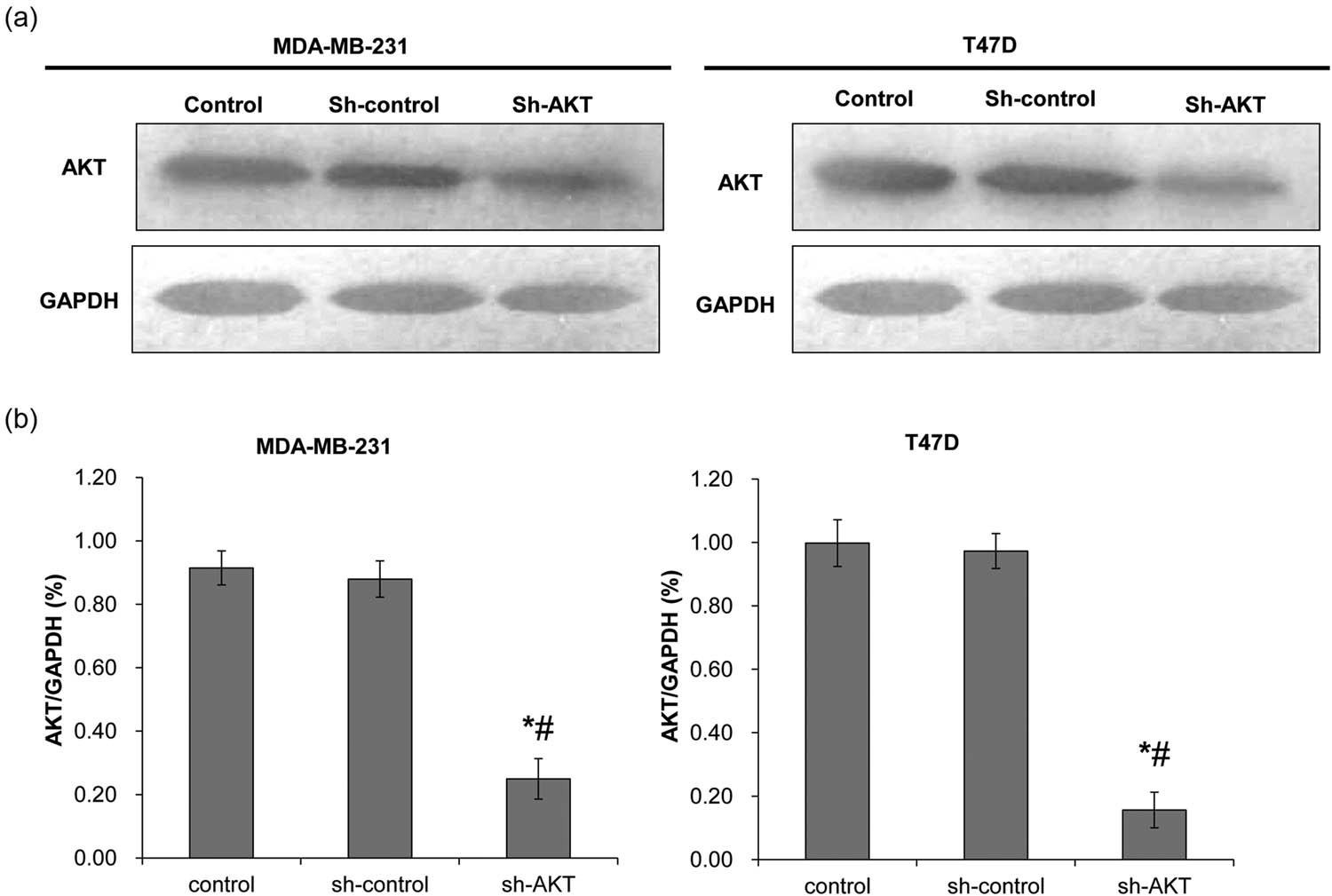
Determination of lentiviral transfection efficiency. Western blot was used to detect AKT protein expression after shRNA transfection. The control group was not transfected with shRNA; the sh-control group was transfected with void vector; and the sh-AKT group was transfected with shRNA of AKT. (a) Representative Western blot results. (b) The quantified expression of AKT. Left: MDA-MB-231 cells; right: T47D cells. *P < 0.05, compared with the control group; # P < 0.05, compared with the sh-control group.
3.2 Regulation of BRCA1 subcellular localization by the key molecules of the PI3K signaling pathway
To determine the subcellular localization of BRCA1, immunofluorescence was performed. As shown in Figure 2, in MDA-MB-231 cells, BRCA1 was mainly located in the cytoplasm, while only the HRG group had a very small number of BRCA1 located in the nucleus, while the other groups of cells had no obvious BRCA1 in the nucleus. In T47D cells, a small amount of BRCA1 was located in the nucleus in the Control group. In the HRG group, BRCA1 was mainly located in the nucleus. In the HRG + LY294002 group, a small amount of BRCA1 was located in the nucleus, while the remaining BRCA1 was mainly located on the edge of nuclear membranes. In the HRG + ShRNA group, BRCA1 was mainly located in the cytoplasm, and only a small part of BRCA1 was located in the nucleus (Figure 2). These results indicate that the nuclear localization of BRCA1 in both MDA-MB-231 cells and T47D cells is increased after PI3K activation, while decreased after PI3K inhibition, especially after the silencing of Akt. Moreover, the nuclear localization of BRCA1 in T47D breast cancer cells is higher than that in the MDA-MB-231 cells.

The subcellular localization of BRCA1 was observed under a fluorescence microscope (400×). The Control group was untreated. The HRG group was treated with 30 ng/mL HRG for 24 h to activate the PI3K signaling pathway. The HRG + LY294002 group was treated with 30 ng/mL HRG for 24 h to activate the PI3K signaling pathway and then treated with 25 μM LY294002 for 48 h to inhibit the PI3K pathway. The HRG + ShRNA group was treated with 30 ng/mL HRG for 24 h to activate the PI3K signaling pathway and then transfected with shRNA to silence Akt. The red arrows indicate the localization of BRCA1 protein in cell nuclei, while the yellow arrows indicate that on the edge of nuclear membranes.
3.3 The effect of the PI3K signaling pathway on the apoptosis of different types of breast cancer cells
To detect cell apoptosis, flow cytometry was performed. The representative flow cytometry results are shown in Figure 3a. The early and total apoptosis rate was calculated, respectively. As shown in Figure 3b, for both MDA-MB-231 and T47D cells, the early apoptosis rates in the HRG group were significantly higher than those in the other groups (P < 0.05), whereas there was no significant difference between the other groups (P > 0.05). In MDA-MB-231 cells, the percentage of total apoptotic cells was not significantly different among all the groups (P > 0.05) (Figure 3c). In T47D cells, the apoptosis rate of the Control group was significantly lower than that in other groups (P < 0.01). However, there was no significant difference among the HRG group, HRG + LY294002 group, and HRG + ShRNA group (P > 0.05) (Figure 3c). This indicates that the activation of the PI3K signaling pathway can significantly increase the early apoptosis rate of cells, while the activation or inhibition of the PI3K signaling pathway has no effect on the total apoptosis rate of MDA-MB-231 cells, but can significantly increase that of the T47D cells.
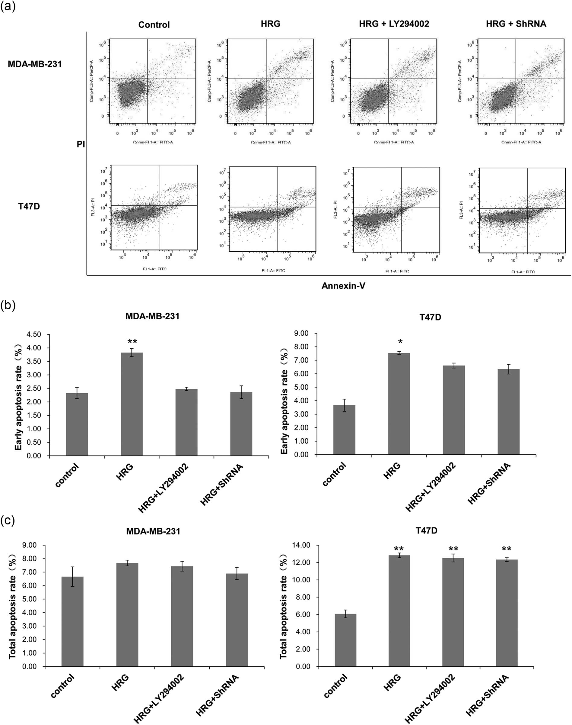
The cell apoptosis rate of different groups detected by flow cytometry. Cell treatment and grouping were described above. (a) Representative flow cytometry results. (b) The early cell apoptosis rates and (c) the total apoptosis rates of MDA-MB-231 and T47D cells. All experiments were performed at least three times. *P < 0.05, **P < 0.01, compared with the control group.
3.4 The phosphorylation and mRNA expressions of Akt and BRCA1 in breast cancer cells
The protein levels of Akt and BRCA1 and their phosphorylation in MDA-MB-231 cells were detected with Western blot. After PI3K activation by HRG, the phosphorylation of Akt and BRCA1 increased significantly in MDA-MB-231 cells (P < 0.05) compared with the Control group and decreased significantly after the inhibition of PI3K pathway or silencing of Akt (P < 0.01) (Figure 4a–e). There was no significant difference in the phosphorylation of Akt and BRCA1 between the HRG + LY294002 and the HRG + ShRNA groups (P > 0.05). This suggests that HRG can promote the phosphorylation of BRCA1 by increasing the phosphorylation of Akt in the PI3K/Akt signaling pathway.

The protein and mRNA expression of Akt and BRCA1 in MDA-MB-231 cells. Cell treatment and grouping were described above. The protein and mRNA expressions were detected with Western blot and RT-PCR, respectively. (a) The protein expressions of Akt and BRCA1 and their phosphorylated counterparts. The protein expressions of (b) Akt and (c) BRCA1 were quantified in relative to that of GAPDH. (d) The percentage of p-Akt in the total Akt protein. (e) The percentage of p-BRCA1 in the total BRCA1 protein. (f) The relative expression of Akt mRNA and (g) the relative expression of BRCA1 mRNA were also detected. All experiments were performed at least three times. *P < 0.05, compared with the control group; # P < 0.05, compared with the HRG group.
The mRNA expression levels of BRCA1 and AKT in MDA-MB-231 were detected by RT-PCR. The results showed that the expression of Akt mRNA (Figure 4f) in MDA-MB-231 cells had no significant changes after the activation of PI3K (P > 0.05) when compared with that in the Control group, but the expression of BRCA1 mRNA significantly increased (P < 0.05) (Figure 4g). Both Akt and BRCA1 expressions significantly decreased after the PI3K pathway was inhibited or silenced (P < 0.05) (Figure 4f and g). This indicates that the phosphorylation and mRNA changing trends of Akt and BRCA1 in the TNBC MDA-MB-231 cells are similar after activation or inhibition of the PI3K signaling pathway.
Compared with the Control group, the phosphorylation of Akt (Figure 5a, b and d) had no significant change when PI3K was activated in T47D cells (P > 0.05), but the phosphorylation of BRCA1 (Figure 5a, c and e) increased significantly (P < 0.05). The silencing of Akt by shRNA decreased the phosphorylation of BRCA1 significantly (P < 0.05) (Figure 5e) when compared with the HRG + LY294002 group. The mRNA expressions of Akt (Figure 5f) and BRCA1 (Figure 5g) both significantly increased in the HRG group when PI3K pathway was activated, whereas both significantly decreased after the PI3K pathway was inhibited or silenced in T47D cells of the HRG + LY294002 group and the HRG + ShRNA group (P < 0.05). This indicates that the phosphorylation and mRNA changing trends of Akt and BRCA1 in the hormone-sensitive T47D cells are similar after activation or inhibition of the PI3K signaling pathway. The above results suggest that the phosphorylation of BRCA1 in MDA-MB-231 and T47D cells are both regulated by the PI3K pathway.

The protein and mRNA expression of Akt and BRCA1 in T47D cells. Cell treatment and grouping were described above. The protein and mRNA expressions were detected with Western blot and RT-PCR, respectively. (a) The protein expressions of Akt and BRCA1 and their phosphorylated counterparts. The protein expressions of (b) Akt and (c) BRCA1 were quantified in relative to that of GAPDH. (d) The percentage of p-Akt in the total Akt protein. (e) The percentage of p-BRCA1 in the total BRCA1 protein. (f) The relative expression of Akt mRNA and (g) the relative expression of BRCA1 mRNA were also detected. All experiments were performed at least three times. *P < 0.05, compared with the control group; # P < 0.05, compared with the HRG group.
4 Discussion
BRCA1 is a tumor suppressor gene that is closely related to breast cancer, ovarian cancer, and other hormone-related cancers [27] and plays a negative regulatory role in tumor growth. It also plays an important role in regulating cell damage repair, cell cycle, and cell apoptosis. However, there are still some controversies concerning the investigation of breast cancer treatment based on BRCA1 mutations [28]. The PI3K signaling pathway is a common signaling pathway mainly involved in biological behaviors such as substance metabolism regulation, cell proliferation, and survival [29,30,31]. The abnormal activation of the PI3K signaling pathway is associated with many cancers such as liver cancer, colon cancer, bladder cancer, and breast cancer [32]. PI3K signaling pathway is one of the main signaling pathways in breast cancer. Altiok et al. [21] showed that the PI3K signaling pathway was involved in phosphorylation, nuclear transportation, and subcellular localization of BRCA1. In this study, it was found that the activation of the PI3K signaling pathway could promote the expression and phosphorylation of BRCA1 and markedly increased its nuclear localization in T47D cells. On the contrary, after the PI3K signaling pathway was inhibited, BRCA1 nuclear localization was significantly reduced in T47D cells, but no obvious changes in nuclear localization were observed in MDA-MB-231 cells. Further experimentation is needed to verify the present results. These results demonstrate that the activation of the PI3K signaling pathway in T47D cells can promote BRCA1 phosphorylation and increase its nuclear localization, which is consistent with previous studies [33,34]. However, the phosphorylation of BRCA1 was increased in MDA-MB-231, but its BRCA1 nuclear localization was not increased. Therefore, whether there are other mechanisms regulating BRCA1 nuclear translocation in MDA-MB-231 cells needs to be further studied. A possible reason may be that the PI3K signaling pathway is activated by extracellular or intracellular abnormal changes, and the expression of the downstream molecules in cells is changed through a wide range of external and internal stimulation [35,36].
The PI3K signaling pathway plays an anti-apoptotic and pro-proliferative role in cells [31,32]. Activated Akt can downregulate the transcription of pro-apoptotic genes, reduce cell apoptosis, and promote cell survival [37]. Akt can also activate a variety of downstream molecules such as mTOR, Bad, ikB, caspase-9, MMP-9, COX-2, etc., thereby stimulating cell differentiation and proliferation and inhibiting apoptosis [38,39,40]. This study found that PI3K inhibition or Akt knockdown significantly increased cell apoptosis in T47D cells, which is consistent with a previous study [41]. However, the apoptotic rate of T47D cells significantly increased after the activation of PI3K, which is possibly due to the tumor suppressor function of phosphorylated BRCA1, leading to an increased apoptosis rate of cells. In MDA-MB-231 cells, there was no significant change in the apoptosis rate, which may be due to the failure of the nuclear translocation of the BRCA1 protein. Akt is the main target gene of PI3K, and its silencing can cause a decrease of BRCA1 protein in different types of breast cancer cells [21,22,42], suggesting that PI3K pathway can regulate the expression level of BRCA1. In the present study, the expression levels of BRCA1 and Akt showed a consistent changing trend.
5 Conclusions
This study found that the regulation of PI3K signaling pathway on BRCA1 subcellular localization was different between MDA-MB-231 (TNBC type) and T47D (hormone-sensitive type) cells. After the activation or inhibition of the PI3K pathway, the changes in nuclear localization of BRCA1, and the rate of apoptosis in T47D cells were more significant when compared with those in MDA-MB-231 cells. The underlying mechanism needs to be further studied. This study provides some clues in the treatment of different types of breast cancer.
Acknowledgments
This work was supported by the National Natural Science Foundation of China (Grant No. 81360391).
-
Conflict of interest: The authors state no conflict of interest.
-
Data availability statement: The datasets generated during and/or analyzed during the current study are available from the corresponding author on reasonable request.
References
[1] Siegel R, Naishadham D, Jemal A. Cancer statistics, 2013. CA Cancer J Clin. 2013;63(1):11–30.10.3322/caac.21166Search in Google Scholar PubMed
[2] Cox DG, Simard J, Sinnett D, Hamdi Y, Soucy P, Ouimet M, et al. Common variants of the BRCA1 wild-type allele modify the risk of breast cancer in BRCA1 mutation carriers. Hum Mol Genet. 2011;20(23):4732–47.10.1093/hmg/ddr388Search in Google Scholar PubMed PubMed Central
[3] Cortez D, Wang Y, Qin J, Elledge SJ. Requirement of ATM-dependent phosphorylation of brca1 in the DNA damage response to double-strand breaks. Science. 1999;286(5442):1162–6.10.1126/science.286.5442.1162Search in Google Scholar PubMed
[4] Shao N, Chai YL, Shyam E, Reddy P, Rao VN. Induction of apoptosis by the tumor suppressor protein BRCA1. Oncogene. 1996;13(1):1–7.Search in Google Scholar
[5] Yan Y, Haas JP, Kim M, Sgagias MK, Cowan KH. BRCA1-induced apoptosis involves inactivation of ERK1/2 activities. J Biol Chem. 2002;277(36):33422–30.10.1074/jbc.M201147200Search in Google Scholar PubMed
[6] Zheng J, Qin W, Jiao D, Ren J, Wei M, Shi S, et al. Knockdown of COUP-TFII inhibits cell proliferation and induces apoptosis through upregulating BRCA1 in renal cell carcinoma cells. Int J Cancer. 2016;139(7):1574–85.10.1002/ijc.30193Search in Google Scholar PubMed
[7] Jiao W, Lin HM, Datta J, Braunschweig T, Chung JY, Hewitt SM, et al. Aberrant nucleocytoplasmic localization of the retinoblastoma tumor suppressor protein in human cancer correlates with moderate/poor tumor differentiation. Oncogene. 2008;27(22):3156–64.10.1038/sj.onc.1210970Search in Google Scholar PubMed
[8] Rakha EA, El-Sheikh SE, Kandil MA, El-Sayed MA, Green AR, Ellis IO. Expression of BRCA1 protein in breast cancer and its prognostic significance. Hum Pathol. 2008;39(6):857–65.10.1016/j.humpath.2007.10.011Search in Google Scholar PubMed
[9] Nanda R. “Targeting” triple-negative breast cancer: the lessons learned from BRCA1-associated breast cancers. Semin Oncol. 2011;38(2):254–62.10.1053/j.seminoncol.2011.01.007Search in Google Scholar PubMed
[10] Chen Y, Chen CF, Riley DJ, Allred DC, Chen PL, Von Hoff D, et al. Aberrant subcellular localization of BRCA1 in breast cancer. Science. 1995;270(5237):789–91.10.1126/science.270.5237.789Search in Google Scholar PubMed
[11] Coene ED, Hollinshead MS, Waeytens AA, Schelfhout VRJ, Eechaute WP, Shaw MK, et al. Phosphorylated BRCA1 is predominantly located in the nucleus and mitochondria. Mol Biol Cell. 2005;16(2):997–1010.10.1091/mbc.e04-10-0895Search in Google Scholar PubMed PubMed Central
[12] Maniccia AW, Lewis C, Begum N, Xu J, Cui J, Chipitsyna G, et al. Mitochondrial localization, ELK-1 transcriptional regulation and growth inhibitory functions of BRCA1, BRCA1a, and BRCA1b proteins. J Cell Physiol. 2009 Jun;219(3):634–41.10.1002/jcp.21708Search in Google Scholar PubMed PubMed Central
[13] Xu J, Song F, Jin T, Qin J, Wu J, Wang M, et al. Prognostic values of Notch receptors in breast cancer. Tumour Biol. 2016;37(2):1871–7.10.1007/s13277-015-3961-6Search in Google Scholar PubMed
[14] Orzechowska M, Jedroszka D, Bednarek AK. Common profiles of Notch signaling differentiate disease-free survival in luminal type A and triple negative breast cancer. Oncotarget. 2017;8(4):6013–32.10.18632/oncotarget.13451Search in Google Scholar PubMed PubMed Central
[15] Bleckmann A, Conradi LC, Menck K, Schmick NA, Schubert A, Rietkötter E, et al. β-catenin-independent WNT signaling and Ki67 in contrast to the estrogen receptor status are prognostic and associated with poor prognosis in breast cancer liver metastases. Clin Exp Metastasis. 2016;33(4):309–23.10.1007/s10585-016-9780-3Search in Google Scholar PubMed PubMed Central
[16] Xu J, Prosperi JR, Choudhury N, Olopade OI, Goss KH. β-catenin is required for the tumorigenic behavior of triple-negative breast cancer cells. PLoS One. 2015;10(2):e0117097.10.1371/journal.pone.0117097Search in Google Scholar PubMed PubMed Central
[17] Harn M, Moon A. Inflammatory and microenvironmental factors involved in breast cancer progression. Arch Pharm Res. 2013;36(12):1419–31.10.1007/s12272-013-0271-7Search in Google Scholar PubMed
[18] Zhang B, Wang Q, Fu C, Jiang C, Ma S. Exploration of the immune-related signature and immune infiltration analysis for breast ductal and lobular carcinoma. Ann Transl Med. 2019;7(23):730.10.21037/atm.2019.11.117Search in Google Scholar PubMed PubMed Central
[19] Jin Y, Feng SJ, Qiu S, Shao N, Zheng JH. LncRNA MALAT1 promotes proliferation and metastasis in epithelial ovarian cancer via the PI3K-AKT pathway. Eur Rev Med Pharmacol Sci. 2017;21:3176–84.Search in Google Scholar
[20] Chen H, Zhou L, Wu X, Li R, Wen J, Sha J, et al. The PI3K/AKT pathway in the pathogenesis of prostate cancer. Front Biosci (Landmark Ed). 2016;21:1084–91.10.2741/4443Search in Google Scholar PubMed
[21] Altiok S, Batt D, Altiok N, Papautsky A, Downward J, Roberts TM, et al. Heregulin induces phosphorylation of BRCA1 through phosphatidylinositol 3-Kinase/AKT in breast cancer cells. J Biol Chem. 1999;274(45):32274–8.10.1074/jbc.274.45.32274Search in Google Scholar PubMed
[22] Hinton CV, Fitzgerald LD, Thompsonab ME. Phosphatidylinositol 3-kinase/Akt signaling enhances nuclear localization and transcriptional activity of BRCA1. Exp Cell Res. 2007;313(9):1735–44.10.1016/j.yexcr.2007.03.008Search in Google Scholar PubMed PubMed Central
[23] Zhang N, Sun G, Ma B. Differential expression of breast cancer susceptibility gene 1 mRNA and protein in sporadic breast cancer. Chinese J Exp Surg. 2016;33(4):884–7.Search in Google Scholar
[24] Prajzendanc K, Domagała P, Hybiak J, Ryś J, Huzarski T, Szwiec M. et al. BRCA1 promoter methylation in peripheral blood is associated with the risk of triple-negative breast cancer. Int J Cancer. 2020 Mar 1;146(5):1293–8.10.1002/ijc.32655Search in Google Scholar PubMed
[25] Radenkovic S, Konjevic G, Isakovic A, Stevanovic T, Gopcevic K, Jurisic V. HER2-positive breast cancer patients: correlation between mammographic and pathological findings. Radiat Prot Dosimetry. 2014 Nov;162(1–2):125–8.10.1093/rpd/ncu243Search in Google Scholar PubMed
[26] Vangangelt KMH, Green AR, Heemskerk IMF, Cohen D, van Pelt GW, Sobral-Leite M, et al. The prognostic value of the tumor-stroma ratio is most discriminative in patients with grade III or triple-negative breast cancer. Int J Cancer. 2020;146(8):2296–304.10.1002/ijc.32857Search in Google Scholar PubMed PubMed Central
[27] Kast K, Rhiem K, Wappenschmidt B, Hahnen E, Hauke J, Bluemcke B, et al. Prevalence of BRCA1/2 germline mutations in 21401 families with breast and ovarian cancer. J Med Genet. 2016;53:465–71.10.1136/jmedgenet-2015-103672Search in Google Scholar PubMed
[28] Co M, Liu T, Leung J, Li CH, Tse T, Wong M, et al. Breast conserving surgery for BRCA mutation carriers – a systematic review. Clin Breast Cancer. 2020 Jun;20(3):e244–50, 2019 Aug 22. pii: S1526-8209(19)30661-5.10.1016/j.clbc.2019.07.014Search in Google Scholar PubMed
[29] Pascual J, Turner NC. Targeting the PI3-kinase pathway in triple negative breast cancer. Ann Oncol. 2019.10.1093/annonc/mdz133Search in Google Scholar PubMed
[30] Guerrero-Zotano A, Mayer IA, Arteaga CL. PI3K/AKT/mTOR: role in breast cancer progression, drug resistance, and treatment. Cancer Metastasis Rev. 2016;35(4):515515.10.1007/s10555-016-9637-xSearch in Google Scholar PubMed
[31] Fruman DA, Chiu H, Hopkins BD, Bagrodia S, Cantley LC, Abraham RT. The PI3K pathway in human disease. Cell. 2017;170(4):605–35.10.1016/j.cell.2017.07.029Search in Google Scholar PubMed PubMed Central
[32] Madsen RR, Vanhaesebroeck B, Semple RK. Cancer-associated PIK3CA mutations in overgrowth disorders. Trends Mol Med. 2018;24(10):856–70.10.1016/j.molmed.2018.08.003Search in Google Scholar PubMed PubMed Central
[33] Kim HJ, Lee SY, Kim CY, Kim YH, Ju W, Kim SC. Subcellular localization of FOXO3a as a potential biomarker of response to combined treatment with inhibitors of PI3K and autophagy in PIK3CA-mutant cancer cells. Oncotarget. 2016;8(4):6608.10.18632/oncotarget.14245Search in Google Scholar PubMed PubMed Central
[34] Yin P. Molecular Mechanism of Cystathionine-γ-lyase Gene Expression Regulated by P13K/Akt Pathway and its Biological Functions in Human Hepatocellular Carcinoma Cell Lines[D]. Shanghai: Fudan University; 2012.Search in Google Scholar
[35] Li L, Bhatia M, Zhu YZ, Zhu YC, Ramnath RD, Wang ZJ. Hydrogen sulfide is a novel mediator of lipopolysaccharide-induced inflammation in the mouse. Feder Am Soc Exp Biol. 2005;19(9):1196–8.10.1096/fj.04-3583fjeSearch in Google Scholar PubMed
[36] Li T, Zhao B, Wang C, Wang H, Liu Z, Li W, et al. Regulatory effects of hydrogen sulfide on IL-6, IL-8 and IL-10 levels in the plasma and pulmonary tissue of rats with acute lung injury. Exp Biol Med (Maywood). 2008;233(9):1081–7.10.3181/0712-RM-354Search in Google Scholar PubMed
[37] Zhao GX, Pan H, Ouyang DY, He XH. The critical molecular interconnections in regulating apoptosis and autophagy. Ann Med. 2015;47:305–15.10.3109/07853890.2015.1040831Search in Google Scholar PubMed
[38] Nitulescu GM, Margina D, Juzenas P, Peng Q, Olaru OT, Saloustros E, et al. Akt inhibitors in cancer treatment: the long journey from drug discovery to clinical use (review). Int J Oncol. 2016;48:869–85.10.3892/ijo.2015.3306Search in Google Scholar PubMed PubMed Central
[39] Ouyang ZH, Wang WJ, Yan YG, Wang B, Lv GH. The PI3K/Akt pathway: a critical player in intervertebral disc degeneration. Oncotarget. 2017;8:57870–81.10.18632/oncotarget.18628Search in Google Scholar PubMed PubMed Central
[40] Huck BR, Mochalkin I. Recent progress towards clinically relevant ATP-competitive Akt inhibitors. Bioorg Med Chem Lett. 2017;27:2838–48.10.1016/j.bmcl.2017.04.090Search in Google Scholar PubMed
[41] Lu H, Yu L, Zhen H, Liu R, Yue ZJ. BMP9 inhibits inflammatory response and apoptosis of degenerative nucleus pulposus cells through activating PI3K/Akt signaling pathway[J]. J Third Mil Med Univ. 2016;38(18):2047–52.Search in Google Scholar
[42] Baek HJ, Kim SE, Kim JK, Shin DH, Kim TH, Kim KG, et al. Inhibition of AKT suppresses the initiation and progression of BRCA1-associated mammary tumors. Int J Biol Sci. 2018;14(13):1769–81.10.7150/ijbs.29242Search in Google Scholar PubMed PubMed Central
© 2020 Bin Ma et al., published by De Gruyter
This work is licensed under the Creative Commons Attribution 4.0 International License.
Articles in the same Issue
- Plant Sciences
- Dependence of the heterosis effect on genetic distance, determined using various molecular markers
- Plant Growth Promoting Rhizobacteria (PGPR) Regulated Phyto and Microbial Beneficial Protein Interactions
- Role of strigolactones: Signalling and crosstalk with other phytohormones
- An efficient protocol for regenerating shoots from paper mulberry (Broussonetia papyrifera) leaf explants
- Functional divergence and adaptive selection of KNOX gene family in plants
- In silico identification of Capsicum type III polyketide synthase genes and expression patterns in Capsicum annuum
- In vitro induction and characterisation of tetraploid drumstick tree (Moringa oleifera Lam.)
- CRISPR/Cas9 or prime editing? – It depends on…
- Study on the optimal antagonistic effect of a bacterial complex against Monilinia fructicola in peach
- Natural variation in stress response induced by low CO2 in Arabidopsis thaliana
- The complete mitogenome sequence of the coral lily (Lilium pumilum) and the Lanzhou lily (Lilium davidii) in China
- Ecology and Environmental Sciences
- Use of phosphatase and dehydrogenase activities in the assessment of calcium peroxide and citric acid effects in soil contaminated with petrol
- Analysis of ethanol dehydration using membrane separation processes
- Activity of Vip3Aa1 against Periplaneta americana
- Thermostable cellulase biosynthesis from Paenibacillus alvei and its utilization in lactic acid production by simultaneous saccharification and fermentation
- Spatiotemporal dynamics of terrestrial invertebrate assemblages in the riparian zone of the Wewe river, Ashanti region, Ghana
- Antifungal activity of selected volatile essential oils against Penicillium sp.
- Toxic effect of three imidazole ionic liquids on two terrestrial plants
- Biosurfactant production by a Bacillus megaterium strain
- Distribution and density of Lutraria rhynchaena Jonas, 1844 relate to sediment while reproduction shows multiple peaks per year in Cat Ba-Ha Long Bay, Vietnam
- Biomedical Sciences
- Treatment of Epilepsy Associated with Common Chromosomal Developmental Diseases
- A Mouse Model for Studying Stem Cell Effects on Regeneration of Hair Follicle Outer Root Sheaths
- Morphine modulates hippocampal neurogenesis and contextual memory extinction via miR-34c/Notch1 pathway in male ICR mice
- Composition, Anticholinesterase and Antipedicular Activities of Satureja capitata L. Volatile Oil
- Weight loss may be unrelated to dietary intake in the imiquimod-induced plaque psoriasis mice model
- Construction of recombinant lentiviral vector containing human stem cell leukemia gene and its expression in interstitial cells of cajal
- Knockdown of lncRNA KCNQ1OT1 inhibits glioma progression by regulating miR-338-3p/RRM2
- Protective effect of asiaticoside on radiation-induced proliferation inhibition and DNA damage of fibroblasts and mice death
- Prevalence of dyslipidemia in Tibetan monks from Gansu Province, Northwest China
- Sevoflurane inhibits proliferation, invasion, but enhances apoptosis of lung cancer cells by Wnt/β-catenin signaling via regulating lncRNA PCAT6/ miR-326 axis
- MiR-542-3p suppresses neuroblastoma cell proliferation and invasion by downregulation of KDM1A and ZNF346
- Calcium Phosphate Cement Causes Nucleus Pulposus Cell Degeneration Through the ERK Signaling Pathway
- Human Dental Pulp Stem Cells Exhibit Osteogenic Differentiation Potential
- MiR-489-3p inhibits cell proliferation, migration, and invasion, and induces apoptosis, by targeting the BDNF-mediated PI3K/AKT pathway in glioblastoma
- Long non-coding RNA TUG1 knockdown hinders the tumorigenesis of multiple myeloma by regulating the microRNA-34a-5p/NOTCH1 signaling pathway
- Large Brunner’s gland adenoma of the duodenum for almost 10 years
- Neurotrophin-3 accelerates reendothelialization through inducing EPC mobilization and homing
- Hepatoprotective effects of chamazulene against alcohol-induced liver damage by alleviation of oxidative stress in rat models
- FXYD6 overexpression in HBV-related hepatocellular carcinoma with cirrhosis
- Risk factors for elevated serum colorectal cancer markers in patients with type 2 diabetes mellitus
- Effect of hepatic sympathetic nerve removal on energy metabolism in an animal model of cognitive impairment and its relationship to Glut2 expression
- Progress in research on the role of fibrinogen in lung cancer
- Advanced glycation end product levels were correlated with inflammation and carotid atherosclerosis in type 2 diabetes patients
- MiR-223-3p regulates cell viability, migration, invasion, and apoptosis of non-small cell lung cancer cells by targeting RHOB
- Knockdown of DDX46 inhibits trophoblast cell proliferation and migration through the PI3K/Akt/mTOR signaling pathway in preeclampsia
- Buformin suppresses osteosarcoma via targeting AMPK signaling pathway
- Effect of FibroScan test in antiviral therapy for HBV-infected patients with ALT <2 upper limit of normal
- LncRNA SNHG15 regulates osteosarcoma progression in vitro and in vivo via sponging miR-346 and regulating TRAF4 expression
- LINC00202 promotes retinoblastoma progression by regulating cell proliferation, apoptosis, and aerobic glycolysis through miR-204-5p/HMGCR axis
- Coexisting flavonoids and administration route effect on pharmacokinetics of Puerarin in MCAO rats
- GeneXpert Technology for the diagnosis of HIV-associated tuberculosis: Is scale-up worth it?
- Circ_001569 regulates FLOT2 expression to promote the proliferation, migration, invasion and EMT of osteosarcoma cells through sponging miR-185-5p
- Lnc-PICSAR contributes to cisplatin resistance by miR-485-5p/REV3L axis in cutaneous squamous cell carcinoma
- BRCA1 subcellular localization regulated by PI3K signaling pathway in triple-negative breast cancer MDA-MB-231 cells and hormone-sensitive T47D cells
- MYL6B drives the capabilities of proliferation, invasion, and migration in rectal adenocarcinoma through the EMT process
- Inhibition of lncRNA LINC00461/miR-216a/aquaporin 4 pathway suppresses cell proliferation, migration, invasion, and chemoresistance in glioma
- Upregulation of miR-150-5p alleviates LPS-induced inflammatory response and apoptosis of RAW264.7 macrophages by targeting Notch1
- Long non-coding RNA LINC00704 promotes cell proliferation, migration, and invasion in papillary thyroid carcinoma via miR-204-5p/HMGB1 axis
- Neuroanatomy of melanocortin-4 receptor pathway in the mouse brain
- Lipopolysaccharides promote pulmonary fibrosis in silicosis through the aggravation of apoptosis and inflammation in alveolar macrophages
- Influences of advanced glycosylation end products on the inner blood–retinal barrier in a co-culture cell model in vitro
- MiR-4328 inhibits proliferation, metastasis and induces apoptosis in keloid fibroblasts by targeting BCL2 expression
- Aberrant expression of microRNA-132-3p and microRNA-146a-5p in Parkinson’s disease patients
- Long non-coding RNA SNHG3 accelerates progression in glioma by modulating miR-384/HDGF axis
- Long non-coding RNA NEAT1 mediates MPTP/MPP+-induced apoptosis via regulating the miR-124/KLF4 axis in Parkinson’s disease
- PCR-detectable Candida DNA exists a short period in the blood of systemic candidiasis murine model
- CircHIPK3/miR-381-3p axis modulates proliferation, migration, and glycolysis of lung cancer cells by regulating the AKT/mTOR signaling pathway
- Reversine and herbal Xiang–Sha–Liu–Jun–Zi decoction ameliorate thioacetamide-induced hepatic injury by regulating the RelA/NF-κB/caspase signaling pathway
- Therapeutic effects of coronary granulocyte colony-stimulating factor on rats with chronic ischemic heart disease
- The effects of yam gruel on lowering fasted blood glucose in T2DM rats
- Circ_0084043 promotes cell proliferation and glycolysis but blocks cell apoptosis in melanoma via circ_0084043-miR-31-KLF3 axis
- CircSAMD4A contributes to cell doxorubicin resistance in osteosarcoma by regulating the miR-218-5p/KLF8 axis
- Relationship of FTO gene variations with NAFLD risk in Chinese men
- The prognostic and predictive value of platelet parameters in diabetic and nondiabetic patients with sudden sensorineural hearing loss
- LncRNA SNHG15 contributes to doxorubicin resistance of osteosarcoma cells through targeting the miR-381-3p/GFRA1 axis
- miR-339-3p regulated acute pancreatitis induced by caerulein through targeting TNF receptor-associated factor 3 in AR42J cells
- LncRNA RP1-85F18.6 affects osteoblast cells by regulating the cell cycle
- MiR-203-3p inhibits the oxidative stress, inflammatory responses and apoptosis of mice podocytes induced by high glucose through regulating Sema3A expression
- MiR-30c-5p/ROCK2 axis regulates cell proliferation, apoptosis and EMT via the PI3K/AKT signaling pathway in HG-induced HK-2 cells
- CTRP9 protects against MIA-induced inflammation and knee cartilage damage by deactivating the MAPK/NF-κB pathway in rats with osteoarthritis
- Relationship between hemodynamic parameters and portal venous pressure in cirrhosis patients with portal hypertension
- Long noncoding RNA FTX ameliorates hydrogen peroxide-induced cardiomyocyte injury by regulating the miR-150/KLF13 axis
- Ropivacaine inhibits proliferation, migration, and invasion while inducing apoptosis of glioma cells by regulating the SNHG16/miR-424-5p axis
- CD11b is involved in coxsackievirus B3-induced viral myocarditis in mice by inducing Th17 cells
- Decitabine shows anti-acute myeloid leukemia potential via regulating the miR-212-5p/CCNT2 axis
- Testosterone aggravates cerebral vascular injury by reducing plasma HDL levels
- Bioengineering and Biotechnology
- PL/Vancomycin/Nano-hydroxyapatite Sustained-release Material to Treat Infectious Bone Defect
- The thickness of surface grafting layer on bio-materials directly mediates the immuno-reacitivity of macrophages in vitro
- Silver nanoparticles: synthesis, characterisation and biomedical applications
- Food Science
- Bread making potential of Triticum aestivum and Triticum spelta species
- Modeling the effect of heat treatment on fatty acid composition in home-made olive oil preparations
- Effect of addition of dried potato pulp on selected quality characteristics of shortcrust pastry cookies
- Preparation of konjac oligoglucomannans with different molecular weights and their in vitro and in vivo antioxidant activities
- Animal Sciences
- Changes in the fecal microbiome of the Yangtze finless porpoise during a short-term therapeutic treatment
- Agriculture
- Influence of inoculation with Lactobacillus on fermentation, production of 1,2-propanediol and 1-propanol as well as Maize silage aerobic stability
- Application of extrusion-cooking technology in hatchery waste management
- In-field screening for host plant resistance to Delia radicum and Brevicoryne brassicae within selected rapeseed cultivars and new interspecific hybrids
- Studying of the promotion mechanism of Bacillus subtilis QM3 on wheat seed germination based on β-amylase
- Rapid visual detection of FecB gene expression in sheep
- Effects of Bacillus megaterium on growth performance, serum biochemical parameters, antioxidant capacity, and immune function in suckling calves
- Effects of center pivot sprinkler fertigation on the yield of continuously cropped soybean
- Special Issue On New Approach To Obtain Bioactive Compounds And New Metabolites From Agro-Industrial By-Products
- Technological and antioxidant properties of proteins obtained from waste potato juice
- The aspects of microbial biomass use in the utilization of selected waste from the agro-food industry
- Special Issue on Computing and Artificial Techniques for Life Science Applications - Part I
- Automatic detection and segmentation of adenomatous colorectal polyps during colonoscopy using Mask R-CNN
- The impedance analysis of small intestine fusion by pulse source
- Errata
- Erratum to “Diagnostic performance of serum CK-MB, TNF-α and hs-CRP in children with viral myocarditis”
- Erratum to “MYL6B drives the capabilities of proliferation, invasion, and migration in rectal adenocarcinoma through the EMT process”
- Erratum to “Thermostable cellulase biosynthesis from Paenibacillus alvei and its utilization in lactic acid production by simultaneous saccharification and fermentation”