LncRNA SNHG15 regulates osteosarcoma progression in vitro and in vivo via sponging miR-346 and regulating TRAF4 expression
-
Xuewu Chen


Abstract
Osteosarcoma (OS) is a common primary malignant bone tumor around the world. It has been reported that long noncoding RNAs (lncRNAs) take part in diverse pathological processes of OS; however, the mechanism remains unknown. This study aimed to uncover the profile of lncRNA small nucleolar RNA host gene 15 (SNHG15), its biological function, and its potential involvement in the mechanism of OS progression in vitro and in vivo. The expression of SNHG15 and TRAF4 was promoted in OS tissues opposite for that of miR-346. The silencing of SNHG15 limited the proliferation, invasion, and enhanced apoptosis of SaoS2 and HOS cells. Moreover, the putative binding sites between miR-346 and SNHG15 or TRAF4 were predicted by starBase and Targetscan software online, individually. Also, miR-346 deletion reversed the positive effects of SNHG15 elimination on proliferation, apoptosis, and invasion in cells. In addition, the upregulation of TRAF4 disrupted the biofunctional results from miR-346 promotion subsequently. Finally, SNHG15 knockdown repressed OS tumor growth in a xenograft tumor model. SNHG15 enhanced the progression of OS by regulating the miR-346/TRAF4 axis in vitro and in vivo.
1 Introduction
Osteosarcoma (OS) is the most common primary malignant bone tumor in children and adolescents [1], which was characterized by strong invasiveness and early metastasis [2]. Although chemotherapy regimens and neoadjuvant techniques have improved, approximately 50% of all patients have metastases at the time of diagnosis, which results in a high mortality rate [3]. Therefore, exploring the mechanism of OS is of great significance for understanding the progress of the disease and for finding potential therapeutic targets.
Long noncoding RNAs (lncRNAs) are a category of long RNAs (more than 200 nucleotides (nts) in length) without translation capacity that can affect gene expression at the transcriptional stage [4]. Emerging evidence suggests that lncRNAs act as functional regulators in tumorigenesis of cancers such as papillary thyroid carcinoma [5], gastric cancer [6], colorectal cancer [7], hepatocellular carcinoma [8], and pancreatic adenocarcinoma [9]. Extensive research indicates that the disordered expression of lncRNA is found in OS [10,11]. Small nucleolar RNA host gene 15 (SNHG15), located on chromosome 7p13, is reported to be involved in the response to environmental stressors [12,13]. Recently, increasing evidence suggests that lncRNA SNHG15 is highly expressed in OS tissues and cells and regulates a range of processes, including proliferation, invasion, migration, and even autophagy [14], but the role of SNHG15 in OS has not been investigated in depth.
Numerous studies have shown that lncRNA can act as a molecular sponge in combination with miRNA to regulate the development of cancer [15,16,17]. It was reported that SNHG15 could mediate the development of cancer by binding a variety of miRNAs [14,18,19]. However, the targeting relationship between SNHG15 and miRNA is still ambiguous in OA. Previous studies have shown that miR-346 plays a role in tumor supõpression in OS [20]. In this experiment, we used starBase software to predict the possible binding of SNHG15 to miR-346.
Tumor necrosis factor receptor-associated factors (TRAFs) were originally discovered as adaptor proteins that regulate the cell life and death [21]. Tumor necrosis factor receptor-associated factor 4 (TRAF4) is a unique member of the TRAF protein family [22]. A recent article showed that TRAF4, working as an oncogene, was highly expressed in OS [23]. In the current study, we used Targetscan software to predict that miR-346 may target TRAF4. However, the molecular mechanisms of SNHG15, miR-346, and TRAF4 in OS remained unclear.
In this study, we first investigated the expression level of SNHG15 on OS tissues and cells and examined the changes in the biological phenotype of OS cells by loss-function of SNHG15. To explain the mechanism, we proposed that SNHG15 functions as a sponge of miR-346, allowing enhanced TRAF4 expression, thereby boosting the development of OS. Our findings provided a certain basic value for clinical diagnosis.
2 Materials and methods
2.1 Clinical samples
Thirty pairs of specimens from OS patients and healthy volunteers were collected from Yijishan Hospital (The First Affiliated Hospital of Wannan Medical College). All samples were preserved at −80°C. The main inclusion criteria were as follows: (1) patients were pathologically diagnosed with OS and (2) OS was the primary tumor, without chemotherapy, surgery, or radiotherapy. The exclusion criteria were as follows: (1) history of other malignant disease, (2) recurrent or previously treated disease, and (3) patients who received treatment within the past 90 days before admission.
Informed consent: Informed consent has been obtained from all individuals included in this study.
Ethical approval: The research related to human use has been complied with all the relevant national regulations, institutional policies, and in accordance with the tenets of the Helsinki Declaration and has been approved by the Ethics Committee of Yijishan Hospital (The First Affiliated Hospital of Wannan Medical College).
2.2 Cell culture and transfection
Human OS cell lines (MG63, U2OS, SaoS2, and HOS) and human osteoblast cell lines (HFOB1.19) were obtained from Shanghai Innovation Biotechnology Co., Ltd (Shanghai, China) and were cultured with 1% penicillin/streptomycin (Beyotime Biotechnology Company, Shanghai, China) as previously described [24]. Small interference RNA (siRNA) targeting SNHG15 (si-SNHG15), SNHG15 overexpression plasmid (SNHG15), TRAF4 overexpression plasmid (TRAF4), miR-346 mimic (miR-346), miR-346 inhibitor (miR-346 inhibitor), and controls (si-control, vector, miR-control, inhibitor-control) were all obtained from GenePharm (Shanghai, China). Lipofectamine 3000 (Invitrogen, Carlsbad, CA, USA) kit was used for transfection according to the manufacturer’s instructions. The sequences were shown as follows: si-SNHG15 (Sense: 5′-ACGGTGGCAACGTGCGTGGCCA-3′, Anti-sense: 3′-GCCTGCAACGGTGCAAAT GCG-5′).
2.3 Quantitative real-time polymerase chain reaction (qRT-PCR)
Total RNA of cells was extracted through TRIzol reagent (Life Technologies Corporation, Carlsbad, CA, USA). Primer-Script one-step RT-PCR kit (Takara, Shiga, Japan) or miRNA Reverse Transcription kit (GeneCopoeia, FulenGen, China) was employed to synthesize the first-strand complementary DNA of miR-346 and TRAF4. The levels of SNHG15, miR-346, and TRAF4 were assessed via SYBR Premix Dimer Eraser Kit (Takara). The primer sequences used are as follows: SNHG15 forward (5′-CAACCATAGCGGTGCAACTGTGC-3′), SNHG15 reverse (5′-GGCTGAACCAAGTTGCAAGTCATG-3′), miR-346 forward (5′-CACGGATCCCTTGTCAGCAAGGAGTG-3′), miR-346 reverse (5′-CGGAATTCTAGGTTGGGAGCGAAGTG-3′), TRAF4 forward (5′-AGGAGTTCGTCTTTGACACCATC-3′), TRAF4 reverse (5′-CTTTGAATGGGCAGAGCACC-3′), U6 forward (5′-GCTTCGGCAGCACATATACTAAAAT-3′), U6 reverse (5′-CGCTTCACGAATTTGCGTGTCAT-3′), GAPDH forward (5′-GACTCATGACCACAGTCCATGC-3′), and GAPDH reverse (5′-AGAGGCAGGGATGATGTTCTG-3′). The expression levels of SNHG15, miR-346, and TRAF4 were calculated by the 2−ΔΔCt method, and glyceraldehyde-3-phosphate dehydrogenase (GAPDH) or U6 snRNA was viewed as an internal control for SNHG15, TRAF4, and miR-346.
2.3.1 Subcellular fractionation assay
In this assay, Nuclear/Cytosol Isolation Kit (BioVision, San Francisco, CA, USA) was applied to isolate nuclear and cytoplasm fractions in line with the supplier’s instructions. RNAs isolated from the nucleus and the cytoplasm were analyzed by the qRT-PCR assay. The expression levels of circNHSL1, U6 (nucleus control), and GAPDH (cytoplasm control) were examined separately.
2.4 3-(4,5-Dimethyl-2-thiazolyl)-2,5-diphenyl-2-H-tetrazolium bromide (MTT) assay
The proliferation capacity of transfected SaoS2 and HOS cells was evaluated by the MTT assay. Briefly, transfected SaoS2 and HOS cells (2.5 × 10³/well) were seeded into 96-well plates (Corning Inc., Corning, NY, USA) and maintained in an incubator with 5% CO2 at 37°C for 24, 48, and 72 h. Then, MTT (20 µL) from Sigma (St Louis, MO, USA) was replenished to each well and kept for 4 h. Following this, the supernatant of each well was discarded, and DMSO (150 µL, Sigma) was added for the dissolution of formazan crystals. Finally the Microplate Absorbance Reader (Thermo Fisher Scientific, Waltham, MA, USA) was executed for the assessment of the optical density value at 490 nm.
2.5 Flow cytometry
Cells were collected by digesting with pancreatin and centrifuging and then resuspended with 1× binding buffer after being washed with iced phosphate-buffered saline (PBS), followed by supplementation with Annexin V-fluorescein isothiocyanate propidium iodide (Annexin V-FITC/PI) kit (BD Pharmingen, San Diego, CA, USA) following the manufacturer’s instructions. Apoptotic cells were examined using the flow cytometer (BD Biosciences, Franklin Lakes, NJ, USA), and then, the apoptosis rate was calculated.
2.6 Western blot
RIPA buffer (Solarbio, Beijing, China) was used to isolate total proteins in cells, and proteins were quantified by a NanoDrop 3000 (Thermo Fisher Scientific). Sodium dodecyl sulfate-polyacrylamide gel electrophoresis (SDS-PAGE) was used to separate proteins, and then, proteins were transferred onto polyvinylidene fluoride (PVDF) membranes. Membranes were then blocked using skim milk for 2 h at 37°C and then incubated with primary antibodies at 4°C overnight. Following 2 h incubation with the secondary antibody [Goat Anti-Rabbit IgG H&L (HRP) (1:1,000; ab205718, Abcam, Cambridge, UK)], the chemiluminescence was performed using an ECL detection kit (Beyotime). The primary antibodies were as follows: anti-Bcl-2 (1:1,000; ab196495, Abcam), anti-Bax (1:1,000; ab53154, Abcam), anti-Cleaved Caspase 3 (1:1,000; ab49822, Abcam), anti-TRAF4 (1:1,000; ab245666, Abcam), and anti-GAPDH (1:5,000; ab37168, Abcam).
2.7 Transwell assay
The rate of cell invasion was investigated by Transwell chamber (Corning) with Matrigel Matrix (Corning). The lower chamber was filled with RPMI-1640 medium with 10% FBS, while the transfected SaoS2 and HOS cells were injected into the upper chamber with 100 µL of serum-free medium, according to the manufacturer’s instructions. Finally, paraformaldehyde (PFA; Sigma) was used to attach cells located on the lower surface of the upper chamber. Cells were analyzed under a microscope after staining with crystal violet.
2.8 Dual-luciferase assay
The starBase or TargetScan database was executed for the prediction of the binding sites between miR-346 and SNHG15 or TRAF4, individually. Following this, the pGL3-control vector (Promega, Madison, WI, USA) with the wild-type SNHG15 sequence (SNHG15 WT) and mutant SNHG15 sequence (SNHG15 MUT) (with predicted miR-346 binding sites or not) was constructed to verify the binding sites between miR-346 and SNHG15. The miR-control or miR-346 was then co-transfected into OS cells with dual-luciferase reporter vectors for the execution of the dual-luciferase reporter assay. Similar steps were used for the identification of binding sites between miR-346 and TRAF4. Finally, the luciferase activities of luciferase reporter vectors were tested through the dual-luciferase reporter assay kit (Promega). The Renilla luciferase reporter vector was used as the internal reference.
2.9 Xenograft tumor model
A total of six BALB/c male nude mice, purchased from Charles River Laboratories (Beijing, China), were randomly divided into two groups (n = 3 per group). A total of 2 × 10⁶ HOS cells stably transfected with sh-SNHG15 were injected subcutaneously into the left hindquarter of the nude mice; the control group received an equal amount of sh-control. The tumor volume was measured using a caliper every 10 days. After 45 days, the mice were euthanized under anesthesia to separate the tumors for weighing, qRT-PCR, and western blot assay.
Ethical approval: The research related to animal use has been complied with all the relevant national regulations and institutional policies for the care and use of animals, and has been approved by the Experimental Animal Ethics Committee of Yijishan Hospital (The First Affiliated Hospital of Wannan Medical College).
2.10 Statistical analysis
All data were expressed as mean ± standard deviation (SD) and analyzed using SPSS 17.0 software. Comparisons among different groups were analyzed using paired Student’s t-test and one-way analysis of variance. A P value less than 0.05 was regarded as statistically significant.
3 Results
3.1 The role of lncRNA SNHG15 in OS tissues and cells
We evaluated the expression level of SNHG15 in OS tissues, and the results revealed that the expression of SNHG15 was significantly upregulated in OS tissues (Figure 1a). We also explored the expression pattern of SNHG15 in four different OS cell lines (MG63, U2OS, SaoS2, and HOS). The similar enhanced expression of SNHG15 could be seen in these cells (Figure 1b). Hence, the SaoS2 and HOS cells were chosen for the next experiments, owing to the ectopic expression of SNHG15 in SaoS2 and HOS cells versus HFOB1.19 cells. Moreover, the subcellular fractionation assay suggested enrichment of SNHG15 mainly in the cytoplasm (Figures 1c and d), implying SNHG15 can exert posttranscriptional regulation. Apart from that, qRT-PCR results indicated that SNHG15 level was decreased in SaoS2 (si-SNHG15#1: 41.2%, si-SNHG15#2: 53.6%) and HOS cells (si-SNHG15#1: 36.2%, si-SNHG15#2: 64.7%) (Figure 1e and f), suggesting that the knockdown efficiency of SNHG15 was successful in OS cells. Meanwhile, the transfection efficiency of SNHG15#1 and si-SNHG15#2 was also measured and presented in Figure 1g and h. These data indicated that SNHG15 might be involved with the progression of OS.
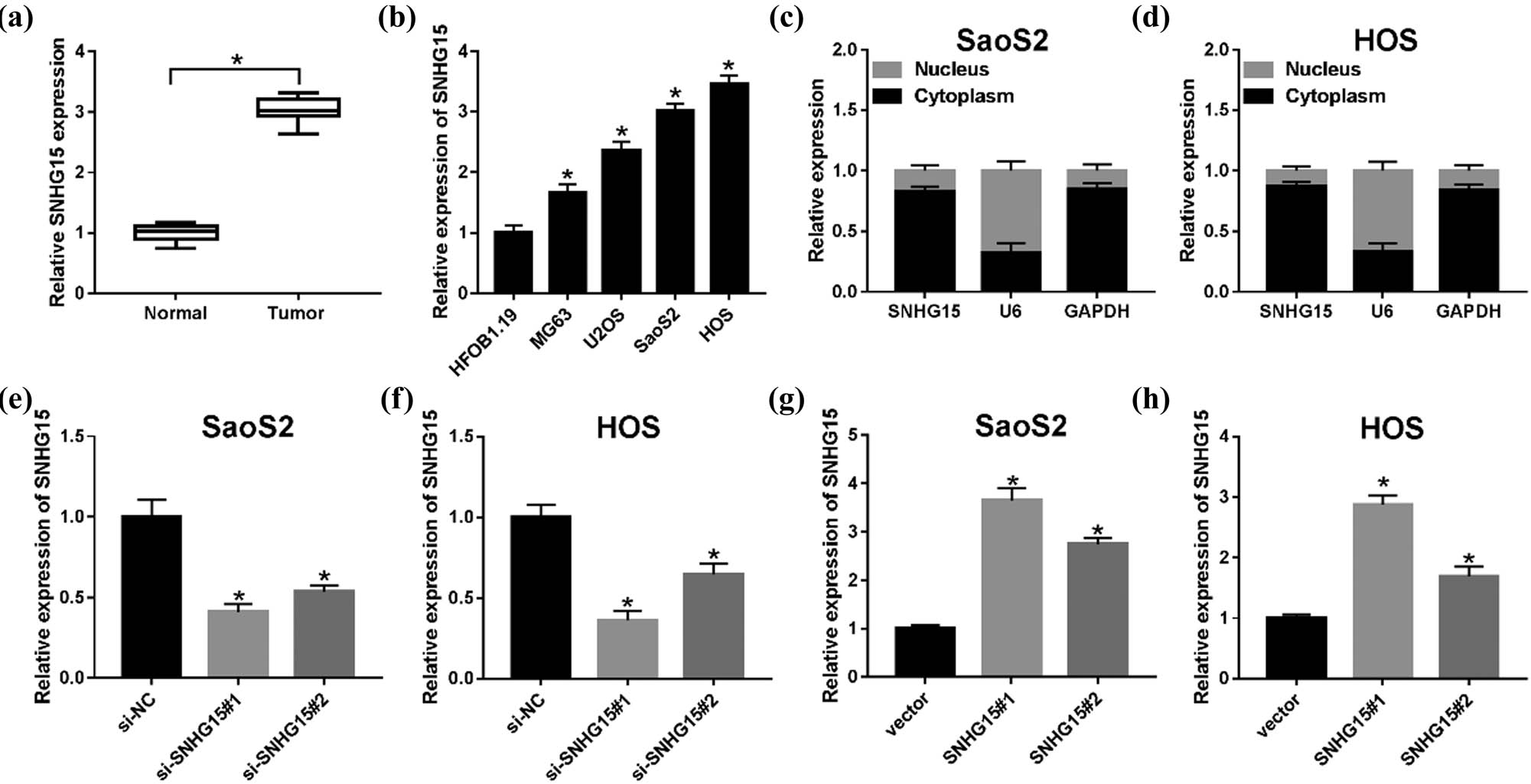
The roles of lncRNA SNHG15 in OS tissues and cells. (a) qRT-PCR was used to detect the expression of lncRNA SNHG15 in tissues from 30 patients with OS compared with the adjacent parts. (b) The expression of SNHG15 in human osteoblast cells (HFOB1.19) and OS cells (MG63, U2OS, SaoS2, and HOS) was measured by the qRT-PCR assay. (c and d) Localization of SNHG15 in SaoS2 and HOS cells was analyzed by the Subcellular fractionation assay. (e and f) The relative expression level of SNHG15 was detected in SaoS2 and HOS cells transfected with si-NC, si-SNHG15#1, and si-SNHG15#2. (g and h) The SNHG15 level was measured in SaoS2 and HOS cells transfected with vector, SNHG15#1, and SNHG15#2. *P < 0.05.
3.2 Knockdown of SNHG15 repressed proliferation and invasion, but induced apoptosis, in OS cells
First, to explore the role of SNHG15 of OS, we detected the biofunctional effects (proliferation, apoptosis, and invasion) (Figure 2) when cells were transfected with si-SNHG15 vector, and the results showed that proliferation and invasion were limited (Figure 2a, b, g and h) with apoptosis being promoted (Figure 2c and d). Second, we also investigated the change of apoptosis-related proteins in OS cells. The western blot assay confirmed that the levels of both Bax and Cleaved Caspase 3 were increased, while that of Bcl-2 was suppressed (Figure 2e and f). These data suggest that the knockdown of SNHG15 ameliorates the proliferation, apoptosis, and invasion abilities of OS cells.

Knockdown of SNHG15 repressed the abilities of proliferation and invasion but induced apoptosis in OS cells. The OS cells were transfected with si-control or si-SNHG15. By the loss-function experiments, the biofunction effects of SNHG15 were performed. (a and b) The cell viability at determined times (24, 48 and 72 h) was analyzed by the MTT assay in OS cells. (c and d) The rate of apoptosis was measured by the flow cytometry assay. (e and f) The levels of apoptosis-related protein (Bcl-2, Bax, and Cleaved Caspase 3) were confirmed by western blot. (g and h) The cell invasion was evaluated by the transwell assay in SaoS2 and HOS cells. *P < 0.05.
3.3 MiR-346 was a target for SNHG15
StarBase v 2.0 software was used to explore the potential binding sites of SNHG15 to miR-346 (Figure 3a). A dual-luciferase activity assay was performed in SaoS2 and HOS cell and showed that co-transfection of miR-346 and SNHG15 wide type (WT) significantly decreased the luciferase activity, whereas co-transfection of miR-control and SNHG15-mutant (MUT) did not change the dual-luciferase activity (Figure 3b and c). Meanwhile, we measured the expression pattern in OS tissues and interaction between miR-346 and SNHG15 and found an obvious decline in miR-346 in the tumor group compared with normal parts (Figure 3d), and a negative correlation could be seen between the expression of miR-346 and SNHG15 (Figure 3e). Then, the qRT-PCR assay was conducted to assess the expression of miR-346 after loss- and gain-function administration. The data suggested that the expression of miR-346 represented high tendency when cells transfected with si-SNHG15 vectors, while the low trend of miR-346 occurred when cells transfected with SNHG15 overexpressed vectors, compared with its own negative controls individually (Figure 3f and g).
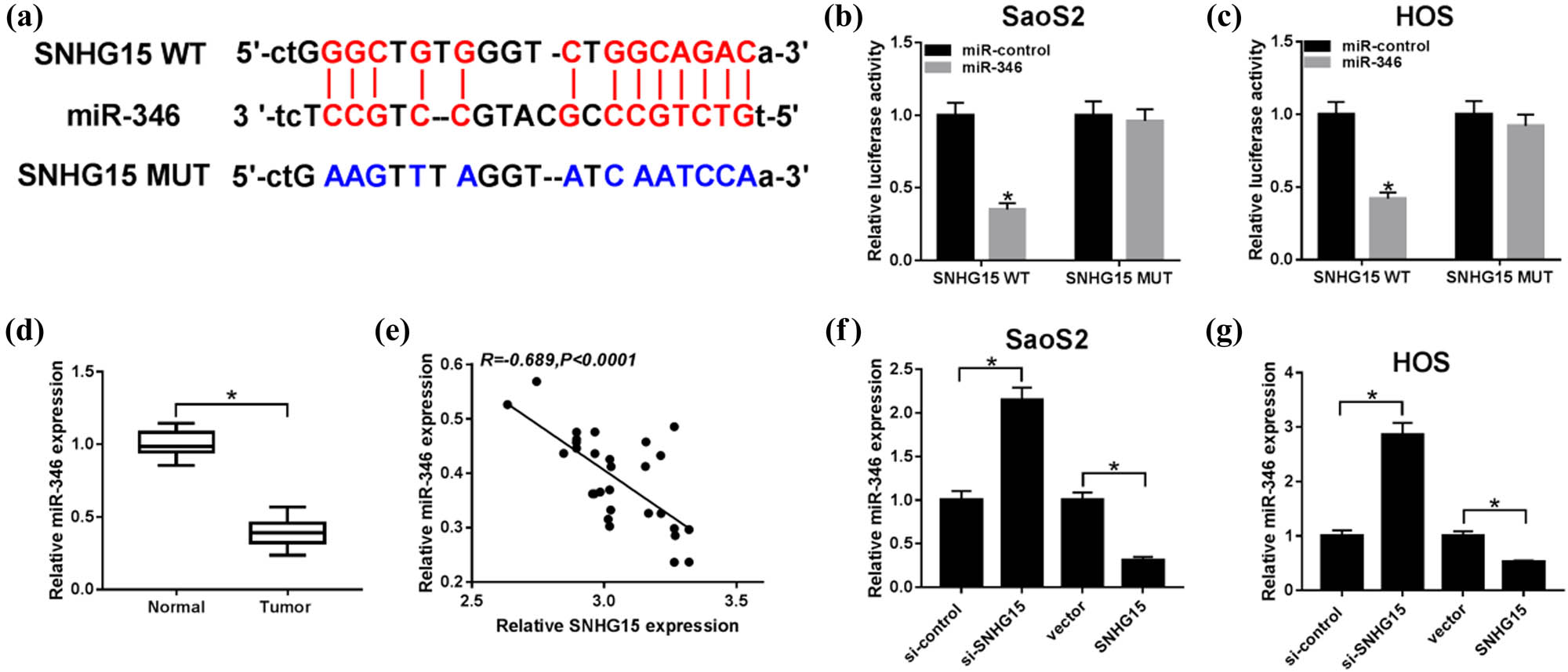
miR-346 was a target for SNHG15. (a) The putative binding sites between miR-346 and SNHG15 were predicted by starBase. (b and c) The predicted sites were identified by the dual-luciferase reporter assay. (d) QRT-PCR was carried out to detect the level of miR-346 in OS tissues. (e) A correlation between the expression of miR-346 and SNHG16 was explored. The SaoS2 and HOS cells were transfected with SNHG15 or vector. (f and g) The expression of miR-346 was detected by qRT-PCR in SaoS2 and HOS cells. *P < 0.05.
3.4 Inhibition of miR-346 reversed the effects of downregulation of SNHG15 of OS in vitro
To figure out how SHNG15 regulates the changes of miR-346 upon biofunctional phenotype, anti-miR-346 plasmid (miR-346 inhibitor) was constructed and transfected into the SaoS2 and HOS cells with downregulated expression of SNHG15, and the qRT-PCR assay confirmed that miR-346 inhibitor successfully suppressed the level of miR-346 in SaoS2 and HOS cells, compared with the transfection with an inhibitor-control (Figure 4a and b). It was observed that the suppression of miR-346 reversed the limitation of proliferation (Figure 4c and d) and invasion (Figure 4i), and the promotion of apoptosis had occurred in the cells where SNHG15 was downregulated (Figure 4e and f). When it referred to the influence on protein biomarkers of apoptosis, western blot results suggested that decreasing miR-346 inverted the enhanced expression pattern on Bax and Cleaved Caspase 3 and limited pattern on Bcl-2 from downregulation of SNHG15 in cells (Figure 4g and h). In addition, our data suggest that upregulating miR-346 could reduce the upregulating effect of SNHG15 on OS cell viability and the decrease in apoptosis rate in SaoS2 and HOS cells (Figure A1). These results show that limitation of miR-346 reversed the effects from downregulation of SNHG15 in OS cells.

Inhibition of miR-346 inverted the effects from the downregulation of SNHG15 of OS in vitro. Si-control, si-SNHG15, si-SNHG15 + inhibitor-control, and si-SNHG15 + miR-346 inhibitor were separately transfected into OS cells. (a and b) The knockdown efficiency of miR-346 was confirmed by qRT-PCR. (c and d) The cell viability was analyzed by the MTT assay at stated times (24, 48, and 72 h) in SaoS2 and HOS cells. (e and f) Flow cytometry assay was used to confirm the rate of apoptosis. (g and h) Apoptosis-related proteins (Bcl-2, Bax, and Cleaved Caspase 3) were evaluated using western blot. (i) The cell invasion was evaluated by the transwell assay. *P < 0.05.
3.5 TRAF4 was a direct target of miR-346
To further figure out the role of miR-346 upon regulatory mechanisms in OS, the online predicted bioinformatics software starBase (http://starbase.sysu.edu.cn/starbase2/) was used to predict the potential gene-binding sites in miR-346. We speculated that TRAF4 had a binding site with miR-346 (Figure 5a). To confirm this relationship between TRAF4 and miR-346, the dual-luciferase reporter assay showed that co-transfection of miR-346 and TRAF4-WT decreased the luciferase activity, while co-transfection of miR-control and TRAF4-MUT did not change the dual-luciferase activity (Figure 5b and c). Interestingly, the expression of TRAF4 on mRNA (Figure 5d) and protein (Figure 5e) levels was elevated in OS tissues as well. Then, we explored the expression of TRAF4 in SaoS2 and HOS cells, by gain-functional experiments, and found that the level of TRAF4 was upregulated with the limited expression of miR-346 (including mRNA (Figure 5f and g) and protein levels (Figure 5h and i)) when the cells were successfully transfected with the miR-346 inhibitor. These data confirmed that TRAF4 is a target of miR-346, and miR-346 regulates the expression of TRAF4 in a negative manner.

TRAF4 was a direct target of miR-346. (a) TRAF4 was predicted by starBase as a potential target for miR-346. (b and c) The dual-luciferase reporter assay was conducted to verify the interaction between miR-346 and TRAF4. (d and e) The expression of TRAF4 on OS tissues was explored at mRNA (d) and protein (e) levels. (f–i) The expression of TRAF4 after the loss-and-gain experiment was measured in OS cells using qRT-PCR (f and g) and western blot (h and i). *P < 0.05.
3.6 Overexpression of TRAF4 restores the biofunctional results from upregulation of miR-346
To confirm that the expression of TRAF4 could be regulated by miR-346, we examined the mRNA level of TRAF4 by the gain-functional experiment. As shown in Figure 6a and b, the mRNA level of TRAF4 was increased when cells were co-transfected with miR-346 and TRAF4, compared with cells co-transfected with miR-346 and vector, which was assayed by the qRT-PCR. Then, the equal results were shown in Figure 6c and d at the protein level. The function assays also affirmed that the inhibition of proliferation (Figure 6e and f) and invasion (Figure 6k and l) by miR-346 was impaired by overexpression of TRAF4 in OS cells. Moreover, the promotion effect of miR-346 overexpression on cell apoptosis was abolished by upregulation of TRAF4 in vitro (Figure 6g and h). The high expression of Bax and Cleaved Caspase 3 and low expression of Bcl-2 were inverted by the enforced expression of TRAF4 in OS cells (Figure 6i and j). The results suggested that overexpression of TRAF4 restored the biofunctional results from upregulation of miR-346.

Overexpression of TRAF4 restored the biofunctional results from the upregulation of miR-346. (a–d) The SaoS2 and HOS cells were administrated by co-overexpression miR-346 and TRAF4, and the TRAF4 expression was measured by qRT-PCR (a and b) and western blot (c and d). (e–l) The functional assays, namely, proliferation (e and f), apoptosis (g and h), and invasion (k and l) were determined by MTT analysis, flow cytometry assays, and transwell invasion assay individually. (i and j) The biomarkers of Bcl-2, Bax, and Cleaved Caspase 3 were assessed by the western blot. *P < 0.05.
3.7 SNHG15 regulates the expression of TRAF4 by sponging with miR-346
To investigate how SNHG15 regulates OS progression via the TRAF4/miR-346 axis, we thus designed the next experiments. We found that the relative expression of TRAF4 and SNHG15 maintain a positive correlation (Figure 7a). The mRNA level and protein level of TRAF4 were decreased in OS cells treated with si-SNHG15, while miR-346 inhibitor could mitigate the suppression effect (Figure 7b–d). These data identified that SNHG15 could regulate the expression of TRAF4 by targeting miR-346 in OS cells.

SNHG15 regulated the expression of TRAF4 by sponging with miR-346. (a) The relationship between TRAF4 and SNHG15 was uncovered using the qRT-PCR analysis. (b–d) The SaoS2 and HOS cells were co-transfected with si-SNHG15and miR-346 inhibitor, and the expression pattern of TRAF4 was investigated by the qRT-PCR (b) and western blot separately (c and d). *P < 0.05.
3.8 Silencing of SNHG15 inhibits OS growth in vivo
To further examine the efficacy of SNHG15 in vivo, as shown in Figure 8a, the tumor in sh-SNHG15-transfected group grew more slowly compared to that in the sh-control group. A significant decrease in the tumor weight was observed between sh-control and sh-SNHG15 groups (Figure 8b). Excised tumor tissues exhibited the improved expression of miR-346 and the reduced expression of SNHG15 in the sh-SNHG15 group (Figure 8c and d). In addition, downregulation of SNHG15 significantly inhibited the expression of TRAF4 at mRNA and protein levels (Figure 8e and f). These results suggested that silencing of SNHG15 inhibits the OS growth in vivo.

Silencing of SNHG15 inhibited the growth of OS in vivo. Mice (3 mice/group) were subcutaneously injected with HOS cells (2 × 10⁶) stably transfected with sh-SNHG15 or an equal volume of the vehicle into the left flank; the animals were sacrificed after 45 days. (a and b) Tumor volume (a) and tumor weight (b) were measured. (c–e) The qRT-PCR analysis was performed to measure the expression levels of SNHG15 (c), miR-346 (d), and TRAF4 (e) in excised tumor tissues. (f) Western blotting was performed to measure the expression of TRAF4. *P < 0.05.
4 Discussion
LncRNA SNHG15 has been reported to be a cancer-promoting lncRNA in a variety of tumors [25], for instance, prostate cancer [26], colorectal cancer [27], ovarian cancer [28], lung cancer [29,30], and renal cell carcinoma [19]. Zhang et al. identified that SNHG15 was elevated and promoted cell proliferation in prostate cancer cells [26]. Jin et al. reported that SNHG15 promotes proliferation, apoptosis, cell cycle, and tumor growth in non-small cell lung cancer in vitro and in vivo [30].
In this experiment, we first examined the expression level of lncRNA SNHG15 in OS cells and tissues, and the results showed that the expression of lncRNA SNHG15 is upregulated in both tissues and cells, consistent with the previous reports [14].
In previous studies, downregulation of SNHG15 was shown to inhibit tumor cell proliferation and invasion and promote apoptosis [14,31]. Through loss-functional experiments, similar results were found in our experiments that the downregulated expression of SNHG15 slowed down the abilities of proliferation and invasion in OS cells and promoted the rate of cell apoptosis. Subsequently, the miR-346 was predicted and proved to be the potential target for SNHG15. Interestingly, the expression pattern of miR-346 was attenuated in OS tissues and negatively correlated with that of SNHG15. Hence, we conducted further exploration by constructing inhibitor plasmid and uncovered that the knockdown of miR-346 could reverse the effect of decreasing SNHG15.
MiRNAs are a variety of endogenous low-molecular-weight compound, with about 22 nucleotides in length. Evidence is increasingly supporting that some miRNAs exert a tumor-suppressing role through targeting and inhibiting the expression of multiple oncogenes. The oncogene TRAF4 has been widely studied in a variety of cancers, such as hepatocellular carcinoma [32] cholangiocarcinoma [33], and non-small cell lung cancer [34]. Kang et al. showed that TRAF4 was upregulated in cholangiocarcinoma tissues and positively correlated with tumor differentiation and TNM (Tumor, Lymph Node, Metastasis) stage [33]. Yang et al. showed that the level of TRAF4 mRNA was inversely correlated with miR-302c-3p expression in hepatocellular carcinoma specimens, and TRAF4 restoration reversed the inhibitory effect of miR-302c-3p on AKT-induced EMT and hepatocellular carcinoma cell metastasis [32]. It has been reported that the upregulated TRAF4 was present in cholangiocarcinoma [33]. Consistent with the previous reports, the expression level of TRAF4 was promoted in OS tissues at both mRNA and protein levels in our study. By targeted prediction and biological verification, we found that overexpression of TRAF4 reversed the effects of miRNA-346 on OS cells. In vivo experiments also showed that the lncRNA SNHG15 knockdown promotes the OS tumor growth through sponging miR-346 to allow TRAF4 expression.
There were some limitations in this study. First, the interaction between miR-346 and SNHG15 or TRAF4 was initially detected by the dual-luciferase reporter assay, and it should be confirmed by RNA immunoprecipitation or RNA pull-down. Besides, the results and conclusions obtained using commercial cell lines could not fully represent the actual situation in vivo.
In summary, we identified that the highly expressed cancer-promoting lncRNA SNHG15 plays an essential role in the development of OS. Furthermore, our study first highlighted the role of lncRNA SNHG15 in promoting TRAF4 by sponging with miR-346, thereby promoting the progression of OS. Thus, this molecule is a new therapeutic target for OS.
Appendix

Overexpression of miR-346 alleviated SNHG15 upregulation-mediated cell viability and apoptosis in OS cells. (a and b) MiR-346 level was detected in SaoS2 and HOS cells transfected with vector, SNHG15, SNHG15+miR-NC, and SNHG15+miR-346. (c and d) Cell viability was assessed in transfected SaoS2 and HOS cells. (e and f) Apoptosis was examined in transfected SaoS2 and HOS cells.*P < 0.05.
Conflict of interest: The authors state no conflict of interest.
Data availability statement: The datasets generated during and/or analyzed during the current study are available from the corresponding author on reasonable request.
References
[1] Vijayamurugan N, Bakhshi S. Review of management issues in relapsed osteosarcoma. Expert Rev Anticancer Ther. 2014;14:151–61.10.1586/14737140.2014.863453Suche in Google Scholar PubMed
[2] Harrison DJ, Geller DS, Gill JD, Lewis VO, Gorlick R. Current and future therapeutic approaches for osteosarcoma. Expert Rev Anticancer Ther. 2018;18:39–50.10.1080/14737140.2018.1413939Suche in Google Scholar PubMed
[3] Toki S, Kobayashi E, Yoshida A, Ogura K, Wakai S, Yoshimot S, et al. A clinical comparison between dedifferentiated low-grade osteosarcoma and conventional osteosarcoma. Bone Joint J. 2019;101-B(6):745–52.10.1302/0301-620X.101B6.BJJ-2018-1207.R1Suche in Google Scholar PubMed
[4] Mercer TR, Dinger ME, Mattick JS. Long non-coding RNAs: insights into functions. Nat Rev Genet. 2009;10:155–9.10.1038/nrg2521Suche in Google Scholar PubMed
[5] Teng H, Mao F, Liang J, Xue M, Wei W, Li X, et al. Transcriptomic signature associated with carcinogenesis and aggressiveness of papillary thyroid carcinoma. Theranostics. 2018;8:4345–58.10.7150/thno.26862Suche in Google Scholar PubMed PubMed Central
[6] Huang Y, Zhang J, Hou L, Wang G, Liu H, Zhang R, et al. LncRNA AK023391 promotes tumorigenesis and invasion of gastric cancer through activation of the PI3K/Akt signaling pathway. J Exp Clin Cancer Res. 2017;36:194.10.1186/s13046-017-0666-2Suche in Google Scholar PubMed PubMed Central
[7] Yu J, Han Z, Sun Z, Wang Y, Zheng M, Song C. LncRNA SLCO4A1-AS1 facilitates growth and metastasis of colorectal cancer through beta-catenin-dependent Wnt pathway. J Exp Clin Cancer Res. 2018;37:222.10.1186/s13046-018-0896-ySuche in Google Scholar PubMed PubMed Central
[8] Ding H, Liu J, Zou R, Cheng P, Su Y. Long non-coding RNA TPTEP1 inhibits hepatocellular carcinoma progression by suppressing STAT3 phosphorylation. J Exp Clin Cancer Res. 2019;38:189.10.1186/s13046-019-1193-0Suche in Google Scholar PubMed PubMed Central
[9] Ling J, Wang F, Liu C, Dong X, Xue Y, Jia X, et al. FOXO1-regulated lncRNA LINC01197 inhibits pancreatic adenocarcinoma cell proliferation by restraining Wnt/beta-catenin signaling. J Exp Clin Cancer Res. 2019;38:179.10.1186/s13046-019-1174-3Suche in Google Scholar PubMed PubMed Central
[10] Wang Y, Zeng X, Wang N, Zhao W, Zhang X, Teng S, et al. Long noncoding RNA DANCR, working as a competitive endogenous RNA, promotes ROCK1-mediated proliferation and metastasis via decoying of miR-335-5p and miR-1972 in osteosarcoma. Mol Cancer. 2018;17:89.10.1186/s12943-018-0837-6Suche in Google Scholar PubMed PubMed Central
[11] Ozawa T, Matsuyama T, Toiyama Y, Takahashi N, Ishikawa T, Uetake H, et al. CCAT1 and CCAT2 long noncoding RNAs, located within the 8q.24.21 ‘gene desert’, serve as important prognostic biomarkers in colorectal cancer. Ann Oncol. 2017;28:1882–8.10.1093/annonc/mdx248Suche in Google Scholar PubMed PubMed Central
[12] Tani H, Torimura M. Development of cytotoxicity-sensitive human cells using overexpression of long non-coding RNAs. J Biosci Bioeng. 2015;119:604–8.10.1016/j.jbiosc.2014.10.012Suche in Google Scholar PubMed
[13] Tani H, Torimura M. Identification of short-lived long non-coding RNAs as surrogate indicators for chemical stress response. Biochem Biophys Res Commun. 2013;439:547–51.10.1016/j.bbrc.2013.09.006Suche in Google Scholar PubMed
[14] Liu K, Hou Y, Liu Y, Zheng J. LncRNA SNHG15 contributes to proliferation, invasion and autophagy in osteosarcoma cells by sponging miR-141. J Biomed Sci. 2017;24:46.10.1186/s12929-017-0353-9Suche in Google Scholar PubMed PubMed Central
[15] Chen DL, Ju HQ, Lu YX, Chen LZ, Zeng ZL, Zhang DS, et al. Long non-coding RNA XIST regulates gastric cancer progression by acting as a molecular sponge of miR-101 to modulate EZH2 expression. J Exp Clin Cancer Res. 2016;35:142.10.1186/s13046-016-0420-1Suche in Google Scholar PubMed PubMed Central
[16] Wu XS, Wang F, Li HF, Hu YP, Jiang L, Zhang F, et al. LncRNA-PAGBC acts as a microRNA sponge and promotes gallbladder tumorigenesis. EMBO Rep. 2017;18:1837–53.10.15252/embr.201744147Suche in Google Scholar PubMed PubMed Central
[17] Li T, Qin Y, Zhen Z, Shen H, Cong T, Schiferle E, et al. Long non-coding RNA HOTAIR/microRNA-206 sponge regulates STC2 and further influences cell biological functions in head and neck squamous cell carcinoma. Cell Prolif. 2019;52:e12651.10.1111/cpr.12651Suche in Google Scholar PubMed PubMed Central
[18] Li Z, Zhang J, Zheng H, Li C, Xiong J, Wang W, et al. Modulating lncRNA SNHG15/CDK6/miR-627 circuit by palbociclib, overcomes temozolomide resistance and reduces M2-polarization of glioma associated microglia in glioblastoma multiforme. J Exp Clin Cancer Res. 2019;38:380.10.1186/s13046-019-1371-0Suche in Google Scholar PubMed PubMed Central
[19] Du Y, Kong C, Zhu Y, Yu M, Li Z, Bi J, et al. Knockdown of SNHG15 suppresses renal cell carcinoma proliferation and EMT by regulating the NF-kappaB signaling pathway. Int J Oncol. 2018;53:384–94.Suche in Google Scholar
[20] Pan R, He Z, Ruan W, Li S, Chen H, Chen Z, et al. lncRNA FBXL19-AS1 regulates osteosarcoma cell proliferation, migration and invasion by sponging miR-346. OncoTargets Ther. 2018;11:8409–20.10.2147/OTT.S160963Suche in Google Scholar PubMed PubMed Central
[21] EI Hokayem J, Brittain GC, Nawaz Z, Bethea JR. Tumor necrosis factor receptor associated factors (TRAFs) 2 and 3 form a transcriptional complex with phosho-RNA polymerase II and p65 in CD40 ligand activated Neuro2a cells. Mol Neurobiol. 2017;54:1301–13.10.1007/s12035-016-9742-4Suche in Google Scholar PubMed PubMed Central
[22] Kedinger V, Rio MC. TRAF4, the unique family member. Adv Exp Med Biol. 2007;579:60–71.10.1007/978-0-387-70630-6_5Suche in Google Scholar PubMed
[23] Yao W, Wang X, Cai Q, Gao S, Wang J, Zhang P. TRAF4 enhances osteosarcoma cell proliferation and invasion by Akt signaling pathway. Oncol Res. 2014;22:21–8.10.3727/096504014X14077751730351Suche in Google Scholar PubMed PubMed Central
[24] Liu Q, Liu H, Cheng H, Li Y, Li X, Zhu C. Downregulation of long noncoding RNA TUG1 inhibits proliferation and induces apoptosis through the TUG1/miR-142/ZEB2 axis in bladder cancer cells. Onco Targets Ther. 2017;10:2461–71.10.2147/OTT.S124595Suche in Google Scholar PubMed PubMed Central
[25] Tong J, Ma X, Yu H, Yang J. SNHG15: a promising cancer-related long noncoding RNA. Cancer Manage Res. 2019;11:5961–9.10.2147/CMAR.S208054Suche in Google Scholar PubMed PubMed Central
[26] Zhang Y, Zhang D, Lv J, Wang S, Zhang Q. LncRNA SNHG15 acts as an oncogene in prostate cancer by regulating miR-338-3p/FKBP1A axis. Gene. 2019;705:44–50.10.1016/j.gene.2019.04.033Suche in Google Scholar PubMed
[27] Saeinasab M, Bahrami AR, Gonzalez J, Marchese FP, Martinez D, Mowla SJ, et al. SNHG15 is a bifunctional MYC-regulated noncoding locus encoding a lncRNA that promotes cell proliferation, invasion and drug resistance in colorectal cancer by interacting with AIF. J Exp Clin Cancer Res. 2019;38:172.10.1186/s13046-019-1169-0Suche in Google Scholar PubMed PubMed Central
[28] Qu C, Dai C, Guo Y, Qin R, Liu J. Long noncoding RNA SNHG15 serves as an oncogene and predicts poor prognosis in epithelial ovarian cancer. OncoTargets Ther. 2019;12:101–11.10.2147/OTT.S182657Suche in Google Scholar PubMed PubMed Central
[29] Cui HX, Zhang MY, Liu K, Liu J, Zhang ZL, Fu L. LncRNA SNHG15 promotes proliferation and migration of lung cancer via targeting microRNA-211-3p. Eur Rev Med Pharmacol Sci. 2018;22:6838–44.Suche in Google Scholar
[30] Jin B, Jin H, Wu HB, Xu JJ, Li B. Long non-coding RNA SNHG15 promotes CDK14 expression via miR-486 to accelerate non-small cell lung cancer cells progression and metastasis. J Cell Physiol. 2018;233:7164–72.10.1002/jcp.26543Suche in Google Scholar PubMed PubMed Central
[31] Sun X, Bai Y, Yang C, Hu S, Hou Z, Wang G. Long noncoding RNA SNHG15 enhances the development of colorectal carcinoma via functioning as a ceRNA through miR-141/SIRT1/Wnt/beta-catenin axis. Artif Cells Nanomed Biotechnol. 2019;47:2536–44.10.1080/21691401.2019.1621328Suche in Google Scholar PubMed
[32] Yang L, Guo Y, Liu X, Wang T, Tong X, Lei K, et al. The tumor suppressive miR-302c-3p inhibits migration and invasion of hepatocellular carcinoma cells by targeting TRAF4. J Cancer. 2018;9:2693–701.10.7150/jca.25569Suche in Google Scholar PubMed PubMed Central
[33] Kang Q, Zou H, Zhou L, Liu LX, Cai JB, Xie N, et al. Role of the overexpression of TRAF4 in predicting the prognosis of intrahepatic cholangiocarcinoma. Int J Oncol. 2018;53:286–96.Suche in Google Scholar
[34] Chen T, Gao F, Feng S, Yang T, Chen M. MicroRNA-370 inhibits the progression of non-small cell lung cancer by downregulating oncogene TRAF4. Oncol Rep. 2015;34:461–8.10.3892/or.2015.3978Suche in Google Scholar PubMed
© 2020 Xuewu Chen and Hongguang Xu, published by De Gruyter
This work is licensed under the Creative Commons Attribution 4.0 International License.
Artikel in diesem Heft
- Plant Sciences
- Dependence of the heterosis effect on genetic distance, determined using various molecular markers
- Plant Growth Promoting Rhizobacteria (PGPR) Regulated Phyto and Microbial Beneficial Protein Interactions
- Role of strigolactones: Signalling and crosstalk with other phytohormones
- An efficient protocol for regenerating shoots from paper mulberry (Broussonetia papyrifera) leaf explants
- Functional divergence and adaptive selection of KNOX gene family in plants
- In silico identification of Capsicum type III polyketide synthase genes and expression patterns in Capsicum annuum
- In vitro induction and characterisation of tetraploid drumstick tree (Moringa oleifera Lam.)
- CRISPR/Cas9 or prime editing? – It depends on…
- Study on the optimal antagonistic effect of a bacterial complex against Monilinia fructicola in peach
- Natural variation in stress response induced by low CO2 in Arabidopsis thaliana
- The complete mitogenome sequence of the coral lily (Lilium pumilum) and the Lanzhou lily (Lilium davidii) in China
- Ecology and Environmental Sciences
- Use of phosphatase and dehydrogenase activities in the assessment of calcium peroxide and citric acid effects in soil contaminated with petrol
- Analysis of ethanol dehydration using membrane separation processes
- Activity of Vip3Aa1 against Periplaneta americana
- Thermostable cellulase biosynthesis from Paenibacillus alvei and its utilization in lactic acid production by simultaneous saccharification and fermentation
- Spatiotemporal dynamics of terrestrial invertebrate assemblages in the riparian zone of the Wewe river, Ashanti region, Ghana
- Antifungal activity of selected volatile essential oils against Penicillium sp.
- Toxic effect of three imidazole ionic liquids on two terrestrial plants
- Biosurfactant production by a Bacillus megaterium strain
- Distribution and density of Lutraria rhynchaena Jonas, 1844 relate to sediment while reproduction shows multiple peaks per year in Cat Ba-Ha Long Bay, Vietnam
- Biomedical Sciences
- Treatment of Epilepsy Associated with Common Chromosomal Developmental Diseases
- A Mouse Model for Studying Stem Cell Effects on Regeneration of Hair Follicle Outer Root Sheaths
- Morphine modulates hippocampal neurogenesis and contextual memory extinction via miR-34c/Notch1 pathway in male ICR mice
- Composition, Anticholinesterase and Antipedicular Activities of Satureja capitata L. Volatile Oil
- Weight loss may be unrelated to dietary intake in the imiquimod-induced plaque psoriasis mice model
- Construction of recombinant lentiviral vector containing human stem cell leukemia gene and its expression in interstitial cells of cajal
- Knockdown of lncRNA KCNQ1OT1 inhibits glioma progression by regulating miR-338-3p/RRM2
- Protective effect of asiaticoside on radiation-induced proliferation inhibition and DNA damage of fibroblasts and mice death
- Prevalence of dyslipidemia in Tibetan monks from Gansu Province, Northwest China
- Sevoflurane inhibits proliferation, invasion, but enhances apoptosis of lung cancer cells by Wnt/β-catenin signaling via regulating lncRNA PCAT6/ miR-326 axis
- MiR-542-3p suppresses neuroblastoma cell proliferation and invasion by downregulation of KDM1A and ZNF346
- Calcium Phosphate Cement Causes Nucleus Pulposus Cell Degeneration Through the ERK Signaling Pathway
- Human Dental Pulp Stem Cells Exhibit Osteogenic Differentiation Potential
- MiR-489-3p inhibits cell proliferation, migration, and invasion, and induces apoptosis, by targeting the BDNF-mediated PI3K/AKT pathway in glioblastoma
- Long non-coding RNA TUG1 knockdown hinders the tumorigenesis of multiple myeloma by regulating the microRNA-34a-5p/NOTCH1 signaling pathway
- Large Brunner’s gland adenoma of the duodenum for almost 10 years
- Neurotrophin-3 accelerates reendothelialization through inducing EPC mobilization and homing
- Hepatoprotective effects of chamazulene against alcohol-induced liver damage by alleviation of oxidative stress in rat models
- FXYD6 overexpression in HBV-related hepatocellular carcinoma with cirrhosis
- Risk factors for elevated serum colorectal cancer markers in patients with type 2 diabetes mellitus
- Effect of hepatic sympathetic nerve removal on energy metabolism in an animal model of cognitive impairment and its relationship to Glut2 expression
- Progress in research on the role of fibrinogen in lung cancer
- Advanced glycation end product levels were correlated with inflammation and carotid atherosclerosis in type 2 diabetes patients
- MiR-223-3p regulates cell viability, migration, invasion, and apoptosis of non-small cell lung cancer cells by targeting RHOB
- Knockdown of DDX46 inhibits trophoblast cell proliferation and migration through the PI3K/Akt/mTOR signaling pathway in preeclampsia
- Buformin suppresses osteosarcoma via targeting AMPK signaling pathway
- Effect of FibroScan test in antiviral therapy for HBV-infected patients with ALT <2 upper limit of normal
- LncRNA SNHG15 regulates osteosarcoma progression in vitro and in vivo via sponging miR-346 and regulating TRAF4 expression
- LINC00202 promotes retinoblastoma progression by regulating cell proliferation, apoptosis, and aerobic glycolysis through miR-204-5p/HMGCR axis
- Coexisting flavonoids and administration route effect on pharmacokinetics of Puerarin in MCAO rats
- GeneXpert Technology for the diagnosis of HIV-associated tuberculosis: Is scale-up worth it?
- Circ_001569 regulates FLOT2 expression to promote the proliferation, migration, invasion and EMT of osteosarcoma cells through sponging miR-185-5p
- Lnc-PICSAR contributes to cisplatin resistance by miR-485-5p/REV3L axis in cutaneous squamous cell carcinoma
- BRCA1 subcellular localization regulated by PI3K signaling pathway in triple-negative breast cancer MDA-MB-231 cells and hormone-sensitive T47D cells
- MYL6B drives the capabilities of proliferation, invasion, and migration in rectal adenocarcinoma through the EMT process
- Inhibition of lncRNA LINC00461/miR-216a/aquaporin 4 pathway suppresses cell proliferation, migration, invasion, and chemoresistance in glioma
- Upregulation of miR-150-5p alleviates LPS-induced inflammatory response and apoptosis of RAW264.7 macrophages by targeting Notch1
- Long non-coding RNA LINC00704 promotes cell proliferation, migration, and invasion in papillary thyroid carcinoma via miR-204-5p/HMGB1 axis
- Neuroanatomy of melanocortin-4 receptor pathway in the mouse brain
- Lipopolysaccharides promote pulmonary fibrosis in silicosis through the aggravation of apoptosis and inflammation in alveolar macrophages
- Influences of advanced glycosylation end products on the inner blood–retinal barrier in a co-culture cell model in vitro
- MiR-4328 inhibits proliferation, metastasis and induces apoptosis in keloid fibroblasts by targeting BCL2 expression
- Aberrant expression of microRNA-132-3p and microRNA-146a-5p in Parkinson’s disease patients
- Long non-coding RNA SNHG3 accelerates progression in glioma by modulating miR-384/HDGF axis
- Long non-coding RNA NEAT1 mediates MPTP/MPP+-induced apoptosis via regulating the miR-124/KLF4 axis in Parkinson’s disease
- PCR-detectable Candida DNA exists a short period in the blood of systemic candidiasis murine model
- CircHIPK3/miR-381-3p axis modulates proliferation, migration, and glycolysis of lung cancer cells by regulating the AKT/mTOR signaling pathway
- Reversine and herbal Xiang–Sha–Liu–Jun–Zi decoction ameliorate thioacetamide-induced hepatic injury by regulating the RelA/NF-κB/caspase signaling pathway
- Therapeutic effects of coronary granulocyte colony-stimulating factor on rats with chronic ischemic heart disease
- The effects of yam gruel on lowering fasted blood glucose in T2DM rats
- Circ_0084043 promotes cell proliferation and glycolysis but blocks cell apoptosis in melanoma via circ_0084043-miR-31-KLF3 axis
- CircSAMD4A contributes to cell doxorubicin resistance in osteosarcoma by regulating the miR-218-5p/KLF8 axis
- Relationship of FTO gene variations with NAFLD risk in Chinese men
- The prognostic and predictive value of platelet parameters in diabetic and nondiabetic patients with sudden sensorineural hearing loss
- LncRNA SNHG15 contributes to doxorubicin resistance of osteosarcoma cells through targeting the miR-381-3p/GFRA1 axis
- miR-339-3p regulated acute pancreatitis induced by caerulein through targeting TNF receptor-associated factor 3 in AR42J cells
- LncRNA RP1-85F18.6 affects osteoblast cells by regulating the cell cycle
- MiR-203-3p inhibits the oxidative stress, inflammatory responses and apoptosis of mice podocytes induced by high glucose through regulating Sema3A expression
- MiR-30c-5p/ROCK2 axis regulates cell proliferation, apoptosis and EMT via the PI3K/AKT signaling pathway in HG-induced HK-2 cells
- CTRP9 protects against MIA-induced inflammation and knee cartilage damage by deactivating the MAPK/NF-κB pathway in rats with osteoarthritis
- Relationship between hemodynamic parameters and portal venous pressure in cirrhosis patients with portal hypertension
- Long noncoding RNA FTX ameliorates hydrogen peroxide-induced cardiomyocyte injury by regulating the miR-150/KLF13 axis
- Ropivacaine inhibits proliferation, migration, and invasion while inducing apoptosis of glioma cells by regulating the SNHG16/miR-424-5p axis
- CD11b is involved in coxsackievirus B3-induced viral myocarditis in mice by inducing Th17 cells
- Decitabine shows anti-acute myeloid leukemia potential via regulating the miR-212-5p/CCNT2 axis
- Testosterone aggravates cerebral vascular injury by reducing plasma HDL levels
- Bioengineering and Biotechnology
- PL/Vancomycin/Nano-hydroxyapatite Sustained-release Material to Treat Infectious Bone Defect
- The thickness of surface grafting layer on bio-materials directly mediates the immuno-reacitivity of macrophages in vitro
- Silver nanoparticles: synthesis, characterisation and biomedical applications
- Food Science
- Bread making potential of Triticum aestivum and Triticum spelta species
- Modeling the effect of heat treatment on fatty acid composition in home-made olive oil preparations
- Effect of addition of dried potato pulp on selected quality characteristics of shortcrust pastry cookies
- Preparation of konjac oligoglucomannans with different molecular weights and their in vitro and in vivo antioxidant activities
- Animal Sciences
- Changes in the fecal microbiome of the Yangtze finless porpoise during a short-term therapeutic treatment
- Agriculture
- Influence of inoculation with Lactobacillus on fermentation, production of 1,2-propanediol and 1-propanol as well as Maize silage aerobic stability
- Application of extrusion-cooking technology in hatchery waste management
- In-field screening for host plant resistance to Delia radicum and Brevicoryne brassicae within selected rapeseed cultivars and new interspecific hybrids
- Studying of the promotion mechanism of Bacillus subtilis QM3 on wheat seed germination based on β-amylase
- Rapid visual detection of FecB gene expression in sheep
- Effects of Bacillus megaterium on growth performance, serum biochemical parameters, antioxidant capacity, and immune function in suckling calves
- Effects of center pivot sprinkler fertigation on the yield of continuously cropped soybean
- Special Issue On New Approach To Obtain Bioactive Compounds And New Metabolites From Agro-Industrial By-Products
- Technological and antioxidant properties of proteins obtained from waste potato juice
- The aspects of microbial biomass use in the utilization of selected waste from the agro-food industry
- Special Issue on Computing and Artificial Techniques for Life Science Applications - Part I
- Automatic detection and segmentation of adenomatous colorectal polyps during colonoscopy using Mask R-CNN
- The impedance analysis of small intestine fusion by pulse source
- Errata
- Erratum to “Diagnostic performance of serum CK-MB, TNF-α and hs-CRP in children with viral myocarditis”
- Erratum to “MYL6B drives the capabilities of proliferation, invasion, and migration in rectal adenocarcinoma through the EMT process”
- Erratum to “Thermostable cellulase biosynthesis from Paenibacillus alvei and its utilization in lactic acid production by simultaneous saccharification and fermentation”