Gold nanocrystals and nanorods functionalized with protein and polymeric ligands for environmental, energy storage, and diagnostic applications: A review
-
Nadiah Aldaleeli
 ,
Amr M. Elbasiony
,
Amr M. Elbasiony

Abstract
This comprehensive review explores the utilization of gold nanocrystals (Au NCs) and gold nanorods (Au NRs) functionalized with protein and polymeric ligands for various environmental applications. These applications include drug delivery, diagnostics, and environmental monitoring. This review also explores the biogenic synthesis of Au nanocrystals and nanostars, characterization techniques, and their analytical applications in environmental contexts. Moreover, integrating Au nanocrystals with ligand–receptor chemistry enables rapid and efficient cell detection and separation. Notably, fluorescent polymer–Au nanocrystals exhibit a unique characteristic that renders them highly valuable for energy storage devices such as batteries and supercapacitors. Incorporating these NCs into electrode materials can enhance charge storage capacity, electrochemical performance, and cycling stability. Furthermore, they can enhance light absorption, charge separation, and transfer process in solar cells. The fluorescence properties of these NCs also enable them to serve as specific probes for identifying contaminants, including heavy metals, organic compounds, and environmental toxins. The fluorescence can be tuned to interact with particular analytes, enabling accurate detection and measurement. Additionally, these NCs can be functionalized to enhance pollutant removal, thereby increasing their adsorption capacity and catalytic activity. Collectively, fluorescent polymers incorporating Au nanocrystals hold immense potential in energy storage systems and environmental applications. They offer improved performance, sensitivity, and efficiency. Consequently, researchers actively explore ways to enhance and expand their utilization to address energy and environmental challenges.
Abbreviations
- AGGP
-
aminoglycoside-polyglutamate
- AGR
-
anti-galvanic reduction
- BSA
-
bovine serum albumin
- CT
-
computed tomography
- CTAB
-
cetyltrimethylammonium bromide
- DBAM
-
dodecyloxybenzalacetal
- EPR
-
enhanced permeability and retention effect
- FDTD
-
finite-difference time-domain
- FLIM
-
fluorescence lifetime imaging microscopy
- GLY
-
glyphosate
- GSH
-
glutathione
- HEMA
-
hydroxyethyl methacrylate
- HOMO
-
highest occupied molecular orbital
- HRP
-
horseradish peroxidase
- HSA
-
human serum albumin
- HEXA
-
hexaconazole
- LSPR
-
longitudinal surface plasmon resonance
- LUMO
-
lowest occupied molecular orbital
- MBA
-
mercaptobenzoic acid
- MEF
-
metal-enhanced fluorescence
- MHDA
-
mercapto hexadecanoic acid
- MPS
-
mononuclear phagocyte system
- MRI
-
magnetic resonance imaging
- MUA
-
mercaptoundecanoic acid
- NAS
-
transparent styrene acrylic copolymers
- NCrys
-
nanocrystal
- ND
-
nanodot
- NIRF
-
near-infrared fluorescence
- NR
-
nanorod
- NSp
-
nanosphere
- PCa
-
prostate cancer
- PCM
-
phase change material
- PEDOT
-
poly(3,4-ethylene dioxythiophene)
- PES
-
potential energy surface
- QD
-
quantum dot
- QY
-
quantum yield
- SAIL
-
surface-active ionic liquid
- SERS
-
surface-enhanced Raman scattering
- SPR
-
surface plasmon resonance
- TDDFPT
-
time-dependent density functional perturbation theory
- TES
-
thermal energy storage
1 Introduction
The emergence of gold nanocrystals (Au NCs) and gold nanorods (Au NRs) functionalized with protein and polymeric ligands has sparked immense interest in various environmental applications [1]. Recent studies have addressed various topics, including the regulatory aspects and impact modification techniques of polymer-based blends, interpenetrating polymer networks, and gels, highlighting their industrial applications and advancements [2,3]. Other areas of focus include greener synthesis approaches for polymer-based carbon dots, cross-linking methods for biomedical nano gels [4], the influence of sulfonated groups on proton and methanol transport in irradiated membranes [5], and the in situ polymerization of magnetic polymer nanocomposites, demonstrating the breadth of research in materials science and nanotechnology [6].
This comprehensive review delves into utilizing these functionalized nanocrystals, exploring their roles in drug delivery, diagnostics, environmental detection, and energy storage applications. Additionally, the biogenic synthesis of Au nanocrystals and nanostars and their characterization techniques and analytical uses in environmental contexts are thoroughly examined. Integrating Au nanocrystals with ligand–receptor chemistry has proven to be a rapid and efficient cell detection and separation platform, holding significant promise in biomedical applications [7]. Particularly noteworthy are fluorescent polymer–Au nanocrystals, which possess unique characteristics that render them highly valuable in energy storage devices such as batteries and supercapacitors [8]. Incorporating these nanocrystals into electrode materials has the potential to enhance the charge storage capacity, electrochemical performance, and cycling stability of these devices. Furthermore, their ability to enhance light absorption, charge separation, and transfer process in solar cells positions them as promising candidates for solar energy harvesting and utilization [9]. One striking property of these Au nanocrystals is their fluorescence. This enables them to function as specific probes for identifying environmental contaminants, including heavy metals, organic compounds, and environmental toxins. The tunability of their fluorescence allows for interactions with particular analytes, facilitating accurate detection and measurement in environmental monitoring applications. Tiyyagura et al. [10] investigated the efficacy of polyvinylpyrrolidone (PVP)-stabilized gold nanoparticle (AuNP) coatings in preventing the nonspecific adsorption of blood proteins, a critical factor in the performance of blood-contacting medical devices such as vascular grafts and stents. The research utilized a quartz crystal microbalance to monitor real-time interactions between the AuNP-coated sensors and bovine serum albumin (BSA), a model blood protein. The findings demonstrated that PVP-stabilized AuNP coatings effectively inhibited BSA adsorption, suggesting their potential to reduce bacterial biofilm formation on medical implants. Characterization of the coatings included assessments of wettability, surface morphology, and chemical composition, confirming the successful stabilization of AuNPs by PVP and their suitability for biomedical applications.
Moreover, researchers have explored further functionalizing these NCs to enhance their pollutant removal capabilities, adsorption capacity, and catalytic activity. This versatility makes fluorescent polymers incorporating Au nanocrystals highly appealing for addressing environmental challenges and advancing sustainable solutions. Throughout this review, we explore the potential of Au nanocrystals and NRs functionalized with protein and polymeric ligands in various environmental applications, shedding light on their promising roles in drug delivery, diagnostics, and environmental detection. Additionally, the review article explores their outstanding potential in energy storage systems and their effectiveness as highly sensitive probes for ecological monitoring.
2 State-of-the-art for Au nanocluster
Gold nanoclusters (Au NCs) have emerged as fascinating and highly promising nanomaterials with unique properties and diverse applications. These nanoclusters typically consist of a small number of gold atoms, usually ranging from a few to several dozens, and their physical and chemical characteristics differ from those of individual gold atoms and bulk gold materials [11].
One significant area of research focuses on synthesizing and characterizing Au nanoclusters. Various methods, such as chemical reduction [12], photochemical synthesis [13], and template-assisted approaches [14], have been developed to create Au NCs with precise control over their size, shape, and surface chemistry. Researchers continue to explore novel synthetic routes to achieve monodisperse and stable nanoclusters, as well as to understand the factors that influence their growth and stability.
The optical and electronic properties of Au nanoclusters have garnered considerable attention due to their size-dependent behaviours. Quantum confinement effects at the nanoscale result in discrete electronic energy levels that resemble molecular orbitals rather than the continuous band structure observed in bulk gold. As a result, Au nanoclusters exhibit unique fluorescence, photoluminescence, and light absorption properties, which vary with their size and surface ligands.
In recent years, Au nanoclusters have found applications in various fields, including catalysis [15], sensing [16], imaging [17], and nanoelectronics [18]. Their enhanced catalytic activity, tuneable fluorescence, and ability to serve as efficient probes make them valuable tools in analytical and biomedical applications. Moreover, researchers have explored their potential in energy storage systems, where they may be integrated into electrode materials to improve charge storage capacity and electrochemical performance.
In environmental applications, these nanoclusters are promising and highly efficient sensors for detecting environmental contaminants, such as heavy metals and organic compounds. Their tuneable fluorescence allows for specific interactions with target analytes, enabling accurate and sensitive detection.
Despite the progress made in understanding and utilizing Au nanoclusters, there are still challenges to overcome. Developing reproducible and scalable synthesis methods remains challenging, especially for large-scale production. Additionally, understanding the stability and long-term behaviour of Au nanoclusters in various environments is crucial for their practical applications.
Generally, state-of-the-art Au nanocluster research is a dynamic field that continues to evolve rapidly. As researchers gain deeper insights into their properties and develop innovative synthesis techniques, the applications of these nanoclusters are expected to expand further, opening new avenues in nanotechnology, energy storage, environmental sensing, and beyond.
2.1 Advancements in Au nanocluster synthesis techniques
Advancements in Au nanocluster synthesis techniques have led to the development of a wide range of methods to produce nanoclusters with precise control over their size, shape, and surface properties. These techniques have enabled researchers to tailor the properties of Au nanoclusters for specific applications, including energy storage, catalysis, sensing, and biomedicine. Some of the notable advancements in Au nanocluster synthesis techniques are as follows:
Ligand-assisted synthesis : Ligand-assisted synthesis methods involve using specific ligands as stabilizing agents during the Au nanocluster formation. By carefully selecting the ligands, researchers can control the size and shape of the nanoclusters. This approach allows for the production of monodisperse Au nanoclusters with well-defined structures and enhanced stability [19].
Seed-mediated growth : Seed-mediated growth is a technique that involves using preformed Au nanocrystals as seeds for the controlled growth of larger nanoclusters. By adjusting the growth conditions, researchers can precisely control the size and morphology of the resulting nanoclusters. This method enables the synthesis of Au nanoclusters with uniform size and shape distributions [20].
Microfluidic synthesis : Microfluidic platforms offer a highly controlled and reproducible environment for synthesizing Au nanoclusters. The precisely controlled flow conditions and reaction parameters in microfluidic channels allow for the synthesis of uniform and monodisperse nanoclusters with tuneable properties [21].
Bottom-up approaches : Bottom-up approaches involve the reduction in Au ions or Au complexes in the presence of reducing and stabilizing agents. These methods enable the formation of Au nanoclusters from atomic or molecular precursors. The careful selection of reactants and reaction conditions allows for fine-tuning nanocluster properties [20,22].
Biogenic synthesis : Biogenic synthesis involves using biological entities, such as microorganisms or plant extracts, as reducing and stabilizing agents for Au nanocluster formation. This environmentally friendly approach offers a sustainable and green route to produce nanoclusters with unique properties [23].
Plasma synthesis : Plasma-based methods, such as plasma sputtering or plasma-induced nucleation, provide a controlled environment for synthesizing Au nanoclusters. These techniques allow for the production of nanoclusters with well-defined sizes and structures.
Template-assisted synthesis : Template-assisted synthesis involves using templates or scaffolds with specific shapes and sizes to guide the nucleation and growth of Au nanoclusters. This method enables the production of nanoclusters with tailored shapes and arrangements [24].
Photochemical synthesis : Photochemical synthesis methods utilize light irradiation to drive the reduction and nucleation of Au ions, leading to the formation of Au nanoclusters. Light-controlled synthesis allows for the precise control of nanocluster properties [25].
Gas-phase synthesis : Gas-phase synthesis techniques involve the nucleation and growth of Au nanoclusters in the gas phase, followed by deposition onto a substrate. This approach offers high purity and control over nanocluster size and composition [26].
Advancements in Au nanocluster synthesis techniques have opened up new opportunities for tailoring their properties for various applications. Researchers continue exploring novel approaches and fine-tuning existing methods to produce Au nanoclusters with enhanced performance and functionality in energy storage, catalysis, sensing, and biomedical applications. These advancements in Au nanocluster synthesis techniques offer researchers the ability to tailor the properties of nanoclusters for specific applications in energy storage, catalysis, sensing, and biomedicine. Each method has advantages and disadvantages, and ongoing research aims to optimize these techniques further for improved performance and functionality (Table 1).
Summary of advantages and disadvantages of gold synthesis methods
| Synthesis technique | Advantages | Disadvantages |
|---|---|---|
| Ligand-assisted synthesis | Precise control over size and shape | Potential ligand interactions affecting properties |
| Monodisperse nanoclusters with enhanced stability | Complexity | |
| Seed-mediated growth | Controlled growth with uniform size and morphology | Seed preparation and purification can be challenging |
| Tailored nanoclusters with desired properties | Requires additional purification steps | |
| Microfluidic synthesis | Highly controlled and reproducible environment | Initial setup and optimization can be time-consuming |
| Synthesis of uniform and tuneable nanoclusters | Is complex and expensive, requiring specialized equipment and expertise. Scaling challenge | |
| Bottom-up approaches | Fine-tuning of nanocluster properties | Requirement of precise reaction conditions |
| Formation of nanoclusters from atomic/molecular precursors | The purification of nanoclusters from excess reagents can be cumbersome | |
| Biogenic synthesis | Environmentally friendly and sustainable approach | Limited control over nanocluster size and shape |
| Unique properties from biological reducing agents | Using biological materials might also introduce impurities that require additional purification steps | |
| Plasma synthesis | Controlled environment for precise synthesis | Complex equipment and experimental setup |
| Well-defined sizes and structures | The formation of unwanted by-products | |
| Template-assisted synthesis | Tailored shapes and arrangements | Template removal can be challenging |
| Controlled nucleation and growth | Time-consuming and may limit the variety of shapes that can be achieved | |
| Photochemical synthesis | Precise control of properties through light irradiation | Limited scalability for large-scale production |
| Light-induced reduction and nucleation of Au ions | It may be challenging to precisely control nanocluster size and properties through light irradiation | |
| Gas-phase synthesis | High purity and control over size and composition | Complex gas-phase synthesis setups |
| Deposition onto substrates for various applications | Achieving a high yield of nanoclusters might be challenging due to loss during deposition |
2.2 Emerging applications of Au nanoclusters
Emerging applications of Au nanoclusters have garnered significant attention in recent years due to their unique properties and versatile nature. These tiny clusters of a few to several dozens of Au atoms exhibit distinct electronic, optical, and catalytic features that make them attractive for various cutting-edge applications. Some of the emerging applications of Au nanoclusters are as follows:
Energy storage : Au nanoclusters have shown promise in energy storage applications, particularly in developing advanced batteries and supercapacitors. Their small size and high surface area-to-volume ratio allow efficient charge storage and fast charge–discharge rates. Incorporating Au nanoclusters into electrode materials can enhance energy storage devices’ electrochemical performance and cycling stability [27].
Sensing and detection : Au nanoclusters have exceptional sensing capabilities, making them valuable in environmental monitoring, disease diagnostics, and food safety. Their fluorescence and plasmonic properties enable sensitive and selective detection of target analytes, such as heavy metals, environmental toxins, and biomolecules [28].
Catalysis : Au nanoclusters possess immense potential as catalysts for various chemical reactions. Their unique electronic and geometric structures allow for efficient activation of reactants and selective catalytic transformations. These nanoclusters find applications in green chemistry, hydrogen generation, and pollutant degradation [29].
Biomedical imaging and therapy : Au nanoclusters have been explored for bioimaging and therapeutic medical applications. Their biocompatibility, tuneable fluorescence, and surface chemistry are ideal candidates for targeted imaging and drug delivery systems. Additionally, their photothermal properties enable localized hyperthermia for cancer therapy [30].
Optoelectronics and photonics : Au nanoclusters have shown potential in optoelectronics and photonics, contributing to advancements in light-emitting devices, lasers, and sensors. Their size-dependent emission and tuneable fluorescence allow for the design of novel optical devices with enhanced efficiency and functionality [31].
Nanoelectronics : Au nanoclusters are being explored for applications in nanoelectronics, such as single-electron transistors and molecular electronic devices. Their discrete energy levels and quantum confinement effects offer opportunities for developing nanoscale electronic components [32].
Environmental remediation : Au nanoclusters can be functionalized to enhance their pollutant removal capabilities. Their high surface area and catalytic activity enable the degradation of organic pollutants and the removal of contaminants from water and air [33].
Photovoltaics : Au nanoclusters have the potential to enhance light absorption and charge separation in solar cells. By incorporating these nanoclusters into solar cell materials, researchers aim to improve the efficiency and performance of photovoltaic devices [34].
Nanomedicine : Au nanoclusters are being investigated for drug delivery and theranostic applications. Their ability to carry and release therapeutic agents and their imaging capabilities allow personalized medicine and targeted treatments [35].
As research on Au nanoclusters continues to progress, these tiny nanoparticles are expected to find even more innovative applications across various fields, driving advancements in technology, medicine, and environmental sustainability. Table 2 summarizes the emerging applications of Au nanoclusters and their potential advantages and disadvantages.
Summary of advantages and disadvantages of emerging applications of Au nanoclusters
| Application | Advantages | Disadvantages |
|---|---|---|
| Energy storage | High-charge storage capacity | Synthesis challenges, cost |
| Sensing and detection | High sensitivity and selectivity | Limited stability in harsh conditions |
| Catalysis | Efficient activation of reactants | Limited scalability for industrial processes |
| Biomedical imaging | Targeted imaging and drug delivery | Potential toxicity and clearance issues |
| Biomedical therapy | Localized hyperthermia for cancer treatment | Safety concerns for clinical applications |
| Optoelectronics | Enhanced efficiency in light-emitting devices | Integration challenges with existing devices |
| Nanoelectronics | Potential for nanoscale electronic components | Stability and reproducibility issues |
| Environmental remediation | Effective pollutant removal and degradation | Optimization for specific pollutants needed |
| Photovoltaics | Improved light absorption in solar cells | High cost and scalability challenges |
| Nanomedicine | Targeted drug delivery and imaging | Biocompatibility and clearance concerns |
2.3 Unique characteristics and surface influences of Au nanocrystal
The transition from the atomic or molecular system to like material is considerable when considering nanoscale plans [36,37]. At the nanoscale, materials implement unique characterization and behaviours that vary from their bulk counterparts due to the dominance of surface influences, quantum confinement, and increased surface-to-volume ratio [38,39,40]. Materials and particles possess distinct electronic and optical features at an atomic and molecular scale [41]. Quantum mechanical influences become pronounced. Physiological features like band gaps, energy levels, and optical transitions may be sensitively tuned by monitoring the nano diameter, nanoparticles’ shape, and nanoscale materials’ composition [42]. These unique features have been used in electronics, photonics, catalysis, energy storage, and biomedicine. However, as the diameter of the particles increases, the collective behaviour of a large atom or molecule number becomes more relevant, leading to the emergence of bulk features. The transition from atomic and molecular systems to bulk systems occurs when the number of molecules or atoms reaches a sufficiently large scale [37]. Macroscopic features like density, conductivity, and thermal stability may describe the material’s behaviour. In bulk matter, the influence of individual atoms or molecules becomes less pronounced, and the statistical behaviour of large ensembles becomes more prominent. It involves considering the inter implement among nanoscale influences and bulk features and the scalability of nanoscale phenomena to macroscopic usages.
2.4 Harnessing the potential of Au nanocrystals for enhanced fluorescence imaging and sensing
By harnessing the unique features of nanoscale materials and influencing and integrating them into bulk systems, it is possible to develop innovative technologies with enhanced performance and functionality [43]. Clusters of a little Au atom, often referred to as Au clusters, may implement features that resemble those of molecules. These clusters typically consist of a few Au atoms, ranging from a little to several dozens of atoms. Due to its small diameter, Au clusters may implement discrete electronic energy levels resembling molecular orbitals in molecules. This leads to unique electronic and optical features that vary from individual Au atoms to bulk Au materials [44]. Indeed, in the nanoscale diameter range, there is a fascinating transition zone where Au nanoclusters of approximately 100 metal atoms may be found. The behaviour and features of these nanoclusters are influenced by their electronic structure, which determines their stability and reactivity. When considering the lowest-energy configurations of Au nanoclusters, disordered and ordered structures may be produced on the cluster potential energy surface (PES). The PES represents the potential energy of the cluster as a function of its atomic positions, and finding the lowest-energy configuration corresponds to the most stable structure. A series of mono-cuboctahedral Au nanoclusters has been observed, showing a correlation among its architecture and features. “Mono-cuboctahedral” refers to a specific arrangement of atoms resembling a cube and octahedron combination. This series of nanoclusters implements enhanced features and photoluminescence, which is the emission of light upon excitation. In contrast to nanoclusters, where the interactions among metal atoms dominate, Au complexes demonstrate considerable charge transfer among metal atoms. Au complexes refer to larger assemblies or molecules containing Au atoms bonded to other ligands or atoms. The existence of ligands may influence these complexes’ electronic architecture and charge transfer features. Understanding the electronic architecture and features of Au nanoclusters and Au complexes is pivotal for exploiting their unique characteristics in various usages, including catalysis, sensing, and nanoelectronics. These nanoscale systems offer opportunities for tailoring their features through precisely monitoring their diameter, shape, and surface chemistry, leading to potential advancements in materials science and technology [45].
2.5 Gold nanoclusters: Bridging the gap between molecules and bulk materials
It is pivotal to comprehend the electronic level of energy architecture of Au nanoclusters to comprehend the molecular or metallic behaviour for energy storage usages. Little-atom Au nanoclusters implement unique features due to the influence of quantum confinement, which arises from the confinement of electrons within the small diameter of the cluster. This influence leads to discrete energy levels, similar to molecular orbitals, rather than a continuous band architecture typically seen in bulk metals. A mono-cub octahedral series of Au nanoclusters implements structure–property correlation, considerable enhancement, and photoluminescence origin. These gaps may reduce and slow the transport of nonradiative electrons among excited states with various features. Au nanoclusters, like the Au156 molecular Au nanocluster, demonstrate distinct energy levels resembling molecular orbitals and metallic bands in their excited state electron dynamics. This behaviour arises from combining molecular-like characteristics and metallic features within the same nanocluster. By gathering a small number of Au ions and stabilizing agents, the molecules with a ligand layer Au, NPs have also been utilized for novel energy storage usages [37]. The Au nanocluster behaves like a molecule because the thermal energy is insufficient to excite the electrons as long as there is a considerable gap among it and the thermal energy. Excitons are formed in Au nanoclusters by photons with energies greater than the HOMO–LUMO gap. Excitons may split apart and produce free electrons and holes. In the matter of Au nanoclusters, the thermal excitation of valence electrons from fully occupied levels to upper levels becomes more dominant when the energy gap among the two bands is small. This thermal excitation is responsible for the absorption of photons and the promotion of electrons to higher energy levels. The existence of energy gaps in Au nanoclusters may considerably influence their electronic and optical features. These energy gaps are pivotal in slowing down the nonradiative electron transport among excited states with various features. Nonradiative electron transport refers to processes where excited electrons lose excess energy through mechanisms other than emitting light. As the diameter of the Au nanoclusters diminishes, the energy gap among distinct energy levels tends to increase. This broadening of the energy gap results in a shift towards higher energy levels and, in optical absorption, a blue shift. In other words, smaller Au nanoclusters tend to absorb light at shorter λ max, leading to a blue-shifted absorption spectrum. This phenomenon is attributed to the influence of quantum confinement, where the diameter of the nanoclusters restricts the motion of electrons, causing discrete energy levels to emerge. As the nanoclusters become smaller, the quantum confinement influence becomes more pronounced, resulting in considerable alterations in their electronic and optical feature.s Understanding and monitoring the diameter-dependent optical feature of Au nanoclusters is pivotal for various usages, like sensing, imaging, and optoelectronics. Researchers may tailor their absorption and emission features by manipulating the diameter and architecture of Au nanoclusters, opening up possibilities for designing nanoscale devices with desired optical functionalities [46]. Due to their distinctive physical and chemical characteristics, Au and silver NPs are preferred over metallic NPs for various nanotechnology usages. In the matter of AuNPs, as the diameter diminishes and the nanoparticle becomes more “metallic,” the symmetry of the Au core tends to diminish. This symmetry diminishes due to the crystal lattice distortion as the number of Au atoms diminishes and the surface-to-volume ratio increases. In larger AuNPs, the Au core may implement a high degree of symmetry, often approximating a spherical shape. As the diameter diminishes and the number of Au atoms becomes smaller, the surface atoms have a more considerable impact on the overall architecture and features of the nanoparticle. These surface atoms experience various local environments compared to the core atoms, leading to deviations from perfect symmetry. The distortion of the Au core’s symmetry may result in various shapes, like rod-like, triangular, or branched structures, depending on the specific conditions during nanoparticle synthesis and the interactions with stabilizing agents or ligands. These shape deviations from perfect symmetry may considerably influence the electronic, optical, and catalytic features of the AuNPs. The lower symmetry of the Au core in smaller AuNP introduces new electronic states and energy levels, which may affect its chemical reactivity and physical features. These unique electronic states are often responsible for the diameter-dependent feature and enhanced catalytic activity observed in little AuNPs. Understanding the relationship among the diameter, symmetry, and features of AuNP is essential for tailoring its behaviour and designing novel usages. Researchers continue exploring synthesis methods and characterization techniques to precisely monitor the diameter, shape, and symmetry of AuNP for various usages in catalysis, sensing, imaging, and nanoelectronics. The fundamental mechanism results from distributing the NP electronic states near Fermi energy sensitive to NP symmetry. Gold nanoclusters, such as Au144, exhibit molecular-like properties, whilelarger clusters, like Au333, begin to display metallic characteristics, highlighting the size-dependent transition in AuNP behavior. AuNPs may alter from non-metallic to metallic in just 33 atoms [47,48]. AuNPs and silver NPs are the most accepted among other metallic NPs due to their distinctive physical and chemical characteristics. AuNPs have been extensively explored for a variety of uses in nanotechnology. The nanoparticle is more “metallic” with the lower symmetry of the Au centre. The fundamental mechanism results from the symmetry-sensitive distribution of the nanoparticle’s electronic states near Fermi energy [49]. This confirmed that Au atoms have photophysical features [50]. Figure 1 shows that AuNPs were created by collecting a small number of Au atoms. For example, Mustalahti et al. [51,52] used a small number of Au nanoclusters as a temporary energy storage system. An analogous potential would be to store an electric charge for use with molecular electronics, which might be used for bio-imaging in medical research. He and his co-authors employed 102 Au atoms to create the water-soluble cluster Au102(pMBA)44, which is stabilized by a layer of para-mercaptobenzoic acid (pMBA) molecules. But when phenylethanethiolate (SC2H4Ph) is employed as a ligand layer, the Au atom becomes 144, resulting in a somewhat bigger cluster of Au144(SC2H4Ph)60 that may be categorized as a “metal.” At the molecular level at the nanoscale, molecules behave quite differently from real metal. Transient mid-IR spectroscopy is a technique employed to investigate the energy relaxation dynamics of atomically accurate Au systems. It supplies insights into the behaviour of Au atoms or clusters at the atomic scale and its interaction with light. For example, Au atom at 102 and 144 of Au102(pMBA)44 and Au144(SC2H4Ph)60 in the molecular and metallic state, respectively. Excitation at 652 nm corresponds to a photon energy of approximately 1.9 electron volts (eV). When AuNP or systems are excited with photons at this energy, it may lead to various electronic and vibrational transitions within the system and it will be used in the experiment to simultaneously examine electronic and vibrational dynamics.
![Figure 1
(a) The schematic demonstrates the core–shell architecture of Au NCs representing an NC with a gold core surrounded by a shell made of various materials, like PVP. The core–shell architecture may supply additional functionalities and features to the NC. For example, the shell material may act as a protective layer, preventing oxidation or degradation of the core material. It may also enable surface modifications and enhance the stability, optical feature, or catalytic activity of the nanocrystal, and (b) as the number of the atoms in a nanomaterial diminishes, like in nanoscale materials or NPs, the material’s band gap may alter. In bulk materials, the band gap remains relatively constant. However, quantum confinement influences start to operate when the material is shrunk to the nanoscale. Researchers have discovered Au NCs with the “magic number” of atoms implement exceptional stability and comparable photophysical capabilities. The quantity of Au atoms in atomically accurate Au nanoclusters directly correlates with its photophysical characteristics. Delocalized “super atomic orbitals,” like 1S, 1P, 1D, 2S, 1F, and more, are present in the core of Au NCs. The electronic shell model has been researched and may supply a qualitative explanation for the “magic number” of Au clusters stabilized by the shell of PVP. The atomically precise NCs, often called nanoscale noble metals with magic atom numbers, have a core–shell structure – copyright 2021 American Chemical Society [53].](/document/doi/10.1515/ntrev-2025-0170/asset/graphic/j_ntrev-2025-0170_fig_001.jpg)
(a) The schematic demonstrates the core–shell architecture of Au NCs representing an NC with a gold core surrounded by a shell made of various materials, like PVP. The core–shell architecture may supply additional functionalities and features to the NC. For example, the shell material may act as a protective layer, preventing oxidation or degradation of the core material. It may also enable surface modifications and enhance the stability, optical feature, or catalytic activity of the nanocrystal, and (b) as the number of the atoms in a nanomaterial diminishes, like in nanoscale materials or NPs, the material’s band gap may alter. In bulk materials, the band gap remains relatively constant. However, quantum confinement influences start to operate when the material is shrunk to the nanoscale. Researchers have discovered Au NCs with the “magic number” of atoms implement exceptional stability and comparable photophysical capabilities. The quantity of Au atoms in atomically accurate Au nanoclusters directly correlates with its photophysical characteristics. Delocalized “super atomic orbitals,” like 1S, 1P, 1D, 2S, 1F, and more, are present in the core of Au NCs. The electronic shell model has been researched and may supply a qualitative explanation for the “magic number” of Au clusters stabilized by the shell of PVP. The atomically precise NCs, often called nanoscale noble metals with magic atom numbers, have a core–shell structure – copyright 2021 American Chemical Society [53].
2.6 Time-dependent density functional perturbation theory (TDDFPT) for studying vibrational dynamics in Au nanocrystal and ligand molecules
In the context of studying the stretching vibration of a ligand molecule and the relaxation processes in the AuNP system, TDDFPT may be a valuable tool. TDDFPT allows a system to calculate electronically excited states and their corresponding transitions, including vibrations and electronic excitations. In the experiment, the sample is electronically activated using visible or near-infrared (NIR) light, which excites the system to higher energy states. This excitation may create electronic states localized on the Au core and induce alterations in the ligand molecule, like stretching vibrations. After the excitation, the relaxation processes may be monitored by observing the transient absorption of a specific vibrational mode in the ligands. Transient absorption spectroscopy is a technique that measures the alteration in the absorption of a sample as a function of time following an initial excitation. The relaxation dynamics, energy transfer, and interactions among the ligand and Au core may be studied by monitoring the transient absorption of the targeted vibrational mode. By combining experimental measurements of transient absorption with theoretical calculations using TDDFPT, researchers may gain insights into the relaxation pathways, electronic states, and vibrational dynamics of the ligand–AuNP system. This approach supplies a detailed understanding of the inter-implement among electronic and vibrational features and may help elucidate the processes governing energy transfer and relaxation in these systems. These studies provide a comprehensive picture of the dynamics of energy relaxation, including the following time constants: (1) 0.5–1.5 ps electronic relaxation; (2) 6.8 ps vibrational cooling; (3) 84 ps intersystem crossing from the lowest triplet state to the ground state; and (4) 3.5 ns internal conversion to the ground state. The supplied search results contain information about the behaviour of AuNPs and the influence of metal-enhanced fluorescence (MEF). Unlike the previously studied bigger Au144(SC2H4Ph)60 cluster, which showed relaxation typical of metallic particles, the enormous cluster with 102 metal atoms reportedly behaves like a little molecule. These demonstrate that metallic property results from a molecular behaviour shift among the Au of 102 and 144 species.
Standard AuNPs do not usually glow, although little AuNP shows prominent surface plasmon resonance (SPR) peaks. The MEF influence is one technique that may be used to make AuNP luminous. The phenomenon was familiar as “metal-enhanced fluorescence” is made possible by the remarkable plasmonic characteristics of AuNPs, which are predicted to help dye fluorescence intensification (MEF). The creation of ultra-bright fluorescent Au core–shell NP is based on the phenomenon of MEF. This approach uses AuNPs as the core and tagged with fluorophores to achieve enhanced fluorescence signals. These fluorescent AuNPs may be employed in multimodal imaging techniques like dark-field scattering, fluorescence, and electron microscopy. The diameter of the metal core, i.e. the AuNPs, plays a pivotal role in MEF. The plasmonic features of the AuNPs, including their diameter and shape, determine the extent of light absorption and scattering by the metal core. Larger AuNPs implement stronger plasmonic influences, enhancing light–matter interactions, including increased absorption and scattering. The separation space among the fluorescent molecules (fluorophores) and the metal core also influences the fluorescence enhancement. When fluorophores are positioned near a metal surface, they experience a significant enhancement in the local electromagnetic field, resulting in increased fluorescence emission through mechanisms such as elevated radiative decay rates and reduced non-radiative losses.
The fluorescence enhancement may be optimized by carefully monitoring the diameter of the Au core and the separation space among the fluorophores and the metal surface. Various factors need to be considered to optimize fluorescence enhancement, including the diameter of the Au core and the separation distance between the fluorophores and the metal surface. Table 3 is a comparison table illustrating how different core diameters and separation distances can affect fluorescence enhancement.
Crucial role of Au nanocrystals’ core diameter and the separation distance in fluorescence enhancement
| Core diameter (Au) | Separation distance | Fluorescence enhancement | Application/remarks |
|---|---|---|---|
| Small | Far | High | Efficient energy transfer |
| (Few nm) | (Tens of nm) | Strong plasmonic coupling | |
| Medium | Moderate | Very high | Enhanced fluorescence |
| (10–30 nm) | (A few nm) | Significant plasmon-exciton coupling | Used in sensing and imaging applications |
| Large | Close | Moderate to high | Weaker plasmon-exciton coupling |
| (Over 50 nm) | (Sub-nanometre) | Strong quenching effect on fluorophores | Used in studying biomolecular interactions |
Generally, more minor AuNPs with monitoring diameters and shapes are preferred to achieve higher fluorescence enhancements. The separation space may be monitored by functionalizing the surface of the AuNP with appropriate linkers or spacers, allowing for precise positioning of the fluorophores. By studying the diameter-dependent and space-dependent influences, researchers may gain insights into the fundamental mechanisms underlying MEF and optimize the design of fluorescent AuNPs for various imaging usages. These NPs offer the combined benefits of strong Au nanoparticle absorption, scattering features, and enhanced fluorescence, making them promising candidates for advanced imaging techniques. A straightforward procedure in glycerol makes it possible to label individual 20 nm AuNPs fluorescently. Fluorescent AuNP usage includes radio sensitization, fluorescent nanocomposites, and cancer cell imaging [54].
Regarding its photostability and biocompatibility, metal Au nanoclusters (NCs) are superior to colloidal semiconductor quantum dots (QDs) and organic dyes. Although AuNPs are frequently applied as labelling materials, QDs are more appealing and sensitive than AuNPs. Compared to organic dyes, metal chelates, and semiconductor QDs, luminescent Au nanocrystals, a recently created material that may be utilized for energy transfer sensitization, offer benefits (QDs). Fluorescent Au nanoclusters or nanodots (NDs) are a unique class of Au nanomaterials with diameters typically smaller than 3 nm. These nanoclusters implement fascinating fluorescence features, making them highly valuable for various uses. The small diameter of Au NCs results in quantum confinement influences, giving rise to its distinct optical and electronic features. These nanoclusters often implement diameter-dependent fluorescence emission, with emission λ max ranging from visible to NIR parts. The tunability of its fluorescence emission makes it suitable for a broad range of usages. A novel technique for creating Au NCs with red emissions uses a popular protein to capture and diminish Au precursors [55].
By customizing their diameter, structure, composition, and surface chemistry, metal Au nanoclusters have undergone numerous attempts to increase fluorescence efficiency. Ligand exchange with electron-rich species has proven to be a successful approach for increasing the fluorescence intensity of gold nanoclusters (Au NCs). Bright NIR fluorescence is produced, and the great coverage of zwitterionic ligands improves surface stiffness. A novel technique for creating Au NCs with red emissions uses a typical protein to adsorb and diminish Au precursors. Broad-spread interest in biological usages has been generated by monitoring the optical features of Au nanoclusters and enhancing their performance [56]. According to the search findings, ligand-to-metal or metal–metal charge transfer may be improved by exchanging capping ligands with a high electron donation capability. The “ligand exaltation” procedure frequently swaps out the native capping ligand for the wanted one. This process is typically prompted by the various binding strengths of the two capping ligands. Electron transfer rates monotonically diminish with longer alkanethiol capping ligands. The efficient removal of many commonly used capping ligands from the surface of noblemetal nanoclusters (NCs) can be achieved through a customizable ligand exchange process. This process enables substitution with a wide range of ligands to tailor the NCs surface properties. Ligand exchange typically occurs during phase transfer, where the NCs are moved from a non-polar to a polar solvent system [57].
3 Magic number of Au clusters stabilized by the shell of the polymer
The magic number in clusters refers to specific cluster diameters that implement enhanced stability and unique features due to a complete or nearly complete electronic shell filling. The magic number may vary depending on the particular cluster and polymer system for Au clusters stabilized by a polymer shell, like PVP or other polymers. Stabilizing Au clusters with a polymer shell may affect the electronic architecture and features of the clusters. The existence of the polymer shell may supply stability, monitor the growth and aggregation of the clusters, and influence its optical and electronic features. Studies have identified specific magic numbers in PVP-stabilized Au clusters. For example, in some research, Au clusters with diameters like Au25, Au38, Au102, and Au144 stabilized by PVP have shown enhanced stability and unique features associated with their specific electronic shell configurations. However, it is important to note that the exact magic number may be based on various factors, including the particular polymer used, the synthesis method, and the experimental conditions. Therefore, further research and investigation are necessary to fully understand and determine the magic number of Au clusters stabilized by a polymer shell. Figure 2 shows how the magic number of Au clusters stabilized by a polymer shell offers unique and advantageous properties, making them promising candidates for nanotechnology, materials science, biomedicine, and beyond applications.
Well-defined size : The magic number of Au clusters stabilized by a shell of the polymer allows for precise control over the cluster size, leading to uniform and monodisperse clusters with specific properties.
Enhanced stability : The polymer shell provides stability to the Au clusters, preventing agglomeration and maintaining their structural integrity under different environmental conditions.
Tailored properties : The magic number of Au clusters with a polymer shell can exhibit unique electronic and optical properties, making them suitable for applications such as sensing and catalysis.
Biocompatibility : Polymer-stabilized Au clusters are often biocompatible, making them promising candidates for biomedical applications, including imaging and drug delivery.
Tuneable surface chemistry : The polymer shell allows for functionalization and modification of the Au cluster surface, enabling the attachment of different molecules for specific applications and interactions with other materials.
Unique optical properties : Au clusters with a polymer shell exhibit distinct optical properties, including enhanced fluorescence and tuneable plasmonic resonances, making them valuable for various optical and sensing applications.
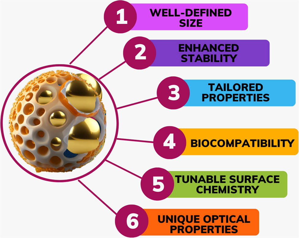
Polymer shell offers six benefits to magic numbers of gold clusters.
The “magic number” refers to a specific cluster diameter that implements unique stability and electronic features due to a complete or nearly complete electronic shell filling. In the matter of Au clusters stabilized by a shell of PVP, specific cluster diameters have been identified as having enhanced stability and distinct features. Overall, stabilizing Au clusters by a polymer shell may lead to the emergence of unique magic number diameters with improved stability and characteristic features, offering potential usages in areas like catalysis, sensing, and nanoelectronics. The supplied search results contain information about the “magic number” of Au clusters stabilized by a shell of PVP. According to the search results, a magic number smaller than 70 agrees with those of free Au clusters and may be explained qualitatively by the electronic shell model. An unprecedented magic number cluster, Au24Clx (x = 0–3), was selectively synthesized through kinetic control by monitoring reaction conditions, enabling the reduction of the Au precursor in the presence of polyvinylpyrrolidone (PVP). The magic stability of naked Au clusters is governed by electronic shell closure based on a jellium model. The same model explains the construction of Au34 and Au58 in PVP. In contrast, Au24Clx in Au24:PVP corresponds to an electronic shell closure. The cavity volume created by the multiple PVP chains determines the preferable and most minor diameter of Au clusters to be stabilized [58].
3.1 Polymer stabilization of Au clusters and its effects on electronic architecture
The protective ligand implements a pivotal role in the Au nanoclusters’ fluorescence. In fundamental scientific research, the inherent nanoparticle fluorescence, like nanoclusters, unique from its large crystals, has drawn considerable interest for practical use in many disciplines, including electronic and environmental usages [57]. Ligands may passivate the surface of the metal core, preventing aggregation and providing stability to the nanoclusters. Various ligands may interact with the metal core, affecting the nanoclusters’ electronic architecture and energy levels. This, in turn, may impact its fluorescence feature [59,60]. To illustrate this, innovative synthesis techniques should be created to create distinctive structures that would otherwise be challenging to produce using the present, broadly used techniques (mostly Brust–Schiffrin and ligand-exaltation techniques) [59]. Recently discovered anti-galvanic reduction (AGR), originally used to create bimetal nanoclusters (like Au25Ag2), has also been used to create mono-metal nanoparticles, like Au44(SC2H4Ph)32 [59]. Inspired by this, Yao et al. [59] synthesized the fluorescent phenylethanethiolated Au nanoclusters of Au24(SC2H4Ph)20, Au25(SC2H4Ph)18, Au38(SC2H4Ph)24, and Au144(SC2H4Ph)60 by AGR. The fluorescence quantum yield (QY) of Au24, approximately 40 times more than Au25 molecules, is the highest of all samples of phenylethanethiolated Au nanoclusters (including Au24, Au25, Au38, and Au144). Additionally, Au24 has three fluorescence lifetimes, each measuring 1.16, 45.25, and 267.63 ns. The ground state and the first singlet’s lowest vibrational level in the Au24 molecule have significant energy gaps. In Figure 3, the effect of quantum confinement on the optical absorption of gold nanoclusters (Au NCs) is illustrated. As the diameter of the Au NCs decreases, the spacing between discrete energy levels increases, leading to a blue shift in their optical absorption. For example, the Au10-12(SG)10-12, Au15(SG)13, Au18(SG)14, and Au25(SG)18 clusters exhibit absorption onsets at approximately 450 nm, 650 nm, 700 nm, and 900 nm, respectively. cluster, for instance, have absorption onsets that are 450, 650, 700, and 900 nm, respectively. For small-diameter Au NCs, solid quantum confinement influences lead to relaxation dynamics that are greatly influenced by atomic packing, shape, and diameter. The conversion of Au-sulphide (Au2S) particles to AuNPs may lead to considerable alterations in the optical absorption feature, including a blueshift in the plasmon absorption peak. This phenomenon may be attributed to the quantum diameter influences displayed by the AuNPs. Quantum diameter influences arise from the confinement of electrons within the nanoscale dimensions of the NPs. When the diameter of a metallic NP becomes comparable to or smaller than the characteristic length scale of the electrons, like the Fermi λ max, its electronic architecture and energy levels undergo quantization. This quantization leads to discrete energy levels and a modification of the electronic density of states. In the matter of AuNPs, as the diameter diminishes, the quantization of electronic states becomes more pronounced. This shifts the plasmon absorption peak to higher energies, leading to a blueshift in the observed optical absorption spectrum. The plasmon absorption peak corresponds to the collective oscillation of conduction electrons in the NP, and its energy is strongly influenced by the diameter and shape of the NP [61].
![Figure 3
(a) The structural diagram and variations in the optical absorption spectra of Au NCs of various diameters, like Au12, Au15, Au18, and Au25. The space among distinct energy levels broadens as the diameter of the Au NCs shrinks, and the clusters’ optical absorption turns blue. The number of Au atoms in atomically exact Au nanoclusters is intimately related to its photophysical characteristics; hence, the “magic number” of Au atoms was discovered to be present in Au NCs. The Au nanoclusters with magic numbers show high photophysical stability and resemblance to one another. The molecular Au nanocluster Au156 has a unique electronic structure among the molecular and metallic states, with distinct energy levels like molecular orbitals and metallic bands. Also given are the optical absorption spectra of identical-diameter Au NCs shielded by various ligands and the Tauc plot. Copyright 2014 American Chemical Society [62]. (b) The optical absorption spectra of charge-neutral and anionic Au25 clusters in solution are presented, highlighting the effect of cluster charge on their optical properties Copyright 2008 [63].](/document/doi/10.1515/ntrev-2025-0170/asset/graphic/j_ntrev-2025-0170_fig_003.jpg)
(a) The structural diagram and variations in the optical absorption spectra of Au NCs of various diameters, like Au12, Au15, Au18, and Au25. The space among distinct energy levels broadens as the diameter of the Au NCs shrinks, and the clusters’ optical absorption turns blue. The number of Au atoms in atomically exact Au nanoclusters is intimately related to its photophysical characteristics; hence, the “magic number” of Au atoms was discovered to be present in Au NCs. The Au nanoclusters with magic numbers show high photophysical stability and resemblance to one another. The molecular Au nanocluster Au156 has a unique electronic structure among the molecular and metallic states, with distinct energy levels like molecular orbitals and metallic bands. Also given are the optical absorption spectra of identical-diameter Au NCs shielded by various ligands and the Tauc plot. Copyright 2014 American Chemical Society [62]. (b) The optical absorption spectra of charge-neutral and anionic Au25 clusters in solution are presented, highlighting the effect of cluster charge on their optical properties Copyright 2008 [63].
4 Fluorescent protein-Au nanocrystal for environmental usage
AuNPs have potential environmental applications; here is an example of their usage. AuNPs have been used to detect environmental pollutants, such as mercury and lead. The AuNPs are functionalized with specific ligands, such as proteins that bind to the target pollutants, resulting in a colour change that can be detected visually or using spectrophotometry. Other environmental applications of AuNPs include nanoclusters for imaging and therapy in cancer theranostics [64]. Gold nanoclusters have been used for cell imaging and Fe3+ sensing [65]. Gold nanoclusters have been used for localized multimodal therapy and imaging of tumoural cells. Gold nanoclusters have been loaded onto albumin nanoparticles for tumour detection and ablation [66]. AuNPs are biosensors for detecting environmental toxins, such as pesticides and heavy metals [67]. AuNPs have potential ecological applications due to their unique properties and ease of functionalization. They have been used for detecting environmental pollutants, imaging, and therapy in cancer theranostics, and biosensors for environmental toxins. The benefit of protein as a stabilizer of Au NCs is that it has intrinsic biological activity, which makes it possible to create Au NCs with intriguing bio functions [68]. Fluorescent protein-Au nanocrystals hold significant potential for various bio-applications. One notable benefit of using proteins as stabilizers for Au nanocrystals is their intrinsic biological activity. This property enables the creation of Au nanocrystals with intriguing bio functions, making them highly versatile and suitable for a wide range of environmental usages.
Incorporating fluorescent proteins into Au nanocrystals allows for easy detection and monitoring of environmental pollutants and contaminants. These protein-stabilized NCs can serve as highly sensitive sensors for detecting changes in environmental conditions, such as the presence of specific chemicals or pollutants. Furthermore, the biocompatibility of protein-stabilized Au nanocrystals makes them suitable for various biological applications in environmental studies. They can be used as non-toxic labels or tracers to track biological processes in environmental samples or living organisms. In addition, the ability to engineer and modify the surface properties of these NCs by using different proteins opens up possibilities for the targeted delivery of environmental agents, such as pollutants or therapeutic compounds, to specific sites for remediation or treatment purposes. Table 4 lists various synthesis methods of protein and gold nanocrystal composites and their applications.
Summary of different synthesis methods of Au nanocrystals stabilized by protein
| Synthesis methods | Description | Application | Ref. |
|---|---|---|---|
| Co-precipitation | Simultaneous precipitation of gold ions and proteins | Bioimaging | [67] |
| Chemical reduction | Reduction of gold ions using chemical agents in the protein solution | Drug delivery | [70] |
| Green synthesis | Environmentally friendly methods using plant extracts | Photothermal therapy | [69,70] |
| Microemulsion | Formation of Au nanocrystals in a stabilized emulsion | Biosensing | [71,72,73] |
| Pulsed laser ablation | Laser-induced ablation of gold in the protein solution | Catalysis | [74,75,76] |
| Seed-mediated growth | Seeding growth of Au nanocrystals in the protein solution | Biomedical Imaging | [77,78,79] |
| Protein-directed synthesis | Proteins act as templates to control AuNP growth | Nanomedicine | [77,80,81] |
| Electrochemical synthesis | Formation of Au nanocrystals through electrochemical methods | Optoelectronic devices | [82,83,84] |
| Sol-Gel method | Formation of Au nanocrystals in a sol–gel matrix | Environmental remediation | [85,86,87] |
| Microwave-assisted synthesis | Rapid synthesis of gold NPs using microwave radiation | Antimicrobial coatings | [88,89 90] |
4.1 Lysozyme-stabilized Au NCs for bio functions
Lysozyme-stabilized Au NCs have emerged as a fascinating class of nanomaterials with diverse biofunctions. Lysozyme, an enzyme naturally found in various biological systems, is an excellent stabilizer for Au NCs due to its unique structural and functional properties. One prominent bio-function of lysozyme-stabilized Au NCs is their inherent antimicrobial activity. Lysozyme can disrupt bacterial cell walls by hydrolysing peptidoglycan, an essential component of bacterial cell walls. When combined with Au NCs, these nanoclusters can exhibit enhanced antimicrobial properties, making them valuable candidates for applications in antibacterial coatings, disinfectants, and wound dressings, contributing to environmental health and safety.
Moreover, lysozyme-stabilized Au NCs have shown remarkable potential in biomedical imaging. Their strong fluorescence properties make them suitable as fluorescent probes for imaging biological structures and processes, aiding in the visualization and study of cellular and molecular events in environmental samples or living organisms. In addition to their bioimaging applications, lysozyme-stabilized Au NCs have demonstrated promising results in drug delivery. These nanoclusters can be engineered to encapsulate therapeutic agents, protecting them from degradation and facilitating targeted delivery to specific sites in the body. This capability holds the potential for environmentally friendly and precise drug delivery systems, reducing the environmental impact of pharmaceutical compounds.
Furthermore, lysozyme-stabilized Au NCs have attracted attention as potential sensors for detecting various analytes in environmental samples. Their unique fluorescence properties can be exploited to create sensitive and selective sensing platforms for monitoring environmental pollutants or detecting biological molecules. Lysozyme (Lys) has been widely used by various researchers to stabilize gold nanoclusters (Au NCs). Lysozyme-stabilized Au nanoclusters (Lys-Au NCs) have been developed and applied as fluorescent probes for selective cyanide detection. The search results highlight the versatility of Au nanoclusters as functional nanomaterials in sensing and analytical chemistry. The average Lys-Au NC diameter was 4 nm, and they emitted a red light at 650 nm. Lys-Au NCs have also been used as a nanomedicine to induce osteogenic variolation and diminish osteoclast activity. The enzymatic activities of lysozyme may be modulated by the existence of surface ligands on ultra-low Au nanoclusters. This opens up possibilities for regulating enzymatic functions using these nanoclusters as molecular tools. In the context of Lys-Au NCs, these nanoclusters have been employed for label-free ratiometric fluorescent pH sensing. pH is a pivotal parameter in various biological and chemical processes, and the ability to monitor pH alterations in real-time is of great significance in many usages. Lys-Au NCs have also been used to develop photoactivated multifunctional nanoplatforms with improved antibacterial abilities compared to pure curcumin and Lys-Au NCs. Lysozyme NP-encapsulated Au nanoclusters (LysNP-Au NCs) have been designed as a dual-emission probe for ratiometric fluorescent detection of cyanide (CN−) in various environmental samples. Cyanide is a toxic substance that may be found in contaminated water sources, certain plants containing cyanogenic glycosides, and soil [91].
The lysozyme-capped Au NCs, which have a nano diameter of about 4 nm and were created in a previous study, have much potential for use in cyanide ion diagnostics. Cyanide ions linearly reduced the lysozyme-Au NCs’ emissive qualities. Combining NIR fluorescence with CT imaging in vivo makes bimodal bioimaging easier to distinguish between cancerous and healthy tissues. They found that when folic acid was added as a targeting agent, the lysozyme-capped Au NCs gathered in the tumour site after being delivered intravenously to HeLa tumour-bearing mice. When the folic acid alteration was not applied, the Au NCs’ fluorescence did not show up at the tumour site. The liver and kidney showed positive signal increases an hour after receiving the Au NCs injections for use in CT imaging, showing that the Au NCs mostly gather in these organs without tumour tissue [92].
4.2 Lactotransferrin (Lf) and horseradish peroxidase (HRP) gold nanoclusters in biomedical applications
Au NCs Lf refers to Lf protein that has been conjugated or encapsulated with Au nanoclusters. This hybrid nano material combines the unique feature of Lf and Au nanoclusters, opening up potential usages in various fields, including biomedical and biotechnological research [93]. HRP Au NCs refer to the conjugation or encapsulation of HRP enzyme with Au nanoclusters. HRP is an enzyme popularly found in horseradish roots and is broadly used in various biochemical and analytical usages due to its catalytic activity [94]. Human serum albumin (HSA) Au NCs refer to the conjugation or encapsulation of HSA protein with Au nanoclusters. HSA is a highly abundant protein found in human blood plasma. It plays important roles in maintaining osmotic pressure, transporting various molecules, and regulating the distribution of nutrients and drugs in the body [95]. Combining pepsin with Au NCs, the resulting nanomaterials may possess fluorescence features, enabling their use as fluorescent probes or markers in various usages [96]. Trypsin Au NCs with fluorescence capabilities may be applied for cellular or tissue imaging. The nanoclusters may be specifically targeted to certain cells or tissues by conjugating them with targeting ligands or antibodies. This allows for the visualization and tracking of specific biological targets in real-time [97] and egg white [98]. Production of photoluminescent Au NCs – several methods may produce photoluminescent Au nanoclusters. One popular approach is the reduction of Au ions in the existence of stabilizing ligands or capping agents. Here is a general outline of the process: Preparation of Au NCs precursor: Start with a suitable Au precursor, like Au salts (e.g. Au chloride, Au acetate, or AuNPs. These precursors supply the source of Au ions to construct Au NCs. Reducing agents: Introduce a reducing agent to reduce Au ions into Au NCs. Popular reducing agents include sodium borohydride (NaBH4), ascorbic acid, or other suitable reducing agents. The choice of reducing agent depends on the specific synthesis method and conditions. Stabilization with ligands: To monitor the diameter, shape, and stability of the Au NCs, add stabilizing ligands or capping agents. These ligands may supply surface passivation, prevent aggregation, and impart photoluminescent feature to the Au NCs. Popular ligands used for Au NC synthesis include thiols (e.g. thiolated organic molecules), polymers, proteins, or other surface-active molecules. Reaction and growth: Allow the reduction reaction to proceed under suitable conditions like temperature and reaction time. The reduction process leads to the growth and construction of photoluminescent Au NCs stabilized by the ligands. The specific reaction conditions may influence the diameter, shape, and photoluminescent feature of the resulting Au NCs. Purification and characterization: After the synthesis, the Au NCs need to be purified to remove any unreacted precursors, by-products, or excess ligands. Purification techniques may include centrifugation, dialysis, or other methods based on the features of the Au NCs. The purified Au NCs may then be characterized using UV-Vis spectroscopy, fluorescence spectroscopy, transmission electron microscopy (TEM), or other analytical methods to assess their diameter, morphology, and photoluminescent feature. Generally speaking, proteins with plenty of cysteines and tyrosines are thought to be excellent candidates for producing protein-Au NCs [91]. For example, due to their catalytic and optical properties, AuNP clusters like HRP-Au NCs (horseradish peroxidase-AuNP clusters) have potential applications as biocatalysts and biosensors. Biosensors for hydrogen peroxide detection (HRP-Au NCs) can be used as catalysts for reducing hydrogen peroxide, generating an optical or electrochemical signal that can indicate the presence of H2O2. This could be used to develop sensitive biosensors for hydrogen peroxide [99]. Further, HRP-Au NCs could act as catalytic labels in immunoassays, providing signal amplification through their peroxidase-like catalytic activity. This could enhance the sensitivity of assays for detecting biomarkers, proteins, etc. [100]. Additionally, the combination of enzymatic activity and photoluminescence opens up possibilities for advanced catalysis and sensing usages (HRP is an enzyme familiar for its peroxidase activity, which involves the catalytic reduction/oxidation of hydrogen peroxide (H2O2) [91]. In an in vivo study, insulin-stabilized Au NCs were employed to maintain their bioactivity in monitoring blood glucose like natural insulin [101]. No extra reducing agent is required because some proteins may serve as capping and reducing agents [102]. Alternately, the creation of Au NCs may benefit from the etching of larger Au NPs (typically 2–4 nm) by thiol compounds in an alkaline solution. The etching of larger Au NP (typically 2–4 nm) by thiol compounds in an alkaline solution is a popularly used method for the creation of smallest Au nanoclusters. This process is often called “etching-induced synthesis” or “ligand-mediated etching.” Here is a general outline of the etching-induced synthesis process: Preparation of larger Au nanoparticles: Initially, larger Au NPs are prepared through conventional synthesis methods, like chemical reduction or colloidal synthesis. These NP typically have diameters in the range of 2–4 nm. Thiol compounds: Thiol compounds, like thiols or thiolated ligands, are introduced to the solution containing the larger AuNPs. The thiol compounds act as etchants and attach to the surface of the AuNPs. Etching process: The thiol compounds in the alkaline solution undergo a redox reaction with Au atoms on the surface of the larger NPs. This reaction results in removing Au atoms from the surface and constructing Au-thiol complexes in the solution. Construction of Au nanoclusters: As the etching process continues, the Au NP gradually transforms into smaller-diameter Au nanoclusters. The diameter and shape of the resulting Au NCs may be monitored by adjusting the etching time, the concentration of thiol compounds, and reaction conditions. Purification and characterization: The Au NCs are typically purified by centrifugation or other separation techniques to remove excess thiol compounds and by-products after the etching process. The purified Au NCs may then be characterized using techniques like UV-Vis spectroscopy, TEM, or X-ray diffraction (XRD) to determine their diameter, morphology, and optical features [68].
Manjunath et al. [103] developed an electrochemical sensor using a nickel-activated carbon/PEDOT composite derived from coffee silver skin for detecting glyphosate (GLY) and hexaconazole (HEXA). The composite leverages activated carbon’s porosity, nickel’s electron-transfer enhancement, and PEDOT’s redox activity, achieving ultra-low detection limits of 0.8 ppt for GLY and 0.613 ppt for HEXA. Real-sample analysis in coffee beans validated the sensor’s practicality, with liquid chromatography–mass spectrometry corroborating its potential for precise pesticide monitoring in environmental and agricultural settings. On comparison, the AC/Ni/PEDOT sensor outperforms methods like Sadhu et al.’s [104] fluorescent gold nanocluster-based approach for HEXA detection, which reported limit of detection (LOD) of 21.94 nM (∼6,760 ppt). The composite’s ppt-level sensitivity demonstrates superior trace detection capabilities, making it more suitable for analysing complex agricultural matrices. The study by Nguyen Thi Nhat et al. [105] investigates the application of gold nanospheres (AuNSps) as a SPR-based sensor for in situ detection of residual fungicides, focusing on thiophanate methyl. AuNSps were synthesized via a seed-mediated method, where gold nanoseeds, formed by reducing chloroauric acid with trisodium citrate dihydrate (TSC), were grown into 53 nm spherical NPs using HAuCl4, TSC, and ethylenediaminetetraacetic acid. UV-vis spectroscopy, XRD, SEM-EDX, and TEM characterized their uniform size, crystallinity, and plasmonic properties with a resonance peak at 560 nm. When deposited on a glass substrate and coated with thiophanate methyl, the AuNSps demonstrated significant surface-enhanced Raman spectroscopy (SERS) effects, amplifying the fungicide’s Raman signal intensity compared to bare glass. This enhancement highlights the nanoparticles’ ability to improve trace-level detection sensitivity. The research underscores the potential of AuNSps-based sensors for rapid, on-site monitoring of fungicide residues in agricultural products, offering a practical solution to ensure food safety and reduce human exposure to harmful chemicals through cost-effective, field-deployable technology.
4.3 Fluorescent gold nanoclusters stabilized by BSA for biomedical applications
Numerous multifunctional nanocomposites have been used to construct numerous protein-stabilized Au NCs that may be used for medicinal, sensing, and targeting usages. Based on a one-pot synthetic route, Xie et al. used BSA to prepare Au NCs (37°C) with red emission λ em max and QY of 640 nm and 6%, respectively [106]. The protein molecules absorbed and sequestered the Au ions when Au(iii) ions were introduced to the aqueous BSA solution [107]. When the pH of the reaction was raised to 12 to activate the reduction ability of BSA molecules, the trapped ions gradually reduced to form Au NCs in situ. The 25 Au atoms in the as-prepared Au NCs were stabilized inside BSA molecules as BSA-Au NC bioconjugates. The use of BSA coating layer on Au nanoclusters supplies several advantages, including the ability to make post-synthesis surface modifications using functional ligands and the economic and environmental benefits associated with BSA as a biocompatible and cost-influence protein. When exposed to ultraviolet (UV) light (365 nm), the dark brown solution of BSA-coated Au NCs implements red fluorescence (Figure 4a, item no. 3 in the lower image). The mild blue fluorescence observed in the monitoring BSA solution under UV light may be attributed to the existence of aromatic side groups in the amino acid residues of BSA. BSA contains several aromatic amino acids, like tryptophan, tyrosine, and phenylalanine, which are familiar to implement intrinsic fluorescence (Figure 4a, item no. 2 in the lower image). The monitoring BSA solution was a pale-yellow colour when viewed with the naked eye (Figure 4a, item no. 2 in the upper image). Excitation and emission peaks for the fluorescent Au NCs were seen at 480 and 640 nm, respectively (Figure 4b). The QY for red photoluminescence was 6%.
![Figure 4
(a) Photographs of BSA and BSA-Au NCs (gold nanoclusters): 1. BSA powder: A sample of dry BSA powder, a common form of storage for proteins like BSA. 2. BSA aqueous solution: A clear or slightly cloudy liquid containing BSA dissolved in water. BSA is soluble in water and other aqueous solutions. 3. BSA-Au NCs aqueous solution under visible light: An aqueous solution containing BSA-Au NCs (gold nanoclusters) under regular visible light. The colour of the solution may vary depending on the size and concentration of the gold nanoclusters. 4. BSA-Au NCs powder under visible light: A dry powder or solid form of BSA-Au NCs under visible light. (b) Optical absorption and photoemission spectra: The graph represents the optical absorption and photoemission (fluorescence) spectra of two different samples: Blue curve represents the aqueous solution of BSA. The dashed line indicates the absorption spectrum, which shows the wavelengths of light absorbed by BSA. The solid line represents the photoemission spectrum, which shows the wavelengths of light emitted by BSA after photoexcitation. Red curve represents the aqueous solution of BSA-Au NCs. The dashed line indicates the absorption spectrum of the BSA-Au NCs, showing the wavelengths of light absorbed by the gold nanoclusters. The solid line represents the photoemission spectrum of BSA-Au NCs, showing the wavelengths of light emitted by the gold nanoclusters after photoexcitation at a wavelength of 470 nm (λ
ex = 470 nm). The inset in the graph likely shows the photoexcitation spectrum of BSA-Au NCs. This spectrum reveals the light absorption efficiency of the gold nanoclusters at different excitation wavelengths. Overall, the graph provides valuable information about the optical properties of BSA and BSA-Au NCs, demonstrating how adding gold nanoclusters can alter BSA’s absorption and photoemission properties. Copyright 2009 American Chemical Society [106].](/document/doi/10.1515/ntrev-2025-0170/asset/graphic/j_ntrev-2025-0170_fig_004.jpg)
(a) Photographs of BSA and BSA-Au NCs (gold nanoclusters): 1. BSA powder: A sample of dry BSA powder, a common form of storage for proteins like BSA. 2. BSA aqueous solution: A clear or slightly cloudy liquid containing BSA dissolved in water. BSA is soluble in water and other aqueous solutions. 3. BSA-Au NCs aqueous solution under visible light: An aqueous solution containing BSA-Au NCs (gold nanoclusters) under regular visible light. The colour of the solution may vary depending on the size and concentration of the gold nanoclusters. 4. BSA-Au NCs powder under visible light: A dry powder or solid form of BSA-Au NCs under visible light. (b) Optical absorption and photoemission spectra: The graph represents the optical absorption and photoemission (fluorescence) spectra of two different samples: Blue curve represents the aqueous solution of BSA. The dashed line indicates the absorption spectrum, which shows the wavelengths of light absorbed by BSA. The solid line represents the photoemission spectrum, which shows the wavelengths of light emitted by BSA after photoexcitation. Red curve represents the aqueous solution of BSA-Au NCs. The dashed line indicates the absorption spectrum of the BSA-Au NCs, showing the wavelengths of light absorbed by the gold nanoclusters. The solid line represents the photoemission spectrum of BSA-Au NCs, showing the wavelengths of light emitted by the gold nanoclusters after photoexcitation at a wavelength of 470 nm (λ ex = 470 nm). The inset in the graph likely shows the photoexcitation spectrum of BSA-Au NCs. This spectrum reveals the light absorption efficiency of the gold nanoclusters at different excitation wavelengths. Overall, the graph provides valuable information about the optical properties of BSA and BSA-Au NCs, demonstrating how adding gold nanoclusters can alter BSA’s absorption and photoemission properties. Copyright 2009 American Chemical Society [106].
Ivleva et al. [108] produced and stabilized bimetallic Au-Cd NCs with diameters less than 1.6 nm using BSA. In Figure 5, the maximum fluorescence emission λ max observed for various types of nanoclusters indicates its unique optical feature and behaviour. Figure 5 shows some characteristics of the maximum fluorescence emissions for Au NCs, Cd NCs, and mixed Au-Cd NCs.

The TEM images and mass spectra of (a) Au NCs, (b) Cd NCs under UV light at 365 nm, and (c) fluorescent emission spectra of Au NCs. (d) Fluorescent emission spectra of Cd NCs. (e) Fluorescent emission spectra of Au/Cd NCs.
For Au NCs, maximum emission λ max was observed at 325, 410, and 650 nm. This emission λ max indicates that Au NCs implement fluorescence in the spectrum’s UV, blue, and red parts. The excitation at 325 nm suggests the existence of specific electronic transitions or energy levels in the Au NCs that result in emission at shorter λ max (UV part). The 410 and 650 nm emissions indicate fluorescence in the blue and red parts, respectively. For Cd NCs, the maximum emission λ max was observed at 370 and 500 nm. These emission λ max values suggest that Cd NCs implement fluorescence in the UV and green parts of the spectrum. The excitation at 370 nm indicates electronic transitions or energy levels in the Cd NCs, resulting in emission at shorter λ max (UV part). The emission at 500 nm suggests fluorescence in the green part.
For mixed Au-Cd NCs, two emission peaks were observed at 440 and 640 nm. These dual emission peaks indicate the existence of various chromophores or energy levels within the mixed Au-Cd NCs. The excitation at 365–380 nm results in the simultaneous emission at 440 and 640 nm, suggesting the existence of distinct emission mechanisms or transitions. Compared to conventional organic fluorophores and QDs, the stabilized fluorescent NCs offer several advantages, such as minor diameter: NCs are typically on the nanometre scale, allowing for better penetration and interaction with biological systems. Considerable Stokes shift: The difference among the excitation and emission λ max, familiar as the Stokes shift, is often more prominent in NCs. This feature helps reduce self-absorption and improves detection sensitivity. Biocompatibility: NCs may be engineered biocompatible, making them suitable for various biological and biomedical usages. High stability: NCs may implement excellent chemical and photostability, ensuring long-term fluorescence performance. Prolonged fluorescence lifetime: NCs often have longer than organic fluorophores, which may be advantageous for time-resolved measurements and imaging techniques. These advantages make stabilized fluorescent NCs promising candidates for various usages, including bioimaging, biosensing, and optoelectronics.
4.4 Glutathione (GSH)-coated gold nanoclusters with enhanced fluorescence
The tripeptide GSH, which has a low affinity for and interaction with biological proteins, has frequently been used as an Au NC surface ligand [109].
GSH-coated AU NCs (GSH-Au NCs) have emerged as a remarkable class of nanomaterials with enhanced fluorescence properties, making them highly valuable for various applications. The unique combination of gold nanoclusters with GSH, a naturally occurring tripeptide found in living organisms, enhances fluorescence emission. GSH is an excellent stabilizer for gold nanoclusters, preventing aggregation and enhancing their fluorescence intensity.
One of the key advantages of GSH-Au NCs is their excellent biocompatibility. GSH is a vital antioxidant in cells, and its presence in the nanoclusters makes them biologically compatible and safe for use in various biological applications, including bioimaging and biosensing.
In bioimaging, GSH-Au NCs have shown great promise as fluorescent probes for cellular imaging. Their enhanced fluorescence emission allows for high-resolution imaging of cellular structures and processes, enabling researchers to gain insights into cellular behaviour and functions in environmental studies.
Moreover, GSH-Au NCs have demonstrated potential in biosensing applications. Their strong and stable fluorescence can be exploited to design sensitive and selective sensors for detecting various analytes in environmental samples, such as heavy metals, pollutants, or biomolecules. This capability offers a powerful tool for environmental monitoring and detecting contaminants in different environmental settings.
Another advantage of GSH-Au NCs is their potential as drug delivery vehicles. The biocompatibility and stability of GSH-Au NCs make them suitable for loading therapeutic agents and delivering them to specific targets in the body. This could lead to environmentally friendly and targeted drug delivery systems with reduced side effects. GSH has been observed to improve renal clearance and diminish the buildup of Au NCs in the liver and spleen, causing at least 50% of GSH-Au NCs to be successfully eliminated from the body via the urinary systems within 24 h following IV injection [110]. Zhou et al. observed a QY of 3.5% and photoluminescence at about 560 nm in the NIR band [110]. Further to this, Luo et al. [111] found that aggregation by solvent mixing and a lower thiol-to-Au ratio (1.5:1 instead of 2:1) during the synthesis of Au NCs may result in a higher QY of about 15% in the setting of GSH-Au complexes [68,111]. A now-promising and well-recognized phenomenon is that metal nanocluster aggregation-induced emission enables the influence synthesis of extremely luminous nanoclusters. The fluorescence QYs of the Au nanocrystals stabilized by GSH-Au NCs dispersed in an aqueous solution are only 1%. Still, when Zn NP is added, the QYs of the Zn-GSH-Au NCs go up to 40%. A now-promising and well-recognized phenomenon is that metal nanocluster aggregation-induced emission enables the influence synthesis of extremely luminous nanoclusters. The fluorescence QYs of the Au nanocrystals stabilized by GSH-Au NCs dispersed in an aqueous solution are only 1%. Still, when Zn NP added, the QYs of the Zn-GSH-Au NCs go up to 40% [112]. Poly(amidoamine) is a dendrimer with a sphere-like form that has been the subject of the most research due to its robust production, availability, dendritic structure, and protein and peptide mimic feature [113].
5 Fluorescent polymer Au nanocrystals
Fluorescent Au nanoclusters and fluorescent Au NDs are both terms used to describe Au nanomaterials with diameters smaller than 3 nm [114]. Unlike larger AuNPs, which implement SPR absorption in the visible part, Au nanoclusters typically do not implement strong SPR absorption due to their minor diameter and unique electronic structure. However, they may implement fluorescence emission in the visible to NIR range, making them attractive for bioimaging, sensing, and optoelectronics. The fluorescence feature of Au NCs arises from quantum confinement influences and the interactions among the Au core and surface ligands [115].
In Figure 6, the synthesis of Au nanoclusters may be achieved by selecting appropriate capping ligands acting as both capping and reducing agents. In some matters, polymers, proteins, or biomolecules may influence capping and reducing agents, eliminating the need for a separate reducing agent in the synthesis process. The existence of these capping ligands helps stabilize the Au NCs and monitor the diameter and features. By choosing various capping ligands, it is possible to tune the fluorescence emission of the Au NCs across a range of spectral λ max, allowing for versatile usages in multiple fields, including sensing, imaging, and catalysis.
![Figure 6
The choice of ligands has a considerable impact on the fluorescence feature of Au NCs. Ligands may influence the diameter, shape, and surface feature of the Au NCs, which in turn affect its fluorescence emission. Selecting specific ligands allows monitoring of the energy levels and electronic transitions within the Au NCs, leading to a tuneable fluorescence feature. Ligands may also implement a role in stabilizing the Au NCs and preventing aggregation, which is pivotal for maintaining its fluorescence efficiency [68].](/document/doi/10.1515/ntrev-2025-0170/asset/graphic/j_ntrev-2025-0170_fig_006.jpg)
The choice of ligands has a considerable impact on the fluorescence feature of Au NCs. Ligands may influence the diameter, shape, and surface feature of the Au NCs, which in turn affect its fluorescence emission. Selecting specific ligands allows monitoring of the energy levels and electronic transitions within the Au NCs, leading to a tuneable fluorescence feature. Ligands may also implement a role in stabilizing the Au NCs and preventing aggregation, which is pivotal for maintaining its fluorescence efficiency [68].
Fluorescent Au NCs were created using a template polymer and reducing agents like poly(amidoamine) and NaBH4. Changing the molar ratio of the template polymer to Au3+ ions from 1:1 to 1:15 allows various diameters of Au NCs. The atom numbers mentioned (5, 8, 13, 23, and 31) represent the diameters of the resulting Au NCs, with each number indicating the number of Au atoms in the cluster. The resulting Au NCs implement fluorescence emission in the NIR part. The QY, which represents the efficiency of fluorescence emission, may vary depending on the diameter and architecture of the Au NCs. In this matter, the QY ranges from 10 to 70%, indicating the varying fluorescence efficiencies of the various-diameters Au NCs [116,117]. Fluorescent Au NCs have been produced using polymers like poly(tert-butyl methacrylate), poly(n-butyl methacrylate), and poly(methyl methacrylate). These Au NCs emit blue light with QY of 20.1, 14.3, and 3.8%, respectively. The molar ratio of the template polymer poly(amidoamine) dendrimer (PAMAM) to Au ions may be used to monitor the diameter and QY of Au NCs. By adjusting this ratio, the amount of polymer available for capping and stabilizing the Au NCs may be modulated, affecting the Au clusters’ growth and organization [118]. A polymer-induced etching procedure may also create Au NCs from polymer-stabilized NCs. For instance, the production of Au8 NCs with a 445 nm emission and a QY of 10–20% occurred when polyethyleneimine containing multivalent imine groups was employed in place of the initial capping agent dodecyl amine and to etch the Au NPs. A nanocomposite is formed by combining the amphiphilic copolymer, GSH-stabilized Au NCs, and the targeted ligand. This nanocomposite may implement enhanced features like improved stability, biocompatibility, and targeted delivery, making it suitable for various biomedical usages. Using an amphiphilic copolymer, poly(DBAM-co-NAS-co-HEMA), GSH-stabilized Au NCs, and a targeted ligand (folic acid) allows for a nanocomposite. The amphiphilic copolymer, poly(DBAM-co-NAS-co-HEMA), possesses both hydrophilic and hydrophobic segments within its structure. This property makes it compatible with aqueous and organic environments, allowing it to interact with the Au NCs. GSH serves as a stabilizing agent for the Au NCs, preventing its aggregation and maintaining its structural integrity. GSH is a tripeptide molecule composed of three amino acids (glutamate, cysteine, and glycine) and is familiar for its antioxidant features. The targeted ligand, folic acid, is incorporated into the nanocomposite to supply specific recognition and binding to its corresponding receptor on the target cells. Folic acid receptors are often overexpressed in specific matter cells, making it a functional targeting ligand for selective delivery of the nanocomposite to master cells [68]. The hydrophobic medication (paclitaxel) was then added to the Au nanocomposite, which self-assembled to create a core-satellites nanocomposite. The medications encapsulated in the polymer were released in mildly acidic endosomal/lysosomal compartments by breaking the pH-labile links [119,120].
Figure 7 shows an increase in fluorescent intensity with an increase in the diameter of the Au NRs. The maximum fluorescent intensity was observed for Au NRs with a diameter of 10 nm. Specifically, the fluorescence intensity was enhanced 120-fold for Au NRs measuring (113 × 38) nm2 and 40-fold for Au NRs measuring (46 × 14) nm2. This finding suggests that the Au nanorods’ diameter is pivotal in determining its fluorescence feature. As the diameter of the nanorods increases, the fluorescent intensity also increases. The observed enhancement in fluorescence may be attributed to factors like the increased surface area of the larger nanorods, which allows for more interaction with the fluorophores or localized surface plasmon resonance (LSPR) influences that enhance the fluorescence signal. The considerable enhancement in fluorescence, as demonstrated by the high-fold increase, highlights the potential of Au NRs as efficient fluorescent probes or imaging agents. Its unique diameter-dependent fluorescence feature makes them promising candidates for various usages, including biological imaging, sensing, and diagnostic assays.
![Figure 7
(a) TEM images of the plasmonic alterations with the density and diameter of Au NRs. The Au NRs’ dimensions from left to right is (46 × 14) nm, (57 × 18) nm, (62 × 18) nm, (85 × 24) nm, (105 × 31) nm, (113 × 38) nm, (130 × 40) nm, and (155 × 48) nm. (b) The electromagnetic wave enhancement of Au NRs at various diameters. (c) Finite-difference time-domain simulation, displaying the electromagnetic wave around Au NRs with multiple diameters (the scale bar represents 50 nm). Copyright © 2021 American Chemical Society [121].](/document/doi/10.1515/ntrev-2025-0170/asset/graphic/j_ntrev-2025-0170_fig_007.jpg)
(a) TEM images of the plasmonic alterations with the density and diameter of Au NRs. The Au NRs’ dimensions from left to right is (46 × 14) nm, (57 × 18) nm, (62 × 18) nm, (85 × 24) nm, (105 × 31) nm, (113 × 38) nm, (130 × 40) nm, and (155 × 48) nm. (b) The electromagnetic wave enhancement of Au NRs at various diameters. (c) Finite-difference time-domain simulation, displaying the electromagnetic wave around Au NRs with multiple diameters (the scale bar represents 50 nm). Copyright © 2021 American Chemical Society [121].
6 Effects of synthetic precursors on the diameter of Au NRs and their relationship with fluorescence intensity
The relationship between the fluorescence intensity and the diameter of Au NRs is complex and multifaceted, depending on various factors. Au NRs exhibit unique optical properties, including plasmon resonance, strongly influencing their fluorescence behaviour [54]. Here are some general trends and considerations regarding the fluorescence intensity and nanorod diameter:
Plasmon resonance : Au NRs have a characteristic plasmon resonance peak, which depends on their aspect ratio (length-to-width ratio). The plasmon resonance peak determines the wavelength at which the nanorods efficiently absorb and scatter light. It is essential to note that plasmon resonance is not directly related to fluorescence but plays a significant role in the excitation and enhancement of fluorescence [122]. Au NRs have a plasmon resonance peak that depends on their aspect ratio. This peak determines the wavelengths of light they most strongly absorb and scatter.
When the plasmon resonance peak overlaps with the excitation wavelength of a fluorophore [123], it can efficiently excite the fluorophore and enhance its absorption. This leads to higher fluorescence. The localized plasmonic fields generated near the nanorod surface can also enhance the emission rate of fluorophores, leading to higher fluorescence intensities [124]. Wider nanorods with red-shifted plasmon resonance peaks provide better spectral overlap with fluorophores’ excitation and emission wavelengths [125]. Plasmon resonance’s strong local electromagnetic fields can effectively increase nearby fluorophores’ absorption and emission rates [126]. Both of these effects contribute to enhanced fluorescence. However, plasmon resonance itself does not directly produce fluorescence – it requires the presence of fluorophores whose excitation and emission it can enhance [127].
Size-dependent fluorescence : The fluorescence intensity of Au NRs can be influenced by their size and precisely their diameter. In some cases, smaller NRs may exhibit higher fluorescence intensity due to quantum confinement effects, which can enhance the exciton behaviour. However, larger NRs may show higher scattering and absorption efficiency, leading to more efficient excitation and, subsequently, higher fluorescence signals. The relationship between Au NR diameter and fluorescence intensity is complex and depends on several factors. Smaller NRs can show higher fluorescence due to quantum confinement effects. As NR diameter decreases towards the quantum regime, quantum confinement of electrons can enhance exciton behaviour and radiative recombination rates. This can lead to higher fluorescence intensities. However, larger NRs tend to have higher scattering and absorption efficiencies due to their plasmon resonances. So, the fluorescence of Au NRs is influenced by a combination of quantum confinement effects (which favour smaller NRs) and plasmonic enhancement effects (which favour larger NRs). The relative strengths of these two opposing trends depend strongly on the specific nanorod-fluorophore system and solution conditions. The net result is that the fluorescence-diameter relationship is often non-monotonic, with either smaller or larger NRs (within a given synthetic batch) exhibiting the highest fluorescence intensities, depending on the situation.
Ligand effects : The surface chemistry and functionalization of Au NR attachment with a specific ligand can impact their fluorescence behaviour. Surface ligands or capping agents can affect the charge transfer and interactions at the nanorod surface, influencing the fluorescence intensity. Surface charge – ligands that impart a net positive or negative surface charge to the NRs can differentially influence specific fluorophores’ association and exciton coupling. The following parameters can impact fluorescence intensities such as the nature of surface ligands. Some ligands can facilitate chargetransfer between the nanorod (NR) surface and thesurrounding environment, thereby enhancing orquenching fluorescence and fluorophores, eitherquenching or enhancing their fluorescence. Themagnitude of this effect depends on the specificligands used. Certain ligands can bind or conjugatefluorophores to the NR surface more efficiently, leading structural changes. This can enhance fluorescence intensities. Ligands that passivate the NR surface more uniformly can minimize fluorescence quenching effects related to high surface curvature. This tends to increase fluorescence. More extended/linear ligands can allow better access of fluorophores to the plasmonic “hot spots” near the nanorod surface, enhancing fluorescence. More stable ligands can minimize the aggregation and precipitation of NRs, preserving their fluorescence over time. Less stable ligands can quench fluorescence. So, the choice of surface ligands on Au NRs can impact their fluorescence in various ways related to surface charge, charge transfer, fluorophore binding/access, ligand rigidity, and nanorod stability. Engineering the surface chemistry of NRs allows for fine-tuning of their fluorescence properties.
Fluorophore attachment : Au NRs can be functionalized with various fluorophores or fluorescent molecules. The choice of the fluorophore and its attachment method can significantly affect the fluorescence intensity, irrespective of the nanorod diameter, regarding the following factors: Covalently attaching fluorophores ensures they are stable and positioned optimally for plasmonic enhancement. Non-covalent attachment can be less stable and less well-defined. Fluorophores closely and uniformly attached to the NR surface can best take advantage of plasmon-enhanced fluorescence. Those that are further away may show lower enhancement. The relative orientation of fluorophores to the NR can impact how strongly they couple to the plasmonic near-fields. More parallel orientations tend to produce higher enhancement. Fluorophores with excitation and emission spectra that best overlap the plasmon resonance bandwidth of the NRs show the most remarkable enhancement. Non-overlapping fluorophores may show minor enhancement. The emission wavelength of fluorophores relative to the plasmon resonance also influences how well the NR can re-radiate and enhance the emission. Matching wavelengths provides optimal enhancement. Intrinsically brighter fluorophores with higher QYs produce higher fluorescence signals, all else equal. Darker fluorophores result in lower signals. So, factors like the conjugation method, distance, orientation, spectral overlap, emission wavelength, and QY of attached fluorophores can significantly impact the overall fluorescence intensity of Au NRs – irrespective of their actual diameter. Effectively functionalizing NRs with appropriate fluorophores is crucial for maximizing fluorescence signals.
Aggregation : NR aggregation can occur, especially in solution, and may affect the fluorescence behaviour. Aggregation-induced changes in the local electromagnetic environment can alter the fluorescence intensity. The shapes and configurations of NRs within aggregates can differ significantly from isolated NRs. This can change both plasmonic and fluorescence properties. When NRs aggregate, their plasmon resonances can couple together and shift. This can change the local electromagnetic environment and plasmonic enhancement of attached fluorophores, altering the fluorescence intensity. Aggregated NRs have a higher proportion of surface atoms that can quench the fluorescence of nearby fluorophores. This can reduce the overall fluorescence intensity. The local concentration of fluorophores increases within aggregates, which can influence their excitation and emission rates. This can impact fluorescence intensities in either direction. Aggregation-induced spectral changes in the NRs and fluorophores can alter the degree of spectral overlap and plasmonic enhancement. This again impacts fluorescence intensities. Excitons can migrate further within aggregates before recombining radiatively. This can either enhance or quench fluorescence, depending on the system.
Solution conditions: The fluorescence of Au NRs can be influenced by their surroundings, including the refractive index of the medium and the presence of other molecules. Changes in the local environment can lead to alterations in fluorescence intensity. (i) As the surrounding medium’s refractive index increases, the NRs’ plasmon resonance red-shifts. This can alter the degree of spectral overlap and plasmonic enhancement of attached fluorophores, impacting fluorescence intensities. (ii) More polarizable environments can enhance the near-field interaction between NRs and fluorophores, increasing fluorescence intensities. (iii) The presence of other molecules that can associate with NRs or fluorophores can impact fluorescence through factors like aggregation, energy transfer, charge transfer, etc. (iv) Higher ion concentrations can screen the surface charge of NRs, altering their association with fluorophores and resulting fluorescence intensities. (v) Changes in pH can protonate or deprotonate surface ligands and fluorophores in different ways, impacting their interaction with NRs and fluorescence properties. (vi) Oxygen quenches the triplet state of many fluorophores, influencing their fluorescence QY. Higher oxygen levels tend to increase fluorescence. So, aspects of the local environment, like refractive index, polarizability, molecular composition, ionic strength, pH, and oxygen content, can all impact the fluorescence of Au NRs. Any changes to the surrounding medium that influence the system’s plasmonic or fluorescent properties can alter the observed fluorescence intensities. Engineering the NR environment – through choice of solvent, additives, pH adjustment, degassing, etc. – provides a helpful way to control and optimize their fluorescence-based applications.
In summary, the relationship between the fluorescence intensity and the diameter of Au NRs is influenced by a combination of factors, including plasmon resonance, size-dependent effects, surface chemistry, fluorophore attachment, aggregation, and the local environment. Researchers often explore and optimize these parameters to tailor the fluorescence behaviour of Au NRs for specific applications, such as bioimaging, sensing, and nanomedicine.
The final factors of solution conditions and their influence on the diameter of Au NRs can be controlled by various synthetic precursors and reaction conditions during their synthesis. Some of the additional factors can affect synthetic precursors on the diameter of Au NRs as follows:
Gold salt concentration : The concentration of the gold salt, typically HAuCl4, used in the synthesis plays a crucial role in determining the final diameter of the NRs. Higher concentrations of gold salt can lead to larger NRs, as more gold ions are available for growth. Conversely, lower concentrations may result in smaller NRs.
Silver nitrate concentration : In seed-mediated growth methods, the concentration of silver nitrate (AgNO3) influences the aspect ratio of the NRs, which is the ratio of their length to diameter. Higher concentrations of AgNO3 can lead to longer NRs with a constant diameter, while lower concentrations may result in shorter NRs.
Surfactant type and concentration : Surfactants, such as cetyltrimethylammonium bromide (CTAB), play a crucial role in controlling the growth and stabilization of Au NRs. The surfactant’s type and concentration can influence the NRs’ diameter. For example, higher CTAB concentrations are often used to obtain smaller-diameter NRs.
Reducing agent : The choice and concentration of the reducing agent used in the synthesis can also affect the diameter of the Au NRs. Different reducing agents may lead to varying rates of gold ion reduction and subsequent nanorod growth, influencing the final diameter.
Seed concentration : In seed-mediated growth methods, the concentration of the seed nanoparticles used as nucleation sites can impact the final diameter of the NRs. Higher seed concentrations typically result in smaller NRs due to a higher density of nucleation sites.
pH of the reaction : The reaction mixture’s pH can influence the NRs growth kinetics, affecting their diameter. Adjusting the pH can be a way to control the size of the NRs.
Reaction time and temperature : The reaction time and temperature also play significant roles in the growth of Au NRs. Longer reaction times or higher temperatures may result in larger NRs, while shorter or lower temperatures may lead to smaller NRs.
Overall, the diameter of Au NRs can be finely tuned and controlled by adjusting the concentrations of gold salt, AgNO3, surfactants, reducing agents, seed NPs, pH, reaction time, and temperature. Understanding the effects of these synthetic precursors is essential for producing Au NRs with desired dimensions and properties for various applications, including biomedical imaging, catalysis, and sensing.
Finally, there is a relationship between the fluorescence intensity and the diameter of Au NRs. As the diameter of Au NRs increases, their fluorescence intensity also tends to increase. This is due to the following reasons: As the diameter increases, the NRs’ aspect ratio (length/diameter) decreases. This leads to a red shift in the NRs’ longitudinal surface plasmon resonance (LSPR) peak, which correlates with increased fluorescence intensity. Large-diameter NRs have a higher absorption cross-section, which absorbs more light. This increased light absorption contributes to the higher fluorescence intensity. Small-diameter NRs have a higher percentage of surface atoms than their bulk atoms. These surface atoms can quench the fluorescence more efficiently, leading to lower fluorescence intensities for small-diameter NRs. Large-diameter NRs have more “space” inside for fluorescent molecules to bind and emit light. Smaller NRs have a higher surface curvature that can hinder fluorescent molecule binding and fluorescence. So, as the diameter of Au NRs increases, their fluorescence intensity also tends to increase. This is due to factors like red-shifting of the LSPR peak, higher light absorption, less fluorescence quenching, and more space for fluorescent molecules – all of which contribute to the observed trend. However, the relationship is not strictly linear; other factors also play a role in a nanorod-fluorophore system.
7 Environmental and bio-usages of simultaneous fluorescent Au nanocrystals
Fluorescent Au nanocrystals have attracted considerable attention for their potential applications in environmental and biological fields, such as biosensing, bioimaging, and biolabeling. These NCs exhibit unique optical properties, such as tuneable emission wavelength, high photostability, and biocompatibility, which make them superior to conventional organic fluorophores and QDs. Moreover, the simultaneous fluorescence and plasmon resonance of Au nanocrystals enable the integration of multiple functions in a single nanostructure, such as fluorescence enhancement, surface-enhanced Raman scattering (SERS), and photothermal therapy. This section summarizes the recent advances in the synthesis, characterization, and environmental and bio-usages of simultaneous fluorescent Au nanocrystals, emphasizing their structure–property relationships and functionalization strategies. We also discuss the challenges and perspectives for the future development of these nanomaterials.
7.1 Multimodal imaging using Au nanoclusters: Advantages and applications
Researchers have tested the feasibility of Au NCs and hybrid materials, including Au NCs as an imaging agent in vivo, using X-ray computed tomography (X-ray CT), magnetic resonance imaging (MRI), positron emission tomography (PET), and NIR fluorescence imaging [128]. Each imaging technique has advantages and disadvantages of its own (Table 5). Multimodal imaging integrates many imaging techniques, fusing the best features of each [129]. As a result, employing more than one imaging technique with a single imaging agent is beneficial. More imaging agents should be collected in the tumour than other organs during imaging tests to diagnose cancer. Therefore, the platform should target the tumour actively or inactively [130].
Advantages and disadvantages of each imaging technique
| Imaging technique | Advantages | Disadvantages |
|---|---|---|
| X-ray CT | High spatial resolution | Ionizing radiation exposure |
| Quick image acquisition | Limited soft tissue contrast | |
| Suitable for bone and dense tissues | Not ideal for functional or molecular imaging | |
| Potential risks for pregnant patients and children | ||
| MRI | Excellent soft tissue contrast | Expensive and time-consuming |
| No ionizing radiation | Lower spatial resolution compared to CT | |
| Wide range of applications | Can be affected by metal implants | |
| Multiplanar imaging capabilities | It may not be suitable for patients with claustrophobia. | |
| High sensitivity and molecular imaging | Radiation exposure | |
| PET | Quantitative measurement of metabolic | Expensive radiopharmaceuticals |
| and biochemical processes | Limited availability of radiotracers | |
| Functional and molecular imaging | Requires cyclotron for radiotracer production | |
| Wide range of applications | ||
| NIR fluorescence imaging | Non-invasive and real-time imaging | Limited tissue penetration depth |
| High sensitivity and specificity | Autofluorescence from tissues | |
| Multiplexing potential | Limited depth for in vivo imaging | |
| Safe and cost-effective | Requires fluorescent contrast agents |
7.2 Passive and active targeting strategies for Au nanoclusters in cancer imaging
Passive targeting is made possible by using the enhanced permeability and retention influence in body areas where there is a lot of hypoxia and/or inflammation, both of which are features of the tumour microenvironment [131]. Extravasation of Au NC-containing NP platforms from 10 to 500 nm in the blood serum into the tissue is conceivable because the tumour’s unpredictable growth and holes in the endothelium cause irregularities in the vasculature [132]. In vivo, FA- or HA-coated BSA-stabilized Au NCs displayed similar fluorescent features and gathered in HeLa or Hep-2 tumours, respectively. MRI typically uses gadolinium-functionalized Au NCs [128]. Au MRI multimodal imaging studies have also used silica quantum rattles and mesoporous silica NP packed with Au NCs and AuNPs. NIR fluorescence imaging and CT imaging may be done depending on the various modifications or conjugations made to the Au NC. Shortwave infrared emitting Au NCs have demonstrated outstanding potential for in vivo imaging with more excellent contrast than current NIR imaging and support for PET [128]. Additionally, the acquired system could be exploited for PET and fluorescence dual-imaging in lung cancer by coupling iodine-124 to a peptide-protected Au NC, as done by Daems et al. [133]. The use of radiolabelled Au for image-guided therapy is also discussed, as well as the improvement of targeted radionuclide therapy and nano brachytherapy through an increased dose deposition and radio sensitization, as demonstrated by numerous Monte Carlo studies and experimental in vitro and in vivo studies [133]. In 2023, Borse et al. [134] prepared bimetallic nanoclusters of copper and Au with papain as a ligand for fluorescence detection of cortisone in vitro biological samples.
Its investigation discovered that the hybrid nanoclusters displayed outstanding signals for NIR fluorescence with an impressive detection limit of 1.89 nM at a concentration as low as 0.031–1.00 M. Bimetallic Au and copper nanoclusters (Au-Cu NCs) were produced using papain as a ligand. The papain-Au-Cu NCs displayed red fluorescence at 365 nm of UV light. The papain-Au-Cu NCs, as they are generated, show a high fluorescence at 656 nm when activated at 390 nm. Red fluorescence “switches off” in papain-encapsulated Au-Cu NCs due to a particular corticosteroid interaction site. When cortisone was added as a biomarker, papain-Au-Cu NCs demonstrated rapid response and outstanding fluorescence quenching. The constructed probe is revealed to be capable of quantifying cortisone by creating the calibration graph among the fluorescence ratio (I 0/I) and cortisone concentration (0.031–1.00 M) with an astounding detection limit of 1.89 nM.
Interestingly, papain-Au-Cu NCs are highly selective for cortisone detection, as seen by their relatively poor responsiveness to frequent interfering species and other biomarkers. The probe was successfully used to check for the cortisone biomarker in plasma and urine samples. Furthermore, by assessing the analytical validity of the probe by assaying cortisone in intra- and inter-day, it was demonstrated that papain-Au-Cu NCs might be a promising probe for the fast detection of cortisone in biological materials.
Another triple-modal imaging platform was described in 2023 by Wang et al. [135]. The development of a non-invasive multimodal contrast agent (aminoglycoside-polyglutamate [AGGP]) coordinated with a high-affinity prostate-specific membrane antigen (PSMA) ligand (PSMA1) offers promising opportunities for the accurate detection and characterization of prostate cancer (PCa). The AGGP agent combines MR, CT, and near-infrared fluorescence (NIRF) imaging modalities, allowing for the comprehensive and precise imaging of PSMA expression in PCa lesions. Existing contrast agents, particularly Gd-containing NPs, have encountered limitations like nonspecific redistribution in the mononuclear phagocyte system (MPS) and insufficient perfusion to target locations. Moreover, the intrinsic limitations of MR tools have made it challenging to depict PSMA features accurately using these techniques alone. By utilizing AGGP, researchers have successfully demonstrated potent signal augmentations in PCa lesions by integrating MR, CT, and NIRF imaging. The AGGP agent implements specific targeting to PSMA, efficient evasion of the MPS, and beneficial renal-clearable behaviour in living mice, thus overcoming the limitations of previous contrast agents. Biocompatibility and histopathology tests have also supported the high safety of AGGP in vivo, making it a promising tool for more accurate early-stage PCa detection and the development of influence multifunctional nanotherapeutics.
7.3 Temperature-sensitive fluorescence properties of Au nanoclusters for biological thermometry
Furthermore, the AGGP agent allows for active targeting by coupling targeting ligands to the nanoclusters, enhancing its specificity towards PSMA-expressing cells or tissues. This multimodal approach enables the use of a single agent for NIRF, MR, and photoacoustic imaging in both in vitro and in vivo settings, providing a comprehensive assessment of PSMA expression and distribution. Au nanoclusters also possess temperature-sensitive features besides their imaging capabilities.
The fluorescence lifetime and emission intensity of Au nanoclusters undergo significant changes within the physiologically relevant temperature range of 15–45°C. This temperature-dependent behaviour can be attributed to the influence of temperature on the electronic structure and optical properties of the Au NCs.
At lower temperatures, such as 15°C, the fluorescence lifetime of Au NCs may increase, leading to longer emission decay times. This could be due to changes in the excited-state relaxation processes or energy transfer mechanisms within the nanoclusters. Additionally, the emission intensity of Au NCs may be lower at lower temperatures, potentially caused by decreased radiative transition rates or increased nonradiative pathways.
As the temperature increases within the physiological range, the fluorescence lifetime of Au NCs tends to decrease. This decrease in lifetime is often associated with increased thermal energy, which promotes faster relaxation of the excited states and shorter emission decay times. Moreover, the emission intensity of Au NCs may also increase at higher temperatures, indicating enhanced radiative transition rates and potentially reduced nonradiative processes.
These temperature-dependent alterations in the fluorescence properties of Au NCs can be leveraged for various applications, including temperature sensing or imaging in biological systems. By monitoring the changes in fluorescence lifetime and emission intensity of Au NCs within the physiological temperature range, it is possible to obtain valuable information about local temperature variations in biological environments or during specific processes.
It is worth noting that the exact temperature sensitivity and behaviour of Au NCs may vary depending on factors such as their specific size, composition, surface chemistry, and surrounding environment. Therefore, comprehensive studies and characterization are necessary to understand and optimize the temperature-responsive properties of Au NCs for specific applications in multimodal nuclear medicine imaging or other fields.
Fluorescence lifetime imaging microscopy (FLIM) has indeed been applied to investigate the thermometric features of Au NCs, enabling temperature mapping and monitoring of biological processes. FLIM is a powerful imaging technique that measures the fluorescence lifetime of fluorophores, which is the average time the fluorophore spends in the excited state before returning to the ground state.
In the context of Au NCs, FLIM can be utilized to probe their temperature-dependent fluorescence lifetime changes. A calibration curve relating fluorescence lifetime to temperature can be established by measuring the fluorescence lifetime at different temperatures. This calibration curve can then map and quantify temperature variations within biological samples.
The principle behind this technique is that temperature-induced changes influence the fluorescence lifetime of Au NCs in their electronic and vibrational states. As the temperature increases, the increased thermal energy affects the relaxation dynamics of the excited states, leading to changes in the fluorescence lifetime. By quantifying these changes through FLIM, temperature variations can be visualized and measured with high spatial resolution.
FLIM-based thermometry using Au NCs has several advantages. First, Au NCs exhibit excellent photostability and brightness, making them suitable for long-term imaging and monitoring of temperature changes in biological systems. Second, Au NCs can be functionalized and targeted to specific cellular or subcellular locations, allowing for precise temperature measurements in localized regions of interest. Additionally, the non-invasive nature of FLIM imaging makes it compatible with live-cell and in vivo experiments, enabling the study of dynamic temperature changes during various biological processes.
The combination of FLIM and Au NCs as a thermometric tool holds promise for studying temperature gradients in cells and tissues, monitoring heat generation during treatments, and investigating temperature-dependent biological reactions. This approach provides valuable insights into the spatiotemporal dynamics of temperature within biological systems and contributes to our understanding of temperature-sensitive processes.
In summary, the development of AGGP as a noninvasive multimodal contrast agent offers excellent potential for accurately detecting and characterizing PSMA expression in PCa lesions. Its integration of MR, CT, and NIRF imaging modalities, along with the temperature-sensitive feature of Au NCs, opens up new avenues for more precise early-stage PCa detection, imaging-guided therapy, and the development of multifunctional nanotherapeutics [135]. Fluorescence imaging is broadly used in human clinical bioimaging due to its high sensitivity, multidirectional detection, and relatively low cost compared to other imaging modalities. Traditional fluorescent probes, like semiconductor QDs and organic dye-doped NPs, have been extensively studied for bioimaging usages. However, these probes have some limitations, including potential toxicity and photobleaching.
In contrast, Au nanoclusters offer several advantages that make them promising candidates for fluorescent imaging probes in clinical bioimaging. First, Au NCs have an ultra-little diameter, typically ranging from a little to tens of nanometres, which allows them to easily penetrate biological barriers and reach the target sites in the body. This minor diameter also contributes to enhanced tissue penetration depth and improved biodistribution.
Biocompatibility is another pivotal aspect of Au NCs, meaning they are well-tolerated by biological systems and do not cause considerable cytotoxicity or adverse influences. This characteristic is significant for clinical usages where safety is a primary concern. Au NCs also possess remarkable brightness and photostability, enabling long-term imaging without the considerable signal intensity or photobleaching loss. This stability ensures reliable and accurate imaging results over extended periods. Furthermore, Au NCs may be easily functionalized with targeting ligands, like antibodies or peptides, allowing specific binding to cellular or molecular targets. This capability enables selective imaging of specific tissues, cells, or biomarkers, facilitating precise diagnostic and therapeutic usage. Overall, the unique combination of ultra-little diameter, biocompatibility, brightness, and photostability make Au NCs highly attractive as fluorescent imaging probes for clinical bioimaging of humans. Ongoing research and development in this field aim to optimize its features further and expand its usage in medical diagnostics, imaging-guided therapy, and personalized medicine [136]. Au nanoclusters are one of the newest materials because of their luminosity. However, the internal vibration and rotation of the capping ligands considerably lower the fluorescence intensity of the Au NCs in an aqueous media. Bera et al. [137] described the behaviour and characteristics of capped Au nanoclusters in various conditions and by adding certain compounds. Au NCs are capped with ligands like 16-mercapto hexadecanoic acid (MHDA) and 11-mercaptoundecanoic acid (MUA), which allow them to be dispersed in aqueous solutions. These capped Au NCs have QYs of 3.01 and 6.27%, respectively. Zeta potential measurements indicate that these Au NCs carry a negative charge. When a positively charged long hydrocarbon chain containing an imidazolium surface-active ionic liquid called 1-hexadecyl-3-methylimidazolium chloride (C16mimCl) is added to these Au NC solutions, the fluorescence intensities increase. This enhancement is attributed to forming rigid Au NC aggregates induced by adding C16mimCl. The construction of these aggregates minimizes the intramolecular vibration and rotation of the MHDA and MUA capping ligands, leading to increased QYs of the Au NCs to 5.78 and 6.87%, respectively. The concentration of additional C16mimCl in the solution plays a pivotal role in the aggregation-induced emission. The solution undergoes three distinct zones: first turbid, precipitate, and second turbid. The aggregation-induced emission enhancement is pronounced in MHDA-capped Au NCs compared to MUA-capped Au NCs due to stronger hydrophobic interactions with MHDA. Furthermore, as the pH of the solution diminishes, the carboxylate (−COO−) groups of the capping ligands get protonated, losing their ability to interact electrostatically with Au NCs and C16mimCl. This pH-dependent behaviour results in a linear relationship among emission intensity and solution pH in the range of 7.5–1.5, with a progressive decline in emission intensity. Overall, adding C16mimCl to the capped Au NCs induces the construction of rigid aggregates, leading to enhanced fluorescence intensities. The pH of the solution also affects the emission intensity due to the protonation of carboxylate groups on the capping ligands. These findings supply valuable insights into the behaviour and potential usages of capped Au NCs in various fields, like sensing, imaging, and optoelectronics.
8 Laser Au in optical information and thermal energy storage (TES)
Laser Au is a novel material that can be used for optical information and TES applications. Laser Au is composed of AuNPs embedded in a transparent matrix, which can be activated by laser irradiation. The laser-induced plasmonic resonance of the AuNPs generates localized heating and changes the optical properties of the material, such as refractive index, absorption, and fluorescence. These changes can encode, store, and retrieve optical information through holograms, gratings, or patterns. Moreover, the thermal energy generated by the laser can be stored in the material and released on demand, providing a potential solution for renewable energy harvesting and storage.
8.1 Laser interaction with AuNPs for nanotechnology and medical applications
Indeed, the interaction between lasers and AuNPs is a fascinating area of research with broad implications for nanotechnology, engineering, and medical usage. One of the key factors in this interaction is the efficiency of absorption, scattering, and extinction of laser light by AuNPs. The efficiency factors of absorption, scattering, and extinction describe how much of the incident laser light is absorbed, scattered, or combined in both processes. These factors are based on several parameters, including the AuNPs’ diameter and shape and the laser light’s λ max.
The efficiency factors may be estimated for specific laser λ max for spherical Au with radii between 5 and 100 nm. As the diameter of the AuNP changes, its optical features, including absorption, scattering, and extinction, also vary. AuNP implements LSPR arising from the collective oscillation of conduction electrons in response to the incident electromagnetic field. The LSPR strongly depends on the NPs’ diameter, shape, composition, and surrounding medium. For example, resonance occurs when the diameter of the AuNP matches the λ max of the incident laser light, leading to enhanced absorption and scattering. By adjusting the size and shape of the nanoparticles, the resonance behavior can be manipulated to achieve desired optical characteristics.
By studying the absorption, scattering, and extinction efficiency factors at various laser λ max, researchers may optimize the interaction among Au and laser light for various usages. These usages include targeted photothermal therapy for cancer treatment, enhanced imaging techniques, optical sensing, and plasmonic-enhanced catalysis. Understanding AuNPs’ optical features and interaction mechanisms with lasers is pivotal for designing and developing efficient and precise nanoscale systems for various disciplines. Ongoing research continues to advance our knowledge and open up new possibilities for technological advancements in nanotechnology, engineering, and medicine. Figure 8 estimates the maximum temperatures and the amount of laser energy that Au NP may absorb. A wealth of physics and chemistry follows the findings. For NPs, it is critical to use the correct optical parameter values. It is possible to achieve laser action by using laser pulses with a λ max considerably absorbed by AuNP but not by the media around them and a pulse duration that is brief enough to minimize heat transfer away from the absorbing particles.
Pulsed lasers interacted with plasmonic AuNP and related influences.
For instance, the SPR of AuNP at roughly 530 nm causes them to absorb laser light at 532 nm. The absorbed energy is then released as heat into the environment. Utilizing lasers to modify and possibly monitor NP diameter might be revolutionary. Studies have shown that laser-induced heating may modify NPs’ diameter, shape, and structure. Kurita et al. [138] irradiated AuNP by laser irradiation (532 nm Nd:Yag laser), and it was found that the diameter of AuNP was reduced. This is considered a photothermal process, with thermal energy driving it instead of the laser energy initially absorbed by the particles.
According to Figure 8, four possibilities have been identified for consideration when AuNP interacts with laser light. The creation of optical recording media used this approach [139]. The simultaneous occurrence of particle photochemical growth and laser-induced diameter reduction (308 nm excimer laser) is a novel diameter-monitoring-led production technique published in numerous works of literature [140]. There has been much interest in studying nanometre-diameter particles’ vibrational modes over the past few years [141,142]. The symmetric breathing mode of Au particles in an aqueous solution was investigated using laser ultrafast transient absorption spectroscopy (400, 780–800 nm, Ti-sapphire laser) [139] – the existence of an external energy source, like a laser pulse. The ultrafast pump laser pulse transfers energy to the electrons within the Au colloid, increasing its temperature. The lattice temperature gradually rises over a little picosecond as the heated electrons equilibrate with the phonon modes. During this process, the particles experience expansion due to the increased lattice temperature. The expansion of the Au colloid particles is a consequence of the thermal energy being transferred from the excited electrons to the surrounding lattice. This expansion may be observed and studied through various experimental techniques, including transient absorption spectroscopy. In systems with Au colloid, the transient absorption signal primarily arises from the excitation of the symmetric breathing mode. The symmetric breathing mode is a collective oscillation of the Au colloid particles, where the entire particle expands and contracts symmetrically. This mode is particularly pronounced in spherical particles due to its symmetric shape. By analysing the transient absorption signal, researchers may gain insights into the dynamics of the energy transfer processes and the behaviour of the Au colloid system. This understanding is pivotal for optimizing the use of Au colloids in various usages, including energy storage, catalysis, and biomedical imaging.
Overall, the ultrafast pump laser pulse selectively deposits energy into the electronic degrees of freedom of the Au colloid, leading to equilibration among electrons and phonons and subsequent lattice heating.
The expansion of particles and the observation of transient absorption signals can provide valuable information about the behaviour of Au colloids, leading to further studies and applications. Transient absorption spectroscopy is a powerful technique to investigate the dynamics of photo-excited states in materials.
When Au colloids are photoexcited, the absorbed energy generates excited states within the particles. This excitation can result in various processes, such as electron transfer, energy transfer, or structural changes. The expansion of particles is one of the possible outcomes, where the absorbed energy causes the particles to undergo an expansion or contraction on a transient timescale. By monitoring the transient absorption signals, which correspond to the changes in the absorption spectrum of the Au colloids over time after excitation, valuable insights can be gained about the behaviour of the particles. The temporal evolution of the transient absorption signal provides information about the dynamics of the excited states, relaxation pathways, and interparticle interactions.
The expansion of particles observed in the transient absorption signal can be related to the structural changes occurring within the Au colloids. This expansion could indicate interatomic distances, bond lengths, or particle morphology changes. By studying and analysing these expansion dynamics, researchers can better understand the underlying processes and properties of Au colloids.
The information obtained from transient absorption studies of Au colloids can be applied in various areas. For example, it can contribute to developing and optimizing Au colloid-based materials for sensing, catalysis, and energy conversion applications. Understanding the expansion behaviour can help design and engineer colloidal systems with enhanced properties and functionalities.
Furthermore, transient absorption spectroscopy can investigate the interactions between Au colloids and other materials or molecules. By studying the transient absorption signals in the presence of different environments or additives, researchers can gain insights into the processes of charge transfer, energy transfer, or surface interactions in these systems. In summary, observing particle expansion and analysing transient absorption signals provide valuable information about the behaviour of Au colloids. This knowledge can be utilized for further studies and applications, contributing to the advancement of colloidal science and the development of new materials and technologies. The transient absorption signal in systems with Au colloids is mainly attributed to the symmetric breathing mode, which is excited for spherical particles in Figure 8. In laser medicine, AuNPs may generate highly targeted cell damage. These NPs may absorb light energy from short laser pulses, which leads to localized heating. When the AuNPs absorb the laser energy, they convert it into thermal energy, increasing the temperature in its vicinity. This localized heating may be precisely monitored to induce thermal damage to specific cells or tissues while minimizing harm to the surrounding healthy cells. AuNPs are also engaging in the context of TES systems. TES systems are designed to store heat or cold for later use at various temperatures, locations, or power levels. While AuNPs may not serve as TES materials, they may be employed with other TES technologies. For example, in sensible heat storage, AuNP may be incorporated into heat transfer fluids or storage materials to enhance its thermal feature. The high thermal conductivity and efficient heat emission of AuNP may contribute to improving the overall performance and efficiency of the TES system.
Absolutely, AuNPs can be utilized in latent heat storage systems by integrating them into phase change materials (PCMs). PCMs are substances that undergo a phase transition, such as melting or solidification, during TES and release. By incorporating AuNPs into PCMs, the thermal properties and performance of the system can be enhanced.
The presence of AuNPs in PCMs can significantly impact their phase transition behaviour and TES capacity. The NPs act as nucleation sites during the phase transition process, facilitating the formation of stable and uniform crystal structures. This nucleation effect can lead to improved heat transfer and reduced supercooling or superheating, enhancing the efficiency of the TES system.
Moreover, the high surface-to-volume ratio of AuNPs provides many active sites for heat exchange. This increased surface area allows for enhanced heat transfer between the NPs and the PCM, enabling faster and more efficient TES and release.
In addition to their role in promoting nucleation and facilitating heat transfer, AuNPs can also contribute to the stability and durability of the PCM. The NPs can act as stabilizers, preventing phase separation or agglomeration of the PCM during repeated thermal cycling.
Furthermore, the plasmonic properties of AuNPs can be harnessed in latent heat storage systems. The NPs’ localized SPR (LSPR) can be tuned to match the solar spectrum, allowing for efficient solar energy absorption. This absorbed solar energy can then be stored in the PCM during the phase transition, providing a sustainable and renewable approach to TES.
Integrating AuNPs into PCMs offers latent heat storage systems several advantages. It improves nucleation, enhances heat transfer, increases stability, and enables efficient solar energy utilization. These advancements contribute to developing advanced TES technologies with improved performance and sustainability.
Further, AuNP may enhance heat transfer and improve the heat storage/retrieval rates of the PCM, thereby optimizing the efficiency of the latent heat storage system. In summary, AuNPs are valuable in various electrochemical energy storage usages and may enhance TES systems’ thermal features and performance. Its exceptional chemical stability, high electronic conductivity, and efficient heat emission make it well-suited for these purposes. Ongoing research and development in this area aim to explore further the potential of AuNP in energy storage and related usages [143,144].
Furthermore, AuNPs may be optimized by reducing their quantity and evenly spreading them over various electrode materials. This approach helps to maximize its efficiency and reduce the overall cost of the system. Conducting polymers, like polyaniline or poly(3,4-ethylene dioxythiophene) (PEDOT), are broadly used as electrode materials due to their low cost, ease of synthesis, and good electrical conductivity. Incorporating a small amount of AuNP into these conducting polymers may enhance its electrical feature, improving energy storage usage performance [145,146], transition metal oxides, and nano carbons.
8.2 Exploiting the longitudinal SPR of Au NRs for therapies, sensing, and data storage
Indeed, the unique feature of Au NRs, particularly its longitudinal surface plasmon resonance (LSPR), may be harnessed for various usages, including heat-based therapies, SERS, and optical data storage.
Heat-based therapies: When absorption energy is increased in Au NRs, the longitudinal LSPR is enhanced, releasing considerable energy. This energy may rapidly and nonradiatively transfer to the surrounding environment, generating localized heat. This property may be applied in heat-based therapies, like cancer treatment. Targeting and selectively heating cancer cells using Au NRs makes it possible to induce thermal damage and destroy the malignant cells while minimizing damage to healthy tissues.
SERS: The solid electric fields generated by the longitudinal LSPR in Au NRs may significantly enhance the Raman scattering signals of nearby molecules. This influence, known as SERS, enables highly sensitive molecular detection and imaging. Au NRs may serve as excellent substrate materials for SERS, providing enhanced Raman signals that allow for detecting and analysing trace amounts of molecules. This has usages in various fields, including biological sensing, environmental monitoring, and chemical analysis.
Optical data storage: Au NRs’ tunability of LSPR λ max and polarization dependency may be applied in constructing high-capacity information storage devices. Structural deformation may be recorded and encoded by exploiting the sensitivity of Au NRs’ aspect ratios to its longitudinal LSPR λ max. Laser irradiation of Au NRs achieves high-density optical storage, and alterations in the NRs’ dimensions may be detected by modulating its longitudinal LSPR spectra. This enables the encoding and retrieval of information at a high storage density.
These usages demonstrate the versatility of Au NRs in harnessing its unique feature, particularly the longitudinal LSPR, for various technological advancements. Ongoing research and development in this field aim to optimize further and explore the potential of Au NRs in biomedical usages, optical sensing, and data storage systems [147]. Pérez-Juste et al. [148], with the use of a PVA-Au nanorod composite film, which may be used for optical printing, found that when exposed to laser radiation, the irradiated and un-irradiated sections differed noticeably depending on the polarization angle of the incident light. In the study by Qu et al. [149], silver NP and Au NRs were joined, and the composite was then cross-linked to the Pt/n-Si/Ag photon-electrode pipeline. The spectrum dependence demonstrated that adding Au NRs to the longitudinal LSPR improved photocatalytic activity. Wang et al. [150] placed a Pt submono layer on Au NRs to improve formic acid oxidation catalysis and AgPt alloy on Au NRs to reduce CO poisoning [151].
9 How Au nanocrystals could improve solar energy storage
The application of Au nanocrystals holds great potential in various energy-related fields due to their unique properties and versatility. Some of the key energy-related applications of Au nanocrystals are summarized in Table 6. AuNPs have great potential for enhancing solar energy storage and utilization due to their unique properties and functionalities. Here are some ways in which AuNPs can contribute to improving solar energy storage:
Water splitting catalysts: AuNPs can serve as efficient catalysts for water splitting. When exposed to sunlight, AuNPs can absorb photons and create LSPRs, generating hot electrons. These hot electrons can drive the reduction in water molecules, leading to the production of hydrogen gas (H2) and oxygen gas (O2) through the reactions:
The generated hydrogen can be used as a clean and renewable fuel for various applications, including fuel cells, transportation, and energy storage.
Enhanced solar cells: Incorporating AuNPs into the design of solar cells can lead to improved efficiency. AuNPs can act as light-trapping elements, enhancing light absorption in the active layer of the solar cell. They can also facilitate efficient charge transfer between the photoactive material and the electrodes, reducing recombination losses and increasing the overall conversion efficiency of the solar cell.
TES: AuNPs can be utilized in nanocomposites or thin films to store solar energy as heat. When exposed to sunlight, AuNPs can absorb light and convert it into heat. This heat can be stored within the material and released on demand, enabling solar TES. Such materials find applications in thermal management systems, energy-efficient buildings, and solar-driven heating systems.
Electrical energy storage: Besides TES, AuNPs can be incorporated into nanocomposite materials to enable electrical energy storage. These materials can be designed to store charge carriers and release the stored energy as electricity when needed. Such systems can be used for flexible and portable solar energy storage devices.
Smart windows: AuNP-based nanocomposites can also be used to develop smart windows that can dynamically control light transmission and heat gain. By incorporating AuNPs into the window coatings, it is possible to modulate their optical properties in response to sunlight, improving energy efficiency in buildings by reducing the need for air conditioning or heating.
Application of Au nanocrystals in the context of energy can be classified into various energy-related fields
| Application | Description |
|---|---|
| Batteries | Au nanocrystals can be incorporated into the electrode materials of batteries to enhance charge storage capacity, electrochemical performance, and cycling stability. They offer improved energy storage and efficient charge transfer processes |
| Supercapacitors | Au nanocrystals integrated into supercapacitors can improve energy storage and increase capacitance. Their unique properties, such as tuneable fluorescence and photoluminescence, make them valuable components in advanced energy storage devices |
| Solar cells | Au nanocrystals can enhance light absorption and solar cells’ charge separation and transfer process. Their size-dependent optical properties allow for efficient energy conversion and better performance of photovoltaic devices |
| Energy harvesting | Au nanocrystals can be used in energy-harvesting devices to capture and store energy from various sources, such as solar radiation or mechanical vibrations. They offer a high surface area for efficient energy conversion and storage |
| Flexible electronics | Au nanocrystals can be incorporated into flexible electronics like wearable devices or bendable displays. Their small size and unique electronic properties make them suitable for integration into flexible, lightweight electronic components |
| Hybrid energy storage systems | To optimize performance, Au nanocrystals can be combined with other nanomaterials in hybrid energy storage systems. Their ability to enhance charge transfer and storage makes them valuable components in advanced hybrid storage devices |
| Environmental sensing | Fluorescent Au nanocrystals can be used as specific environmental sensing probes, detecting contaminants like heavy metals and organic compounds. Their tuneable fluorescence enables accurate and sensitive detection of pollutants |
| Catalysis | Au nanocrystals have shown enhanced catalytic activity for various chemical reactions. They can be employed as catalysts in energy storage systems, facilitating energy conversion and storage processes with high efficiency |
| Water splitting | Au nanocrystals have been explored as catalysts for water splitting, converting solar energy into hydrogen and oxygen. Their high surface area and unique electronic properties enhance the efficiency of water-splitting reactions |
| Biomedical devices | In biomedical applications, Au nanocrystals can be integrated into implantable devices or drug delivery systems for energy storage. Their biocompatibility and tuneable fluorescence offer potential in advanced biomedical devices |
9.1 Enhancing solar energy storage with AuNPs
Plasmonic enhancement: AuNP implements a phenomenon familiar as LSPR, where the collective oscillation of conduction electrons in the NP generates intense and localized electromagnetic fields. These enhanced electromagnetic fields may enhance light absorption in solar cells. By incorporating AuNP into the active layer of solar cells, the LSPR influence may increase light absorption and improve the overall energy conversion efficiency of the device.
SERS: AuNPs, particularly those with star-shaped configurations, have been broadly used in SERS usages. SERS is a sensitive spectroscopic technique that detects and identifies molecules based on their unique vibrational fingerprint. The existence of Au NP enhances the Raman scattering signal of molecules adsorbed on its surfaces, improving detection sensitivity. In the context of solar energy storage, SERS may be applied for real-time monitoring of the chemical processes occurring in the solar cell, providing valuable insights for optimization and performance enhancement.
Catalytic activity: AuNP implements excellent catalytic activity that may be leveraged for solar energy storage usage. For example, Au may be the catalyst for converting solar energy into chemical fuels, like hydrogen production through water splitting. The existence of AuNP may considerably enhance the efficiency of these catalytic reactions, enabling more efficient energy storage and utilization.
Optical feature: AuNP possesses unique optical features, including tuneable plasmon resonances and strong light absorption and scattering capabilities. These features may be harnessed to improve light management in solar cells. By strategically designing and incorporating Au into the solar cell structure, the absorption of sunlight may be optimized, allowing for increased energy generation and storage.
Biocompatibility and stability: AuNPs are biocompatible and chemically stable, making them suitable for long-term use in solar energy storage systems. They may withstand harsh environmental conditions and maintain their catalytic and optical feature over time. This stability and biocompatibility make AuNP promising for integration into practical and durable solar energy storage devices.
In conclusion, AuNP offers exciting opportunities to enhance solar energy storage. Its plasmonic feature, catalytic activity, optical capabilities, and stability make them valuable tools for improving the efficiency and performance of solar cells. Continued research and development in this field hold great potential for advancing renewable energy technologies and addressing the challenges of climate alteration.
9.2 Intriguing phenomena and effects of pulsed laser interactions with plasmonic AuNPs
Then, pulsed lasers interact with plasmonic AuNPs, and several intriguing phenomena and effects can occur. The plasmonic properties of AuNPs arise from the collective oscillation of conduction electrons in response to incident electromagnetic radiation. This collective oscillation, known as LSPR, can be excited and manipulated using pulsed laser sources, leading to various interesting outcomes. Here are some of the key interactions and influences observed when pulsed lasers are applied to plasmonic AuNPs:
Enhanced optical fields: Plasmonic AuNPs can concentrate electromagnetic fields around them due to their resonant response. When pulsed lasers with appropriate wavelengths are incident on the NPs, the LSPR can significantly enhance the local optical field. This enhanced field can be utilized for SERS or enhanced fluorescence applications [152].
Nonlinear optical effects: The high intensity of pulsed lasers can induce nonlinear optical effects in plasmonic Au NPs. These effects can include multiphoton absorption, harmonic generation, or even the generation of optical pulses at new frequencies. Nonlinear optical processes can be exploited for imaging, sensing, and optical signal processing applications [153].
Photoacoustic effect: When plasmonic AuNPs absorb laser energy, they can rapidly convert it into heat, leading to localized heating of the surrounding medium. This localized heating can induce a rapid expansion and subsequent medium contraction, generating acoustic waves. This phenomenon, known as the photoacoustic effect, has applications in imaging, sensing, and therapy [154].
Photothermal therapy: The ability of plasmonic AuNPs to efficiently convert light into heat makes them promising agents for photothermal therapy. Pulsed lasers can heat the NPs, leading to selectively localized hyperthermia and targeted destruction of cancer cells or diseased tissues [155].
Optoacoustic imaging: Plasmonic NPs can also be employed as contrast agents in optoacoustic imaging. Pulsed laser irradiation of the NPs results in rapid expansion and contraction, generating acoustic waves that can be detected and used to reconstruct high-resolution images of biological tissues [156].
Nanosurgery and manipulation: Pulsed lasers can be focused to a diffraction-limited spot size, allowing for precise nanosurgery and manipulation of plasmonic gold NPs. Controlling the laser parameters makes it possible to selectively ablate or modify individual NPs or nearby structures [140].
These are just a few examples of the interactions and influences that occur when pulsed lasers interact with plasmonic AuNPs. The precise outcomes depend on the laser parameters, NP properties, and the surrounding environment. The ability to control and harness these interactions opens up exciting possibilities for various applications in biomedicine, sensing, imaging, and nanophotonics.
10 Challenges and future perspectives of Au nanocrystals and Au NRs
Au nanocrystals and Au NRs have gained significant attention due to their unique optical, electronic, and chemical properties. They are widely used in biomedical applications, environmental sensing, catalysis, and energy storage [157]. However, despite their promising potential, several challenges must be addressed to fully harness their capabilities and ensure their safe and efficient implementation.
One of the major challenges in utilizing Au NCs and Au NRs is the difficulty in achieving precise and reproducible synthesis. The properties of gold nanomaterials are highly dependent on their size, shape, and surface chemistry, but even minor variations in synthesis conditions can lead to inconsistent results [158]. Furthermore, the large-scale production of high-quality NCs remains expensive and difficult, limiting their commercial applications. Using toxic reducing agents, such as CTAB, raises environmental and health concerns, making it essential to develop eco-friendly synthesis approaches that reduce toxicity while maintaining efficiency [36].
Stability is another key challenge for Au nanocrystals and Au NRs, especially in biological and environmental applications. These NPs tend to aggregate over time, which alters their optical and electronic properties, potentially affecting their performance. To address this, surface functionalization with stabilizing agents, such as polymers, biomolecules, or silica coatings, has been employed [159]. However, the functionalization process must be optimized to avoid affecting the nanomaterial’s desired properties. Additionally, ensuring the long-term stability of these NPs in physiological conditions is crucial for biomedical applications such as drug delivery and biosensing.
In biomedical applications, the biocompatibility and potential toxicity of Au nanocrystals and Au NRs remain areas of concern. While gold is biologically inert, surface coatings and stabilizers can influence toxicity. Some surface modifications, such as CTAB coatings, have been shown to cause cytotoxicity, necessitating the development of safer alternatives [160]. Moreover, the biodistribution and clearance of gold nanomaterials in the human body require further investigation. Long-term accumulation in organs such as the liver and spleen could pose health risks, making it essential to design NPs that can be effectively excreted after use [161].
Another challenge lies in the optical and electronic limitations of Au NCs and Au NRs. While these nanomaterials exhibit strong SPR properties, their sensitivity in complex environments can be affected by surrounding media and non-specific interactions. In biosensing applications, for example, changes in refractive index due to protein adsorption can shift the SPR peak, reducing detection accuracy. Similarly, Au nanocrystals are promising in enhancing charge storage capacity for energy storage applications, but their relatively low electrical conductivity limits their efficiency in supercapacitors and batteries.
Despite these challenges, the future of Au nanocrystals and Au NRs is highly promising. One of the key areas of future research involves the development of green and scalable synthesis methods. Scientists are exploring biogenic synthesis approaches using plant extracts, enzymes, and microorganisms to produce gold nanomaterials in an environmentally friendly manner. Additionally, advances in microfluidic synthesis techniques can enable more precise control over NP properties while improving scalability.
Advanced surface engineering techniques can also improve the functionalization and stability of Au NCs and Au NRs. The development of stimuli-responsive coatings, such as pH-sensitive or temperature-responsive polymers, can enhance their functionality for drug delivery and biosensing applications. Furthermore, hybrid nanomaterials that combine gold with other materials, such as graphene or silica, can improve stability, conductivity, and biocompatibility.
Integrating Au nanocrystals into personalized medicine is an exciting future direction in biomedical applications. Researchers are working on targeted drug delivery systems using AuNPs conjugated with ligands or antibodies to selectively deliver therapeutics to diseased cells, such as cancer cells. Additionally, using Au nanocrystals in multiplexed biosensing platforms could enable rapid and highly sensitive detection of multiple disease biomarkers in a single test.
For energy storage applications, Au nanocrystals and Au NRs are being explored as components of hybrid electrodes to improve the performance of supercapacitors and lithium-ion batteries. Researchers aim to enhance charge transfer efficiency and increase energy storage capacity by integrating gold nanomaterials with conductive polymers or carbon-based materials. These advancements could develop next-generation energy storage devices with improved stability and performance.
Finally, addressing gold nanomaterials’ regulatory and safety concerns will be critical for widespread adoption. More comprehensive toxicity studies are needed to understand their long-term effects on biological systems. Standardized safety guidelines and regulatory frameworks must be established to ensure their safe use in medical and environmental applications.
In conclusion, while Au nanocrystals and Au NRs face several challenges related to synthesis, stability, biocompatibility, and scalability, ongoing research is paving the way for innovative solutions. Advances in green synthesis, functionalization techniques, and biomedical and energy applications hold great promise for the future of these nanomaterials. With continued development, Au nanocrystals and Au NRs could revolutionize multiple fields, offering safer, more efficient, and highly functional solutions for healthcare, environmental monitoring, and energy storage.
Table 7 provides a comprehensive overview of various applications of Au NCs and Au NRs in biosensing, environmental monitoring, and biomedical fields. Au NCs synthesized via green and microwave-assisted methods exhibit strong fluorescence properties, making them effective for detecting GLY, HEXA, and heavy metals like Hg2+ with impressive detection limits (LOD) in the nanomolar to picomolar range. Notably, plant extract-capped Au NCs have been successfully employed for GLY detection, offering high sensitivity with a 96–102% recovery rate in rice samples. Similarly, GSH-functionalized Au NCs demonstrate significant selectivity for Hg2+ detection, tolerating interference from other heavy metals. CTAB-capped Au NRs synthesized via seed-mediated growth in biomedical applications show dual SPR peaks. They are highly effective in photothermal cancer therapy, with 90% tumour ablation efficiency under NIR laser irradiation. Furthermore, electrodeposited Au NRs optimized for SERS detection provide an enhancement factor of 108, allowing for rapid and highly specific identification of E. coli with a detection time of under 15 min. BSA-modified Au NCs have also been tailored for folic acid sensing, demonstrating high accuracy in pharmaceutical samples. These findings highlight the versatility of Au NCs and Au NRs in analytical applications, offering exceptional sensitivity, specificity, and stability across diverse fields.
Summary of Au NCs and Au NRs applications with spectral and analytical data
| Application | Material | Synthesis method | Spectral properties | Analytical data | References |
|---|---|---|---|---|---|
| Biosensing of GLY | Au NCs (plant extract-capped) | Green synthesis using Senna auriculata leaf extract | SPR peak at 520 nm and fluorescence: λ ex = 370 nm, λ em = 650 nm (red emission) | LOD: 0.032 nM (5.88 × 10⁻8 M), linear range: 0.05–100 μM, recovery rate: 96–102% in rice samples | [162] |
| Detection of HEXA | Au NCs (Argyreia nervosa-capped) | Microwave-assisted reduction | UV-vis: Broad absorption at 350–450 nm, fluorescence: λ ex = 370 nm, λ em = 445 nm (blue emission) | LOD: 21.94 nM, linear range: 0.025–180 μM, selectivity: No cross-reactivity with other triazoles | [104] |
| Cancer photothermal therapy | Au NRs (CTAB-capped) | Seed-mediated growth (AgNO3-assisted) | UV-vis: Dual SPR peaks at 520 nm (transverse) and 800 nm (longitudinal, NIR), Photothermal efficiency: 85% | Tumour ablation: ∼90% at 1.5 W/cm2 (808 nm laser), stability: No aggregation after 5 cycles | [163] |
| SERS pathogen detection | Au NRs (electrodeposited) | Electrochemical deposition on ITO substrate | SPR: Tuneable peak at 700–900 nm (optimized for 785 nm laser), SERS enhancement factor: 108 | LOD: 10⁻12 M for E. coli, detection time: <15 min, specificity: 95% accuracy in sputum | [164] |
| Heavy metal detection | Au NCs (GSH-functionalized) | Microwave-assisted one-pot synthesis | Fluorescence: λ em = 560 nm (quenched by Hg2+), QY: 18% | LOD: 0.5 ppb Hg2+, linear range: 0.5–100 ppb, interference: Tolerates 10 × Cu2+/Pb2+ | [165] |
| Detection of Hg2+ | BSA-stabilized Au NCs (optimized) | Varying BSA/HAuCl4 ratios (pH 12, 38°C for 12 h); optimized for QY = 74.3% | Fluorescence: λ em = 650 nm (λ ex = 500 nm), UV-vis: LSPR at 520 nm (Au NPs) | LOD: 0.01 μM Hg2, linear range: 0.01–4.5 μM, specificity: No interference from other ions | [166] |
| Detection of folic acid | BSA-modified Au NCs | Chemical reduction of HAuCl4 with BSA (NaOH activation, 37°C for 12 h) | Fluorescence: λ em = 629 nm (λ ex = 370 nm), UV-vis: No distinct absorption peaks for small NCs | LOD: 18.3 ng/mL linear range: 120 ng/mL–33.12 μg/mL, linear range: 120 ng/mL–33.12 μg/mL, recovery: 93.5–95.7% in tablets | [167] |
11 Conclusion
The past two decades have witnessed thorough research on the genesis and adjustment of NIR fluorescence in Au nanoclusters. Scientists have focused on maintaining the beneficial photoluminescent feature of Au NCs while enhancing their biocompatibility. Design strategies using proteins, peptides, or other biological molecules as structural scaffolds for Au NC synthesis have been developed to achieve this. These advancements have led to the emergence of Au NCs as valuable tools for NIR fluorescent bioimaging. Moreover, investigations have explored the potential medicinal usage of Au NCs, including drug delivery, phototherapy, and radiation. Researchers have also concentrated on integrating imaging and therapeutic functionalities into a single platform, termed theranostics. A theranostic platform combines imaging and therapeutic capabilities validated through cellular studies or in vivo experiments. Au NCs possess several attributes that make them attractive for biomedical usage. They have implemented versatility in sensing intracellular temperature and detecting biological compounds and heavy metals.
Additionally, Au NCs may be applied in surgical procedures like fluorescence-guided surgery with robotic assistance. Au NCs and other metal nanoclusters as bactericides have also gained popularity. However, it is essential to acknowledge the limitations of using Au NCs as diagnostic and therapeutic tools. For instance, the intrinsic fluorescence peaks of Au NCs are often in the NIR part, which poses challenges for deep tissue imaging. Tissue autofluorescence and limited penetration depth of NIR light restrict imaging capabilities. Other imaging techniques like CT, MRI, and PET are limited despite providing infinite tissue penetration.
Multimodal fluorescent imaging, which combines fluorescence imaging with other modalities, holds promise for overcoming the constraints of NIR light. Various biomolecules, including peptides, proteins, and polymers, have been explored as capping ligands for Au NCs, offering opportunities for toxicity reduction, improved biocompatibility, and enhanced targeting. The growing interest in Au NCs for biological usage is evident from the increasing number of publications on imaging, sensing, and therapy using Au NCs. This suggests that clinical translation of Au NC technology may be on the horizon. However, translating laboratory findings into usable clinical technology requires further time and research. Active targeting ligands, like folic acid, hyaluronic acid, or the aptamer AS1411, may enhance the imaging of cancer tissue by increasing absorption in tumour cells. Combination approaches that integrate CT and fluorescence imaging may lead to accurate tumour diagnosis. Additional modifications may enable the use of PET and MRI techniques. The ideal approach would involve Au NC-based diagnostics before initiating local treatment to halt tumour growth with minimal adverse influences. Localized release facilitated by acid-, enzyme-, or redox-sensitive linkers and externally triggered drug release or therapy may be explored. Photothermal, photodynamic, and radiation treatments are alternative therapeutic options. Achieving a balance among photoluminescence and photothermal conversion is pivotal for optimal outcomes in photothermal therapy. Ultimately, combination therapy and multimodal imaging will likely be part of the most influencing diagnostic strategy. Au NCs have the potential to surpass existing Au standards in cancer diagnosis and therapy and find usages in infectious diseases and neurological disorders. However, it is essential to acknowledge the limitations and explore the benefits and challenges associated with its use as a diagnostic and therapeutic tool. Further research and development are required to realize the potential of Au NCs in clinical settings fully.
In conclusion, developing fluorescent polymer Au nanocrystals has opened up new possibilities for various usages, particularly in energy storage and environmental usages. Combining polymer matrices with Au nanocrystals has enhanced optical features, improved stability, and increased biocompatibility, making them promising materials for various usages. Fluorescent polymer Au nanocrystals have demonstrated considerable potential in energy storage systems. Its unique optical features, like high photoluminescence efficiency and tuneable emission λ max, make it suitable for photovoltaics and light-emitting devices. By incorporating Au NCs into the active layer of solar cells, their light-harvesting capabilities may be enhanced, leading to improved energy conversion efficiencies.
Similarly, incorporating Au NCs into light-emitting devices may result in brighter and more efficient light emissions. Furthermore, fluorescent polymer Au nanocrystals have shown great promise in environmental usage. Its fluorescence feature may detect various environmental pollutants, like heavy metals, organic compounds, and biological analytes. Au NC-based sensors offer high sensitivity, selectivity, and real-time monitoring capabilities, enabling efficient detection and monitoring of environmental contaminants. Additionally, Au NCs may be functionalized with specific ligands or receptors to target particular pollutants, enhancing their sensing performance.
12 Future work
While fluorescent polymer Au nanocrystals have shown tremendous potential, there are still several avenues for future research and development. Some areas that warrant further investigation include the following:
Synthesis and characterization: Continued efforts should focus on developing new synthesis methods and strategies to produce Au NCs with improved optical features, stability, and scalability. Additionally, comprehensive characterization techniques should be employed to understand the structure-property relationships of Au NCs and optimize their performance.
Energy storage systems: Further research is needed to explore the full potential of fluorescent polymer Au nanocrystals in energy storage usages. This includes investigating its performance as active materials in batteries, supercapacitors, and other energy storage devices. Efforts should be made to enhance its charge storage capacity, cycling stability, and overall energy conversion efficiency.
Environmental sensing and detection: The development of Au NC-based sensors for environmental monitoring should be expanded to target a broader range of pollutants and analytes. Research should focus on improving these sensors’ sensitivity, selectivity, and response time. Additionally, efforts should be made to integrate Au NC sensors into portable and field-deployable devices for on-site environmental monitoring.
Biomedical usages: Au NCs have shown promise in various biomedical usages, including imaging, drug delivery, and therapeutics. Further research is needed to explore its biocompatibility, pharmacokinetics, and therapeutic efficacy in vivo. Strategies for targeted delivery and monitoring that led to the release of therapeutic agents using Au NCs should also be investigated.
Environmental impact: As with any nanomaterial, it is essential to understand the potential environmental impact of fluorescent polymer Au nanocrystals. Studies should be conducted to evaluate its long-term stability, possible toxicity, and environmental fate to ensure its safe and sustainable use.
In summary, fluorescent polymer Au nanocrystals are promising for energy storage systems and environmental usage. Further research and development in these areas contribute to advancing these materials and translating them into practical and sustainable technologies.
Acknowledgments
The authors extend their appreciation to the Deanship of Scientific Research at Northern Border University, Arar, KSA for funding this research work through the project number “NBU-FFR-2025-3049-02.”
-
Funding information: This work was funded by the Deanship of Scientific Research at Northern Border University, Arar, KSA through the project number “NBU-FFR-2025-3049-02.”
-
Author contributions: All authors have accepted responsibility for the entire content of this manuscript and approved its submission.
-
Conflict of interest: The authors state no conflict of interest.
-
Data availability statement: The datasets generated and/or analysed during the current study are available from the corresponding author on reasonable request.
References
[1] Yao C, Liang XX, Wang S, Xin J, Zhang L, Zhang Z. Optical theranostics based on gold nanoparticles. Biomedical Photonic Technologies. Wiley; 2023. p. 245–84.10.1002/9783527823550.ch8Suche in Google Scholar
[2] Ghobashy MM. Regulatory aspects of the use of PVC and its blends, gels, and IPNs. Poly (vinyl chloride)-based blends, IPNs, and gels. Amsterdam, Netherlands: Elsevier; 2024. p. 551–76.10.1016/B978-0-323-99474-3.00022-7Suche in Google Scholar
[3] Ghobashy MM. Impact modification in PVC blends, IPNs, and gels. Poly (vinyl chloride)-based blends, IPNs, and gels. Amsterdam, Netherlands: Elsevier; 2024. p. 315–37.10.1016/B978-0-323-99474-3.00013-6Suche in Google Scholar
[4] Ghobashy MM. Cross-linking method-based nanogels for biomedical applications. Handbook of nanomaterials and nanocomposites for energy and environmental applications. Berlin/Heidelberg, Germany: Springer; 2021. p. 3289–305.10.1007/978-3-030-36268-3_199Suche in Google Scholar
[5] Ghobashy MM. Effect of sulfonated groups on the proton and methanol transport behavior of irradiated PS/PEVA membrane. Int J Plast Technol. 2017;21:130–43.10.1007/s12588-017-9176-5Suche in Google Scholar
[6] Ghobashy MM. In-situ core-shell polymerization of magnetic polymer nanocomposite (PAAc/Fe3O4) particles via gamma radiation. Nanocomposites. 2017;3:42–6.10.1080/20550324.2017.1316600Suche in Google Scholar
[7] Sun X, Yang X, Chen Y, Sun J, He Z, Zhang S, et al. In situ self-assembled nanomedicines for cancer treatment. Chem Eng J. 2023;466:143365.10.1016/j.cej.2023.143365Suche in Google Scholar
[8] Sanyal G, Jaiswal R, Chakraborty B. 2D materials-conducting polymers-based hybrids for electrochemical sensing. 2D materials-based electrochemical sensors. Amsterdam, Netherlands: Elsevier; 2023. p. 325–54.10.1016/B978-0-443-15293-1.00012-4Suche in Google Scholar
[9] Reddy CV, Reddy IN, Ravindranadh K, Reddy KR, Shim J, Cheolho B. Au-doped BiVO4 nanostructure-based photoanode with enhanced photoelectrochemical solar water splitting and electrochemical energy storage ability. Appl Surf Sci. 2021;545:149030.10.1016/j.apsusc.2021.149030Suche in Google Scholar
[10] Tiyyagura HR, Rudolf R, Bracic M. Polyvinylpyrrolidone-stabilised gold nanoparticle coatings inhibit blood protein adsorption. Nanotechnol Rev. 2024;13:20230176.10.1515/ntrev-2023-0176Suche in Google Scholar
[11] Srinivasulu YG, Yao Q, Goswami N, Xie J. Interfacial engineering of gold nanoclusters for biomedical applications. Mater Horiz. 2020;7:2596–618.10.1039/D0MH00827CSuche in Google Scholar
[12] Hasnat A, Hasan S, Bano S, Sultana S, Ibhadon AO, Khan MZ. Development of nanozyme based sensors as diagnostic tools in clinic applications: a review. J Mater Chem B. 2023;11(29):6762–81.10.1039/D3TB00451ASuche in Google Scholar PubMed
[13] Wang Y-X, Zhang J, Su H-F, Cui X, Wei C-Y, Li H, et al. Photochemical synthesis of atomically precise Ag nanoclusters. ACS Nano. 2023;17(12):11607–15.10.1021/acsnano.3c02005Suche in Google Scholar PubMed
[14] Tan SCL, He Z, Wang G, Yu Y, Yang L. Protein-templated metal nanoclusters: molecular-like hybrids for biosensing, diagnostics and pharmaceutics. Molecules. 2023;28:5531.10.3390/molecules28145531Suche in Google Scholar PubMed PubMed Central
[15] Matus MF, Häkkinen H. Understanding ligand-protected noble metal nanoclusters at work. Nat Rev Mater. 2023;8:372–89.10.1038/s41578-023-00537-1Suche in Google Scholar
[16] Liu F, Han L, Yang Y, Xue Z, Lu X, Liu X. Designable synthesis of a novel layered MXene loaded gold nanocluster composite for efficient electrochemical sensing of homocysteine in biological samples. Chem Eng J. 2023;461:141928.10.1016/j.cej.2023.141928Suche in Google Scholar
[17] Pourmadadi M, Eshaghi MM, Ostovar S, Mohammadi Z, Sharma RK, Paiva-Santos AC, et al. Innovative nanomaterials for cancer diagnosis, imaging, and therapy: Drug delivery applications. J Drug Delivery Sci Technol. 2023;82:104357.10.1016/j.jddst.2023.104357Suche in Google Scholar
[18] Harinisri K, Jayanthi N, Kumar RS. Diverse application of green nanotechnology–A review. Materials today: Proceedings. Washington, DC, USA: American Chemical Society; 2023.10.1016/j.matpr.2023.06.085Suche in Google Scholar
[19] Wang L, Li Z, Wang Y, Gao M, He T, Zhan Y, et al. Surface ligand-assisted synthesis and biomedical applications of metal-organic framework nanocomposites. Nanoscale. 2023;15(25):10529–57.10.1039/D3NR01723KSuche in Google Scholar
[20] Vinnacombe-Willson GA, Conti Y, Stefancu A, Weiss PS, Cortés E, Scarabelli L. Direct bottom-up in situ growth: a paradigm shift for studies in wet-chemical synthesis of gold nanoparticles. Chem Rev. 2023;123(13):8488–529.10.1021/acs.chemrev.2c00914Suche in Google Scholar PubMed PubMed Central
[21] Kapoor A, Vaishampayan V, Awasthi A, Banerjee S. Microfluidic synthesis of nanoparticles. Advances in MEMS and microfluidic systems. PA, USA: IGI Global; 2023. p. 173–97.10.4018/978-1-6684-6952-1.ch009Suche in Google Scholar
[22] Sahu U. Bio-inspired synthesis and applications of gold and silver nanoparticles using plants: a comprehensive review. Modern nanotechnology: Volume 2: Green synthesis, sustainable energy and impacts. Berlin/Heidelberg, Germany: Springer; 2023. p. 175–202.10.1007/978-3-031-31104-8_8Suche in Google Scholar
[23] Sanjivkumar M, Silambarasan TS. Exploration on green synthesis of nanoparticles from plants and microorganisms and their biological applications. Modern nanotechnology: Volume 2: Green synthesis, sustainable energy and impacts. Berlin/Heidelberg, Germany: Springer; 2023. p. 149–73.10.1007/978-3-031-31104-8_7Suche in Google Scholar
[24] Harish V, Ansari MM, Tewari D, Yadav AB, Sharma N, Bawarig S, et al. Cutting-edge advances in tailoring size, shape, and functionality of nanoparticles and nanostructures: A review. J Taiwan Inst Chem Eng. 2023;149:105010.10.1016/j.jtice.2023.105010Suche in Google Scholar
[25] Alnafisah AS, Alqrairy E, Tar H, M Alminderej F, Aroua LM, Graff B, et al. Light-assisted synthesis of silver and gold nanoparticles by new benzophenone derivatives. ACS Omega. 2023;8:3207–20.10.1021/acsomega.2c06655Suche in Google Scholar PubMed PubMed Central
[26] Chrystie RSM. A review on 1‐D nanomaterials: Scaling‐up with gas‐phase synthesis. Chem Rec. 2023;23:e202300087.10.1002/tcr.202300087Suche in Google Scholar PubMed
[27] Chakrabarti MH, Mjalli FS, AlNashef IM, Hashim MA, Hussain MA, Bahadori L, et al. Prospects of applying ionic liquids and deep eutectic solvents for renewable energy storage by means of redox flow batteries. Renew Sustain Energy Rev. 2014;30:254–70.10.1016/j.rser.2013.10.004Suche in Google Scholar
[28] Wassie AT, Bachheti RK, Bachheti A. Recent advances in nanoparticles for environmental monitoring and sensing: an overview. Nanomater Environ Agric Sect. 2023;1:107–22.10.1007/978-981-99-2874-3_6Suche in Google Scholar
[29] Liu X, Peng F, Li G, Diao K. Dynamic metal nanoclusters: a review on accurate crystal structures. Molecules. 2023;28:5306.10.3390/molecules28145306Suche in Google Scholar PubMed PubMed Central
[30] Patil T, Gambhir R, Vibhute A, Tiwari AP. Gold nanoparticles: Synthesis methods, functionalization and biological applications. J Clust Sci. 2023;34:705–25.10.1007/s10876-022-02287-6Suche in Google Scholar
[31] Chen S-H, Hsieh W-J, Hong Y-W, Huang HJ, Chiang L-M, Kao TS, et al. Gold nanohole arrays with ring-shaped silver nanoparticles for highly efficient plasmon-enhanced fluorescence. Results Phys. 2023;51:106740.10.1016/j.rinp.2023.106740Suche in Google Scholar
[32] Wei C, Xu W, Ji S, Huang R, Liu J, Su W, et al. Single-cluster electronics using metallic clusters: fabrications, regulations, and applications. Nano Res. 2023;17:65–78.10.1007/s12274-023-5774-zSuche in Google Scholar
[33] Nandini R, Amuthavallinayaki M, Sangameswaran R, Arthe R. A review on nano enhanced bioremediation of toxic contaminants in the environment. Int J Mod Dev Eng Sci. 2023;2:28–34.Suche in Google Scholar
[34] Selvapriya R, Vinodhini J, Abhijith T, Sasirekha V, Ragavendran V, Pearce JM, et al. Fabrication of bimetallic inlaid working electrode for highly efficient dye sensitized solar cells. J Alloy Compd. 2023;939:168634.10.1016/j.jallcom.2022.168634Suche in Google Scholar
[35] Lincy SA, Richard YA, Vinitha T, Balamurugan K, Dharuman V. Streptavidin Fe2O3-gold nanoparticles functionalized theranostic liposome for antibiotic resistant bacteria and biotin sensing. Biosens Bioelectron. 2023;219:114849.10.1016/j.bios.2022.114849Suche in Google Scholar PubMed
[36] Madani M, Hosny S, Alshangiti DM, Nady N, Alkhursani SA, Alkhaldi H, et al. Green synthesis of nanoparticles for varied applications: Green renewable resources and energy-efficient synthetic routes. Nanotechnol Rev. 2022;11:731–59.10.1515/ntrev-2022-0034Suche in Google Scholar
[37] Ghobashy MM, Mohamed TM. Radiation preparation of conducting nanocomposite membrane based on (copper/polyacrylic acid/poly vinyl alcohol) for rapid colorimetric sensor of mercury and silver ions. J Inorg Organomet Polym Mater. 2018;28:2297–305.10.1007/s10904-018-0882-zSuche in Google Scholar
[38] Ghobashy MM, Elkodous MA, Shabaka SH, Younis SA, Alshangiti DM, Madani M, et al. An overview of methods for production and detection of silver nanoparticles, with emphasis on their fate and toxicological effects on human, soil, and aquatic environment. Nanotechnol Rev. 2021;10:954–77.10.1515/ntrev-2021-0066Suche in Google Scholar
[39] Ghobashy MM, El-Sawy NM, Kodous AS. Nanocomposite of cosubstituted carbonated hydroxyapatite fabricated inside Poly (sodium hyaluronate-acrylamide) hydrogel template prepared by gamma radiation for osteoblast cell regeneration. Radiat Phys Chem. 2021;183:109408.10.1016/j.radphyschem.2021.109408Suche in Google Scholar
[40] Ghobashy MM, Elbarbary AM, Hegazy DE, Maziad NA. Radiation synthesis of pH-sensitive 2-(dimethylamino) ethyl methacrylate/polyethylene oxide/ZnS nanocomposite hydrogel membrane for wound dressing application. J Drug Delivery Sci Technol. 2022;73:103399.10.1016/j.jddst.2022.103399Suche in Google Scholar
[41] Abdelhamied MM, Ghobashy MM, Hadia NMA, Mohamed WS, Sharshir AI, Nady N, et al. Chemical deposition of Ag and Ag2O on grafting film of PET-COOH by photografting polymerization for optoelectronic application. J Mater Sci: Mater Electron. 2023;34:41.10.1007/s10854-022-09474-1Suche in Google Scholar
[42] Abdel Maksoud MIA, Ghobashy MM, Kodous AS, Fahim RA, Osman AI, Al-Muhtaseb AA, et al. Insights on magnetic spinel ferrites for targeted drug delivery and hyperthermia applications. Nanotechnol Rev. 2022;11:372–413.10.1515/ntrev-2022-0027Suche in Google Scholar
[43] Solov’yov IA, Verkhovtsev AV, Korol AV, Solov’yov AV. Dynamics of systems on the nanoscale. Berlin/Heidelberg, Germany: Springer; 2022.Suche in Google Scholar
[44] Boronat M, Climent MJ, Concepción P, Díaz U, García H, Iborra S, et al. A career in catalysis: Avelino Corma. ACS Catal. 2022;12:7054–123.10.1021/acscatal.2c01043Suche in Google Scholar
[45] El-Sayed MA. Some interesting properties of metals confined in time and nanometer space of different shapes. Acc Chem Res. 2001;34:257–64.10.1021/ar960016nSuche in Google Scholar PubMed
[46] Few S, Frost JM, Nelson J. Models of charge pair generation in organic solar cells. Phys Chem Chem Phys. 2015;17:2311–25.10.1039/C4CP03663HSuche in Google Scholar
[47] Zhou M, Zeng C, Chen Y, Zhao S, Sfeir MY, Zhu M, et al. Evolution from the plasmon to exciton state in ligand-protected atomically precise gold nanoparticles. Nat Commun. 2016;7:13240.10.1038/ncomms13240Suche in Google Scholar PubMed PubMed Central
[48] Neukirch AJ, Hyeon-Deuk K, Prezhdo OV. Time-domain ab initio modeling of excitation dynamics in quantum dots. Coord Chem Rev. 2014;263:161–81.10.1016/j.ccr.2013.08.035Suche in Google Scholar
[49] Basu S, Bhandari S, Pan UN, Paul A, Chattopadhyay A. Crystalline nanoscale assembly of gold clusters for reversible storage and sensing of CO2 via modulation of photoluminescence intermittency. J Mater Chem C. 2018;6:8205–11.10.1039/C8TC02225ASuche in Google Scholar
[50] Forward JM, Fackler JP, Assefa Z. Photophysical and photochemical properties of gold (l) complexes. Optoelectronic properties of inorganic compounds. Modern inorganic chemistry. Boston, MA: Springer; 1999. p. 195–229.10.1007/978-1-4757-6101-6_6Suche in Google Scholar
[51] Mustalahti S, Myllyperkio P, Malola S, Lahtinen T, Salorinne K, Koivisto J, et al. Molecule-like photodynamics of Au102 (p MBA) 44 nanocluster. ACS Nano. 2015;9:2328–35.10.1021/nn506711aSuche in Google Scholar PubMed
[52] Mustalahti S, Myllyperkio P, Lahtinen T, Salorinne K, Malola S, Koivisto J, et al. Ultrafast electronic relaxation and vibrational cooling dynamics of Au144 (SC2H4Ph) 60 nanocluster probed by transient mid-IR spectroscopy. J Phys Chem C. 2014;118:18233–9.10.1021/jp505464zSuche in Google Scholar
[53] Zhou M, Du X, Wang H, Jin R. The critical number of gold atoms for a metallic state nanocluster: Resolving a decades-long question. ACS Nano. 2021;15:13980–92.10.1021/acsnano.1c04705Suche in Google Scholar PubMed
[54] Ghosh SK, Pal T. Interparticle coupling effect on the surface plasmon resonance of gold nanoparticles: from theory to applications. Chem Rev. 2007;107:4797–862.10.1021/cr0680282Suche in Google Scholar PubMed
[55] Vasudevan D, Gaddam RR, Trinchi A, Cole I. Core–shell quantum dots: Properties and applications. J Alloy Compd. 2015;636:395–404.10.1016/j.jallcom.2015.02.102Suche in Google Scholar
[56] Chang H, Karan NS, Shin K, Bootharaju MS, Nah S, Chae SI, et al. Highly fluorescent gold cluster assembly. J Am Chem Soc. 2020;143:326–34.10.1021/jacs.0c10907Suche in Google Scholar PubMed
[57] Wu Z, Jin R. On the ligand’s role in the fluorescence of gold nanoclusters. Nano Lett. 2010;10:2568–73.10.1021/nl101225fSuche in Google Scholar PubMed
[58] Hasegawa S, Masuda S, Takano S, Harano K, Tsukuda T. Polymer-stabilized Au38 cluster: Atomically precise synthesis by digestive ripening and characterization of the atomic structure and oxidation catalysis. ACS Catal. 2022;12:6550–8.10.1021/acscatal.2c01355Suche in Google Scholar
[59] Yao C, Tian S, Liao L, Liu X, Xia N, Yan N, et al. Synthesis of fluorescent phenylethanethiolated gold nanoclusters via pseudo-AGR method. Nanoscale. 2015;7:16200–3.10.1039/C5NR04760ASuche in Google Scholar PubMed
[60] Yu P, Wen X, Toh YR, Ma X, Tang J. Fluorescent metallic nanoclusters: electron dynamics, structure, and applications. Part Part Syst Charact. 2015;32:142–63.10.1002/ppsc.201400040Suche in Google Scholar
[61] Zhou HS, Honma I, Komiyama H, Haus JW. Controlled synthesis and quantum-size effect in gold-coated nanoparticles. Phys Rev B. 1994;50:12052.10.1103/PhysRevB.50.12052Suche in Google Scholar
[62] Stamplecoskie KG, Kamat PV. Size-dependent excited state behavior of glutathione-capped gold clusters and their light-harvesting capacity. J Am Chem Soc. 2014;136:11093–9.10.1021/ja505361nSuche in Google Scholar PubMed
[63] Zhu M, Aikens CM, Hollander FJ, Schatz GC, Jin R. Correlating the crystal structure of a thiol-protected Au25 cluster and optical properties. J Am Chem Soc. 2008;130:5883–5.10.1021/ja801173rSuche in Google Scholar PubMed
[64] Kumar A, Das N, Rayavarapu RG. Role of tunable gold nanostructures in cancer nanotheranostics: implications on synthesis, toxicity, clinical applications and their associated opportunities and challenges. J Nanotheranostics. 2023;4:1–34.10.3390/jnt4010001Suche in Google Scholar
[65] Ding H, Chen Z. Nanotheranostic application of fluorescent protein-gold nanocluster hybrid materials: A mini-review. Nanotheranostics. 2021;5:461.10.7150/ntno.58060Suche in Google Scholar PubMed PubMed Central
[66] Park S, Kim H, Lim SC, Lim K, Lee ES, Oh KT, et al. Gold nanocluster-loaded hybrid albumin nanoparticles with fluorescence-based optical visualization and photothermal conversion for tumor detection/ablation. J Controlled Rel. 2019;304:7–18.10.1016/j.jconrel.2019.04.036Suche in Google Scholar PubMed
[67] Geleta GS. A colorimetric aptasensor based on two dimensional (2D) nanomaterial and gold nanoparticles for detection of toxic heavy metal ions: a review. Food Chem Adv. 2023;2:100184.10.1016/j.focha.2023.100184Suche in Google Scholar
[68] Chen L-Y, Wang C-W, Yuan Z, Chang H-T. Fluorescent gold nanoclusters: recent advances in sensing and imaging. Anal Chem. 2015;87:216–29.10.1021/ac503636jSuche in Google Scholar PubMed
[69] Vijayakumar S. Eco-friendly synthesis of gold nanoparticles using fruit extracts and in vitro anticancer studies. J Saudi Chem Soc. 2019;23:753–61.10.1016/j.jscs.2018.12.002Suche in Google Scholar
[70] Cai F, Li S, Huang H, Iqbal J, Wang C, Jiang X. Green synthesis of gold nanoparticles for immune response regulation: Mechanisms, applications, and perspectives. J Biomed Mater Res Part A. 2022;110:424–42.10.1002/jbm.a.37281Suche in Google Scholar PubMed
[71] Chio WIK, Xie H, Zhang Y, Lan Y, Lee TC. SERS biosensors based on cucurbituril-mediated nanoaggregates for wastewater-based epidemiology. TrAC Trends Anal Chem. 2022;146:116485.10.1016/j.trac.2021.116485Suche in Google Scholar
[72] Raju RR, Koetz J. Pickering Janus emulsions stabilized with gold nanoparticles. Langmuir. 2021;38:147–55.10.1021/acs.langmuir.1c02256Suche in Google Scholar PubMed
[73] Mahalingam S, Xu Z, Edirisinghe M. Antibacterial activity and biosensing of PVA-lysozyme microbubbles formed by pressurized gyration. Langmuir. 2015;31:9771–80.10.1021/acs.langmuir.5b02005Suche in Google Scholar PubMed
[74] Tarasenko NV, Butsen AV, Nevar EA, Savastenko NA. Synthesis of nanosized particles during laser ablation of gold in water. Appl Surf Sci. 2006;252:4439–44.10.1016/j.apsusc.2005.07.150Suche in Google Scholar
[75] Ziefuß AR, Haxhiaj I, Müller S, Gharib M, Gridina O, Rehbock C, et al. Origin of laser-induced colloidal gold surface oxidation and charge density, and its role in oxidation catalysis. J Phys Chem C. 2020;124:20981–90.10.1021/acs.jpcc.0c06257Suche in Google Scholar
[76] Dell’Aglio M, Salajková Z, Mallardi A, Mezzenga R, Van’t Hag L, Cioffi N, et al. Application of gold nanoparticles embedded in the amyloids fibrils as enhancers in the laser induced breakdown spectroscopy for the metal quantification in microdroplets. Spectrochim Acta Part B: At Spectrosc. 2019;155:115–22.10.1016/j.sab.2019.04.002Suche in Google Scholar
[77] Zhang X. Gold nanoparticles: recent advances in the biomedical applications. Cell Biochem Biophys. 2015;72:771–5.10.1007/s12013-015-0529-4Suche in Google Scholar PubMed
[78] Rayavarapu RG, Petersen W, Ungureanu C, Post JN, van Leeuwen TG, Manohar S. Synthesis and bioconjugation of gold nanoparticles as potential molecular probes for light-based imaging techniques. Int J Biomed Imaging. 2007;2007(1):029817.10.1155/2007/29817Suche in Google Scholar PubMed PubMed Central
[79] Fabris L. Gold-based SERS tags for biomedical imaging. J Opt. 2015;17:114002.10.1088/2040-8978/17/11/114002Suche in Google Scholar
[80] Amina SJ, Guo B. A review on the synthesis and functionalization of gold nanoparticles as a drug delivery vehicle. Int J Nanomed. 2020;15:9823–57.10.2147/IJN.S279094Suche in Google Scholar PubMed PubMed Central
[81] Zhang L, Han F. Protein coated gold nanoparticles as template for the directed synthesis of highly fluorescent gold nanoclusters. Nanotechnology. 2018;29:165702.10.1088/1361-6528/aaae47Suche in Google Scholar PubMed
[82] Sadowski Z. Biosynthesis and application of silver and gold nanoparticles. Silver Nanopart. 2010;22:257–77.10.5772/8508Suche in Google Scholar
[83] Stavila V, Talin AA, Allendorf MD. MOF-based electronic and opto-electronic devices. Chem Soc Rev. 2014;43:5994–6010.10.1039/C4CS00096JSuche in Google Scholar PubMed
[84] Ma H, Yin B, Wang S, Jiao Y, Pan W, Huang S, et al. Synthesis of silver and gold nanoparticles by a novel electrochemical method. ChemPhysChem. 2004;5:68–75.10.1002/cphc.200300900Suche in Google Scholar PubMed
[85] Eremenko A, Smirnova N, Gnatiuk I, Linnik O, Vityuk N, Mukha Y, et al. Silver and gold nanoparticles on sol-gel TiO2, ZrO2, SiO2 surfaces: optical spectra, photocatalytic activity, bactericide properties. Nanocomposites Polym Anal Methods. 2011;3:52–81.10.5772/18252Suche in Google Scholar
[86] Madhura TR, Kumar GG, Ramaraj R. Gold nanoparticles decorated silicate sol-gel matrix embedded reduced graphene oxide and manganese ferrite nanocomposite-materials-modified electrode for glucose sensor application. J Chem Sci. 2019;131:1–11.10.1007/s12039-019-1611-zSuche in Google Scholar
[87] Singh S, Jain DVS, Singla ML. Sol–gel based composite of gold nanoparticles as matix for tyrosinase for amperometric catechol biosensor. Sens Actuators B: Chem. 2013;182:161–9.10.1016/j.snb.2013.02.111Suche in Google Scholar
[88] Sadeghi B. Zizyphus mauritiana extract-mediated green and rapid synthesis of gold nanoparticles and its antibacterial activity. J Nanostruct Chem. 2015;5:265–73.10.1007/s40097-015-0157-ySuche in Google Scholar
[89] El-Naggar ME, Shaheen TI, Fouda MMG, Hebeish AA. Eco-friendly microwave-assisted green and rapid synthesis of well-stabilized gold and core–shell silver–gold nanoparticles. Carbohydr Polym. 2016;136:1128–36.10.1016/j.carbpol.2015.10.003Suche in Google Scholar PubMed
[90] Bhuvanasree SR, Harini D, Rajaram A, Rajaram R. Rapid synthesis of gold nanoparticles with Cissus quadrangularis extract using microwave irradiation. Spectrochim Acta Part A: Mol Biomol Spectrosc. 2013;106:190–6.10.1016/j.saa.2012.12.076Suche in Google Scholar PubMed
[91] Li H, Zhu W, Wan A, Liu L. The mechanism and application of the protein-stabilized gold nanocluster sensing system. Analyst. 2017;142:567–81.10.1039/C6AN02112CSuche in Google Scholar PubMed
[92] Bhattacharyya K, Mukherjee S. 90th commemorative account. Washington, DC, USA: American Chemical Society.Suche in Google Scholar
[93] Li C, Chen H, Chen B, Zhao G. Highly fluorescent gold nanoclusters stabilized by food proteins: From preparation to application in detection of food contaminants and bioactive nutrients. Crit Rev Food Sci Nutr. 2018;58:689–99.10.1080/10408398.2016.1213698Suche in Google Scholar PubMed
[94] An X, Tan Q, Pan S, Zhen S, Hu Y, Hu X. Determination of xanthine using a ratiometric fluorescence probe based on boron-doped carbon quantum dots and gold nanoclusters. Microchim Acta. 2022;189:148.10.1007/s00604-021-05139-8Suche in Google Scholar PubMed
[95] Liu W, Zhang H, Dong X, Sun Y. Composite of gold nanoclusters and basified human serum albumin significantly boosts the inhibition of Alzheimer’s β-amyloid by photo-oxygenation. Acta Biomater. 2022;144:157–67.10.1016/j.actbio.2022.03.019Suche in Google Scholar PubMed
[96] Jiang Q, Chen Z, Huang Y, Gao Q, Luo C, Mehdi M, et al. A bovine serum albumin and squaraine dye assembly fluorescent probe for pepsin detection. Microchem J. 2023;186:108361.10.1016/j.microc.2022.108361Suche in Google Scholar
[97] Zheng X, Chen S, Fu B, Cao Y, Li H, Wang F, et al. Improved sensitivity of gold nanoclusters toward trypsin under synergistic adsorption of CdTe quantum dots. Microchem J. 2023;187:108457.10.1016/j.microc.2023.108457Suche in Google Scholar
[98] Li W, Zhou X, Yan W, Wang R, Yang Z, Hu Y, et al. Lysozyme-encapsulated gold nanoclusters for ultrasensitive detection of folic acid and in vivo imaging. Talanta. 2023;251:123789.10.1016/j.talanta.2022.123789Suche in Google Scholar PubMed
[99] Burmistrova NA, Kolontaeva OA, Duerkop A. New nanomaterials and luminescent optical sensors for detection of hydrogen peroxide. Chemosensors. 2015;3:253–73.10.3390/chemosensors3040253Suche in Google Scholar
[100] Manasa G, Mascarenhas RJ, Malode SJ, Shetti NP. Graphene-based electrochemical immunosensors for early detection of oncomarker carcinoembryonic antigen. Biosens Bioelectron: X. 2022;11:100189.10.1016/j.biosx.2022.100189Suche in Google Scholar
[101] Zheng Y, Lai L, Liu W, Jiang H, Wang X. Recent advances in biomedical applications of fluorescent gold nanoclusters. Adv Colloid Interface Sci. 2017;242:1–16.10.1016/j.cis.2017.02.005Suche in Google Scholar PubMed
[102] Sharma VK, Yngard RA, Lin Y. Silver nanoparticles: green synthesis and their antimicrobial activities. Adv Colloid Interface Sci. 2009;145:83–96.10.1016/j.cis.2008.09.002Suche in Google Scholar PubMed
[103] Manjunath AP, Desai N, Sudhakar YN, Mudoi T. Electrochemical detection of glyphosate and hexaconazole using a nickel-activated carbon/PEDOT composite derived from coffee silver skin. Microchem J. 2025;211:113092.10.1016/j.microc.2025.113092Suche in Google Scholar
[104] Sadhu VA, Jha S, Mehta VN, Miditana SR, Park TJ, Kailasa SK. Green synthetic approach for the preparation of blue emitting gold nanoclusters: a simple analytical method for detection of hexaconazole fungicide. J Fluoresc. 2024;35:1–14.10.1007/s10895-024-03714-9Suche in Google Scholar PubMed
[105] Nguyen Thi Nhat H, Le NTT, Phuong Phong NT, Nguyen DH, Nguyen-Le M-T. Potential application of gold nanospheres as a surface plasmon resonance based sensor for in-situ detection of residual fungicides. Sensors. 2020;20:2229.10.3390/s20082229Suche in Google Scholar PubMed PubMed Central
[106] Xie J, Zheng Y, Ying JY. Protein-directed synthesis of highly fluorescent gold nanoclusters. J Am Chem Soc. 2009;131:888–9.10.1021/ja806804uSuche in Google Scholar PubMed
[107] Pu Z-F, Peng J, Wen Q-L, Li Y, Ling J, Liu P, et al. Photocatalytic synthesis of BSA-Au nanoclusters with tunable fluorescence for highly selective detection of silver ion. Dye Pigment. 2021;193:109533.10.1016/j.dyepig.2021.109533Suche in Google Scholar
[108] Ivleva EA, Obraztsova EA, Pavlova ER, Morozova OV, Ivanov DG, Kononikhin AS, et al. Albumin-stabilized fluorescent metal nanoclusters: Fabrication, physico-chemical properties and cytotoxicity. Mater Des. 2020;192:108771.10.1016/j.matdes.2020.108771Suche in Google Scholar
[109] Li H, Li H, Wan A. Luminescent gold nanoclusters for in vivo tumor imaging. Analyst. 2020;145:348–63.10.1039/C9AN01598ASuche in Google Scholar PubMed
[110] Zhou C, Long M, Qin Y, Sun X, Zheng J. Luminescent gold nanoparticles with efficient renal clearance. Angew Chem Int Ed. 2011;50:3168–72.10.1002/anie.201007321Suche in Google Scholar PubMed PubMed Central
[111] Luo Z, Yuan X, Yu Y, Zhang Q, Leong DT, Lee JY, et al. From aggregation-induced emission of Au (I)–thiolate complexes to ultrabright Au (0)@ Au (I)–thiolate core–shell nanoclusters. J Am Chem Soc. 2012;134:16662–70.10.1021/ja306199pSuche in Google Scholar PubMed
[112] Huang H-Y, Cai K-B, Talite MJ, Chou W-C, Chen P-W, Yuan C-T. Coordination-induced emission enhancement in gold-nanoclusters with solid-state quantum yields up to 40% for eco-friendly, low-reabsorption nano-phosphors. Sci Rep. 2019;9:1–11.10.1038/s41598-019-40706-3Suche in Google Scholar PubMed PubMed Central
[113] Lyu Z, Ding L, Huang A-T, Kao CL, Peng L. Poly (amidoamine) dendrimers: Covalent and supramolecular synthesis. Mater Today Chem. 2019;13:34–48.10.1016/j.mtchem.2019.04.004Suche in Google Scholar
[114] Yang L, Liu H. Antimicrobial properties of metal nanoclusters. Luminescent metal nanoclusters. Amsterdam, Netherlands: Elsevier; 2022. p. 537–68.10.1016/B978-0-323-88657-4.00022-3Suche in Google Scholar
[115] Wang C, Liu S, Ju H. Electrochemiluminescence nanoemitters for immunoassay of protein biomarkers. Bioelectrochemistry. 2023;149:108281.10.1016/j.bioelechem.2022.108281Suche in Google Scholar PubMed
[116] Zheng J, Petty JT, Dickson RM. High quantum yield blue emission from water-soluble Au8 nanodots. J Am Chem Soc. 2003;125:7780–1.10.1021/ja035473vSuche in Google Scholar PubMed
[117] Zheng J, Zhang C, Dickson RM. Highly fluorescent, water-soluble, size-tunable gold quantum dots. Phys Rev Lett. 2004;93:077402.10.1103/PhysRevLett.93.077402Suche in Google Scholar PubMed
[118] Li L, Li Z, Zhang H, Zhang S, Majeed I, Tan B. Effect of polymer ligand structures on fluorescence of gold clusters prepared by photoreduction. Nanoscale. 2013;5:1986–92.10.1039/c2nr33693fSuche in Google Scholar PubMed
[119] Karimi M, Eslami M, Sahandi‐Zangabad P, Mirab F, Farajisafiloo N, Shafaei Z, et al. pH‐Sensitive stimulus‐responsive nanocarriers for targeted delivery of therapeutic agents. Wiley Interdiscip Reviews: Nanomed Nanobiotechnol. 2016;8:696–716.10.1002/wnan.1389Suche in Google Scholar PubMed PubMed Central
[120] Oh KT, Yin H, Lee ES, Bae YH. Polymeric nanovehicles for anticancer drugs with triggering release mechanisms. J Mater Chem. 2007;17:3987–4001.10.1039/b707142fSuche in Google Scholar
[121] Liang C, Luan J, Wang Z, Jiang Q, Gupta R, Cao S, et al. Gold nanorod size-dependent fluorescence enhancement for ultrasensitive fluoroimmunoassays. ACS Appl Mater Interfaces. 2021;13:11414–23.10.1021/acsami.0c20303Suche in Google Scholar PubMed
[122] Sahu AK, Das A, Ghosh A, Raj S. Understanding blue shift of the longitudinal surface plasmon resonance during growth of gold nanorods. Nano Express. 2021;2:010009.10.1088/2632-959X/abd966Suche in Google Scholar
[123] Abadeer NS, Brennan MR, Wilson WL, Murphy CJ. Distance and plasmon wavelength dependent fluorescence of molecules bound to silica-coated gold nanorods. ACS Nano. 2014;8:8392–406.10.1021/nn502887jSuche in Google Scholar PubMed
[124] Li J-F, Li C-Y, Aroca RF. Plasmon-enhanced fluorescence spectroscopy. Chem Soc Rev. 2017;46:3962–79.10.1039/C7CS00169JSuche in Google Scholar PubMed
[125] Petryayeva E, Krull UJ. Localized surface plasmon resonance: Nanostructures, bioassays and biosensing – A review. Anal Chim Acta. 2011;706:8–24.10.1016/j.aca.2011.08.020Suche in Google Scholar PubMed
[126] Kochuveedu ST, Kim DH. Surface plasmon resonance mediated photoluminescence properties of nanostructured multicomponent fluorophore systems. Nanoscale. 2014;6:4966–84.10.1039/C4NR00241ESuche in Google Scholar PubMed
[127] Sapsford KE, Berti L, Medintz IL. Materials for fluorescence resonance energy transfer analysis: beyond traditional donor–acceptor combinations. Angew Chem Int Ed. 2006;45:4562–89.10.1002/anie.200503873Suche in Google Scholar PubMed
[128] van de Looij SM, Hebels ER, Viola M, Hembury M, Oliveira S, Vermonden T. Gold nanoclusters: imaging, therapy, and theranostic roles in biomedical applications. Bioconjugate Chem. 2021;33:4–23.10.1021/acs.bioconjchem.1c00475Suche in Google Scholar PubMed PubMed Central
[129] Cheon J, Lee J-H. Synergistically integrated nanoparticles as multimodal probes for nanobiotechnology. Acc Chem Res. 2008;41:1630–40.10.1021/ar800045cSuche in Google Scholar PubMed
[130] Mallidi S, Luke GP, Emelianov S. Photoacoustic imaging in cancer detection, diagnosis, and treatment guidance. Trends Biotechnol. 2011;29:213–21.10.1016/j.tibtech.2011.01.006Suche in Google Scholar PubMed PubMed Central
[131] Onzi G, Guterres SS, Pohlmann AR, Frank LA. Passive targeting and the enhanced permeability and retention (EPR) effect. The ADME encyclopedia: a comprehensive guide on biopharmacy and pharmacokinetics. Berlin/Heidelberg, Germany: Springer; 2021. p. 1–13.10.1007/978-3-030-51519-5_108-1Suche in Google Scholar
[132] Sun W, Hu Q, Ji W, Wright G, Gu Z. Leveraging physiology for precision drug delivery. Physiol Rev. 2017;97:189–225.10.1152/physrev.00015.2016Suche in Google Scholar
[133] Daems N, Michiels C, Lucas S, Baatout S, Aerts A. Gold nanoparticles meet medical radionuclides. Nucl Med Biol. 2021;100:61–90.10.1016/j.nucmedbio.2021.06.001Suche in Google Scholar PubMed
[134] Borse S, Murthy ZVP, Kailasa SK. Synthesis of gold and copper bimetallic nanoclusters with papain for fluorescence detection of cortisone in biological samples. Anal Bioanal Chem. 2023;415:335–43.10.1007/s00216-022-04412-wSuche in Google Scholar PubMed
[135] Wang J, Li L, Li Y, Liu L, Li J, Li X, et al. PSMA1-mediated ultrasmall gold nanoparticles facilitate tumor targeting and MR/CT/NIRF multimodal detection of early-stage prostate cancer. Nanomed: Nanotechnol Biol Med. 2023;47:102617.10.1016/j.nano.2022.102617Suche in Google Scholar PubMed
[136] Li Y, Zhai T, Chen J, Shi J, Wang L, Shen J, et al. Water‐dispersible gold nanoclusters: synthesis strategies, optical properties, and biological applications. Chem–A Eur J. 2022;28:e202103736.10.1002/chem.202281062Suche in Google Scholar
[137] Bera N, Nandi PK, Hazra R, Sarkar N. Aggregation induced emission of surface ligand controlled gold nanoclusters employing imidazolium surface active ionic liquid and pH sensitivity. J Photochem Photobiol A: Chem. 2023;437:114471.10.1016/j.jphotochem.2022.114471Suche in Google Scholar
[138] Kurita H, Takami A, Koda S. Size reduction of gold particles in aqueous solution by pulsed laser irradiation. Appl Phys Lett. 1998;72:789–91.10.1063/1.120894Suche in Google Scholar
[139] Chevychelova TA, Zvyagin AI, Perepelitsa AS, Ovchinnikov OV, Smirnov MS, Selyukov AS. Role of photoinduced destruction of gold nanorods in the formation of nonlinear optical response. Optik. 2022;250:168352.10.1016/j.ijleo.2021.168352Suche in Google Scholar
[140] Al-Kattan A, Grojo D, Drouet C, Mouskeftaras A, Delaporte P, Casanova A, et al. Short-pulse lasers: A versatile tool in creating novel nano-/micro-structures and compositional analysis for healthcare and wellbeing challenges. Nanomaterials. 2021;11:712.10.3390/nano11030712Suche in Google Scholar PubMed PubMed Central
[141] Yukselici MH, Torun MK, Bozkurt AA, Patan Alper M, Nassar ZM, Bulut D, et al. II-VI semiconductor quantum dots: the evolution of color purity with structure. Progress in nanoscale and low-dimensional materials and devices. Berlin/Heidelberg, Germany: Springer; 2022. p. 181–221.10.1007/978-3-030-93460-6_5Suche in Google Scholar
[142] Kato R, Moriyama T, Umakoshi T, Yano T-a, Verma P. Ultrastable tip-enhanced hyperspectral optical nanoimaging for defect analysis of large-sized WS2 layers. Sci Adv. 2022;8:eabo4021.10.1126/sciadv.abo4021Suche in Google Scholar PubMed PubMed Central
[143] Babu B, Kim J, Yoo K. Nanocomposite of SnO2 quantum dots and Au nanoparticles as a battery-like supercapacitor electrode material. Mater Lett. 2022;309:131339.10.1016/j.matlet.2021.131339Suche in Google Scholar
[144] Lv Z, Li W, Wei J, Ho F, Cao J, Chen X. Autonomous chemistry enabling environment-adaptive electrochemical energy storage devices. CCS Chem. 2022;5:1–19.10.31635/ccschem.022.202202153Suche in Google Scholar
[145] Ragab HM, Algethami N, Elamin NY, Asnag GM, Rajeh A, Alzahrani HS. An insight into the influence of Ag/Se nanoparticles on the structural, optical, and electrical properties of Cs/PAM nanocomposites films as application in electrochemical devices. J Mol Struct. 2022;1267:133619.10.1016/j.molstruc.2022.133619Suche in Google Scholar
[146] Sumdani MG, Islam MR, Yahaya ANA, Safie SI. Recent advancements in synthesis, properties, and applications of conductive polymers for electrochemical energy storage devices: A review. Polym Eng Sci. 2022;62:269–303.10.1002/pen.25859Suche in Google Scholar
[147] Chon JWM, Bullen C, Zijlstra P, Gu M. Spectral encoding on Gold nanorods doped in a silica sol–gel matrix and its application to high‐density optical data storage. Adv Funct Mater. 2007;17:875–80.10.1002/adfm.200600565Suche in Google Scholar
[148] Pérez‐Juste J, Rodríguez‐González B, Mulvaney P, Liz‐Marzán LM. Optical control and patterning of gold‐nanorod–poly (vinyl alcohol) nanocomposite films. Adv Funct Mater. 2005;15:1065–71.10.1002/adfm.200400591Suche in Google Scholar
[149] Qu Y, Cheng R, Su Q, Duan X. Plasmonic enhancements of photocatalytic activity of Pt/n-Si/Ag photodiodes using Au/Ag core/shell nanorods. J Am Chem Soc. 2011;133:16730–3.10.1021/ja204383qSuche in Google Scholar PubMed PubMed Central
[150] Wang S, Kristian N, Jiang S, Wang X. Controlled deposition of Pt on Au nanorods and their catalytic activity towards formic acid oxidation. Electrochem Commun. 2008;10:961–4.10.1016/j.elecom.2008.04.018Suche in Google Scholar
[151] He W, Wu X, Liu J, Zhang K, Chu W, Feng L, et al. Formation of AgPt alloy nanoislands via chemical etching with tunable optical and catalytic properties. Langmuir. 2010;26:4443–8.10.1021/la9034968Suche in Google Scholar PubMed
[152] Demishkevich E, Zyubin A, Seteikin A, Samusev I, Park I, Hwangbo CK, et al. Synthesis methods and optical sensing applications of plasmonic metal nanoparticles made from rhodium, platinum, gold, or silver. Materials. 2023;16:3342.10.3390/ma16093342Suche in Google Scholar PubMed PubMed Central
[153] Iqbal MA, Malik M, Anwar N, Bakhsh S, Javeed S, Maidin SS, et al. Basic concepts, advances and emerging applications of nanophotonics. Arab J Chem. 2023;105040.10.1016/j.arabjc.2023.105040Suche in Google Scholar
[154] Jooken S, Deschaume O, Bartic C. Nanocomposite hydrogels as functional extracellular matrices. Gels. 2023;9:153.10.3390/gels9020153Suche in Google Scholar PubMed PubMed Central
[155] Dheyab MA, Aziz AA, Khaniabadi PM, Jameel MS, Oladzadabbasabadi N, Rahman AA, et al. Gold nanoparticles-based photothermal therapy for breast cancer. Photodiagn Photodyn Ther. 2023;103312.10.1016/j.pdpdt.2023.103312Suche in Google Scholar PubMed
[156] Hasanzadeh Kafshgari M, Hayden O. Advances in analytical microfluidic workflows for differential cancer diagnosis. Nano Sel. 2023;4:1–47.10.1002/nano.202200158Suche in Google Scholar
[157] Huang A, He Y, Zhou Y, Zhou Y, Yang Y, Zhang J, et al. A review of recent applications of porous metals and metal oxide in energy storage, sensing and catalysis. J Mater Sci. 2019;54:949–73.10.1007/s10853-018-2961-5Suche in Google Scholar
[158] Yang X, Yang M, Pang B, Vara M, Xia Y. Gold nanomaterials at work in biomedicine. Chem Rev. 2015;115:10410–88.10.1021/acs.chemrev.5b00193Suche in Google Scholar PubMed
[159] Zheng J, Cheng X, Zhang H, Bai X, Ai R, Shao L, et al. Gold nanorods: the most versatile plasmonic nanoparticles. Chem Rev. 2021;121:13342–453.10.1021/acs.chemrev.1c00422Suche in Google Scholar PubMed
[160] Kus-Liśkiewicz M, Fickers P, Ben Tahar I. Biocompatibility and cytotoxicity of gold nanoparticles: recent advances in methodologies and regulations. Int J Mol Sci. 2021;22:10952.10.3390/ijms222010952Suche in Google Scholar PubMed PubMed Central
[161] Bourquin J, Milosevic A, Hauser D, Lehner R, Blank F, Petri‐Fink A, et al. Biodistribution, clearance, and long‐term fate of clinically relevant nanomaterials. Adv Mater. 2018;30:1704307.10.1002/adma.201704307Suche in Google Scholar PubMed
[162] Sadhu VA, Jha S, Ghosh S, Mehta VN, Park TJ, Kailasa SK. Senna auriculata extract-assisted biogenic synthesis of yellow emissive gold nanoclusters for quantitative detection of glyphosate in food and environmental samples. Environ Nanotechnol Monit Manag. 2024;22:100964.10.1016/j.enmm.2024.100964Suche in Google Scholar
[163] Lin Z, Fu X, Zheng K, Han S, Chen C, Ye D. Cellulose surface nanoengineering for visualizing food safety. Nano Lett. 2024;24:10016–23.10.1021/acs.nanolett.4c01513Suche in Google Scholar PubMed
[164] Shamraiz U, Raza B, Hussain H, Badshah A, Green IR, Kiani FA, et al. Gold nanotubes and nanorings: Promising candidates for multidisciplinary fields. Int Mater Rev. 2019;64:478–512.10.1080/09506608.2018.1554991Suche in Google Scholar
[165] Chai F, Wang C, Wang T, Li L, Su Z. Colorimetric detection of Pb2+ using glutathione functionalized gold nanoparticles. ACS Appl Mater Interfaces. 2010;2:1466–70.10.1021/am100107kSuche in Google Scholar PubMed
[166] Jayabal S, Pandikumar A, Lim HN, Ramaraj R, Sun T, Huang NM. A gold nanorod-based localized surface plasmon resonance platform for the detection of environmentally toxic metal ions. Analyst. 2015;140:2540–55.10.1039/C4AN02330GSuche in Google Scholar
[167] Hemmateenejad B, Shakerizadeh-shirazi F, Samari F. BSA-modified gold nanoclusters for sensing of folic acid. Sens Actuators B: Chem. 2014;199:42–6.10.1016/j.snb.2014.03.075Suche in Google Scholar
© 2025 the author(s), published by De Gruyter
This work is licensed under the Creative Commons Attribution 4.0 International License.
Artikel in diesem Heft
- Research Articles
- MHD radiative mixed convective flow of a sodium alginate-based hybrid nanofluid over a convectively heated extending sheet with Joule heating
- Experimental study of mortar incorporating nano-magnetite on engineering performance and radiation shielding
- Multicriteria-based optimization and multi-variable non-linear regression analysis of concrete containing blends of nano date palm ash and eggshell powder as cementitious materials
- A promising Ag2S/poly-2-amino-1-mercaptobenzene open-top spherical core–shell nanocomposite for optoelectronic devices: A one-pot technique
- Biogenic synthesized selenium nanoparticles combined chitosan nanoparticles controlled lung cancer growth via ROS generation and mitochondrial damage pathway
- Fabrication of PDMS nano-mold by deposition casting method
- Stimulus-responsive gradient hydrogel micro-actuators fabricated by two-photon polymerization-based 4D printing
- Physical aspects of radiative Carreau nanofluid flow with motile microorganisms movement under yield stress via oblique penetrable wedge
- Effect of polar functional groups on the hydrophobicity of carbon nanotubes-bacterial cellulose nanocomposite
- Review in green synthesis mechanisms, application, and future prospects for Garcinia mangostana L. (mangosteen)-derived nanoparticles
- Entropy generation and heat transfer in nonlinear Buoyancy–driven Darcy–Forchheimer hybrid nanofluids with activation energy
- Green synthesis of silver nanoparticles using Ginkgo biloba seed extract: Evaluation of antioxidant, anticancer, antifungal, and antibacterial activities
- A numerical analysis of heat and mass transfer in water-based hybrid nanofluid flow containing copper and alumina nanoparticles over an extending sheet
- Investigating the behaviour of electro-magneto-hydrodynamic Carreau nanofluid flow with slip effects over a stretching cylinder
- Electrospun thermoplastic polyurethane/nano-Ag-coated clear aligners for the inhibition of Streptococcus mutans and oral biofilm
- Investigation of the optoelectronic properties of a novel polypyrrole-multi-well carbon nanotubes/titanium oxide/aluminum oxide/p-silicon heterojunction
- Novel photothermal magnetic Janus membranes suitable for solar water desalination
- Green synthesis of silver nanoparticles using Ageratum conyzoides for activated carbon compositing to prepare antimicrobial cotton fabric
- Activation energy and Coriolis force impact on three-dimensional dusty nanofluid flow containing gyrotactic microorganisms: Machine learning and numerical approach
- Machine learning analysis of thermo-bioconvection in a micropolar hybrid nanofluid-filled square cavity with oxytactic microorganisms
- Research and improvement of mechanical properties of cement nanocomposites for well cementing
- Thermal and stability analysis of silver–water nanofluid flow over unsteady stretching sheet under the influence of heat generation/absorption at the boundary
- Cobalt iron oxide-infused silicone nanocomposites: Magnetoactive materials for remote actuation and sensing
- Magnesium-reinforced PMMA composite scaffolds: Synthesis, characterization, and 3D printing via stereolithography
- Bayesian inference-based physics-informed neural network for performance study of hybrid nanofluids
- Numerical simulation of non-Newtonian hybrid nanofluid flow subject to a heterogeneous/homogeneous chemical reaction over a Riga surface
- Enhancing the superhydrophobicity, UV-resistance, and antifungal properties of natural wood surfaces via in situ formation of ZnO, TiO2, and SiO2 particles
- Synthesis and electrochemical characterization of iron oxide/poly(2-methylaniline) nanohybrids for supercapacitor application
- Impacts of double stratification on thermally radiative third-grade nanofluid flow on elongating cylinder with homogeneous/heterogeneous reactions by implementing machine learning approach
- Synthesis of Cu4O3 nanoparticles using pumpkin seed extract: Optimization, antimicrobial, and cytotoxicity studies
- Cationic charge influence on the magnetic response of the Fe3O4–[Me2+ 1−y Me3+ y (OH2)] y+(Co3 2−) y/2·mH2O hydrotalcite system
- Pressure sensing intelligent martial arts short soldier combat protection system based on conjugated polymer nanocomposite materials
- Magnetohydrodynamics heat transfer rate under inclined buoyancy force for nano and dusty fluids: Response surface optimization for the thermal transport
- Fly ash and nano-graphene enhanced stabilization of engine oil-contaminated soils
- Enhancing natural fiber-reinforced biopolymer composites with graphene nanoplatelets: Mechanical, morphological, and thermal properties
- Performance evaluation of dual-scale strengthened co-bonded single-lap joints using carbon nanotubes and Z-pins with ANN
- Computational works of blood flow with dust particles and partially ionized containing tiny particles on a moving wedge: Applications of nanotechnology
- Hybridization of biocomposites with oil palm cellulose nanofibrils/graphene nanoplatelets reinforcement in green epoxy: A study of physical, thermal, mechanical, and morphological properties
- Design and preparation of micro-nano dual-scale particle-reinforced Cu–Al–V alloy: Research on the aluminothermic reduction process
- Spectral quasi-linearization and response optimization on magnetohydrodynamic flow via stenosed artery with hybrid and ternary solid nanoparticles: Support vector machine learning
- Ferrite/curcumin hybrid nanocomposite formulation: Physicochemical characterization, anticancer activity, and apoptotic and cell cycle analyses in skin cancer cells
- Enhanced therapeutic efficacy of Tamoxifen against breast cancer using extra virgin olive oil-based nanoemulsion delivery system
- A titanium oxide- and silver-based hybrid nanofluid flow between two Riga walls that converge and diverge through a machine-learning approach
- Enhancing convective heat transfer mechanisms through the rheological analysis of Casson nanofluid flow towards a stagnation point over an electro-magnetized surface
- Intrinsic self-sensing cementitious composites with hybrid nanofillers exhibiting excellent piezoresistivity
- Research on mechanical properties and sulfate erosion resistance of nano-reinforced coal gangue based geopolymer concrete
- Impact of surface and configurational features of chemically synthesized chains of Ni nanostars on the magnetization reversal process
- Porous sponge-like AsOI/poly(2-aminobenzene-1-thiol) nanocomposite photocathode for hydrogen production from artificial and natural seawater
- Multifaceted insights into WO3 nanoparticle-coupled antibiotics to modulate resistance in enteric pathogens of Houbara bustard birds
- Synthesis of sericin-coated silver nanoparticles and their applications for the anti-bacterial finishing of cotton fabric
- Enhancing chloride resistance of freeze–thaw affected concrete through innovative nanomaterial–polymer hybrid cementitious coating
- Development and performance evaluation of green aluminium metal matrix composites reinforced with graphene nanopowder and marble dust
- Morphological, physical, thermal, and mechanical properties of carbon nanotubes reinforced arrowroot starch composites
- Influence of the graphene oxide nanosheet on tensile behavior and failure characteristics of the cement composites after high-temperature treatment
- Central composite design modeling in optimizing heat transfer rate in the dissipative and reactive dynamics of viscoplastic nanomaterials deploying Joule and heat generation aspects
- Double diffusion of nano-enhanced phase change materials in connected porous channels: A hybrid ISPH-XGBoost approach
- Synergistic impacts of Thompson–Troian slip, Stefan blowing, and nonuniform heat generation on Casson nanofluid dynamics through a porous medium
- Optimization of abrasive water jet machining parameters for basalt fiber/SiO2 nanofiller reinforced composites
- Enhancing aesthetic durability of Zisha teapots via TiO2 nanoparticle surface modification: A study on self-cleaning, antimicrobial, and mechanical properties
- Nanocellulose solution based on iron(iii) sodium tartrate complexes
- Combating multidrug-resistant infections: Gold nanoparticles–chitosan–papain-integrated dual-action nanoplatform for enhanced antibacterial activity
- Novel royal jelly-mediated green synthesis of selenium nanoparticles and their multifunctional biological activities
- Direct bandgap transition for emission in GeSn nanowires
- Synthesis of ZnO nanoparticles with different morphologies using a microwave-based method and their antimicrobial activity
- Numerical investigation of convective heat and mass transfer in a trapezoidal cavity filled with ternary hybrid nanofluid and a central obstacle
- Halloysite nanotube enhanced polyurethane nanocomposites for advanced electroinsulating applications
- Low molar mass ionic liquid’s modified carbon nanotubes and its role in PVDF crystalline stress generation
- Green synthesis of polydopamine-functionalized silver nanoparticles conjugated with Ceftazidime: in silico and experimental approach for combating antibiotic-resistant bacteria and reducing toxicity
- Evaluating the influence of graphene nano powder inclusion on mechanical, vibrational and water absorption behaviour of ramie/abaca hybrid composites
- Dynamic-behavior of Casson-type hybrid nanofluids due to a stretching sheet under the coupled impacts of boundary slip and reaction-diffusion processes
- Influence of polyvinyl alcohol on the physicochemical and self-sensing properties of nano carbon black reinforced cement mortar
- Advanced machine learning approaches for predicting compressive and flexural strength of carbon nanotube–reinforced cement composites: a comparative study and model interpretability analysis
- Review Articles
- A comprehensive review on hybrid plasmonic waveguides: Structures, applications, challenges, and future perspectives
- Nanoparticles in low-temperature preservation of biological systems of animal origin
- Fluorescent sulfur quantum dots for environmental monitoring
- Nanoscience systematic review methodology standardization
- Nanotechnology revolutionizing osteosarcoma treatment: Advances in targeted kinase inhibitors
- AFM: An important enabling technology for 2D materials and devices
- Carbon and 2D nanomaterial smart hydrogels for therapeutic applications
- Principles, applications and future prospects in photodegradation systems
- Do gold nanoparticles consistently benefit crop plants under both non-stressed and abiotic stress conditions?
- An updated overview of nanoparticle-induced cardiovascular toxicity
- Arginine as a promising amino acid for functionalized nanosystems: Innovations, challenges, and future directions
- Advancements in the use of cancer nanovaccines: Comprehensive insights with focus on lung and colon cancer
- Membrane-based biomimetic delivery systems for glioblastoma multiforme therapy
- The drug delivery systems based on nanoparticles for spinal cord injury repair
- Green synthesis, biomedical effects, and future trends of Ag/ZnO bimetallic nanoparticles: An update
- Application of magnesium and its compounds in biomaterials for nerve injury repair
- Micro/nanomotors in biomedicine: Construction and applications
- Hydrothermal synthesis of biomass-derived CQDs: Advances and applications
- Research progress in 3D bioprinting of skin: Challenges and opportunities
- Review on bio-selenium nanoparticles: Synthesis, protocols, and applications in biomedical processes
- Gold nanocrystals and nanorods functionalized with protein and polymeric ligands for environmental, energy storage, and diagnostic applications: A review
- An in-depth analysis of rotational and non-rotational piezoelectric energy harvesting beams: A comprehensive review
- Advancements in perovskite/CIGS tandem solar cells: Material synergies, device configurations, and economic viability for sustainable energy
- Deep learning in-depth analysis of crystal graph convolutional neural networks: A new era in materials discovery and its applications
- Review of recent nano TiO2 film coating methods, assessment techniques, and key problems for scaleup
- Antioxidant quantum dots for spinal cord injuries: A review on advancing neuroprotection and regeneration in neurological disorders
- Rise of polycatecholamine ultrathin films: From synthesis to smart applications
- Advancing microencapsulation strategies for bioactive compounds: Enhancing stability, bioavailability, and controlled release in food applications
- Advances in the design and manipulation of self-assembling peptide and protein nanostructures for biomedical applications
- Photocatalytic pervious concrete systems: from classic photocatalysis to luminescent photocatalysis
- Corrigendum
- Corrigendum to “Synthesis and characterization of smart stimuli-responsive herbal drug-encapsulated nanoniosome particles for efficient treatment of breast cancer”
- Special Issue on Advanced Nanomaterials for Carbon Capture, Environment and Utilization for Energy Sustainability - Part III
- Efficiency optimization of quantum dot photovoltaic cell by solar thermophotovoltaic system
- Exploring the diverse nanomaterials employed in dental prosthesis and implant techniques: An overview
- Electrochemical investigation of bismuth-doped anode materials for low‑temperature solid oxide fuel cells with boosted voltage using a DC-DC voltage converter
- Synthesis of HfSe2 and CuHfSe2 crystalline materials using the chemical vapor transport method and their applications in supercapacitor energy storage devices
- Special Issue on Green Nanotechnology and Nano-materials for Environment Sustainability
- Influence of nano-silica and nano-ferrite particles on mechanical and durability of sustainable concrete: A review
- Surfaces and interfaces analysis on different carboxymethylation reaction time of anionic cellulose nanoparticles derived from oil palm biomass
- Processing and effective utilization of lignocellulosic biomass: Nanocellulose, nanolignin, and nanoxylan for wastewater treatment
- Retraction
- Retraction of “Aging assessment of silicone rubber materials under corona discharge accompanied by humidity and UV radiation”