Biosynthesis of silver nanoparticles using leaves of Mentha pulegium, their characterization, and antifungal properties
-
Humaira Rizwana
 and
Mona S. Alwhibi
and
Mona S. Alwhibi

Abstract
Currently, the prime focus in agricultural research is on sustainability and protection of agricultural produce with minimal use of synthetic fungicides. In the present study, silver nanoparticles (AgNPs) were synthesized using fresh leaves of Mentha pulegium (M.p) through a simple, easy, and economical method. The formation of M.p-AgNPs was ascertained with UV-visible spectroscopy that gave a surface plasmon resonance peak at 430 nm. Further, M.p-AgNPs were characterized by energy dispersive X-Ray analysis (EDX), transmission electron microscopy (TEM), dynamic light scattering, and Fourier-transform infrared (FTIR) spectroscopy. The DLS spectrum and TEM microphotographs showed that the M.p-AgNPs were small in size, measuring between 4 and 60 nm. Significant hyphal growth inhibition of some selected fungal phytopathogens was achieved after exposing them to various concentrations of M.p-AgNPs. The M.p-AgNPs (40 µg·mL−1) suppressed the fungal mycelial growth of all the test isolates significantly. When compared to the control, Fusarium solani (63% inhibition) and Alternaria alternata (61% inhibition) showed the highest inhibition. Likewise, spore germination was inhibited in a potent manner. Hence, based on the findings of the present study, M.p-AgNPs can be used to create a low cost, nontoxic, eco-friendly fungicide to control the growth and proliferation of some fungal phytopathogens in the agricultural sector.
1 Introduction
Nanotechnology is a cutting-edge technology that has rapidly evolved in recent years, resulting in a slew of new breakthroughs in medicine, agronomy, engineering, biosciences, and physics [1]. The nanotechnique entails the controlled synthesis of extremely tiny particles using various metals. The nanoparticles (NPs) created have distinct properties, shapes, and sizes range from 1 to 100 nm [2]. However, silver nanoparticles (AgNPs) are the most studied of all metals. AgNPs are being researched and used extensively in various therapies, including bone and wound healing, vaccine adjuvants, and bioimaging [3]. Furthermore, these particles have demonstrated significant inhibition of pathogenic microbes and cancer cells [3,4,5].
The conventional methods of NP synthesis (chemical and physical) are the most used approaches because they allow control over the shape and size of the particles [6]. However, these methods are expensive and use some toxic components that have a negative impact on the environment. All these characteristics limit their use in many scientific applications and have created a great deal of ambiguity. Conversely, the biological method of NP synthesis is a simple, cost-effective, environmentally friendly, and effective method that uses readily available natural materials and does not necessitate elaborate processing [7]. The biological method of synthesis is also referred to as green synthesis, and it involves organisms as whole cells or biomolecules from plant extracts [8].
Plants are a treasure trove for a variety of biomolecules. Plant secondary metabolites play an important role in reduction and capping during the nanosynthesis process [9,10]. The production of NPs from various plant parts is a simple and expeditious process. Furthermore, the NPs created are nontoxic and biocompatible and have improved stability [11]. Recent research has demonstrated the green synthesis of AgNPs, as well as their antimicrobial activities [12,13].
Mentha pulegium L. (M.p) is a perennial aromatic and tomentose herb belonging to the family Lamiaceae [14]. It is native to Central Asia, the Middle East, North Africa, and Europe [15,16]. In the Kingdom of Saudi Arabia, M.p leaves are popularly known as Mint of Al Medina. The M.p leaves grown in Medina are small and ovate and have a very strong aroma and flavor. The leaves are mostly consumed in fresh forms in herbal tea and culinary applications, whereas the dry leaves are mostly used as an additive in several spices. In folk medicine, M.p leaves are used to treat nausea, bloating, dyspepsia, diarrhea, gastrointestinal ailments, headaches, cough and influenza, and tuberculosis [17,18,19]. Its antibacterial [20,21], antigenotoxic [22], and antioxidant activities [23] have also been reported.
To the best of our knowledge, very few studies have documented the antifungal activity of AgNPs synthesized from M.p, especially from the leaves grown in Al-Madinah Al-Munawarah. Hence, with this assumption, this study aimed to synthesize AgNPs using M.p leaves, characterize the synthesized NPs, and evaluate their antifungal activity against some phytogenic fungi.
2 Materials and methods
All the chemicals and reagents used in this study were of analytical grade and were purchased from Sigma Aldrich-Germany. All the experimental preparations were carried out with double-distilled water, while antifungal studies were done with double-sterilized distilled water.
2.1 Collection and identification of the plant
M.p was purchased from a local market in Medina city, Kingdom of Saudi Arabia. The plant was identified by Professor Najat Bukhari and deposited in the herbarium of King Saud University (Department of Botany), College of Science, Saudi Arabia, under the voucher no. 24543.
2.2 Preparation of leaf extract
M.p leaves were separated from the stem, washed, and shade dried at 25°C. Leaves (10 g) were chopped finely and added to 100 mL of distilled water and mixed well. This mixture was heated at 60°C for 20 min, allowed to cool, and then centrifuged at 5,000 rpm for 10 min. The centrifuged solution was filtered through paper (Whatman No. 1). The filtrate was collected and used for further studies.
2.3 Synthesis of silver nanoparticles (M.p-AgNPs)
A 100 mL aqueous silver nitrate solution (1 mM-AgNO3) was prepared by adding AgNO3 powder to a fixed volume of distilled water. By adding 5 mL of M.p leaf extract to 45 mL of aqueous AgNO3 solution, a reaction mixture was prepared. This yellow-colored mixture was incubated at 80°C (pH 8) for 30 min and observed for color change. When the mixture turned brown, it indicated the formation of M.p-AgNPs through the reduction of Ag ions.
2.4 Characterization of the synthesized M.p-AgNPs
The synthesized M.p-AgNPs were characterized and examined with UV-Vis, transmission electron microscope (TEM), DLS, EDX, and Fourier-transform infrared (FTIR) spectroscopy. UV-Vis spectroscopy was used to obtain the absorption spectrum (Perkin Elmer, MA, USA). The morphology of M.p-AgNPs and their size were examined and photographed under a transmission electron microscope (TEM; JEOL-JEM-Plus-1400, Japan). The polydiversity index, hydrodynamic diameter, and size distribution of M.p-AgNPs were determined with a dynamic light scattering (DLS) analyzer (Malvern-model ZEN-3600, Nano series, Malvern-UK). Furthermore, the elemental composition of M.p-AgNPs was examined, and the spectrum was captured at an accelerating voltage (30 kV) with the help of an energy dispersive X-ray detector (EDX-FESEM-model no-JSM-7610F, Japan). FTIR analysis of M.p leaf extract and M.p-AgNPs was investigated using the potassium bromide pellet on a Thermo Fischer Spectrometer (Thermo Scientific Model-Nicolet 6700, USA). The IR spectrum was collected at a wavelength range of 400–4,000 cm−1.
2.5 Phytopathogens
The following phytopathogens were used in the present study: Alternaria alternata, Colletotrichum musae, Fusarium solani, F. oxysporum, Helminthosporium sativum, and Macrophomina phaseolina. The fungal isolates H. sativum, M. phaseolina, and C. musae were provided by the Department of Plant Protection, College of Food and Agricultural Sciences, while A. alternata, F. solani, and F. oxysporum were obtained from the Department of Botany and Microbiology, College of Science, King Saud University.
2.6 Determination of antifungal activity
The synthesized M.p-AgNPs were evaluated against the aforementioned fungal pathogens at different concentrations (5, 10, 20, and 40 µg·mL−1). The assay was conducted according to the method of Kim et al. [24] with minor changes. In brief, different concentrations of M.p-AgNPs were added separately in a fixed volume to potato dextrose agar (PDA), mixed well by swirling, and the media amended with NPs was carefully added to Petri dishes.
After an hour, 6 mm fungal agar discs were removed aseptically from the respective fungal culture plates and placed in the center of each Petri plate. Plates containing PDA and mycelial discs without M.p-AgNPs served as positive controls, while plates amended with the fungicide (instead of M.p-AgNPs) served as negative controls. In this study, a combination of 0.1% carbendazim + mancozeb (C + M) was used as a fungicide. All the inoculated plates were incubated at 28°C for 7 days. The diameter of the mycelial growth (colony) was measured on the seventh day and the percentage (%) of mycelial inhibition was calculated:
where Dic represents the average mycelial growth in control plates and Dit represents the average colony diameter at each treatment.
2.7 Effect of M.p-AgNPs on spore germination of test pathogens
The assay for spore germination was conducted following the method of Ibrahim [25]. Different concentrations of M.p-AgNP in the range of 5, 10, 20, and 40 µg·mL−1 were prepared, tested, and observed for their effect on spore germination. Briefly, 500 µL of each M.p-AgNP and spore suspension (spore density of 1 × 10⁶ spores·mL−1) were added to a test tube to obtain a final test concentration (5–40 µg·mL−1). Similarly, the same volumes of spore suspension and distilled water in a test tube served as controls. All of the test tubes were placed in an incubator (28°C) for 24 h. Postincubation period, the spore germination rate was recorded. The spore germination was calculated by counting 100 spores (on a slide with lactophenol cotton blue) for each control and treatment, and the percentage inhibition rate was determined [26]. The experiment was repeated thrice with three replicates in each set.
2.8 Statistical analysis
The data presented in this study were analyzed by standard deviation and were determined by analysis of variance for significant differences (p ≤ 0.05) and Tukey’s HSD test. The statistical tests were run on XLSTAT (software version 2020.1.1), Addinsoft Inc.
3 Results
3.1 Synthesis of M.p-AgNPs
When leaf extract of M.p (yellow) was added to colorless AgNO3 solution under facile conditions, the mixture turned brown in color after 45 min indicating the formation of M.p-AgNPs (Figure 1). The brown colored colloidal solution corresponds to the surface plasmon resonance (SPR), which is caused by the oscillation of free electrons in the electromagnetic field [27].

Formation of AgNPs using M.p aqueous leaf extract: A – M.p aqueous leaf extract, B – silver nitrate solution, C – synthesized M.p-AgNPs.
3.2 UV-Vis analysis of biosynthesized NPs
Figure 2 shows UV-Vis absorption spectrum of the biosynthesized AgNPs from M.p leaf extract. A distinct SPR peak at 430 nm confirmed the formation of M.p-AgNPs. The position of the peak (SPR) on the spectrum is determined by various factors and properties of NPs, including the refractive index of the medium and the size and shape of NPs [28,29]. Figure 2 depicts the reasonably broad and unique SPR peak, which is indicative of the size distribution of the synthesized M.p-AgNPs over a wide range. Similar findings have been reported earlier [30].
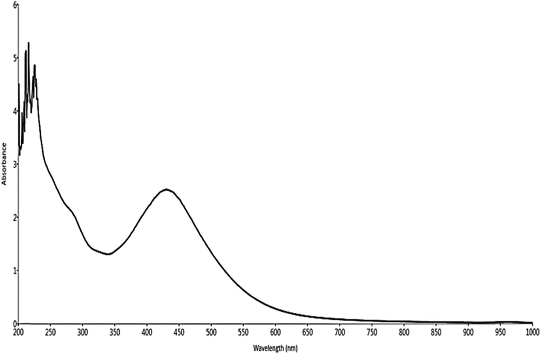
UV-Vis absorption spectrum of the biosynthesized AgNPs from aqueous leaf extracts of M. pulegium.
3.3 FTIR analysis of plant extract and synthesized AgNPs
Figures 3 and 4 depict the IR absorption spectrum of M.p leaf extract and synthesized M.p-AgNPs. The FTIR spectrum of M.p leaf extract (Figure 3) showed a strong broad peak at 3,404 cm−1. This peak could be associated with the OH stretching vibrations of phenols and alcohols. The peaks observed at 2,933, 1,606, and 1,521 cm1 denote the CH stretching of alkanes and symmetric –NH2 bending of amino acids. The peaks at 1,408, 1,265, and 1,067 cm−1 suggest the presence of aromatic compounds (C–C stretching) and symmetric and asymmetric stretches of C–O–C. Rad et al. [31] in a recent study characterized zinc NPs synthesized from M.p leaf extract and showed peaks at similar positions on the IR spectrum.

FTIR spectrum of M.p aqueous leaf extract.
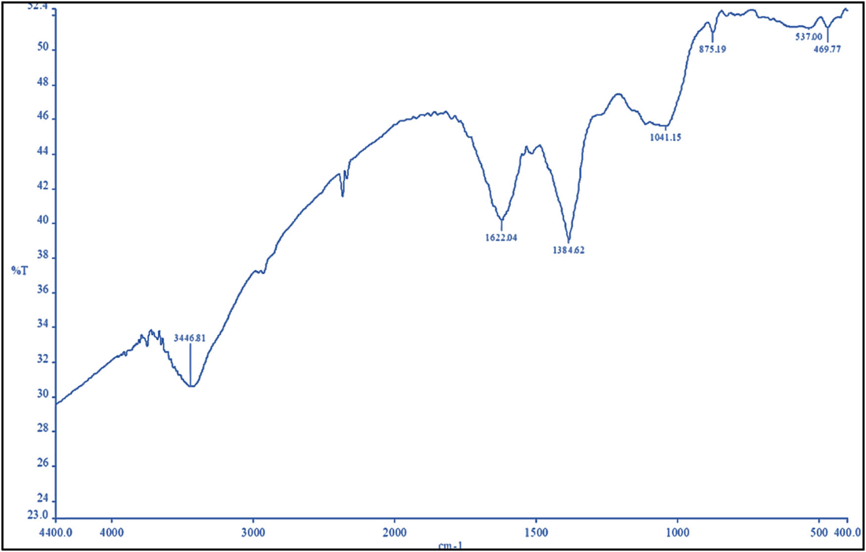
FTIR spectrum of the biosynthesized AgNPs of M.p-AgNPs.
The FTIR spectrum of M.p-AgNP was significantly different from that of M.p leaf extract. The peaks at 3,446, 1,622, 1,384, and 1,041 cm−1 shown in the FTIR spectrum of M.p-AgNP (Figure 4) indicate the presence of an OH group of alcohol or phenols, –NH group of amide or proteins, and carbonyl group of esters. These results strongly indicate the binding of Ag+ to OH groups of phenol and alcohols and C═O of esters or aldehydes and also confirm the capping and reduction of Ag+ to AgNPs. The noticeable alterations in the vibrational bands and stretches in the IR of M.p-AgNP suggest that functional groups play a significant role in the capping and reduction processes during the synthesis of M.p-AgNP. The peaks observed in both spectra in the current study indicate the presence of several bioactive molecules such as amines, proteins, phenols, and aromatic compounds. These biomolecules are well recognized for their role in various processes during the synthesis of NPs, such as reduction, capping, and stabilization. Previous research has demonstrated the role of carbonyl and hydroxyl groups in the aforementioned process involving the biosynthesis of green AgNPs [32,33]. Furthermore, the biomolecules present in plant extract prevent the agglomeration of NPs [33].
3.4 DLS – measurement analysis
The average size of biosynthesized M.p-AgNPs and their distribution were evaluated with the DLS analyzer. The pattern is depicted in Figure 5. The average size of the NPs was 61.81 nm while the polydiversity index was 0.233. The low polydiversity index value and size distribution show that the particles are highly dispersed. DLS is an important technique that determines the hydrodynamic measurements of the NPs, in association with biological molecules and ions attached to the NPs [34,35].

The DLS spectrum exhibiting the size range of synthesized M.p-AgNPs.
3.5 TEM of biosynthesized M.p-AgNP
Figure 6 depicts the microphotograph of biosynthesized M.p-AgNP. The synthesized M.p-AgNPs were chiefly spherical, and the mean diameter of the particles ranged between 4 and 46 nm. The particles were widely separated without agglomeration. The current findings are consistent with those of Kelkawi et al. [36] who reported that the size of AgNPs synthesized by using M.p plant extract was in the 5–50 nm range and that their shape was anisotropic. The average diameter of M.p-AgNP measured with TEM was much smaller than that measured with DLS. This could be because TEM measures the NPs in their dry state, whereas DLS measures them in their hydrated state [37].
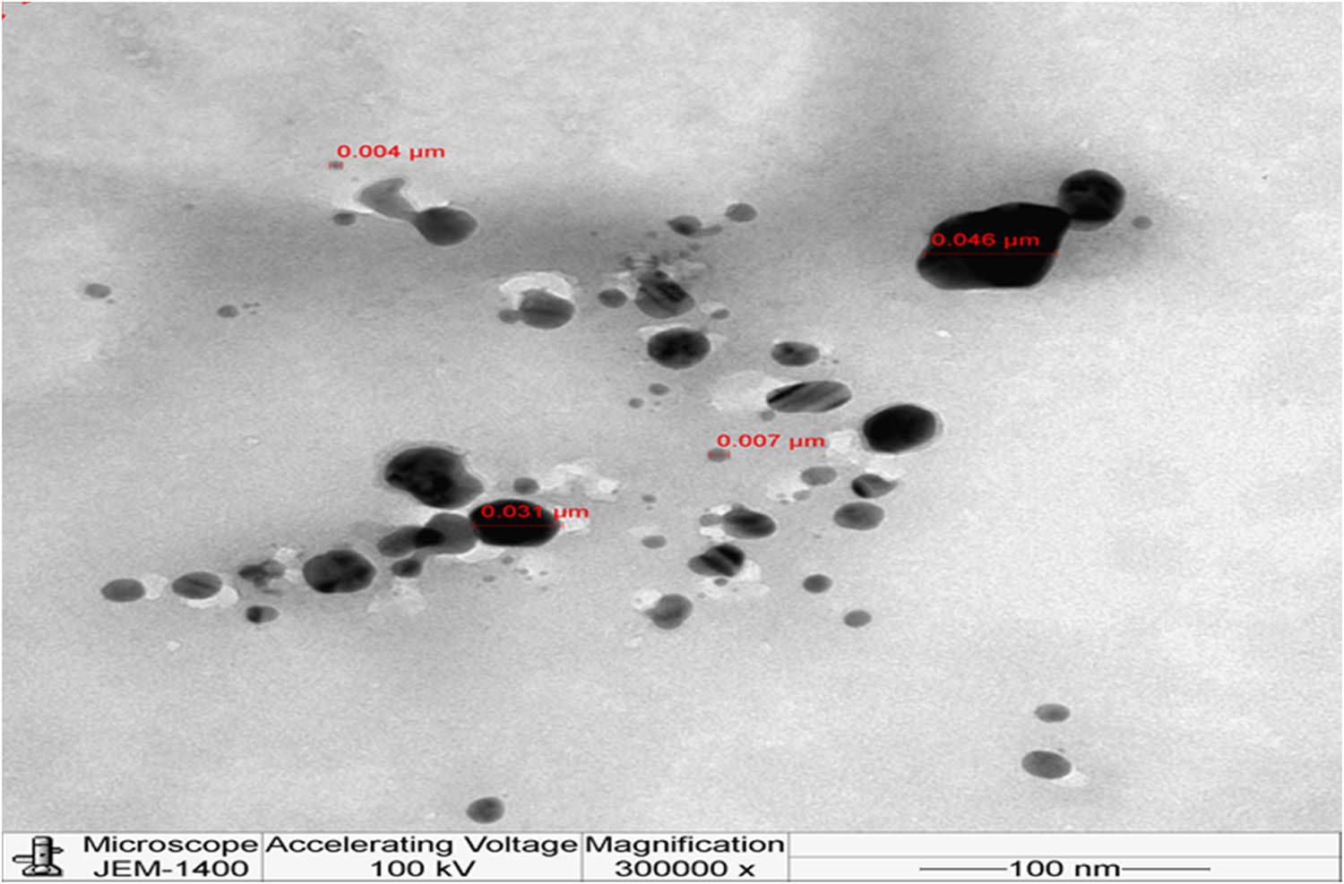
TEM microphotographs of the NPs synthesized using M.p leaf extracts.
3.6 Energy dispersive X-ray analysis-EDX
The EDX spectrum of the synthesized M.p-AgNPs is depicted in Figure 7. The spectrum showed peaks between 2.5 and 3 keV, indicating silver signals and the formation of M.p-AgNP. The peak at 3 keV is an accurate indication of the formation of AgNPs and is ascribed to the SPR [38]. The results indicate that M.p-AgNPs contained silver (23.9%), along with other elements like carbon (42.9%), oxygen (30.7%), calcium (0.04%), and potassium (20%). The presence of elements other than silver could be from the biomolecules of leaf extract that get bound to NPs during synthesis or contaminants from the grid itself.

EDX spectrum of NPs synthesized from the leaves of M.p.
3.7 Mycelial growth inhibition
As depicted in Figures 8 and 9, all the fungal phytopathogens treated with M.p-AgNPs inhibited mycelial growth significantly (p ≥ 0.05). As the concentration of M.p-AgNPs increased, so did the inhibitory activity. F. solani and A. alternaria had the smallest colony diameters at the highest test concentration (40 µg·mL−1; Figure 8). Similarly, at 40 µg·mL−1, F. solani inhibited mycelial growth the most (66%), followed by A. alternata (61%), M. phaseolina (56%), H. sativum (53%), and F. oxysporum (51%). C. musae, however, was least inhibited (36%), even at the highest test concentration. Some isolates, like M. phaseolina and F. oxysporum, showed complete inhibition when treated with the fungicide (Figure 9). However, M.p leaf extract and AgNO3 had minimal growth inhibitory activity on all the test isolates. Overall, for all the plant pathogens tested, M.p-AgNPs caused considerable mycelial growth inhibition as compared to AgNO3 and extracts. M.p-AgNPs’ large surface area and micro size allow them to easily penetrate cells through the cell membrane, causing cell damage [39,40]. However, the antifungal mechanism of NPs against plant pathogenic fungi remains unclear. Biosynthesized NPs are thought to be capable of crossing plant cell membrane barriers very meticulously via a very precise penetration technique. The rapid influx of NPs is thought to cause an electrolyte imbalance. Furthermore, the unchecked entry of NPs causes cell death by targeting the cellular proteins and the nucleic acids (DNA and RNA) [41,42].
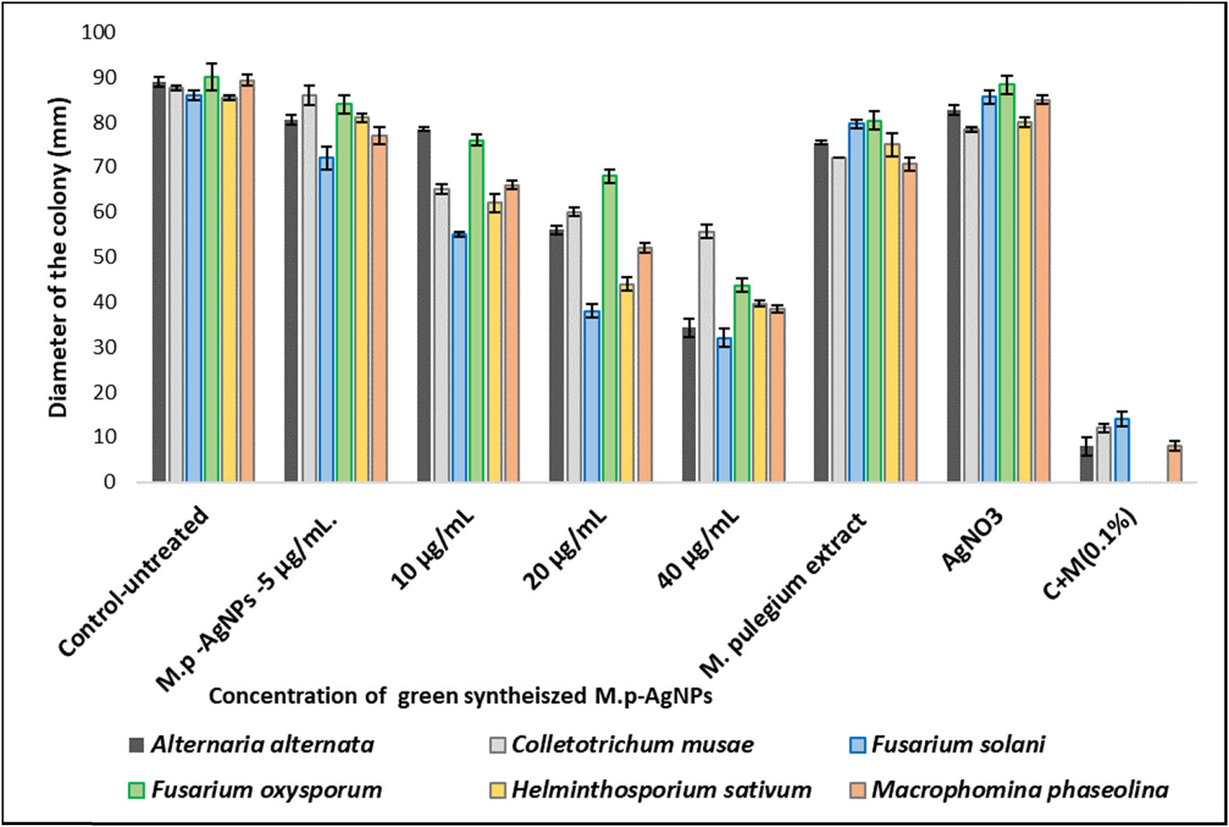
Antifungal activity of green-synthesized M.p-AgNPs.
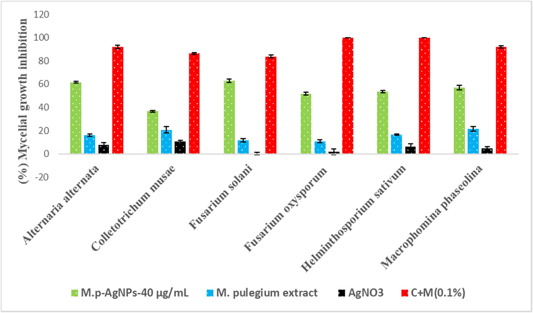
Mycelial growth inhibition of test pathogens treated with M.p-AgNPs at 40 µg·mL−1.
3.8 Spore germination
Spores are very crucial in the growth of fungal pathogens as they germinate, spread, and penetrate the underlying tissue, thereby establishing the pathogen. Impeding the growth and germination of spores is vital for arresting the spread and establishment of any disease. Figure 10 illustrates the effective suppression of spore germination treated with M.p-AgNPs. The figure clearly shows that at a concentration of 40 µg·mL−1, F. solani, A. alternata, and M. phaseolina exhibited considerable inhibition of spore germination (18%, 22%, and 24%, respectively). Sterilized distilled water had no effect on spore germination. AgNO3 and M.p leaf extract had a negligible effect on the inhibition of spore germination (Figure 10).
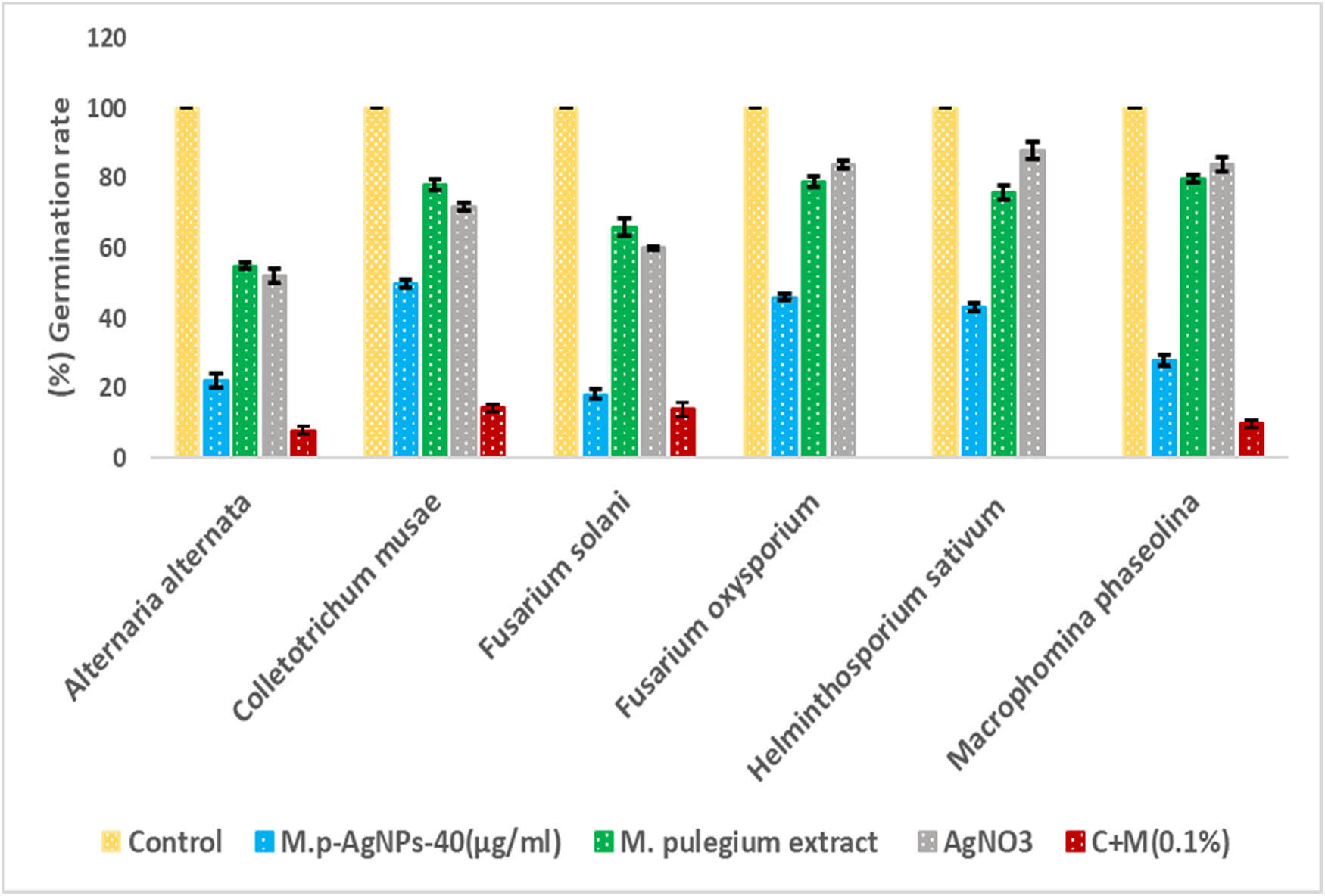
Percentage spore germination of fungal test pathogens treated with M.p-AgNPs at 40 µg·mL−1.
The current study clearly indicates the significant inhibition of mycelial growth and spore germination of plant pathogenic fungi treated with M.p-AgNPs. The small size of M.p-AgNPs along with the secondary metabolites present in the leaf extracts might have facilitated the robust inhibitory activity. In agreement with these findings, previous reports have demonstrated the antifungal activity of plant-derived AgNPs against an array of plant pathogenic fungi, including Colletotrichum sp., Aspergillus sp., and Fusarium sp. [43,44]. The variable inhibitory activity of M.p-AgNPs on the growth of fungal pathogens seen in this study could be due to the resistance offered by the fungal cell barriers that hinder the entry of NPs into the cell, thus reducing the extent of damage to fungal cells and spores.
4 Conclusion
Plant-derived NPs are used widely in a variety of bioscience applications around the world. AgNPs were successfully synthesized using M.p leaves in this study. The synthesized NPs were characterized, and their antifungal properties were evaluated. The biosynthesized M.p-AgNPs inhibited hyphal growth and suppressed spore germination of fungal test pathogens in a significant manner. The growing demand for a safe food supply has compelled the use of nontoxic agents to protect agricultural produce from devastating fungal pathogens. Based on the findings of this study, it is proposed that M.p-AgNPs can be formulated into a nontoxic fungicide to control plant pathogens at a low cost and in an environmentally friendly manner. More research is needed to understand their effect on human cells and to investigate their therapeutic applications.
Acknowledgements
The authors extend their appreciation to the Researchers Support Project (number RSP 2021/173) of King Saud University.
-
Funding information: The authors extend their appreciation to the Researchers Support Project (number RSP 2021/173) of King Saud University, Riyadh, Saudi Arabia, for payment of the charge for publishing this manuscript.
-
Author contributions: Humaira Rizwana: conceptualization, project administration, writing – original draft, writing – review and editing, formal analysis; Mona S. Alwhibi: methodology, resources, writing – original draft, validation, and funding acquisition.
-
Conflict of interest: Authors state no conflict of interest.
References
[1] Nicolas J, Mura S, Brambilla D, Mackiewicz N, Couvreur P. Design, functionalization strategies and biomedical applications of targeted biodegradable/biocompatible polymer-based nanocarriers for drug delivery. Chem Soc Rev. 2013;42(3):1147–235.10.1039/C2CS35265FSearch in Google Scholar PubMed
[2] Ahmed S, Chaudhry SA, Ikram S. A review on biogenic synthesis of ZnO nanoparticles using plant extracts and microbes: a prospect towards green chemistry. J Photochem Photobiol B Biol. 2017;166:272–84.10.1016/j.jphotobiol.2016.12.011Search in Google Scholar PubMed
[3] Xu L, Wang YY, Huang J, Chen CY, Wang ZX, Xie H. Silver nanoparticles: synthesis, medical applications and biosafety. Theranostics. 2020;10(20):8996–9031. 10.7150/thno.45413.Search in Google Scholar PubMed PubMed Central
[4] Khan I, Saeed K, Khan I. Nanoparticles: properties, applications and toxicities. Arab J Chem. 2019;12(7):908–31. 10.1016/j.arabjc.2017.05.011.Search in Google Scholar
[5] Rizwana H, Alwhibi MS, Aldarsone HA, Awad MA, Soliman DA, Bhat RS. Green synthesis, characterization, and antimicrobial activity of silver nanoparticles prepared using Trigonella foenum-graecum L. leaves grown in Saudi Arabia. Green Process Synth. 2021;10:421–9. http://dx.doi.org/10.1515/gps-2021-0043.Search in Google Scholar
[6] Iravani S, Korbekandi H, Mirmohammadi SV, Zolfaghari B. Synthesis of silver nanoparticles: chemical, physical and biological methods. Res Pharm Sci. 2014;9(6):385–406.Search in Google Scholar
[7] Kumari S, Tyagi M, Jagadevan S. Mechanistic removal of environmental contaminants using biogenic nano-materials. Int J Env Sci Technol. 2019;16:7591–606. 10.1007/s13762-019-02468-3.Search in Google Scholar
[8] Rana A, Yadav K, Jagadevan S. A comprehensive review on green synthesis of nature-inspired metal nanoparticles: mechanism, application and toxicity. J Clean Prod. 2020;272:122880. 10.1016/j.jclepro.2020.122880. ISSN 0959-6526.Search in Google Scholar
[9] Jha A, Prasad K, Prasad K, Kulkarni AR. “Plant system: nature’s nanofactory”. Colloids Surf B Biointerfaces. 2009;73:219–23.10.1016/j.colsurfb.2009.05.018Search in Google Scholar PubMed
[10] Ahmed S, Ahmad M, Swami BL, Ikram S. Plants extract mediated synthesis of silver nanoparticles for antimicrobial applications: a green expertise. J Adv Res. 2016;7:17–28.10.1016/j.jare.2015.02.007Search in Google Scholar PubMed PubMed Central
[11] Singh P, Kim YJ, Zhang D, Yang DC. Biological synthesis of nanoparticles from plants and microorganisms. Trends Biotechnol. 2016;34(7):588–99. 10.1016/j.tibtech.2016.02.006.Search in Google Scholar PubMed
[12] Alwhibi MS, Soliman DA, Awad MA, Alangery AB, Al Dehaish H, Alwasel YA. Green synthesis of silver nanoparticles: characterization and its potential biomedical application. Green Process Synth. 2021;10:412–20.10.1515/gps-2021-0039Search in Google Scholar
[13] Rauf A, Ahmad T, Khan A, Maryam M, Uddin G, Ahmad B, et al. Green synthesis and biomedicinal applications of silver and gold nanoparticles functionalized with methanolic extract of Mentha longifolia. Artif Cell Nanomed Biotechnol. 2021;49(1):194–203. To link to this article 10.1080/21691401.2021.1890099.Search in Google Scholar PubMed
[14] Zargari A. Herbal medicines. 1st edn. Tehran: Publication of Tehran University; 1990. p. 14–8.Search in Google Scholar
[15] Chalchat JC, Gorunovic MS, Maksimovic ZA, Petrovic SD. Essential oil of wild growing Mentha pulegium L. from yugoslavia. J Essent Oil Res. 2000;12:598–600.10.1080/10412905.2000.9712166Search in Google Scholar
[16] Harley RM, Atkins S, Budantsev AL, Cantino PD, Conn BJ, Grayer R, et al. Labiatae. In: Kadereit JW, editors. Flowering plants-dicotyledons. the families and genera of vascular plants. Vol. 7. Berlin, Heidelberg: Springer; 2004. p. 167–275. 10.1007/978-3-642-18617-2_11.Search in Google Scholar
[17] Mimica-Dukic N, Bozin B. Mentha l. Species (Lamiaceae) as promising sources of bioactive secondary metabolites. Curr Pharm Des. 2008;14:3141–50.10.2174/138161208786404245Search in Google Scholar PubMed
[18] Khonche A, Fallah Huseini H, Abdi H, Mohtashami R, Nabati F, Kianbakht S. Efficacy of Mentha pulegium extract in the treatment of functional dyspepsia: a randomized double-blind placebo-controlled clinical trial. J Ethnopharmacol. 2017;206:267–73. 10.1016/j.jep.2017.05.026.Search in Google Scholar PubMed
[19] Teixeira B, Marques A, Ramos C, Batista I, Serrano C, Matos O, et al. European pennyroyal (Mentha pulegium) from Portugal: chemical composition of essential oil and antioxidant and antimicrobial properties of extracts and essential oil. Ind Crop Prod. 2012;36:81–7.10.1016/j.indcrop.2011.08.011Search in Google Scholar
[20] Mahboubi M, Haghi G. Antimicrobial activity and chemical composition of Mentha pulegium L. essential oil. J Ethnopharmacol. 2008;119:325–7.10.1016/j.jep.2008.07.023Search in Google Scholar PubMed
[21] Ibrahim AK. New terpenoids from Mentha pulegium L. and their antimicrobial activity. Nat Prod Res. 2013;27:691–6.10.1080/14786419.2012.691488Search in Google Scholar PubMed
[22] Romero-Jiménez M, Campos-Sánchez J, Analla M, Muñoz-Serrano A, Alonso-Moraga A. Genotoxicity and anti-genotoxicity of some traditional medicinal herbs. Mutat Res. 2005;585:147–55.10.1016/j.mrgentox.2005.05.004Search in Google Scholar PubMed
[23] Kogiannou DA, Kalogeropoulos N, Kefalas P, Polissiou MG, Kaliora AC. Herbal infusions; their phenolic profile, antioxidant and anti-inflammatory effects in HT29 and PC3 cells. Food Chem Toxicol. 2013;61:152–9.10.1016/j.fct.2013.05.027Search in Google Scholar PubMed
[24] Kim SW, Jung JH, Lamsal K, Kim YS, Min JS, Lee YS. Antifungal effects of silver nanoparticles (AgNPs) against various plant pathogenic fungi. Mycobiol. 2012;40:53–8.10.5941/MYCO.2012.40.1.053Search in Google Scholar PubMed PubMed Central
[25] Ibrahim E, Luo J, Ahmed T, Wu W, Yan C, Li B. Biosynthesis of silver nanoparticles using onion endophytic bacterium and its antifungal activity against rice pathogen Magnaporthe oryzae. J Fungi (Basel). 2020;6:294. 10.3390/jof6040294.Search in Google Scholar PubMed PubMed Central
[26] Rizwana H. Postharvest control of anthracnose lesions and its causative agent, Colletotrichum musae by some oils. Cell Mol Biol (Noisy-le-grand). 2018;64:52–8. 10.14715/cmb/2018.64.4.9. PMID: 29641375.Search in Google Scholar
[27] Boken J, Khurana P, Thatai S, Kumar D, Prasad S. Plasmonic nanoparticles and their analytical applications: a review. Appl Spectrosc Rev. 2017;52(9):774–820.10.1080/05704928.2017.1312427Search in Google Scholar
[28] Kelly KL, Coronado E, Zhao LL, Schatz GC. -e optical properties of metal nano particles: the influence of size, shape and dielectric environment. J Phys Chem B. 2003;107(3):668–77.10.1021/jp026731ySearch in Google Scholar
[29] Rajkumar T, Sapi A, Das G, Debnath T, Ansari A, Patra JK. Biosynthesis of silver nanoparticle using extract of Zea mays (corn flour) and investigation of its cytotoxicity effect and radical scavenging potential. JPPBEG. 2019;193:1–7.10.1016/j.jphotobiol.2019.01.008Search in Google Scholar PubMed
[30] Awad AM, Salem NM, Abdeen AO. Green synthesis of silver nanoparticles using carob leaf extract and its antibacterial activity. Int J Ind Chem. 2013;4(1):1–6.10.1186/2228-5547-4-29Search in Google Scholar
[31] Rad SS, Sani AM, Mohseni S. Biosynthesis, characterization and antimicrobial activities of zinc oxide nanoparticles from leaf extract of Mentha pulegium (L.). Microb Pathog. 2019;131:239–45.10.1016/j.micpath.2019.04.022Search in Google Scholar PubMed
[32] Kora AJ, Beedu SR, Jayaraman A. Size-controlled green synthesis of silver nanoparticles mediated by gum ghatti (Anogeissus latifolia) and its biological activity. Org Med Chem Lett. 2012;2(1):17. 10.1186/2191-2858-2-17.Search in Google Scholar PubMed PubMed Central
[33] Gupta A, Koirala AR, Gupta B, Parajuli N. Improved method for separation of silver nanoparticles synthesized using the Nyctanthes arbor-tristis shrub. ACMY. 2019;3(1):35–42.10.2478/acmy-2019-0005Search in Google Scholar
[34] Das B, Dash SK, Mandal D, Ghosh T, Chattopadhyay S, Tripathy S, et al. Green synthesized silver nanoparticles destroy multidrug resistant bacteria via reactive oxygen species mediated membrane damage. Arab J Chem. 2017;10(6):862–76. 10.1016/j.arabjc.2015.08.008.Search in Google Scholar
[35] Moteriya P, Chanda S. Green synthesis of silver nanoparticles from Caesalpinia pulcherrima leaf extract and evaluation of their antimicrobial, cytotoxic and genotoxic potential (3-in-1 system). J Inorg Organomet Polym. 2020;30:3920–32. 10.1007/s10904020-01532-7.Search in Google Scholar
[36] Kelkawi AHA, Abbasi Kajani A, Bordbar AK. Green synthesis of silver nanoparticles using Mentha pulegium and investigation of their antibacterial, antifungal and anticancer activity. IET Nanobiotechnol. 2017;11(4):370–6. 10.1049/iet-nbt.2016.0103. PMID: 28530184.Search in Google Scholar
[37] Clayton KN, Salameh JW, Wereley ST, Kinzer-Ursem TL. Physical characterization of nanoparticle size and surface modification using particle scattering diffusometry. Biomicrofluidics. 2016;10(5):054107. 10.1063/1.4962992.Search in Google Scholar PubMed PubMed Central
[38] Kaviya S, Santhanalakshmi J, Viswanathan B, Muthumary J, Srinivasan K. Biosynthesis of silver nanoparticles using citrus sinensis peel extract and its antibacterial activity. Spectrochim Acta A Mol Biomol Spectrosc. 2011;79(3):594–8. 10.1016/j.saa.2011.03.040.Search in Google Scholar PubMed
[39] Duran N, Marcato P, Alves OL, De Sousa GIH, Esposito E. Mechanistic aspects of biosynthesis of silver nanoparticles by several Fusarium oxysporum strains. J Nanobiotechnol. 2005;3:8. 10.1186/1477-3155-3-8.Search in Google Scholar PubMed PubMed Central
[40] Bera RK, Mandal SM, Retna Raj C. Antimicrobial activity of fluorescent Ag nanoparticles. Lett Appl Microbiol. 2014;58:520–6.10.1111/lam.12222Search in Google Scholar PubMed
[41] Khatami M, Sharifi I, Marcos AL. Waste-grass-mediated green synthesis of silver nanoparticles and evaluation of their anticancer, antifungal and antibacterial activity. Green Chem Lett Rev. 2018;11(2):125–34.10.1080/17518253.2018.1444797Search in Google Scholar
[42] Kim KJ, Sung WS, Suh BK, Moon SK, Choi JS, Kim JG, et al. Antifungal activity and mode of action of silver nano-particles on Candida albicans. Biometals. 2009;22(2):235–42.10.1007/s10534-008-9159-2Search in Google Scholar PubMed
[43] Lamsal K, Kim SW, Jung JH, Kim YS, Kim YS, Lee YS. Application of silver nanoparticles for the control of colletotrichum species in vitro and pepper anthracnose disease in field. Mycobiol. 2011;39(3):194–9.10.5941/MYCO.2011.39.3.194Search in Google Scholar PubMed PubMed Central
[44] Gallardo RV, Cruz JFO, Ortíz-Rodriguez OO. Fungicidal effect of silver nanoparticles on toxigenic fungi in cocoa. FITOPATOLOGIA. Pesq Agropec Bras. 2016;51(12):1929–36. 10.1590/S0100-204X2016001200003.Search in Google Scholar
© 2021 Humaira Rizwana and Mona S. Alwhibi, published by De Gruyter
This work is licensed under the Creative Commons Attribution 4.0 International License.
Articles in the same Issue
- Research Articles
- MW irradiation and ionic liquids as green tools in hydrolyses and alcoholyses
- Effect of CaO on catalytic combustion of semi-coke
- Studies of Penicillium species associated with blue mold disease of grapes and management through plant essential oils as non-hazardous botanical fungicides
- Development of leftover rice/gelatin interpenetrating polymer network films for food packaging
- Potent antibacterial action of phycosynthesized selenium nanoparticles using Spirulina platensis extract
- Green synthesized silver and copper nanoparticles induced changes in biomass parameters, secondary metabolites production, and antioxidant activity in callus cultures of Artemisia absinthium L.
- Gold nanoparticles from Celastrus hindsii and HAuCl4: Green synthesis, characteristics, and their cytotoxic effects on HeLa cells
- Green synthesis of silver nanoparticles using Tropaeolum majus: Phytochemical screening and antibacterial studies
- One-step preparation of metal-free phthalocyanine with controllable crystal form
- In vitro and in vivo applications of Euphorbia wallichii shoot extract-mediated gold nanospheres
- Fabrication of green ZnO nanoparticles using walnut leaf extract to develop an antibacterial film based on polyethylene–starch–ZnO NPs
- Preparation of Zn-MOFs by microwave-assisted ball milling for removal of tetracycline hydrochloride and Congo red from wastewater
- Feasibility of fly ash as fluxing agent in mid- and low-grade phosphate rock carbothermal reduction and its reaction kinetics
- Three combined pretreatments for reactive gasification feedstock from wet coffee grounds waste
- Biosynthesis and antioxidation of nano-selenium using lemon juice as a reducing agent
- Combustion and gasification characteristics of low-temperature pyrolytic semi-coke prepared through atmosphere rich in CH4 and H2
- Microwave-assisted reactions: Efficient and versatile one-step synthesis of 8-substituted xanthines and substituted pyrimidopteridine-2,4,6,8-tetraones under controlled microwave heating
- New approach in process intensification based on subcritical water, as green solvent, in propolis oil in water nanoemulsion preparation
- Continuous sulfonation of hexadecylbenzene in a microreactor
- Synthesis, characterization, biological activities, and catalytic applications of alcoholic extract of saffron (Crocus sativus) flower stigma-based gold nanoparticles
- Foliar applications of plant-based titanium dioxide nanoparticles to improve agronomic and physiological attributes of wheat (Triticum aestivum L.) plants under salinity stress
- Simultaneous leaching of rare earth elements and phosphorus from a Chinese phosphate ore using H3PO4
- Silica extraction from bauxite reaction residue and synthesis water glass
- Metal–organic framework-derived nanoporous titanium dioxide–heteropoly acid composites and its application in esterification
- Highly Cr(vi)-tolerant Staphylococcus simulans assisting chromate evacuation from tannery effluent
- A green method for the preparation of phoxim based on high-boiling nitrite
- Silver nanoparticles elicited physiological, biochemical, and antioxidant modifications in rice plants to control Aspergillus flavus
- Mixed gel electrolytes: Synthesis, characterization, and gas release on PbSb electrode
- Supported on mesoporous silica nanospheres, molecularly imprinted polymer for selective adsorption of dichlorophen
- Synthesis of zeolite from fly ash and its adsorption of phosphorus in wastewater
- Development of a continuous PET depolymerization process as a basis for a back-to-monomer recycling method
- Green synthesis of ZnS nanoparticles and fabrication of ZnS–chitosan nanocomposites for the removal of Cr(vi) ion from wastewater
- Synthesis, surface modification, and characterization of Fe3O4@SiO2 core@shell nanostructure
- Antioxidant potential of bulk and nanoparticles of naringenin against cadmium-induced oxidative stress in Nile tilapia, Oreochromis niloticus
- Variability and improvement of optical and antimicrobial performances for CQDs/mesoporous SiO2/Ag NPs composites via in situ synthesis
- Green synthesis of silver nanoparticles: Characterization and its potential biomedical applications
- Green synthesis, characterization, and antimicrobial activity of silver nanoparticles prepared using Trigonella foenum-graecum L. leaves grown in Saudi Arabia
- Intensification process in thyme essential oil nanoemulsion preparation based on subcritical water as green solvent and six different emulsifiers
- Synthesis and biological activities of alcohol extract of black cumin seeds (Bunium persicum)-based gold nanoparticles and their catalytic applications
- Digera muricata (L.) Mart. mediated synthesis of antimicrobial and enzymatic inhibitory zinc oxide bionanoparticles
- Aqueous synthesis of Nb-modified SnO2 quantum dots for efficient photocatalytic degradation of polyethylene for in situ agricultural waste treatment
- Study on the effect of microwave roasting pretreatment on nickel extraction from nickel-containing residue using sulfuric acid
- Green nanotechnology synthesized silver nanoparticles: Characterization and testing its antibacterial activity
- Phyto-fabrication of selenium nanorods using extract of pomegranate rind wastes and their potentialities for inhibiting fish-borne pathogens
- Hydrophilic modification of PVDF membranes by in situ synthesis of nano-Ag with nano-ZrO2
- Paracrine study of adipose tissue-derived mesenchymal stem cells (ADMSCs) in a self-assembling nano-polypeptide hydrogel environment
- Study of the corrosion-inhibiting activity of the green materials of the Posidonia oceanica leaves’ ethanolic extract based on PVP in corrosive media (1 M of HCl)
- Callus-mediated biosynthesis of Ag and ZnO nanoparticles using aqueous callus extract of Cannabis sativa: Their cytotoxic potential and clinical potential against human pathogenic bacteria and fungi
- Ionic liquids as capping agents of silver nanoparticles. Part II: Antimicrobial and cytotoxic study
- CO2 hydrogenation to dimethyl ether over In2O3 catalysts supported on aluminosilicate halloysite nanotubes
- Corylus avellana leaf extract-mediated green synthesis of antifungal silver nanoparticles using microwave irradiation and assessment of their properties
- Novel design and combination strategy of minocycline and OECs-loaded CeO2 nanoparticles with SF for the treatment of spinal cord injury: In vitro and in vivo evaluations
- Fe3+ and Ce3+ modified nano-TiO2 for degradation of exhaust gas in tunnels
- Analysis of enzyme activity and microbial community structure changes in the anaerobic digestion process of cattle manure at sub-mesophilic temperatures
- Synthesis of greener silver nanoparticle-based chitosan nanocomposites and their potential antimicrobial activity against oral pathogens
- Baeyer–Villiger co-oxidation of cyclohexanone with Fe–Sn–O catalysts in an O2/benzaldehyde system
- Increased flexibility to improve the catalytic performance of carbon-based solid acid catalysts
- Study on titanium dioxide nanoparticles as MALDI MS matrix for the determination of lipids in the brain
- Green-synthesized silver nanoparticles with aqueous extract of green algae Chaetomorpha ligustica and its anticancer potential
- Curcumin-removed turmeric oleoresin nano-emulsion as a novel botanical fungicide to control anthracnose (Colletotrichum gloeosporioides) in litchi
- Antibacterial greener silver nanoparticles synthesized using Marsilea quadrifolia extract and their eco-friendly evaluation against Zika virus vector, Aedes aegypti
- Optimization for simultaneous removal of NH3-N and COD from coking wastewater via a three-dimensional electrode system with coal-based electrode materials by RSM method
- Effect of Cu doping on the optical property of green synthesised l-cystein-capped CdSe quantum dots
- Anticandidal potentiality of biosynthesized and decorated nanometals with fucoidan
- Biosynthesis of silver nanoparticles using leaves of Mentha pulegium, their characterization, and antifungal properties
- A study on the coordination of cyclohexanocucurbit[6]uril with copper, zinc, and magnesium ions
- Ultrasound-assisted l-cysteine whole-cell bioconversion by recombinant Escherichia coli with tryptophan synthase
- Green synthesis of silver nanoparticles using aqueous extract of Citrus sinensis peels and evaluation of their antibacterial efficacy
- Preparation and characterization of sodium alginate/acrylic acid composite hydrogels conjugated to silver nanoparticles as an antibiotic delivery system
- Synthesis of tert-amylbenzene for side-chain alkylation of cumene catalyzed by a solid superbase
- Punica granatum peel extracts mediated the green synthesis of gold nanoparticles and their detailed in vivo biological activities
- Simulation and improvement of the separation process of synthesizing vinyl acetate by acetylene gas-phase method
- Review Articles
- Carbon dots: Discovery, structure, fluorescent properties, and applications
- Potential applications of biogenic selenium nanoparticles in alleviating biotic and abiotic stresses in plants: A comprehensive insight on the mechanistic approach and future perspectives
- Review on functionalized magnetic nanoparticles for the pretreatment of organophosphorus pesticides
- Extraction and modification of hemicellulose from lignocellulosic biomass: A review
- Topical Issue: Recent advances in deep eutectic solvents: Fundamentals and applications (Guest Editors: Santiago Aparicio and Mert Atilhan)
- Delignification of unbleached pulp by ternary deep eutectic solvents
- Removal of thiophene from model oil by polyethylene glycol via forming deep eutectic solvents
- Valorization of birch bark using a low transition temperature mixture composed of choline chloride and lactic acid
- Topical Issue: Flow chemistry and microreaction technologies for circular processes (Guest Editor: Gianvito Vilé)
- Stille, Heck, and Sonogashira coupling and hydrogenation catalyzed by porous-silica-gel-supported palladium in batch and flow
- In-flow enantioselective homogeneous organic synthesis