Synthesis and characterization of Ce-doped TiO2 nanoparticles and their enhanced anticancer activity in Y79 retinoblastoma cancer cells
-
Balachandran Kartha
 ,
Naiyf S. Alharbi
,
Naiyf S. Alharbi

Abstract
Rare earth metal cerium-doped titania nanoparticles (titanium dioxide [TiO2]) were produced utilizing a low-cost and straightforward sol–gel technique, and its enhanced photodynamic anticancer activity was tested on Y79 retinoblastoma cancer cells. The structural, optical, morphological, anticancer activity, and cytotoxicity of pure and cerium-doped TiO2 (Ce-doped TiO2) were investigated. In X-ray diffraction (XRD) measurements, apparent doping of cerium in TiO2 was detected, with reported anatase patterns shifting toward a lower angle in the anatase structure. Raman spectra verify the presence of cerium doping in TiO2 by revealing greater wave number shifting. The scanning electron microscope (SEM) and transmission electron microscope (TEM) analysis showed that the synthesized TiO2 and Ce-doped TiO2 nearly spherical. TiO2 and Ce-doped TiO2 were studied for their photodynamic anticancer activities, and the results suggest that cerium doping in TiO2 improves anticancer activity.
1 Introduction
Due to their innovative biological applications, semiconductor nanostructures with diameters smaller than 100 nm have emerged as nanobiomaterials [1,2]. In recent years, a wide range of nanocomposite semiconductor materials has been created to increase the photocatalytic activity efficiency [3,4]. By covering metal or semiconductors nanoclusters with some other layer of appropriate materials, the functional characteristics of such materials can be substantially enhanced [5,6].
Titanium dioxide (TiO2) has been extensively used and demonstrated as a critical perspective photosensitizer [7,8,9], photostability, low cost, and nontoxicity. According to a recent study, the particle size of TiO2 has a significant impact on its photocatalytic activity [10]. The reduction in particle size suggests an increase in surface area and a high redox potential, resulting in a strong photocatalytic activity.
Several methods, including doping, surface modification with metal particles, and particle size reduction to the nanoscale, have been proposed to increase the effectiveness of photocatalytic reactions utilizing TiO2 when exposed to visible light [11]. Chemically modified TiO2 nanoparticles are used in several environmental applications due to their self-cleaning properties [12,13]. Because of their nontoxicity, excellent optical absorption, cheap cost, and good chemical stability, metal and metal oxide nanoparticles have been widely investigated [14,15,16,17,18,19]. Among them, the medical applications of TiO2 are undeniably promising, with the potential to significantly enhance health care, notably cancer therapy. Doping of Ce in TiO2 enhances the photocatalysis and photodegradation of TiO2 [20].
Sol–gel, direct aqueous solution depositions, ultrasonic spray pyrolysis, and sputtering are some of the techniques used [21,22,23,24,25,26,27]. Among these techniques, the sol–gel process has several distinct benefits, including composition control, excellent homogeneity, reduced crystallization temperature, and, when dip coating is used, the capacity to create thin coatings on complex shapes [28].
This study uses the sol–gel technique to make TiO2 and cerium-doped titanium dioxide (Ce-doped TiO2) nanoparticles. XRD, SEM, TEM, and photodynamic anticancer activity analyses were used to characterize the created nanoparticles. Moreover, the anticancer effect of Ce-doped TiO2 nanoparticles in Y79 retinoblastoma cancer cells was thoroughly addressed.
2 Experimental details
2.1 Synthesis and characterization of TiO2 and Ce-doped TiO2 nanoparticles
TiO2 nanoparticles were created using a wet chemical (sol–gel) method [29,30]. TiO2 sol was agitated for 3 h with a 0.1 M cerium nitrate hexahydrate solution. The white Ti(OH)4 precipitate that forms is refluxed for 2 h before being agitated continuously for 12 h. The precipitate is centrifuged with deionized water and ethanol to eliminate contaminants. After centrifugation, the white precipitate is dried for 3 h in a hot air oven at 100°C to eliminate moisture and other solvents. Finally, the dry powder was crushed and calcined in an air tube furnace at 400°C for 2 h.
The crystalline phase construction of TiO2 nanoparticles and Ce-TiO2 nanocomposites was examined utilizing a D8 Advance X-beam diffraction meter (Bruker AXS, Germany) at room temperature. The structural identification of TiO2 and Ce-TiO2 nanocomposites was inspected utilizing an examining electron magnifying instruments SEM (Model JSM 6390LV, JOEL, USA) and TEM (JEOL-TEM 2100) [32].
2.2 Cellular incubation with TiO2 nanoparticles and cell culture
2.2.1 Method of cell culture and counting
The Y79 cell line, a well-known cancer cell line, was used in this investigation. The cells were grown for 3 days in a 60 mm Petri plate at a concentration of around 3 × 10⁵. After the previous culture media was replaced with TiO2 colloidal solution, the cells were recultured in an incubator for 24 h. After removing the colloidal solution, it was rinsed with phosphate-buffered saline (PBS, Invitrogen Corporation, Gibco). The cells were stained with trypan blue after being cleaved by trypsin-ethylenediaminetetraacetic acid (Gibco; Nacalai Tesque, Kyoto, Japan). The cells were then counted using a hemocytometer under a 10× in-bright-field microscope. Cell survival was measured by the proportion of unstained (living) cells compared to control dish cells.
2.2.2 Anticancer activity of pure and Ce-doped TiO2
A continuous wavelength UV lamp with a wavelength of 380 nm was utilized for the UV light irradiation experiment. The wavelength of the light was in the 380 nm region. After 3 days, the media was changed with colloidal TiO2 solution, and the cells were incubated for another 24 h. The light control dish was photoirradiated without using a nanoparticle solution, whereas the other dishes were photoirradiated using TiO2. The colloidal arrangement was then eliminated and washed with PBS before being stained with trypan blue to decide cell feasibility. Live cells were not stained blue; however, dead cells have gathered the color. The pictures were taken with an inverted magnifying instrument (Olympus, CKX41, Japan) equipped with a mathematical light field condenser that produces an exceptionally tight light emission from a tungsten light (sent light of 6V-30W halogen brightening) mounted on top of the examples. A similar system concerning Ce-doped TiO2 nanoparticles was followed.
2.2.3 In vitro cytotoxicity and imaging of pure and Ce-doped TiO2
The provided test sample was subjected to an in vitro cytotoxicity test technique by ISO 10993:5. The cell’s culture media was changed with a new medium. The test sample was applied to the cells in triplicate [31].
The below formula calculated cytotoxicity and cell viability.
3 Results and discussion
3.1 XRD analysis of pure and Ce-doped TiO2
XRD (Figure 1) analysis discovered that the peaks detected at 25.8°, 36.8°, 37.9°, 48°, and 54.5°, correspond to reflections 101, 103, 004, 112, and 200, demonstrate the existence of anatase phase in both samples. Using Scherrer’s equation, the crystallite size of the nanoparticles was calculated to be 19 nm for TiO2 and 15 nm for Ce-doped TiO2, showing that Ce doping in TiO2 inhibits nanoparticle grain development [33].
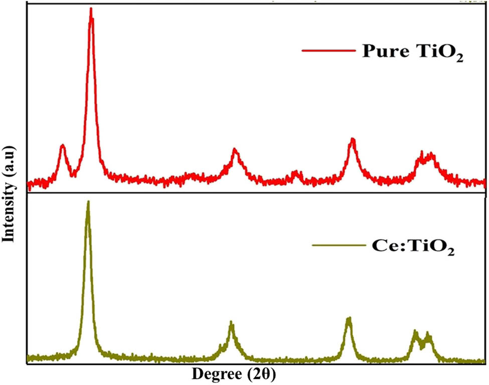
X-ray diffraction analysis of pure and Ce-doped TiO2 nanoparticles.
3.2 Raman analysis of pure and Ce-doped TiO2
Raman analysis was used to detect surface structure changes before and after doping. The spectra demonstrate a blue shift in TiO2, indicating that some small structural deformation happened in TiO2. The observed bands at 150, 400, 510, and 640 cm−1 correspond to the Raman active modes of Eg (1), B1g (1), A1g + B1g (2), and Eg (2), respectively, and Eg (2) shows the presence of anatase phase (Figure 2) [34].
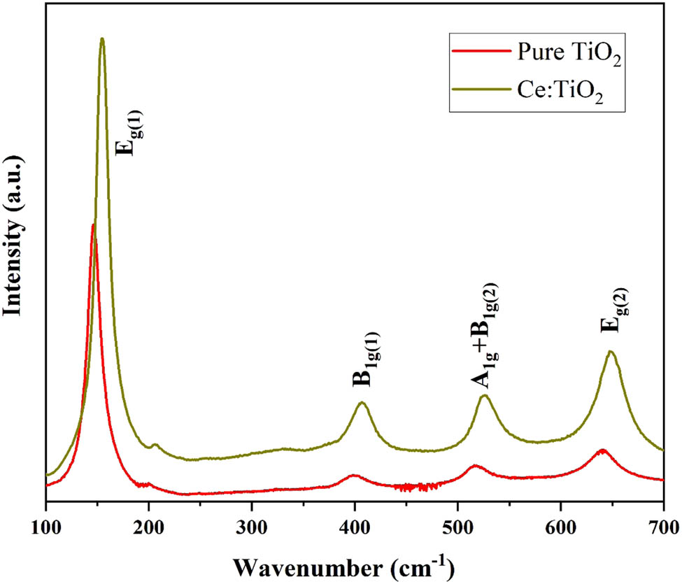
Raman analysis of pure and Ce-doped TiO2.
3.3 Microscopic (SEM and TEM) analysis of pure and Ce-doped TiO2
Figure 3 depicts the SEM and TEM analyses of Ce-doped TiO2 and TiO2 nanoparticles. TiO2 was shown to have an uneven shape, a nonuniform size distribution, and an aggregated character in SEM examination. However, Ce-doped TiO2 has a spherical shape and a homogeneous distribution. Combining materials with high surface energy and a low number of surface states causes nanoparticle aggregation. Aggregation and nonuniform size distribution may be caused by the interaction between anion and cation in the synthesized material [35].
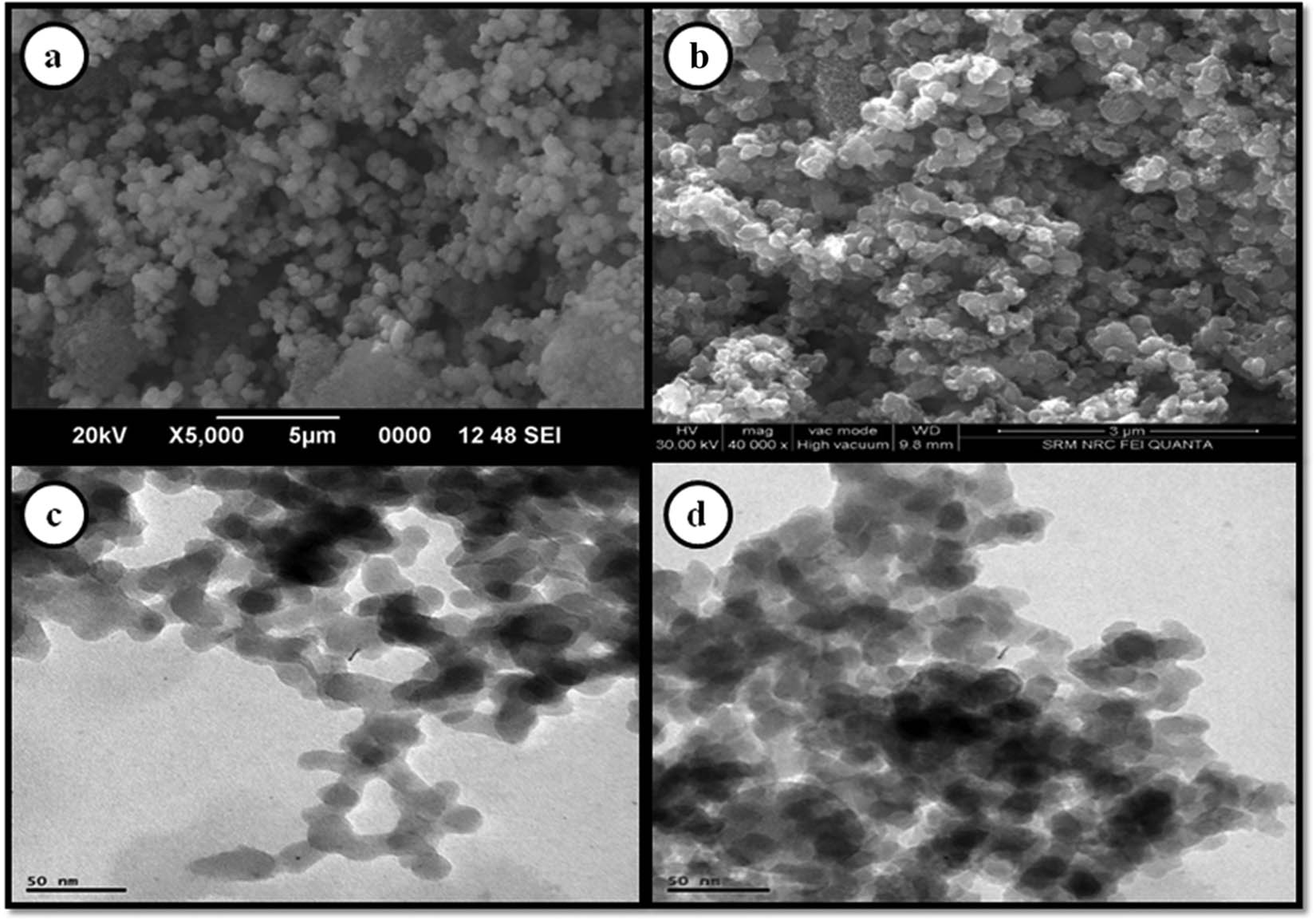
SEM analysis of (a) TiO2 and (b) Ce-doped TiO2. TEM and SAED analysis of (c) TiO2 and (d) Ce-doped TiO2.
A TEM investigation was carried out to establish the precise size of the nanoparticles. The images indicate that all of the particles are on the nanoscale. According to the selected area electron diffraction (SAED) analysis, the more crystallites linked to the surface of the single particles, the more continuous ring patterns emerge from polycrystalline nature. The vivid ring patterns indicated the high density of crystallites in the materials [36].
3.4 Anticancer activity of pure and Ce-doped TiO2
Pure and Ce-doped TiO2 nanoparticle solutions were incorporated into well-grown Y79 retinoblastoma cancer cells, and the nanoparticle-containing cells were treated with UV radiation over various periods ranging from 1 to 6 h. The stage-by-stage irradiation and associated cell condition were documented using a microscopic picture. Figure 4 (TiO2) and Figure 5 (Ce-TiO2) show observed cell pictures; when exposed to UV radiation in the presence of nanoparticles, these cells die regularly. This was corroborated by the control picture, which was exposed to UV radiation for 2 h. The cytotoxicity data demonstrate that UV irradiation increases cytotoxicity and decreases viability in nanoparticle-incorporated cells compared to the control. The proportion of cytotoxicity increased with increasing irradiation time. Pure TiO2 exhibits 66.4% TiO2 and 33.6% viability after 6 h of UV irradiation. The value was raised in Ce-TiO2, which exhibits 69.4% cytotoxicity and 30.6% survivability. The results reveal that Ce-TiO2 has a better anticancer activity than pure TiO2 [37] (Table 1).
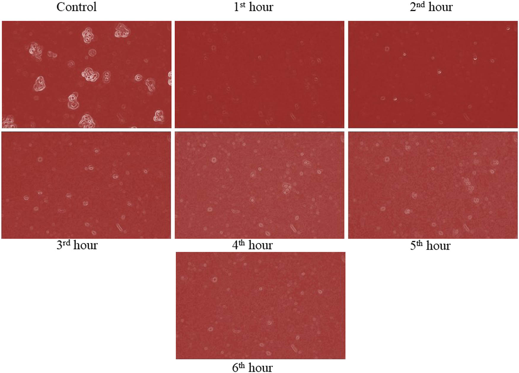
Anticancer activity of pure TiO2.
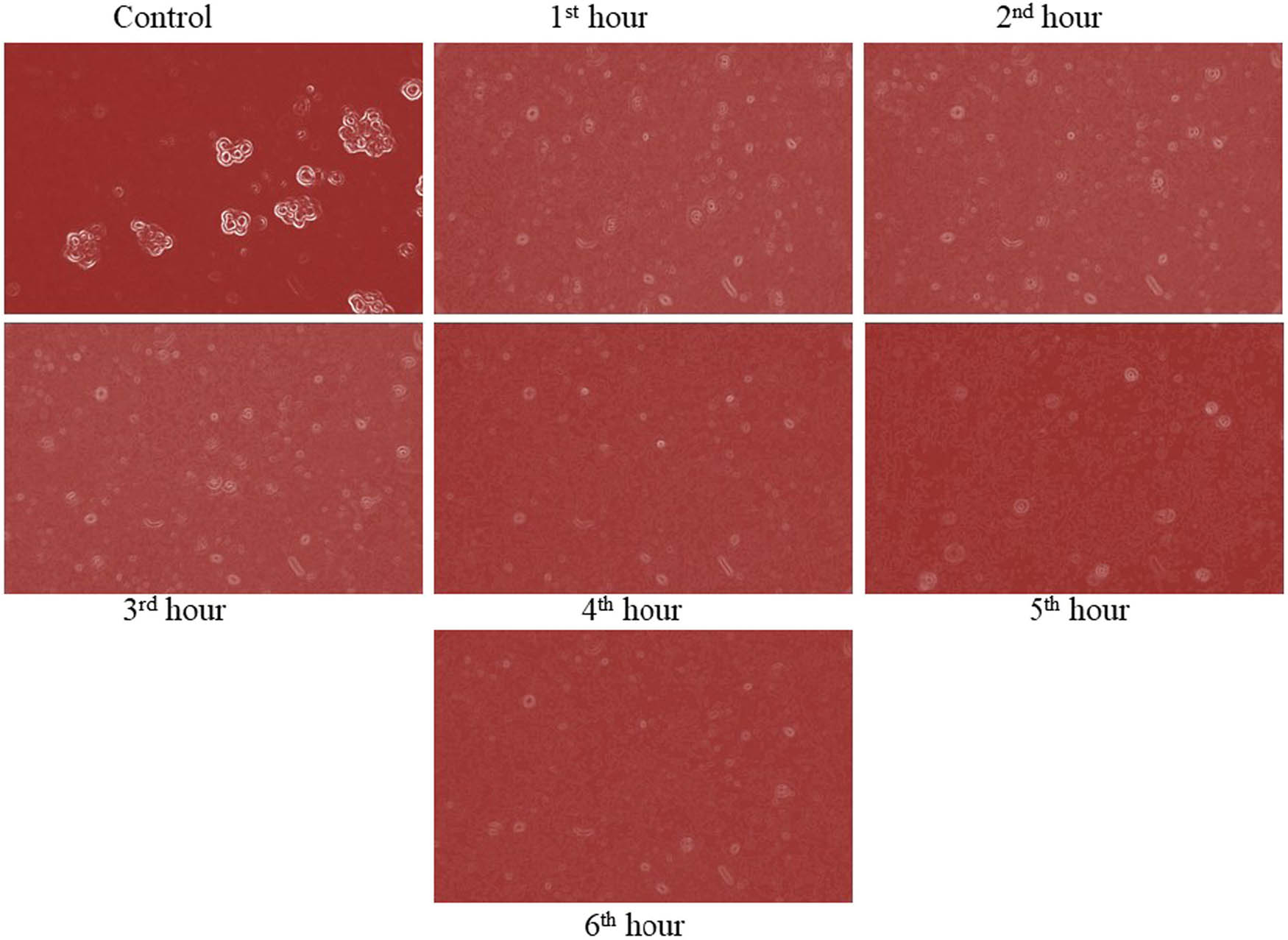
Anticancer activity of Ce-doped TiO2.
Anticancer activity of pure and Ce-doped TiO2
| Sample no. | Sample | Concentration (µg·mL−1) | Time period | Cytotoxicity | Viability | Reactivity |
|---|---|---|---|---|---|---|
| 1 | TiO2 | 200 | 1 | 49.6 | 50.4 | Mild |
| 2 | 52.2 | 47.8 | Moderate | |||
| 3 | 56.5 | 43.5 | Moderate | |||
| 4 | 59.4 | 40.6 | Moderate | |||
| 5 | 61.9 | 38.1 | Moderate | |||
| 6 | 66.4 | 33.6 | Moderate | |||
| 2 | Ce:TiO2 | 200 | 1 | 45.5 | 54.5 | Mild |
| 2 | 48.1 | 51.9 | Mild | |||
| 3 | 56.9 | 43.1 | Moderate | |||
| 4 | 59.3 | 40.7 | Moderate | |||
| 5 | 64.8 | 35.2 | Moderate | |||
| 6 | 69.4 | 30.6 | Moderate |
The anticancer action of Ce-doped TiO2 may be attributed to cerium-doped into TiO2 lattices, which has been postulated to decrease the energy gap that may be activated by UV efficiently [38,39]. Due to its significant catalytic potential and light response extension, cerium has previously been doped into anatase structures to promote photochemical processes [40]. It can also prevent electron–hole pairs from recombination, extending their lifespan. Furthermore, cerium has a higher UV cross section than biological tissue components, resulting in a significant UV interaction with host materials and subsequent reactive oxygen species production [41].
4 Conclusion
The sol–gel technique was successfully utilized to generate pure and rare earth metal Ce-doped TiO2. The structure of the XRD patterns of pure and Ce-TiO2 was anatase, with evident cerium doping in the TiO2 matrix. Raman analysis was used to validate the structural purity and phase purity of pure and Ce-TiO2. SEM and TEM examination verified the spherical shape and nanosize. Ce-doped TiO2 nanoparticles show enhanced photodynamic anticancer activity.
Acknowledgement
The authors express their sincere appreciation to the Researchers Supporting Project Number (RSP-2021/70), King Saud University, Riyadh, Saudi Arabia.
-
Funding information: Researchers Supporting Project Number (RSP-2021/70), King Saud University, Riyadh, Saudi Arabia.
-
Author contributions: Balachandran Kartha: conceptualization, writing – original draft; Kalaivani Thanikachalam: formal analysis; Natesan Vijayakumar: formal analysis; Naiyf S. Alharbi: resources, funding acquisition, validation; Shine Kadaikunnan: funding acquisition, formal analysis; Jamal M. Khaled: formal analysis; Kasi Gopinath: methodology, software; and Marimuthu Govindarajan: writing – review and editing.
-
Conflict of interest: Authors state no conflict of interest.
References
[1] Sungkaworn T, Triampo W, Nalakarn P, Triampo D, Tang IM, Lenbury Y, et al. The effects of TiO2 nanoparticles on tumor cell colonies: fractal dimension and morphological properties. Int J Med Health Biomed Bioeng Pharm Eng. 2008;2:20–7.Suche in Google Scholar
[2] Mohana Roopan S, Bharathi A, Prabhakarn A, Abdul Rahuman A, Velayutham K, Rajakumar G, et al. Efficient phyto-synthesis and structural characterization of rutile TiO2 nanoparticles using Annona squamosa peel extract. Spectrochim Acta A Mol Biomol Spectrosc. 2012;98:86–90.10.1016/j.saa.2012.08.055Suche in Google Scholar PubMed
[3] Xu J, Sun Y, Huang J, Chen C, Liu G, Jiang Y, et al. Photokilling cancer cells using highly cell-specific antibody-TiO2 bioconjugates and electroporation. Bioelectrochemistry. 2007;71:217–22.10.1016/j.bioelechem.2007.06.001Suche in Google Scholar PubMed
[4] Mohana Roopan S, Rahman Nawaz Khan F. SnO2 nanoparticles mediated nontraditional synthesis of biologically active 9-chloro-6,13-dihydro-7-phenyl-5H-indolo [3,2-c]-acridine derivatives. Spectrochim Acta A Mol Biomol Spectrosc. 2011;20:732–7.10.1007/s00044-010-9381-7Suche in Google Scholar
[5] Surendra TV, Roopan SM. Photocatalytic and antibacterial properties of phytosynthesized CeO2 NPs using Moringa oleifera peel extract. J Photochem Photobiol B: Biol. 2016;161:122–8.10.1016/j.jphotobiol.2016.05.019Suche in Google Scholar PubMed
[6] Anand K, Murugan V, Roopan SM, Tammineni V, Surendra A, Muniyasamy CS. Degradation treatment of 4-nitrophenol by Moringa oleifera synthesised GO-CeO2 nanoparticles as catalyst. J Inorg Organomet Polym Mater. 2018;28:2241–8.10.1007/s10904-018-0891-ySuche in Google Scholar
[7] Abdulla-Al-Mamun M, Kusumoto Y, Zannat T, Islam MS. Synergistic enhanced photocatalytic and photothermal activity of Au@TiO2 nanopellets against human epithelial carcinoma cells. Phys Chem Chem Phys. 2011;13:21026–34.10.1039/c1cp22683eSuche in Google Scholar PubMed
[8] Kubota Y, Shuin T, Kawasaki C, Hosaka M, Kitamura H, Cai R, et al. Photokilling of T-24 human bladder cancer cells with titanium dioxide. Br J Cancer. 1994;70:1107–11.10.1038/bjc.1994.456Suche in Google Scholar PubMed PubMed Central
[9] Paszko E, Ehrhardt C, Senge MO, Kelleher DP, Reynolds JV. Nanodrug applications in photodynamic therapy. Photodiag Photodyn Ther. 2011;8:14–29.10.1016/j.pdpdt.2010.12.001Suche in Google Scholar PubMed
[10] Zhou M, Yu J, Cheng B. Effects of Fe-doping on the photocatalytic activity of mesoporous TiO2 powders prepared by an ultrasonic method. J Hazard Mater. 2006;137:1838–47.10.1016/j.jhazmat.2006.05.028Suche in Google Scholar PubMed
[11] Zhang X, Zheng H. Synthesis of TiO2-doped SiO2 composite films and its applications. Bull Mater Sci. 2008;31:787–90.10.1007/s12034-008-0125-ySuche in Google Scholar
[12] Pelaez M, Nolan NT, Pillai SC, Seery MK, Falaras P, Kontos AG, et al. A review on the visible light active titanium dioxide photocatalysts for environmental applications. Appl Catal B Env. 2012;125:331–49.10.1016/j.apcatb.2012.05.036Suche in Google Scholar
[13] Khaki MR, Shafeeyan MS, Raman AA, Daud WM. Application of doped photocatalysts for organic pollutant degradation-A review. J Environ Manag. 2017;198:78–94.10.1016/j.jenvman.2017.04.099Suche in Google Scholar
[14] Balalakshmi C, Gopinath K, Govindarajan M, Lokesh R, Arumugam A, Alharbi NS, et al. Green synthesis of gold nanoparticles using a cheap Sphaeranthus indicus extract: impact on plant cells and the aquatic crustacean Artemia nauplii. J Photochem Photobiol B Biol. 2017;173:598–605.10.1016/j.jphotobiol.2017.06.040Suche in Google Scholar
[15] Govindarajan M, Benelli G. A facile one-pot synthesis of eco-friendly nanoparticles using Carissa carandas: ovicidal and larvicidal potential on malaria, dengue and filariasis mosquito vectors. J Clust Sci. 2017;28(1):15–36.10.1007/s10876-016-1035-6Suche in Google Scholar
[16] Mahboob S, Nivetha R, Gopinath K, Balalakshmi C, Al-Ghanim KA, Al-Misned F, et al. Facile synthesis of gold and platinum doped titanium oxide nanoparticles for antibacterial and photocatalytic activity: a photodynamic approach. Photodiag Photodyn Ther. 2021;33:102148.10.1016/j.pdpdt.2020.102148Suche in Google Scholar
[17] Suganya P, Vaseeharan B, Vijayakumar S, Balan B, Govindarajan M, Alharbi NS, et al. Biopolymer zein-coated gold nanoparticles: synthesis, antibacterial potential, toxicity and histopathological effects against the Zika virus vector Aedes aegypti. J Photochem Photobiol B Biol. 2017;173:404–11.10.1016/j.jphotobiol.2017.06.004Suche in Google Scholar
[18] Jayaseelan C, Rahuman AA, Roopan SM, Kirthi AV, Venkatesan J, Kim SK, et al. Biological approach to synthesize TiO2 nanoparticles using Aeromonas hydrophila and its antibacterial activity. Spectrochim Acta Part A: Spectrochim Acta A Mol Biomol. 2013;107:82–9.10.1016/j.saa.2012.12.083Suche in Google Scholar
[19] Bharathi A, Roopan SM, Kajbafvala A, Padmaja RD, Darsana MS, Nandhini, et al. Catalytic activity of TiO2 nanoparticles in the synthesis of some 2,3-disubstituted dihydroquinazolin-4(1H)-ones. Chin Chem Lett. 2014;25:324–6.10.1016/j.cclet.2013.11.040Suche in Google Scholar
[20] Rekha R, Divya M, Govindarajan M, Alharbi NS, Kadaikunnan S, Khaled JM, et al. Synthesis and characterization of crustin capped titanium dioxide nanoparticles: photocatalytic, antibacterial, antifungal and insecticidal activities. J Photochem Photobiol B Biol. 2019;199:111620.10.1016/j.jphotobiol.2019.111620Suche in Google Scholar
[21] Liu X, Yang J, Wang L, Yang X, Lu L, Wang X. An improvement on sol–gel method for preparing ultrafine and crystallized titania powder. Mater Sci Eng A. 2000;289:241–5.10.1016/S0921-5093(00)00901-1Suche in Google Scholar
[22] Shimizu K, Imai H, Hirashima H, Tsukuma K. Low-temperature synthesis of anatase thin films on glass and organic substrates by direct deposition from aqueous solutions. Thin Solid Films. 1999;351:220–4.10.1016/B978-044450247-6.50067-9Suche in Google Scholar
[23] TeresaViseu MR, Ferreira MIC. Morphological characterisation of TiO2 films. Vacuum. 1999;52:115–20.10.1016/S0042-207X(98)00230-9Suche in Google Scholar
[24] Blesic MD, Saponjic ZV, Nedeljkovic JM, Uskokovic DP. TiO2 films prepared by ultrasonic spray pyrolysis of nanosize precursor. Mater Lett. 2002;54:298–303.10.1016/S0167-577X(01)00581-XSuche in Google Scholar
[25] Carotta MC, Ferroni M, Gnani D, Guidi V, Merli M, Martinelli G, et al. Nanostructured pure and Nb-doped TiO2 as thick film gas sensors for environmental monitoring. Sens Actuators B Chem. 1999;58:310–7.10.1016/S0925-4005(99)00148-3Suche in Google Scholar
[26] Lee DS, Han SD, Huh JS, Lee DD. Nitrogen oxides-sensing characteristics of WO3-Based nanocrystalline thick film gas sensor. Sens Actuators B Chem. 1999;60:57–63.10.1016/S0925-4005(99)00244-0Suche in Google Scholar
[27] Ruiz AM, Arbiol J, Cornet A, Shimanoe K, Morante JR, Yamazoe N. HRTEM/EELS analysis, structural characterisation and sensor performances of hydrothermal nano-TiO2. Mater Res Soc. 2004;828:155–60.10.1557/PROC-828-A4.10Suche in Google Scholar
[28] Mohammadi MR, Fray DJ, Cordero-Cabrera MC. Sensor performance of nanostructuredTiO2 thin films derived from particulate sol–gel route and polymeric fugitive agents. Sens Actuators B Chem. 2007;124:74–83.10.1016/j.snb.2006.11.048Suche in Google Scholar
[29] Marami MB, Farahmandjou M. Water-based sol–gel synthesis of Ce-doped TiO2 nanoparticles. J Electron Mater. 2019;48:4740–7.10.1007/s11664-019-07265-9Suche in Google Scholar
[30] Alijani M, Kaleji BK. Optical and structural properties of TiO2 nanopowders with Ce/Sn doping at various calcinations temperature and time. Opt Quant Electron. 2017;49:34–50.10.1007/s11082-016-0851-0Suche in Google Scholar
[31] Chen YF, Lee CY, Yeng MY, Chiu HT. The effect of calcination temperature on the crystallinity of TiO2 nanopowders. J Cryst Growth. 2003;247:363–70.10.1016/S0022-0248(02)01938-3Suche in Google Scholar
[32] Nakagawa K, Murata Y, Kishida M, Adachi M, Hiro M, Susa K. Formation and reaction activity of CeO2 nanoparticles of cubic structure and various shaped CeO2–TiO2 composite nanostructures. Mater Chem Phys. 2007;104:30–9.10.1016/j.matchemphys.2007.02.047Suche in Google Scholar
[33] Balachandran K, Venckatesh R, Sivaraj R, Hemalatha KV, Mariappan R. Enhancing power conversion efficiency of DSSC by doping SiO2 in TiO2 photoanodes. Mater Sci Semicond Process. 2015;35:59–65.10.1016/j.mssp.2015.02.071Suche in Google Scholar
[34] Balalakshmi C, Alharbi N, Kadaikunnan S, Khaled J, Alanzi K, Gopinath K, et al. Development of chitosan/agar-silver nanoparticles-coated paper for antibacterial application. Green Process Synth. 2020;9(1):751–9.10.1515/gps-2020-0070Suche in Google Scholar
[35] Alwhibi M, Soliman D, Awad M, Alangery A, Al Dehaish H, Alwasel Y. Green synthesis of silver nanoparticles: characterization and its potential biomedical applications. Green Process Synth. 2021;10(1):412–20.10.1515/gps-2021-0039Suche in Google Scholar
[36] Devi R, Francis A, Devasena T. Green-synthesized gold nanocubes functionalized with bisdemethoxycurcumin analog as an ideal anticancer candidate. Green Process Synth. 2014;3(1):47–61.10.1515/gps-2013-0090Suche in Google Scholar
[37] Andleeb S, Tariq F, Muneer A, Nazir T, Shahid B, Latif Z, et al. In vitro bactericidal, antidiabetic, cytotoxic, anticoagulant, and hemolytic effect of green-synthesized silver nanoparticles using Allium sativum clove extract incubated at various temperatures. Green Process Synth. 2020;9(1):538–53.10.1515/gps-2020-0051Suche in Google Scholar
[38] Ma L, Zou X, Chen W. A new X-ray activated nanoparticle photosensitizer for cancer treatment. J Biomed Nanotechnol. 2014;10:1501–8.10.1166/jbn.2014.1954Suche in Google Scholar PubMed
[39] Zalas M, Laniecki M. Photocatalytic hydrogen generation over lanthanides-doped Titania. Sol Energy Mater Sol Cell. 2005;89:287–96.10.1016/j.solmat.2005.02.014Suche in Google Scholar
[40] Li FB, Li XZ, Hou MF, Cheah KW, Choy WCH. Enhanced photocatalytic activity of Ce3+–TiO2 for 2-mercaptobenzothiazole degradation in aqueous suspension for odour control. Appl Catal A: Gen. 2005;285:181–9.10.1016/j.apcata.2005.02.025Suche in Google Scholar
[41] Fairchild RG, Bond VP. Photon activation therapy. Strahlentherapie. 1984;160:758–63.Suche in Google Scholar
© 2022 Balachandran Kartha et al., published by De Gruyter
This work is licensed under the Creative Commons Attribution 4.0 International License.
Artikel in diesem Heft
- Research Articles
- Kinetic study on the reaction between Incoloy 825 alloy and low-fluoride slag for electroslag remelting
- Black pepper (Piper nigrum) fruit-based gold nanoparticles (BP-AuNPs): Synthesis, characterization, biological activities, and catalytic applications – A green approach
- Protective role of foliar application of green-synthesized silver nanoparticles against wheat stripe rust disease caused by Puccinia striiformis
- Effects of nitrogen and phosphorus on Microcystis aeruginosa growth and microcystin production
- Efficient degradation of methyl orange and methylene blue in aqueous solution using a novel Fenton-like catalyst of CuCo-ZIFs
- Synthesis of biological base oils by a green process
- Efficient pilot-scale synthesis of the key cefonicid intermediate at room temperature
- Synthesis and characterization of noble metal/metal oxide nanoparticles and their potential antidiabetic effect on biochemical parameters and wound healing
- Regioselectivity in the reaction of 5-amino-3-anilino-1H-pyrazole-4-carbonitrile with cinnamonitriles and enaminones: Synthesis of functionally substituted pyrazolo[1,5-a]pyrimidine derivatives
- A numerical study on the in-nozzle cavitating flow and near-field atomization of cylindrical, V-type, and Y-type intersecting hole nozzles using the LES-VOF method
- Synthesis and characterization of Ce-doped TiO2 nanoparticles and their enhanced anticancer activity in Y79 retinoblastoma cancer cells
- Aspects of the physiochemical properties of SARS-CoV-2 to prevent S-protein receptor binding using Arabic gum
- Sonochemical synthesis of protein microcapsules loaded with traditional Chinese herb extracts
- MW-assisted hydrolysis of phosphinates in the presence of PTSA as the catalyst, and as a MW absorber
- Fabrication of silicotungstic acid immobilized on Ce-based MOF and embedded in Zr-based MOF matrix for green fatty acid esterification
- Superior photocatalytic degradation performance for gaseous toluene by 3D g-C3N4-reduced graphene oxide gels
- Catalytic performance of Na/Ca-based fluxes for coal char gasification
- Slow pyrolysis of waste navel orange peels with metal oxide catalysts to produce high-grade bio-oil
- Development and butyrylcholinesterase/monoamine oxidase inhibition potential of PVA-Berberis lycium nanofibers
- Influence of biosynthesized silver nanoparticles using red alga Corallina elongata on broiler chicks’ performance
- Green synthesis, characterization, cytotoxicity, and antimicrobial activity of iron oxide nanoparticles using Nigella sativa seed extract
- Vitamin supplements enhance Spirulina platensis biomass and phytochemical contents
- Malachite green dye removal using ceramsite-supported nanoscale zero-valent iron in a fixed-bed reactor
- Green synthesis of manganese-doped superparamagnetic iron oxide nanoparticles for the effective removal of Pb(ii) from aqueous solutions
- Desalination technology for energy-efficient and low-cost water production: A bibliometric analysis
- Biological fabrication of zinc oxide nanoparticles from Nepeta cataria potentially produces apoptosis through inhibition of proliferative markers in ovarian cancer
- Effect of stabilizers on Mn ZnSe quantum dots synthesized by using green method
- Calcium oxide addition and ultrasonic pretreatment-assisted hydrothermal carbonization of granatum for adsorption of lead
- Fe3O4@SiO2 nanoflakes synthesized using biogenic silica from Salacca zalacca leaf ash and the mechanistic insight into adsorption and photocatalytic wet peroxidation of dye
- Facile route of synthesis of silver nanoparticles templated bacterial cellulose, characterization, and its antibacterial application
- Synergistic in vitro anticancer actions of decorated selenium nanoparticles with fucoidan/Reishi extract against colorectal adenocarcinoma cells
- Preparation of the micro-size flake silver powders by using a micro-jet reactor
- Effect of direct coal liquefaction residue on the properties of fine blue-coke-based activated coke
- Integration of microwave co-torrefaction with helical lift for pellet fuel production
- Cytotoxicity of green-synthesized silver nanoparticles by Adansonia digitata fruit extract against HTC116 and SW480 human colon cancer cell lines
- Optimization of biochar preparation process and carbon sequestration effect of pruned wolfberry branches
- Anticancer potential of biogenic silver nanoparticles using the stem extract of Commiphora gileadensis against human colon cancer cells
- Fabrication and characterization of lysine hydrochloride Cu(ii) complexes and their potential for bombing bacterial resistance
- First report of biocellulose production by an indigenous yeast, Pichia kudriavzevii USM-YBP2
- Biosynthesis and characterization of silver nanoparticles prepared using seeds of Sisymbrium irio and evaluation of their antifungal and cytotoxic activities
- Synthesis, characterization, and photocatalysis of a rare-earth cerium/silver/zinc oxide inorganic nanocomposite
- Developing a plastic cycle toward circular economy practice
- Fabrication of CsPb1−xMnxBr3−2xCl2x (x = 0–0.5) quantum dots for near UV photodetector application
- Anti-colon cancer activities of green-synthesized Moringa oleifera–AgNPs against human colon cancer cells
- Phosphorus removal from aqueous solution by adsorption using wetland-based biochar: Batch experiment
- A low-cost and eco-friendly fabrication of an MCDI-utilized PVA/SSA/GA cation exchange membrane
- Synthesis, microstructure, and phase transition characteristics of Gd/Nd-doped nano VO2 powders
- Biomediated synthesis of ZnO quantum dots decorated attapulgite nanocomposites for improved antibacterial properties
- Preparation of metal–organic frameworks by microwave-assisted ball milling for the removal of CR from wastewater
- A green approach in the biological base oil process
- A cost-effective and eco-friendly biosorption technology for complete removal of nickel ions from an aqueous solution: Optimization of process variables
- Protective role of Spirulina platensis liquid extract against salinity stress effects on Triticum aestivum L.
- Comprehensive physical and chemical characterization highlights the uniqueness of enzymatic gelatin in terms of surface properties
- Effectiveness of different accelerated green synthesis methods in zinc oxide nanoparticles using red pepper extract: Synthesis and characterization
- Blueprinting morpho-anatomical episodes via green silver nanoparticles foliation
- A numerical study on the effects of bowl and nozzle geometry on performances of an engine fueled with diesel or bio-diesel fuels
- Liquid-phase hydrogenation of carbon tetrachloride catalyzed by three-dimensional graphene-supported palladium catalyst
- The catalytic performance of acid-modified Hβ molecular sieves for environmentally friendly acylation of 2-methylnaphthalene
- A study of the precipitation of cerium oxide synthesized from rare earth sources used as the catalyst for biodiesel production
- Larvicidal potential of Cipadessa baccifera leaf extract-synthesized zinc nanoparticles against three major mosquito vectors
- Fabrication of green nanoinsecticides from agri-waste of corn silk and its larvicidal and antibiofilm properties
- Palladium-mediated base-free and solvent-free synthesis of aromatic azo compounds from anilines catalyzed by copper acetate
- Study on the functionalization of activated carbon and the effect of binder toward capacitive deionization application
- Co-chlorination of low-density polyethylene in paraffin: An intensified green process alternative to conventional solvent-based chlorination
- Antioxidant and photocatalytic properties of zinc oxide nanoparticles phyto-fabricated using the aqueous leaf extract of Sida acuta
- Recovery of cobalt from spent lithium-ion battery cathode materials by using choline chloride-based deep eutectic solvent
- Synthesis of insoluble sulfur and development of green technology based on Aspen Plus simulation
- Photodegradation of methyl orange under solar irradiation on Fe-doped ZnO nanoparticles synthesized using wild olive leaf extract
- A facile and universal method to purify silica from natural sand
- Green synthesis of silver nanoparticles using Atalantia monophylla: A potential eco-friendly agent for controlling blood-sucking vectors
- Endophytic bacterial strain, Brevibacillus brevis-mediated green synthesis of copper oxide nanoparticles, characterization, antifungal, in vitro cytotoxicity, and larvicidal activity
- Off-gas detection and treatment for green air-plasma process
- Ultrasonic-assisted food grade nanoemulsion preparation from clove bud essential oil and evaluation of its antioxidant and antibacterial activity
- Construction of mercury ion fluorescence system in water samples and art materials and fluorescence detection method for rhodamine B derivatives
- Hydroxyapatite/TPU/PLA nanocomposites: Morphological, dynamic-mechanical, and thermal study
- Potential of anaerobic co-digestion of acidic fruit processing waste and waste-activated sludge for biogas production
- Synthesis and characterization of ZnO–TiO2–chitosan–escin metallic nanocomposites: Evaluation of their antimicrobial and anticancer activities
- Nitrogen removal characteristics of wet–dry alternative constructed wetlands
- Structural properties and reactivity variations of wheat straw char catalysts in volatile reforming
- Microfluidic plasma: Novel process intensification strategy
- Antibacterial and photocatalytic activity of visible-light-induced synthesized gold nanoparticles by using Lantana camara flower extract
- Antimicrobial edible materials via nano-modifications for food safety applications
- Biosynthesis of nano-curcumin/nano-selenium composite and their potentialities as bactericides against fish-borne pathogens
- Exploring the effect of silver nanoparticles on gene expression in colon cancer cell line HCT116
- Chemical synthesis, characterization, and dose optimization of chitosan-based nanoparticles of clodinofop propargyl and fenoxaprop-p-ethyl for management of Phalaris minor (little seed canary grass): First report
- Double [3 + 2] cycloadditions for diastereoselective synthesis of spirooxindole pyrrolizidines
- Green synthesis of silver nanoparticles and their antibacterial activities
- Review Articles
- A comprehensive review on green synthesis of titanium dioxide nanoparticles and their diverse biomedical applications
- Applications of polyaniline-impregnated silica gel-based nanocomposites in wastewater treatment as an efficient adsorbent of some important organic dyes
- Green synthesis of nano-propolis and nanoparticles (Se and Ag) from ethanolic extract of propolis, their biochemical characterization: A review
- Advances in novel activation methods to perform green organic synthesis using recyclable heteropolyacid catalysis
- Limitations of nanomaterials insights in green chemistry sustainable route: Review on novel applications
- Special Issue: Use of magnetic resonance in profiling bioactive metabolites and its applications (Guest Editors: Plalanoivel Velmurugan et al.)
- Stomach-affecting intestinal parasites as a precursor model of Pheretima posthuma treated with anthelmintic drug from Dodonaea viscosa Linn.
- Anti-asthmatic activity of Saudi herbal composites from plants Bacopa monnieri and Euphorbia hirta on Guinea pigs
- Embedding green synthesized zinc oxide nanoparticles in cotton fabrics and assessment of their antibacterial wound healing and cytotoxic properties: An eco-friendly approach
- Synthetic pathway of 2-fluoro-N,N-diphenylbenzamide with opto-electrical properties: NMR, FT-IR, UV-Vis spectroscopic, and DFT computational studies of the first-order nonlinear optical organic single crystal