Synthesis of biogenic silver nanoparticles from the seed coat waste of pistachio (Pistacia vera) and their effect on the growth of eggplant
-
Masudulla Khan


Abstract
Herein, the synthesis of silver nanoparticles using extracts of pistachio seed coat waste is investigated. The surface plasmon resonance peak at 443 nm was observed in the nanoparticles by using ultraviolet-visible spectroscopy (UV-Vis). To identify potential biomolecules involved in the bio-reduction of silver ions, Fourier-transform infrared spectroscopy (FTIR) was used. Scanning and transmission electron microscopy (SEM and TEM) show irregular shapes with an average size of ∼20 nm. The active surface determined by Brunauer, Emmett, and Teller analysis was 22 m2/g. The effect of silver nanoparticles on eggplants sprayed with a nanoparticle suspension of 75 mg/L led to increased plant growth and chlorophyll and carotenoid contents. The fly ash addition to the soil promoted plant growth. The highest increase in plant growth occurs when plants were sprayed with 75 ppm AgNPs in 20% fly ash amended soil.
1 Introduction
Nanotechnology has emerged in recent years as one of the most important areas of modern science associated with green synthesis [1]. The use of modern techniques to synthesize nanoparticles has become a popular application for a variety of products in the biomedical and healthcare fields [2]. Richard Feynman coined the term “nanotechnology” for the first time in 1959, which is widely regarded as the start of modern nanotechnology [3]. The green synthesis approach has been subjected to a reliable, sustainable, and eco-friendly protocol to synthesize a variety of nanoparticle metal oxides and metal oxides in some cases under exposure to solar radiation to accelerate the synthesis of nanoparticles. A great deal of attention has been paid in recent years to the exploration of waste materials in different areas of human application. One of these areas where scientists focus with zeal [4,5] is the use of plant waste materials with different names such as shell, seed coat fruit peel, skin, hull, etc., containing bioactive compounds [6]. These waste materials are a prime example of an undervalued and unused energy source that can be used as a reducing agent in nanoparticle synthesis. The list of waste materials of plants used for silver nanoparticles synthesis and their particle size is given in the Supplementary Information (Table S1). Due to their unique characteristics, nanoparticles are used in many industrial sectors. Of the various types of nanoparticles, silver nanoparticles (AGNPs) are the most commonly used [7,8,9]. Synthesis of nanoparticles from plant parts is preferred because it is simple, environment friendly, and safe [10,11]. The plant materials are more advantageous, feasible, and simple for nanoparticles synthesis than other materials [12,13]. The use of nanoparticles to enhance plants growth is currently being researched all over the world [14]. Silver nanoparticles (AgNPs) have antimicrobial, catalytic, physiochemical, and optical properties [15]. Brinjal (Solanum melongena L.) is a popular nightshade crop grown all over the world. It is one of the most common and widely consumed vegetables in India. In this country, 730,000 hectares of land are under eggplant cultivation with an annual production of 12,801 million tonnes [16]. Metalbased nanomaterials are being developed by scientists to aid plant growth and development. Nanotechnology has the potential to be a useful tool for delivering pesticides and fertilizers to the target site with accuracy and precision. Nanoparticles injected into plants could have a significant impact, and thus, could be used to improve plant growth and yield in agricultural applications [17]. However, more research work is needed to fully comprehend the molecular mechanism of action of nanoparticles in plant physiology [18]. Plants have been found to be capable of producing the natural mineralized nanomaterials required for their growth under certain conditions [19]. To the best of our knowledge, this is the first study to use aqueous seed coat extract of pista (Pistacia vera) to the biosynthesis of AgNPs as part of a green nanoparticle synthesis approach. The novelty of this article is therefore to describe the green synthesis of AgNPs from the waste part of the seed and to investigate their impact on eggplant plant growth capacity, carotenoid, and chlorophyll content.
2 Materials and methods
2.1 Biosynthesis of silver nanoparticles and characterization
Silver nanoparticles were synthesized using the same protocol as in prior research [10,20]. All other chemicals such as sodium hypochlorite (NaOCl), silver nitrate (AgNO3), and the solvents and chemicals used in this study were provided by Merck India Ltd. UV-Vis spectral analysis was used to validate the production of AgNPs using a Shimadzu spectrophotometer UV-Vis 1800 (Japan). Cu K radiation (λ = 0.15405 nm) was used for X-ray diffraction (XRD, PANalytical, X’pert PRO-MPD, The Netherlands). SEM (NOVA nano FE-SEM 450 FEI) and TEM (TECNAI-G-20). were used to examine the morphology and the size of the prepared AgNPs. FT-IR spectra of the plant extract and biosynthesized AgNPs were collected utilizing the KBr pellet technique with PerkinElmer Spectrum 2000 in the 4,000–400/cm range. The specific surface area of biogenic silver nanoparticles using nitrogen adsorption–desorption isotherms (Brunauer–Emmett–Teller (BET)) was calculated with the Nova 3200e.
The seed coat extract of pista (Pistacia vera) was used to synthesize silver nanoparticles (AgNPs). The material was cleaned with distilled water to remove contaminated contents and air-dried in sunlight. The material was ground into a fine powder, and 20 g of the fine powder was placed in a flask with 200 mL double distilled water, which was then refluxed for 30 min. For the next step, the plant extract was cooled and filtered through Whatman filter paper no. 1.
As such, silver nitrate GR was used, which was obtained from Merck (India). In an Erlenmeyer flask, 100 mL of silver nitrate at a concentration of 1 mM was prepared. Then, various concentrations of the seed coat extract (2.5, 5.0, and 10 mL) were added to 90 mL of silver nitrate solution to optimize the rapid reduction of AgNO3 and left at room temperature to observe the color change; the formation was also recognized by using UV-Visible spectroscopy in the range of 443 nm. Centrifugation at 10,000 rpm for 20 min was used to collect biosynthesized AgNPs, which were then carefully washed with double-distilled sterile water. The collected nanoparticles synthesized using 10 mL of the extract based on the rapid reduction of AgNO3 to AgNPs were further investigated by using FT-IR, XRD, UV-Vis, SEM, and TEM spectroscopic techniques.
2.2 Plant culture, total leaf chlorophyll, and the carotenoid content
Eggplant seeds were sterilized for 5 min with 0.1% sodium hypochlorite and then washed twice with distilled water. Fifteen days old seedlings of eggplant were sprayed with biosynthesized AgNPs and one set of plants was left without spraying as control (without AgNPs). Each treatment had five replicates. The experiment results were terminated 100 days after spraying of AgNPs.
The total chlorophyll and carotenoid content of photosynthetic pigments of eggplants were approximated using the method given by Mackinney [21] and Maclachlan and Saul [22], respectively.
2.3 Fly ash
In this experiment, we used loam soil, which was obtained from the fields of Jaipur, India.
Before planting, the soil was autoclaved at 137.9 kPa for 20–25 min, while the fly ash was sun-dried for 10–15 days and mixed (vol:vol) in four proportions with soil: 0 (100), 20, 50, and 100% fly ash. About 3 kg of fly ash and soil mixtures were placed in pots. Fly ash was purchased from a local market in Jaipur, India.
2.4 Statistical analysis
The data were analyzed using ANOVA in GraphPad Prism software, and means were compared using mean and standard deviation including the Tukey post-hoc test and Dunnett’s multiple comparisons test at the p < 0.05 probability level. The mean and standard deviation presented in the graph was designed using Excel software.
3 Results and discussion
Previous research studies have reported on the biosynthesis of silver nanoparticles from plant parts [10,11,20]. Therefore, in continuation with our interest in the preparation of silver nanoparticles from the seed coat waste extract of pista, in the current research, AgNPs have been prepared and their impact on eggplant plants is studied.
3.1 Fourier infrared spectroscopy analysis (FTIR)
FT-IR spectra of the biogenic synthesis of Ag-nanoparticles and the aqueous extracts of Pistachio shells are displayed in Figure 1. FT-IR spectra of plant extracts (Figure 1a) and AgNPs were used to identify the metabolites responsible for Ag ion reduction (Figure 1b). Typical infrared absorption bands at 3446.08 and 3436.70/cm showed an indication of the presence of phenol and alcohol with a free functional group (–OH) [23]. The wide infrared bands of the OH group, generally as shown in Figure 1, were overlaid with the NH stretching band [24]. The absorption bands reflected the presence of alkanes (methyl and methylene groups) in the lipids indicating asymmetric stretching bands at 2,926 and 2930.78/cm [23]. The presence of carboxylic ion in the biosynthesized solution [25] was revealed by the bands at 2434.90/cm. The OH and CO functionality of the extract plays a responsible role in reducing and stabilizing the nanoparticles [26]. The strong bands at 1,611 and 1,630 cm−1 primarily correspond to the CONH stretching associated with the presence of amide groups in the samples while the band at 1,418 cm−1 represents the N–H bending vibrational mode. The peaks at about 1,015 and 1,075/cm indicate that there are C–C and C–O-linkages in the samples. Other major IR absorption bands at 1381.65, 1316.67, 777.94, 617.09, 581.78, and 525.98/cm in the spectra of the extract and the nanoparticles represented various functional groups such as ethers, esters, carbonyl (polyol’s), or aromatic compounds and alkyl halides that served as reducing and limiting agents during the synthesis of the stable silver nanoparticles. Between the FT-IR spectrum of Pistacia vera shell extracts and the FT-IR of biosynthesized nanoparticles, only a minor alteration of the location of the majority of the IR bands is observed. This slight variation in the position of the IR bands of functionalities across the spectrum supported the nanoparticle synthesis using the pista extract.
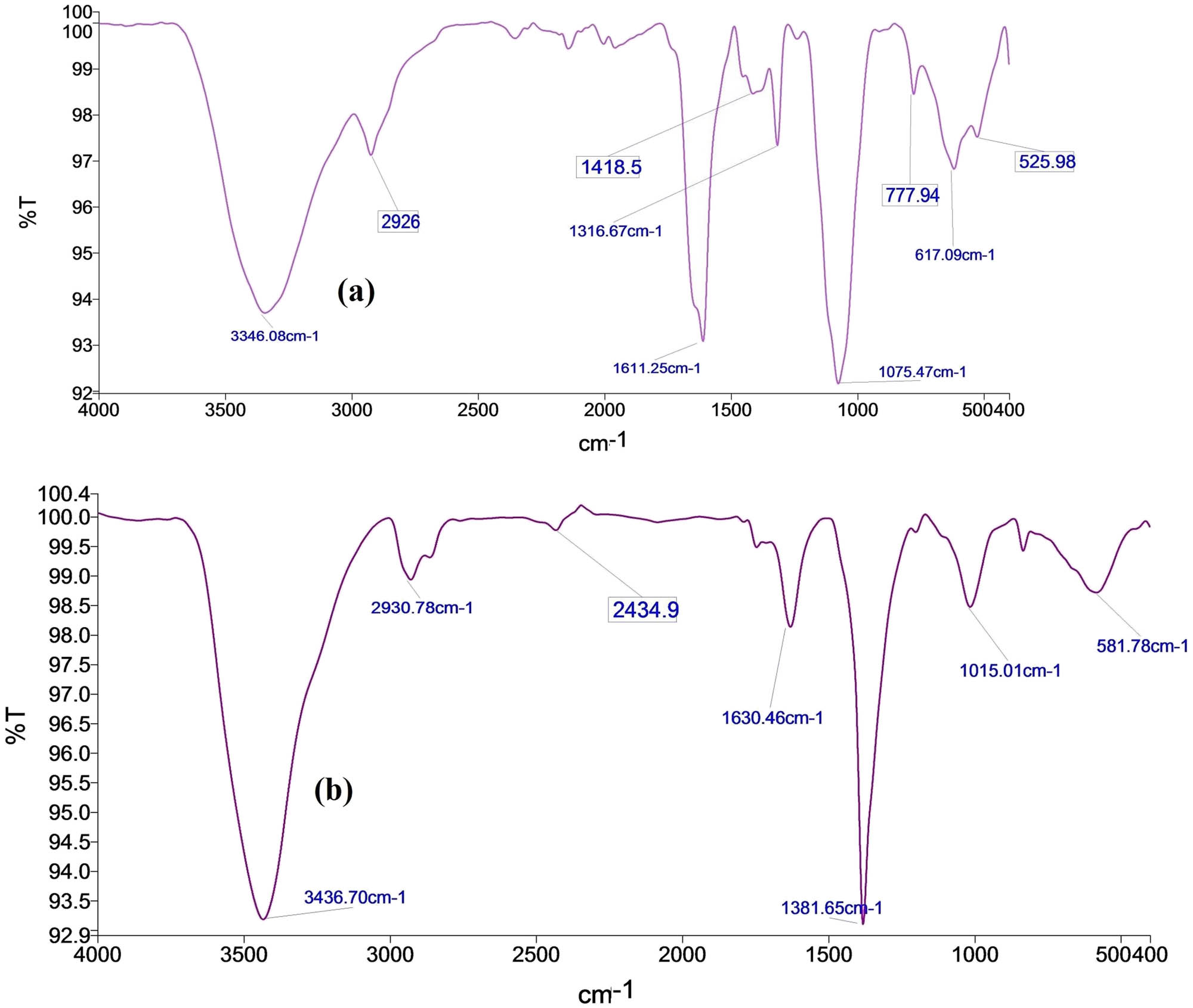
FT-IR spectra of the (a) extract of pista and (b) biosynthesized AgNPs.
3.2 XRD
The X-ray diffraction (XRD) pattern of the green synthesized AgNPs was recorded in the 2θ range 20–80°, as shown in Figure 2. Prominent diffraction peaks at 29.75°, 31.93°, 33.07°, 34.64°, 35.49°, 38.25°, 41.99°, 43.70°, 50.01°, 54.03°, 55.29°, 62.69°, 64.8°, and 77.51° are shown, and the high peak of the XRD pattern signifies active silver composition. In the XRD pattern, characteristic peaks at 29.75°, 38.25°, 43.70°, 64.8°, and 77.51° observed in the experimental diffractogram (Figure 2) are attributed to the silver metal corresponding to hkl values at 210, 111, 200, 220, and 311 silver planes. These peaks are related to the face-centered cubic (fcc) crystal lattice structure of silver (JCPDS Nos. 87-0720, 03-0921, and 04-0783). This indicates that the biosynthesized AgNPs are polycrystalline. Similar peak patterns have also been reported [8,10,20] for AgNPs. The average crystallite size of the synthesized AgNPs was found to be around 20 nm using Scherrer’s formula. The XRD pattern shows that the silver nanoparticles synthesized by the seed coat extract are crystalline [27,28]. In addition, the extra peaks appearing in the diffractogram may be due to the biomass residue capping of AgNPs [29].
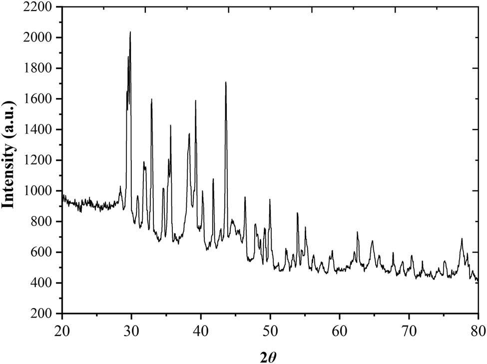
XRD pattern of biosynthesized AgNPs.
3.3 UV-Vis spectroscopy
To confirm the green synthesis of AgNPs and their stability, UV-Vis analysis was performed. Figure 3b depicts the UV-Vis spectrum of the synthesized AgNPs. It had a ∼443 nm absorption band, which is a characteristic band of AgNPs due to the surface plasma resonance (SPR) of silver. The UV-Vis spectrum of the extract of the seed coat (Figure 3a) indicated the presence of polyphenols and flavonoids as reported elsewhere [30]. The absorption peaks at about 260 and 290 nm were attributed to the transition from π to π* located within the polyphenols as the antioxidant content of the shell.
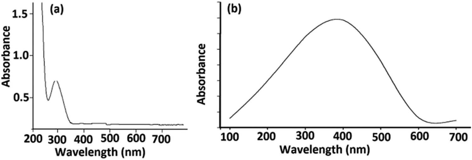
UV-Vis spectrum of (a) the extract of pista shell and (b) AgNPs.
3.4 Mechanism and stabilization
The polyphenols and flavonoids of the shell play an essential part in the biogenic synthesis of silver nanoparticles by reducing and stabilizing the nanoparticles. They also have other unique properties of adhering to the metal surface to form a coat over AgNPs and thus protect them from aggregation. Based on the GC-MS of pistachio shell extracts published earlier [31,32,33], featuring important biomolecules such as polyphenols, flavonoids, glycosides, terpenoids, and anthocyanins, these biomolecules – in particular phenolic compounds – play a major role in the reduction of silver ions and prevent them from aggregation; the tentative mechanism (Figure 4) of silver nanoparticle biosynthesis is drawn based on the screening of literature studies [23,34,35].
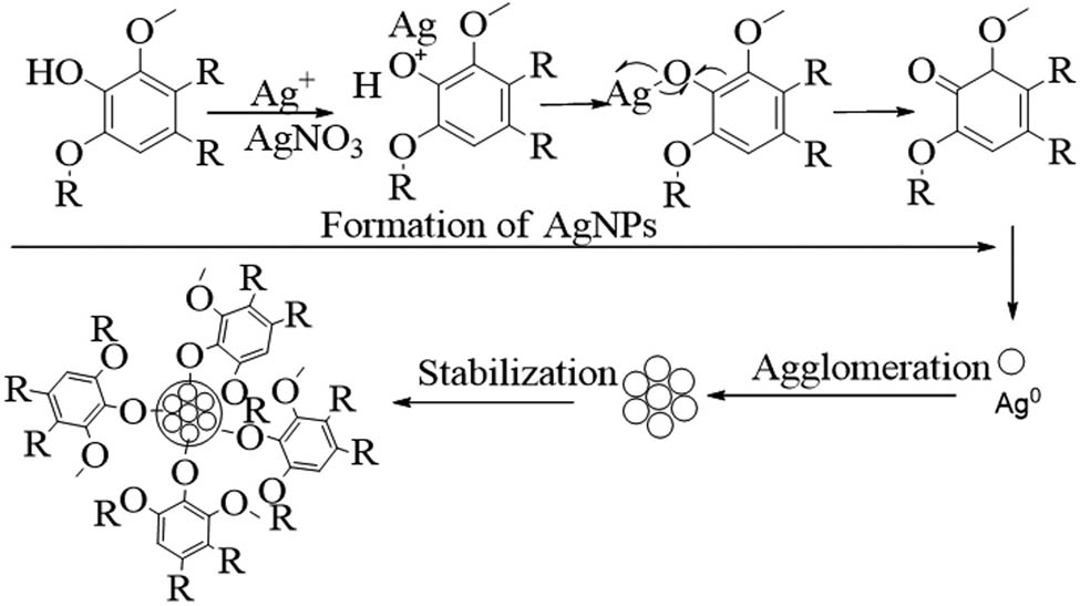
Tentative mechanism and stabilization of biogenic AgNPs.
3.5 SEM, TEM, SAED, and BET studies of biosynthesized AgNPs
SEM data of biosynthesized silver nanoparticles have been used to identify the morphology; it reveals that silver nanoparticles are agglomerated due to induced dehydration but polydispersed with irregular shapes (Figure 5). TEM data confirm the formation of nanoparticles (AgNPs) with irregular shapes, from rectangular to spherical shapes with an average size of 20 nm (Figure 6). BET analysis of AgNPs was performed to know additional information on the surface of nanoparticles obtained from the Brunauer–Emmett–Teller (BET) analysis of the specific surface area. The BET analysis of silver nanoparticles displayed that the surface area of the biogenic AgNPs was 21.73 m2/g at ambient temperature, and the value obtained from BET correlated with the results obtained from TEM findings (Supplementary file S1). A static volumetric absorption analyzer was also used to calculate the isotherms of N2 absorption–desorption of the biogenic NPs. Figure 6b shows a selected area of the electron diffraction (SAED) pattern of the biogenic AgNPs, which confirms the crystalline nature of the synthesized nanoparticles in the sample, as evidenced by the presence of bright dots.
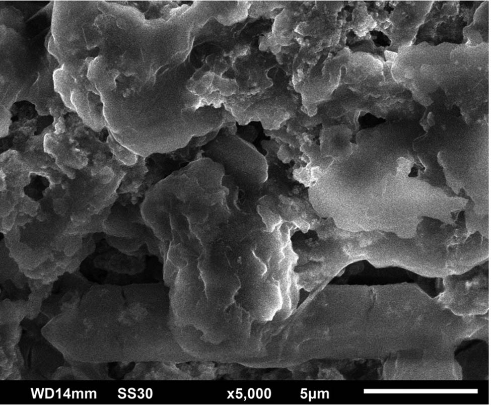
SEM image of the synthesized AgNPs.
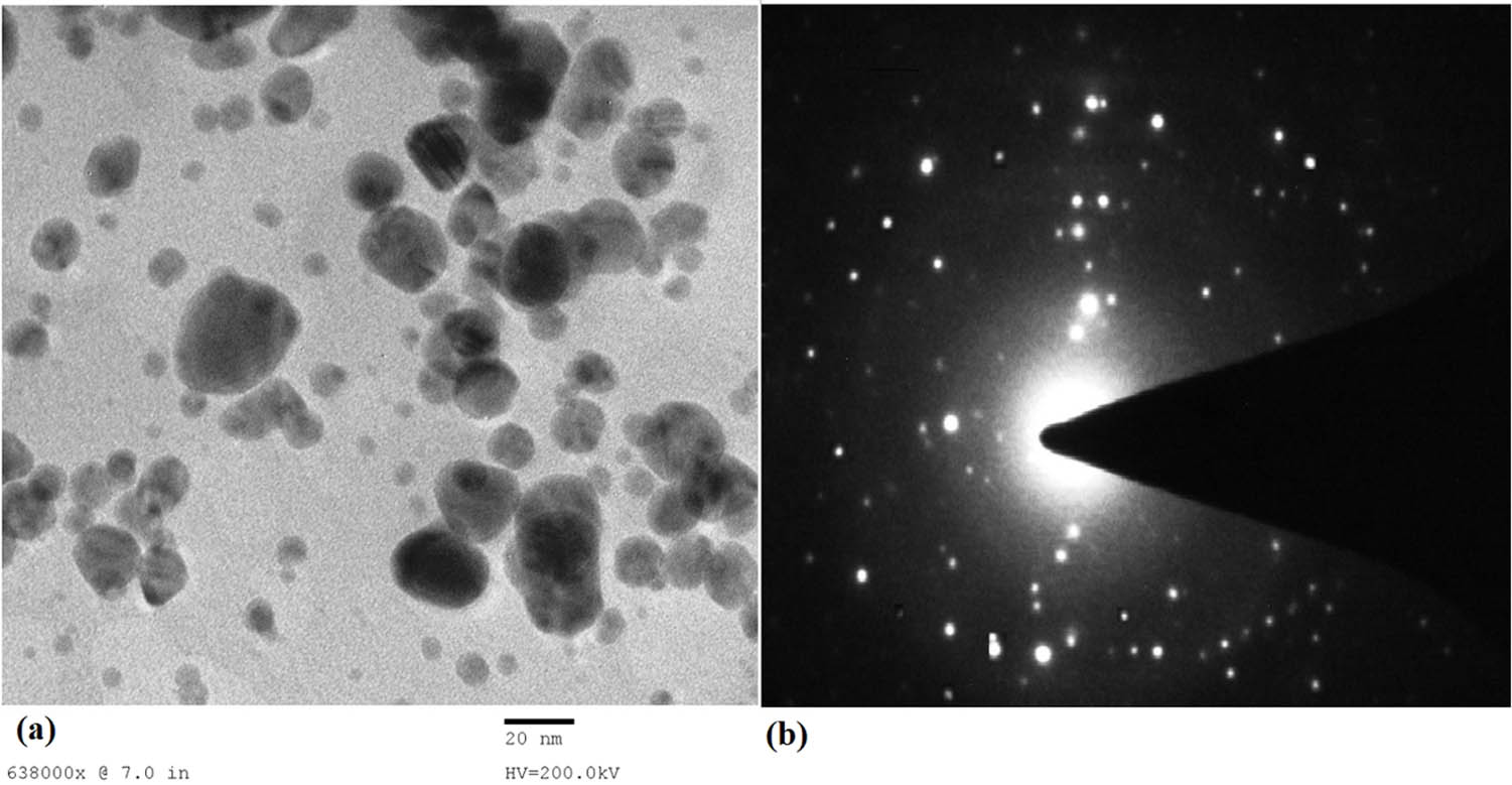
(a) TEM images showing the circular shape of the synthesized AgNPs and (b) the selected area of electron diffraction (SAED) pattern of silver nanoparticles chosen at random.
3.6 Effect of different concentrations of AgNPs and fly ash amendment on plant growth in the pot experiment
Nanoparticle properties, host plant and specific type of nanoparticle interaction, surface coating, size, dosage, exposure time, and a variety of other factors all have an impact on their functional expression. Plants are affected by nanoparticles in a number of ways, including morphological and physiological changes. In our research, foliar spraying of eggplant plants with 25, 50, and 75 ppm AgNPs resulted in significant increases in plant growth, chlorophyll, and carotenoid content. The treatment with 50 ppm AgNPs increased the plant length by 4.65% as compared to the control plant. The highest improvement in plant growth occurs after treatment with 75 ppm AgNPs. An increase in the plant length of 11.90% and an increase in the plant fresh weight of 22.27% occur after treatment with 75 ppm AgNPs. A 21.34% increase in the chlorophyll content occurs. The treatment with 75 ppm AgNPs improves the carotenoid content by 15.47% in treated plants. The effect of AgNPs on eggplants is concentration-dependent (Figure 7). Concentrations of 150, 300, 500, and 1,000 ppm were found to be toxic to the eggplant and decreased the plant growth parameters; the highest toxicity was found at 1,000 ppm for the eggplant (Table 1 and Figure 8). The treatment with 1,000 ppm caused a 30.46% reduction in the plant length. The SEM image of the eggplant leaf sprayed with AgNPs is shown in Figure 9. In our experiments, different concentrations of silver nanoparticles were used to determine the effect of nanoparticles (NPs) on the plant growth parameters and photosynthetic pigments, and it was noticed that the eggplant function is adversely affected after a certain limit of the nanoparticle concentration.
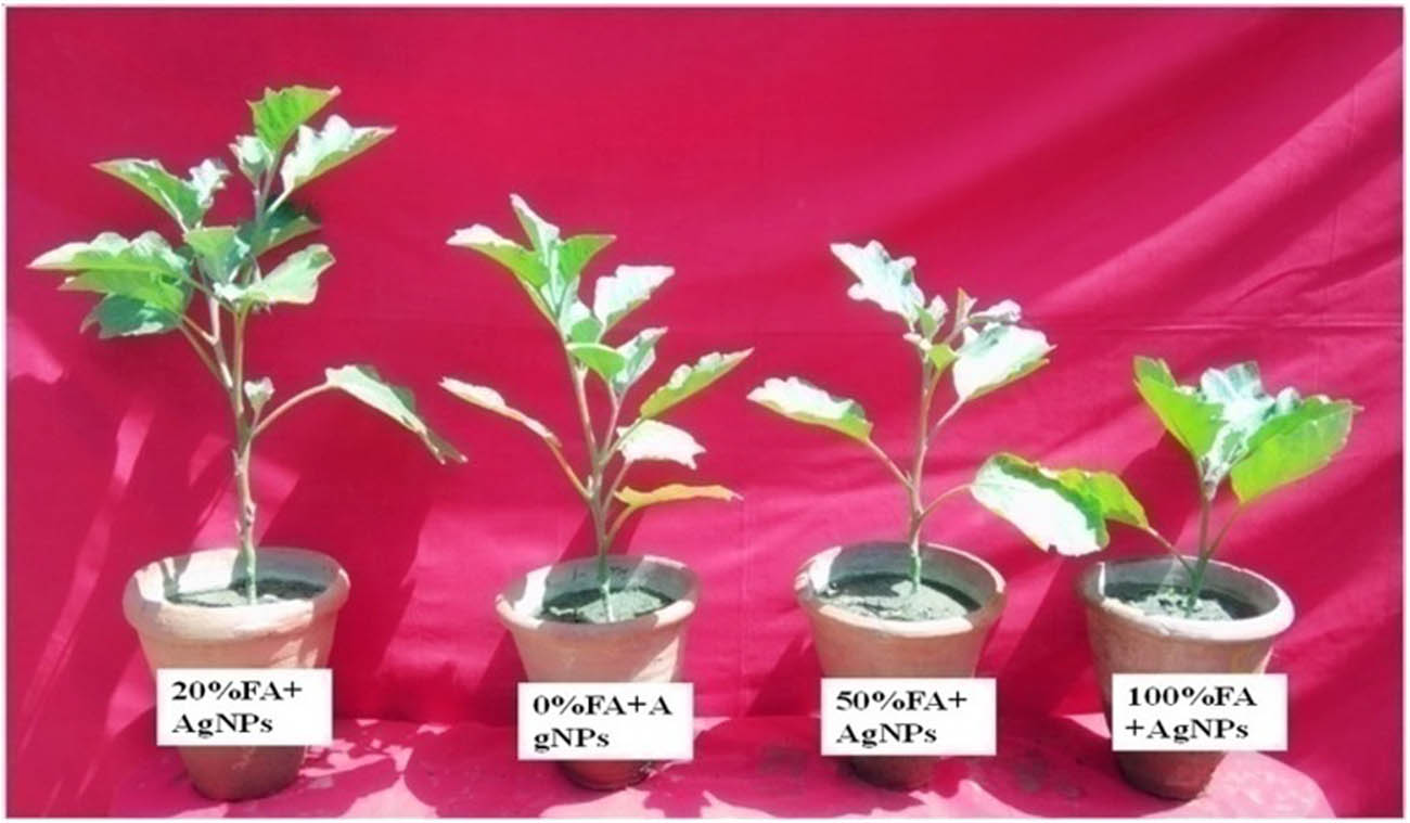
Eggplants showing the effect of AgNPs and fly ash amendment in soil.
Growth profile of eggplants exposed to different concentrations of AgNPs
| Treatment | Length (cm) | Fresh weight (g) | Shoot dry weight (g) | Root dry weight (g) | Chlorophyll in fresh leaves (mg/g) | Carotenoid in fresh leaves (mg/g) |
|---|---|---|---|---|---|---|
| C (No AgNPs) | 66.96c | 50.96c | 8.32c | 1.75c | 1.518c | 0.0504d |
| 25 ppm | 67.08c | 51.12c | 8.73c | 1.89c | 1.527c | 0.0517c |
| 50 ppm | 70.08b | 57.83b | 9.98b | 2.03b | 1.624b | 0.0531b |
| 75 ppm | 74.93a | 62.31a | 11.79a | 2.38a | 1.842a | 0.0582a |
| 150 ppm | 58.61d | 44.75d | 7.37d | 1.47d | 1.281g | 0.0428d |
| 300 ppm | 55.72f | 41.24e | 6.08e | 1.12e | 1.039h | 0.0401e |
| 500 ppm | 51.24f | 38.18f | 5.35f | 0.96f | 0.833i | 0.0396f |
| 1,000 ppm | 46.56g | 34.65g | 5.08f | 0.91f | 0.741j | 0.0387g |
At p ≤ 0.05, values within a column and the same type of treatment followed by the same letter do not differ significantly.
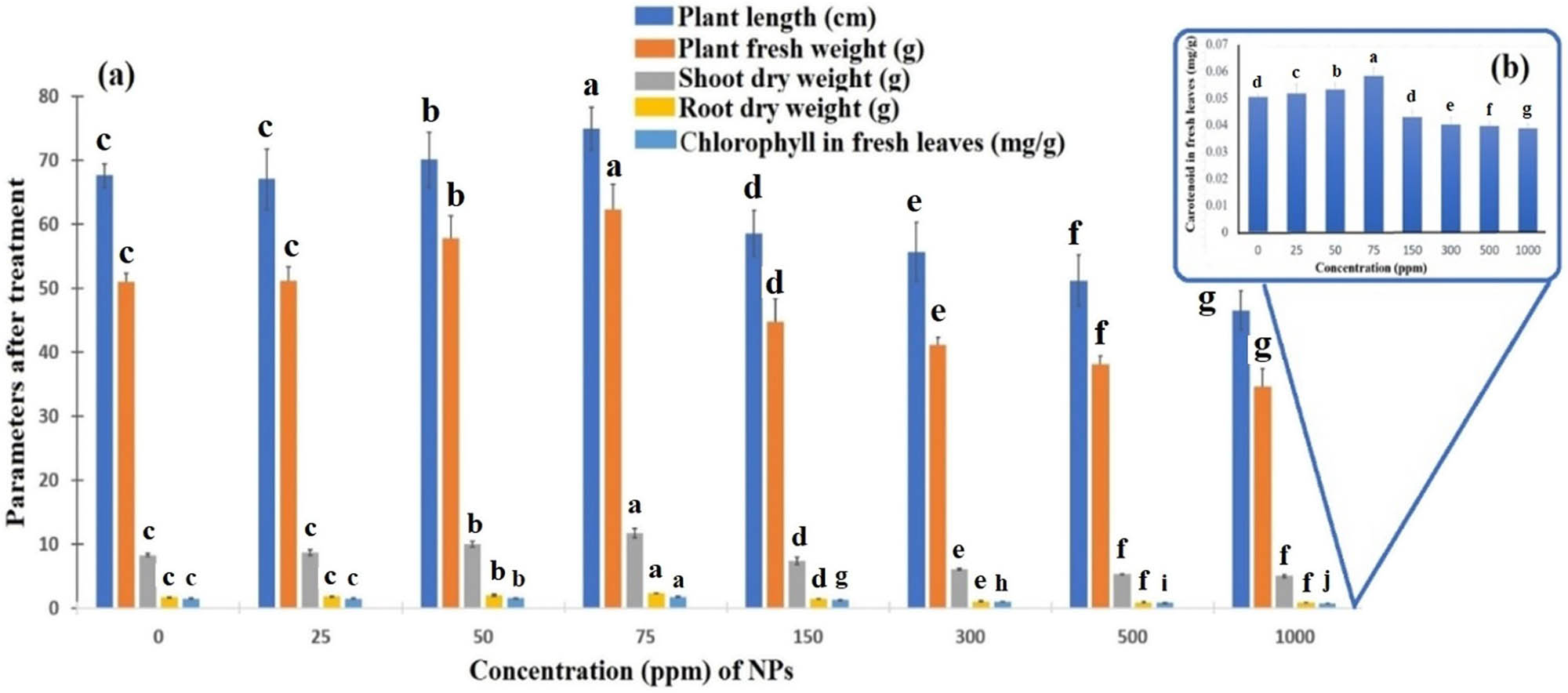
Effect of different concentrations of silver nanoparticles on the growth of the eggplant. C-control (without AgNPs), 25, 50, 75, 150, 300, 500, and 1,000 ppm on the plant length (cm), fresh weight (g), dry weight (g), root dry weight (g), chlorophyll (mg/g), and carotenoid in fresh leaves (mg/g). Graph (b) with fraction data is constructed separately because the graph is not visualized when combined with graph (a) with large data values. There is no significant difference between values within a column and the same type of treatment followed by the same letter. The mean and standard deviation values were used to create the bar graph (Dunnett’s test was used as a post hoc test). Statistically significant level: p < 0.05.
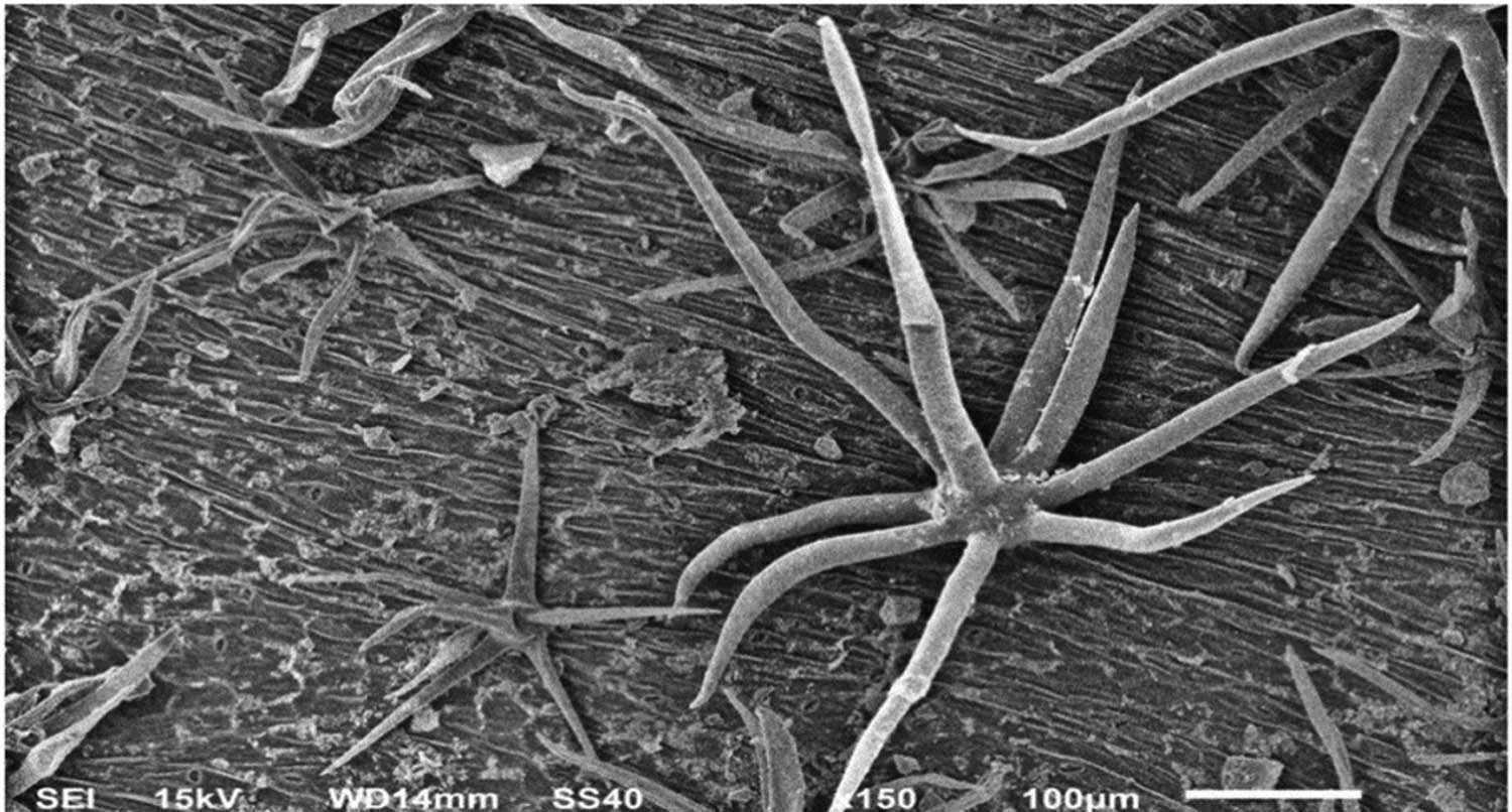
SEM image showing the eggplant leaves sprayed with AgNPs.
Eggplants grown in soil amended with 20% fly ash and sprayed with 75 ppm AgNPs showed a significant increase in the plant growth when compared to plants grown in soil without fly ash (Table 2 and Figure 10). Amendment of the soil with 20% fly ash with 75 ppm AgNPs caused a 10.66% increase in the plant length. However, amendments with 50 and 100% fly ash caused a reduction in the plant growth parameters, and 100% fly ash-amended soil with 75 ppm AgNPs caused a 14.96% decrease in the plant length. A high concentration of AgNPs and also a high concentration of fly ash are not beneficial for the growth of the eggplant. Table 2 shows the positive and negative effects of nanoparticles (NPs) at different concentrations on the plant growth parameters and photosynthetic pigments.
Effect of fly ash and AgNPs on the eggplant plant growth
| Treatment | Length (cm) | Fresh weight (g) | Shoot dry weight (g) | Root dry weight (g) | Chlorophyll in fresh leaves (mg/g) | Carotenoid in fresh leaves (mg/g) |
|---|---|---|---|---|---|---|
| 0% FA + 75 ppm AgNPs | 74.93a* | 62.31a | 11.79a | 2.38a | 1.717c | 0.050cd |
| 20% FA + 75 ppm AgNPs | 82.92a | 69.41a | 14.46a | 2.91a | 1.842c | 0.0582cd |
| 50% FA + 75 ppm AgNPs | 70.48c | 60.43c | 12.21c | 1.93c | 1.477d | 0.046bc |
| 100% FA + 75 ppm AgNPs | 63.72d | 42.72e | 10.04d | 1.18e | 1.397e | 0.039e |
*Values within a column having the same letter are not significantly different at p ≤ 0.05. a a c e a a c d.
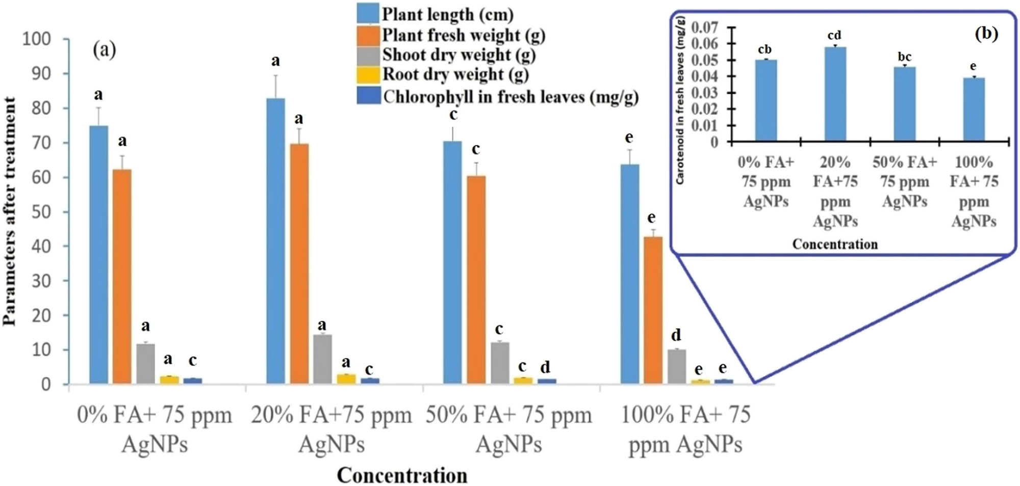
Effect of different concentrations of fly ash with an effective concentration of silver nanoparticles (75 ppm) on the growth of the eggplant plant. 0% FA + 75 ppm AgNPs, 20% FA + 75 ppm AgNPs, 50% FA + 75 ppm AgNPs and 100% FA + 75 ppm AgNPs on the plant length (cm), plant fresh weight (g), shoot dry weight (g), root dry weight (g), chlorophyll in fresh leaves (mg/g), and (b) carotenoid in fresh leaves (mg/g). Graph (b) with fraction data is constructed separately because the graph is not visualized when combined with graph (a) with large data values. The mean and standard deviation values were used to create the bar graph (Dunnett’s test was used as a post hoc test). Statistically significant level: p < 0.05.
Recent research reported that plant response to AgNPs is dependent on the concentration of AgNPs, which can either enhance or inhibit growth. Exposure to specific concentrations of AgNPs may enhance plant growth compared to nonexposed plants, while higher concentrations have a negative effect on plant growth. AgNPs have a concentration-based effect on eggplants, and a high concentration is toxic for plants. Biosynthesized AgNPs at concentrations of 25, 50, and 75 ppm were found beneficial for eggplant plants but 300, 500, and 1,000 ppm were found to be toxic. Higher concentrations of AgNPs resulted in a reduction of biomass in the Arabidopsis plant, according to Kaveh et al. [36]. AgNPs reduced the length of wheat shoots and roots in a dose-dependent manner, as mentioned in the literature [37]. Thuesombat et al. discovered that increased AgNP concentrations in jasmine rice reduced seed germination and subsequent seedling growth [38]. The effect of AgNPs on plant morphology and physiology is determined by their size and shape. Foliar application of AgNPs at different concentrations (20, 40, and 60 mg/L) was found to improve fenugreek plant growth parameters [39]. On mung beans, similar results of improving the role of AgNP treatments were obtained [40]. Growth-promoting AgNP activity on the wheat plant was also registered [41]. Brassica juncea, common bean, and corn all benefit from AgNPs in terms of plant growth and biochemical attributes such as chlorophyll, carbohydrate/protein contents, and antioxidant enzymes [42,43]. Silver is an impressive growth simulator as reported in the literature elsewhere [44]. Furthermore, Krishnaraj et al. [45] discovered that biosynthesized AgNPs influenced seed germination and induced protein and carbohydrate synthesis in Bacopa monnieri. In agriculture, AgNPs were developed as plant-growth stimulators [46] and the report of Khan and Siddiqui [47] states that 25% fly ash-amendment in the soil is better for plant growth, and 50% fly ash amendment and 100% fly ash are harmful to the eggplant plant growth. It is evident from previous studies that AgNPs can affect the plants’ growth attributes and metabolic processes. It was reported that AgNPs improved seed germination in pearl millet [48]. Parveen and Rao [34] reported that AgNPs can increase the germination rate, vigor index, and germination index in Brassica juncea. Sadak [39] found that silver nanoparticles could improve fenugreek plant (Trigonella foenum-graecum) growth, yield, and some biochemical aspects. AgNPs can be used for the breeding of chrysanthemum plants. Treatment of AgNPs (20 ppm) could improve the carotenoid content and induce genetic and phenotypic variation in the chrysanthemum plant [49]. An in-depth investigation is needed to understand the actual mechanism of the mediating effects of AgNPs on crops. Danish et al. [50] synthesized silver nanoparticles (AgNPs) from plants and found that the 50 ppm AgNP treatment in Trachyspermum ammi L. plant improved plant growth, biochemical, and antioxidant enzyme activities. They also reported that the AgNP treatment reduced the nematode infection in treated plants.
3.7 Statistical analysis
The results were provided as mean ± SD and expressed as an average for the measurement. Test values, degrees of freedom, and p values were used to examine statistical differences and significant means in the experimental data. In all statistical studies, the significant difference was determined using the Dunnett test at a level of 0.05 (or 0.01), which is provided in the supplementary information under statistical analysis. Each experimental value was differentiated to its corresponding control value. In nanoparticles-treated plants, a differential correlation pattern was noted between various concentrations of nanoparticles and parameters for plant growth such as plant length (cm), plant fresh weight (g), shoot dry weight (g), root dry weight (g), chlorophyll in fresh leaves (mg/g), and carotenoid in fresh leaves (mg/g). The total chlorophyll in fresh leaves (r 2 = 0.9859), carotenoid in fresh leaves (r 2 = 0.9116), and root dry weight (r 2 = 0.9787) showed positive correlation at different concentrations of silver nanoparticles. However, different concentrations of fly ash with a fixed concentration of silver nanoparticles (75 ppm) show significant difference on various factors of plant growth as indicated by positive R squared values for the plant length (r 2 = 0.7246), plant fresh weight (r 2 = 0.9160), shoot dry weight (r 2 = 0.9551), root dry weight (r 2 = 0.9934), chlorophyll in fresh leaves (r 2 = 0.9508), and carotenoid in fresh leaves (r 2 = 0.9871). The plant length, plant fresh weight, and shoot dry weight did not have a significant difference mean with different concentrations of silver nanoparticles (see supplementary information S1).
4 Conclusion
In this study, 10 mL of the pista seed coat waste extract was found to be suitable for harvesting a larger quantity of AgNPs within 30 min. The results explained in this article are therefore novel, which could emerge as a better alternative to the synthesis of AgNPs using waste plant materials. Biologically active compounds such as alkaloids, amino acids, flavonoids, and glycosides were identified from the extracts by the study of functional groups: –OH, –NH, –C═O, –COOH, and –CONH using IR spectrum; previous reports of GC-MS and these phytochemicals may be responsible for the rapid reduction of Ag+, leading to the AgNP formation. Morphological and particle sizes were confirmed using various techniques, such as SEM, TEM, XRD, and BET, and were found to possess polydispersed irregular shapes from rectangular to spherical shapes with an average size of 20 nm. The BET study also confirmed the surface area of biogenic AgNPs to be 21.73 m2/g at ambient temperature. The effect of AgNPs on the eggplant has been shown to enhance the growth of the eggplant at low concentrations but higher concentrations are toxic to the eggplant plant: 20% fly ash amendment in soil with spraying 75 ppm AgNPs was found most effective in improving the growth of the eggplant and 50% fly ash amendment in the soil was found to be less effective. Further research is required to determine the degree of toxicity, and the mechanistic approach of AgNPs to plant growth for field applications is important for further use.
-
Funding information: Walaa Alsanie would like to acknowledge TURSP (2020/53).
-
Author contributions: All authors have accepted responsibility for the entire content of this manuscript and approved its submission.
-
Conflict of interest: The authors declare no conflict of interest.
References
[1] Mohseniazar M, Barin M, Zarredar H, Alizadeh S, Shanehbandi D. Potential of microalgae and lactobacilli in biosynthesis of silver nanoparticles. BioImpacts. 2011;1:149–52.Suche in Google Scholar
[2] Pal SL, Jana U, Manna PK, Mohanta GP, Manavalan R. Nanoparticle: an overview of preparation and characterization. J Appl Pharm Sci. 2011;1(6):228–34.Suche in Google Scholar
[3] Feynman RP. There’s plenty of room at the bottom. Eng Sci. 1960;23:22–36.Suche in Google Scholar
[4] Azmir J, Zaidul IS, Rahman MM, Sharif KM, Mohamed A, Sahena F, et al. Techniques for extraction of bioactive compounds from plant materials: a review. J Food Eng. 2013;117:426–36.10.1016/j.jfoodeng.2013.01.014Suche in Google Scholar
[5] Pérez C, del Castillo ML, Gil C, Blanch GP, Flores G. Supercritical fluid extraction of grape seeds: extract chemical composition, antioxidant activity and inhibition of nitrite production in LPS-stimulated Raw 264.7 cells. Food Funct. 2015;6:2607–13.10.1039/C5FO00325CSuche in Google Scholar PubMed
[6] de Barros CH, Cruz GC, Mayrink W, Tasic L. Bio-based synthesis of silver nanoparticles from orange waste: effects of distinct biomolecule coatings on size, morphology, and antimicrobial activity. Nanotechnol Sci Appl. 2018;11:1–14.10.2147/NSA.S156115Suche in Google Scholar PubMed PubMed Central
[7] Vance ME, Kuiken T, Vejerano EP, McGinnis SP, Hochella Jr MF, Rejeski D, et al. Nanotechnology in the real world: redeveloping the nanomaterial consumer products inventory. Beilstein J Nanotechnol. 2015;6:1769–80.10.3762/bjnano.6.181Suche in Google Scholar PubMed PubMed Central
[8] Khan M, Khan AU, Alam MJ, Park S, Alam M. Biosynthesis of silver nanoparticles and its application against phytopathogenic bacterium and fungus. Int J Environ Anal Chem. 2020;100(12):1390–401.10.1080/03067319.2019.1654465Suche in Google Scholar
[9] Kaur J, Singh J, Rawat M. An efficient and blistering reduction of 4-nitrophenol by green synthesized silver nanoparticles. SN Appl Sci. 2019;1:1–6.10.1007/s42452-019-1088-xSuche in Google Scholar
[10] Khan AU, Khan M, Khan MM. Antifungal and antibacterial assay by silver nanoparticles synthesized from aqueous leaf extract of Trigonella foenum-graecum. BioNanoScience. 2019;9:597–602.10.1007/s12668-019-00643-xSuche in Google Scholar
[11] Valli JS, Vaseeharan B. Biosynthesis of silver nanoparticles by Cissus quadrangularis extracts. Mater Lett. 2012;82:171–3.10.1016/j.matlet.2012.05.040Suche in Google Scholar
[12] Kumar DA, Palanichamy V, Roopan SM. Green synthesis of silver nanoparticles using Alternanthera dentata leaf extract at room temperature and their antimicrobial activity. Spectrochim Acta A. 2014;127:168–71.10.1016/j.saa.2014.02.058Suche in Google Scholar PubMed
[13] Verma SK, Das AK, Patel MK, Shah A, Kumar V, Gantait S. Engineered nanomaterials for plant growth and development: a perspective analysis. Sci Total Environ. 2018;630:1413–35.10.1016/j.scitotenv.2018.02.313Suche in Google Scholar PubMed
[14] Aslani F, Bagheri S, Julkapli NM, Juraimi AS, Sadat F, Hashemi G, et al. Effects of engineered nanomaterials on plants growth: an overview. Sci World J. 2014;2014:641759.10.1155/2014/641759Suche in Google Scholar
[15] Khan M, Khan AU, Hasan MA, Yadav KK, Pinto M, Malik N, et al. Agro-nanotechnology as an emerging field: a novel sustainable approach for improving plant growth by reducing biotic stress. Appl Sci. 2021;11:2282.10.3390/app11052282Suche in Google Scholar
[16] Glance. Horticultural Statistics Horticulture Statistics Division Department of Agriculture. Cooperation & Farmers’ Welfare Ministry of Agriculture and Farmers’ Welfare Government of India; 2018.Suche in Google Scholar
[17] Izabela J, Oleszczuk P. Influence of soil type and environmental conditions on ZnO, TiO2 and Ni nanoparticles phytotoxicity. Chemosphere. 2013;92:91–9.10.1016/j.chemosphere.2013.02.048Suche in Google Scholar
[18] Khodakovskaya MV, Silva K, Nedosekin DA, Dervishi E, Biris AS, Shashkov EV, et al. Complex genetic, photothermal, and photoacoustic analysis of nanoparticle-plant interactions. Proc Natl Acad Sci. 2011;108:1028–33.10.1073/pnas.1008856108Suche in Google Scholar
[19] Wang LJ, Guo ZM, Li TJ, Li M. The nano structure SiO2 in the plants. Chin Sci Bull. 2001;46:625–31.Suche in Google Scholar
[20] Khan M, Khan AU, Bogdanchikova N, Garibo D. Antibacterial and antifungal studies of biosynthesized silver nanoparticles against plant parasitic nematode Meloidogyne incognita, plant pathogens ralstonia solanacearum and fusarium oxysporum. Molecules. 2021;26:2462.10.3390/molecules26092462Suche in Google Scholar
[21] Mackinney G. Criteria for purity of chlorophyll preparations. J Biol Chem. 1940;132:91–109.10.1016/S0021-9258(18)73399-1Suche in Google Scholar
[22] Maclachlan S, Saul Z. Plastid structure, chlorophyll concentration, and free amino acid composition of a chlorophyll mutant of barley. Can J Bot. 1963;41:1053–62.10.1139/b63-088Suche in Google Scholar
[23] Vanaja M, Gnanajobitha G, Paulkumar K, Rajeshkumar S, Malarkodi C, Annadurai G. Phytosynthesis of silver nanoparticles by Cissus quadrangularis: influence of physicochemical factors. J Nanostruct Chem. 2013;3:1–8.10.1186/2193-8865-3-17Suche in Google Scholar
[24] Suresh S, Karthikeyan S, Jayamoorthy K. FTIR and multivariate analysis to study the effect of bulk and nano copper oxide on peanut plant leaves. J Sci Adv Mater Devices. 2016;1:343–50.10.1016/j.jsamd.2016.08.004Suche in Google Scholar
[25] Ajitha BY, Reddy AK, Reddy PS. Biogenic nano-scale silver particles by Tephrosia purpurea leaf extract and their inborn antimicrobial activity. Spectrochim Acta A. 2014;121:164–72.10.1016/j.saa.2013.10.077Suche in Google Scholar PubMed
[26] Sre PR, Reka RM, Poovazhagi R, Arul Kumar M, Murugesan K. Antibacterial and cytotoxic effect of biologically synthesized silver nanoparticles using aqueous root extract of Erythrina indica lam. Spectrochim Acta A. 2015;135:1137–44.10.1016/j.saa.2014.08.019Suche in Google Scholar PubMed
[27] Venil CK, Malathi M, Velmurugan P, Renuka Devi P. Green synthesis of silver nanoparticles using canthaxanthin from Dietziamaris AURCCBT01 and their cytotoxic properties against human keratinocyte cell line. J Appl Microbiol. 2021;130:1730–44.10.1111/jam.14889Suche in Google Scholar PubMed
[28] Anigol LB, Charantimath JS, Gurubasavaraj PM. Effect of concentration and pH on the size of silver nanoparticles synthesized by green chemistry. Org Med Chem Int J. 2017;3:1–5.10.19080/OMCIJ.2017.03.555622Suche in Google Scholar
[29] Mahiuddin M, Saha P, Ochiai B. Green synthesis and catalytic activity of silver nanoparticles based on piper chaba stem extracts. Nanomaterials. 2020;10:1777.10.3390/nano10091777Suche in Google Scholar PubMed PubMed Central
[30] Taghizadeh A, Rad-Moghadam K. Green fabrication of Cu/pistachio shell nanocomposite using Pistacia Vera L. hull: an efficient catalyst for expedient reduction of 4-nitrophenol and organic dyes. J Clean Prod. 2018;198:1105–19.10.1016/j.jclepro.2018.07.042Suche in Google Scholar
[31] Toldrá F, Nollet LM, editors. Handbook of dairy foods analysis. Oxon, OX14RN. CRC Press; 2021. p. 29.10.1201/9780429342967Suche in Google Scholar
[32] Xinyuan J, Yuanyuan L, Zhong G, An M, Zecai H, Suwen Y. Pyrolysis characteristics and correlation analysis with the major components of seven kinds of nutshell. Sci Silvae Sin. 2015;51:79–86.Suche in Google Scholar
[33] Bordbar M, Mortazavimanesh N. Biosynthesis of waste pistachio shell supported silver nanoparticles for the catalytic reduction processes. IET Nanobiotechnol. 2018;12:939–45.10.1049/iet-nbt.2017.0266Suche in Google Scholar PubMed PubMed Central
[34] Parveen A, Rao S. Effect of nanosilver on seed germination and seedling growth in Pennisetum glaucum. J Clust Sci. 2015;26:693–701.10.1007/s10876-014-0728-ySuche in Google Scholar
[35] Zafar S, Zafar A. Biosynthesis and characterization of silver nanoparticles using Phoenix dactylifera fruits extract and their in vitro antimicrobial and cytotoxic effects. Open Biotechnol J. 2019;13:37–46.10.2174/1874070701913010037Suche in Google Scholar
[36] Kaveh R, Yue-Sheng L, Ranjbar S, Tehrani R, Brueck CL, Aken BV. Changes in Arabidopsis thaliana gene expression in response to silver nanoparticles and silver ions. Environ Sci Technol. 2013;2013(47):10637–44.10.1021/es402209wSuche in Google Scholar PubMed
[37] Dimkpa CO, McLean JE, Martineau N, Britt DW, Haverkamp R, Anderson AJ. Silver nanoparticles disrupt wheat (Triticum aestivum L.) growth in a sand matrix. Environ Sci Technol. 2013;47:1082–90.10.1021/es302973ySuche in Google Scholar PubMed
[38] Thuesombat P, Hannongbua S, Akasit S, Chadchawan S. Effect of silver nanoparticles on rice (Oryza sativa L. cv. KDML 105) seed germination and seedling growth. Ecotoxicol Environ Saf. 2014;104:302–9.10.1016/j.ecoenv.2014.03.022Suche in Google Scholar PubMed
[39] Sadak MS. Impact of silver nanoparticles on plant growth, some biochemical aspects, and yield of fenugreek plant (Trigonella foenum-graecum). Bull Natl Res Cent. 2019;43:1–6.10.1186/s42269-019-0077-ySuche in Google Scholar
[40] Saeideh N, Rashid J. Effect of silver nanoparticles and Pb(NO3)2 on the yield and chemical composition of mung bean (Vigna radiata). J Stress Physiol Biochem. 2014;10:316–25.Suche in Google Scholar
[41] Razzaq A, Ammara R, Jhanzab HM, Mahmood T, Hafeez A, Hussain SA. novel nanomaterial to enhance growth and yield of wheat. J Nanosci Technol. 2016;2:55–8.Suche in Google Scholar
[42] Salama HM. Effects of silver nanoparticles in some crop plants, common bean (Phaseolus vulgaris L.) and corn (Zea mays L.). Int Res J Biotechnol. 2012;3:190–7.Suche in Google Scholar
[43] Sharma P, Bhatt D, Zaidi MG, Saradhi PP, Khanna PK, Arora S. Silver nanoparticle-mediated enhancement in growth and antioxidant status of Brassica juncea. Appl Biochem Biotechnol. 2012;167:2225–33.10.1007/s12010-012-9759-8Suche in Google Scholar PubMed
[44] Sharon M, Choudhary AK, Kumar R. Nanotechnology in agricultural diseases and food safety. J Phytol. 2010;2(4):83–92.Suche in Google Scholar
[45] Krishnaraj C, Jagan EG, Ramachandran R, Abirami SM, Mohan N, Kalaichelvan PT. Effect of biologically synthesized silver nanoparticles on Bacopa monnieri (Linn.) Wettst. plant growth metabolism. Process Biochem. 2012;47(4):651–8.10.1016/j.procbio.2012.01.006Suche in Google Scholar
[46] Monica RC, Cremonini R. Nanoparticles and higher plants. Caryologia. 2009;62:161–5.10.1080/00087114.2004.10589681Suche in Google Scholar
[47] Khan M, Siddiqui ZA. Effects of fly ash amendments, Ralstonia solanacearum, Meloidogyne incognita and Phomopsis vexans on the growth of Solanum melongena. Acta Phytopathol Entomol Hung. 2017;52:145–56.10.1556/038.52.2017.017Suche in Google Scholar
[48] Khan I, Raza MA, Awan SA, Shah GA, Rizwan M, Ali B, et al. Amelioration of salt induced toxicity in pearl millet by seed priming with silver nanoparticles (AgNPs): the oxidative damage, antioxidant enzymes and ions uptake are major determinants of salt tolerant capacity. Plant Physiol Biochem. 2020;156:221–32.10.1016/j.plaphy.2020.09.018Suche in Google Scholar PubMed
[49] Tymoszuk A, Kulus D. Silver nanoparticles induce genetic, biochemical, and phenotype variation in chrysanthemum. Plant Cell Tissue Organ Cult. 2020;143:331–44.10.1007/s11240-020-01920-4Suche in Google Scholar
[50] Danish M, Altaf M, Robab MI, Shahid M, Manoharadas S, Hussain SA, et al. Green synthesized silver nanoparticles mitigate biotic stress induced by meloidogyne incognita in Trachyspermumammi (L.) by improving growth, biochemical, and antioxidant enzyme activities. ACS Omega. 2021;6:11389–403.10.1021/acsomega.1c00375Suche in Google Scholar PubMed PubMed Central
© 2021 Masudulla Khan et al., published by De Gruyter
This work is licensed under the Creative Commons Attribution 4.0 International License.
Artikel in diesem Heft
- Research Articles
- Improved impedance matching by multi-componential metal-hybridized rGO toward high performance of microwave absorption
- Pure-silk fibroin hydrogel with stable aligned micropattern toward peripheral nerve regeneration
- Effective ion pathways and 3D conductive carbon networks in bentonite host enable stable and high-rate lithium–sulfur batteries
- Fabrication and characterization of 3D-printed gellan gum/starch composite scaffold for Schwann cells growth
- Synergistic strengthening mechanism of copper matrix composite reinforced with nano-Al2O3 particles and micro-SiC whiskers
- Deformation mechanisms and plasticity of ultrafine-grained Al under complex stress state revealed by digital image correlation technique
- On the deformation-induced grain rotations in gradient nano-grained copper based on molecular dynamics simulations
- Removal of sulfate from aqueous solution using Mg–Al nano-layered double hydroxides synthesized under different dual solvent systems
- Microwave-assisted sol–gel synthesis of TiO2-mixed metal oxide nanocatalyst for degradation of organic pollutant
- Electrophoretic deposition of graphene on basalt fiber for composite applications
- Polyphenylene sulfide-coated wrench composites by nanopinning effect
- Thermal conductivity and thermoelectric properties in 3D macroscopic pure carbon nanotube materials
- An effective thermal conductivity and thermomechanical homogenization scheme for a multiscale Nb3Sn filaments
- Friction stir spot welding of AA5052 with additional carbon fiber-reinforced polymer composite interlayer
- Improvement of long-term cycling performance of high-nickel cathode materials by ZnO coating
- Quantum effects of gas flow in nanochannels
- An approach to effectively improve the interfacial bonding of nano-perfused composites by in situ growth of CNTs
- Effects of nano-modified polymer cement-based materials on the bending behavior of repaired concrete beams
- Effects of the combined usage of nanomaterials and steel fibres on the workability, compressive strength, and microstructure of ultra-high performance concrete
- One-pot solvothermal synthesis and characterization of highly stable nickel nanoparticles
- Comparative study on mechanisms for improving mechanical properties and microstructure of cement paste modified by different types of nanomaterials
- Effect of in situ graphene-doped nano-CeO2 on microstructure and electrical contact properties of Cu30Cr10W contacts
- The experimental study of CFRP interlayer of dissimilar joint AA7075-T651/Ti-6Al-4V alloys by friction stir spot welding on mechanical and microstructural properties
- Vibration analysis of a sandwich cylindrical shell in hygrothermal environment
- Water barrier and mechanical properties of sugar palm crystalline nanocellulose reinforced thermoplastic sugar palm starch (TPS)/poly(lactic acid) (PLA) blend bionanocomposites
- Strong quadratic acousto-optic coupling in 1D multilayer phoxonic crystal cavity
- Three-dimensional shape analysis of peripapillary retinal pigment epithelium-basement membrane layer based on OCT radial images
- Solvent regulation synthesis of single-component white emission carbon quantum dots for white light-emitting diodes
- Xanthate-modified nanoTiO2 as a novel vulcanization accelerator enhancing mechanical and antibacterial properties of natural rubber
- Effect of steel fiber on impact resistance and durability of concrete containing nano-SiO2
- Ultrasound-enhanced biosynthesis of uniform ZnO nanorice using Swietenia macrophylla seed extract and its in vitro anticancer activity
- Temperature dependence of hardness prediction for high-temperature structural ceramics and their composites
- Study on the frequency of acoustic emission signal during crystal growth of salicylic acid
- Controllable modification of helical carbon nanotubes for high-performance microwave absorption
- Role of dry ozonization of basalt fibers on interfacial properties and fracture toughness of epoxy matrix composites
- Nanosystem’s density functional theory study of the chlorine adsorption on the Fe(100) surface
- A rapid nanobiosensing platform based on herceptin-conjugated graphene for ultrasensitive detection of circulating tumor cells in early breast cancer
- Improving flexural strength of UHPC with sustainably synthesized graphene oxide
- The role of graphene/graphene oxide in cement hydration
- Structural characterization of microcrystalline and nanocrystalline cellulose from Ananas comosus L. leaves: Cytocompatibility and molecular docking studies
- Evaluation of the nanostructure of calcium silicate hydrate based on atomic force microscopy-infrared spectroscopy experiments
- Combined effects of nano-silica and silica fume on the mechanical behavior of recycled aggregate concrete
- Safety study of malapposition of the bio-corrodible nitrided iron stent in vivo
- Triethanolamine interface modification of crystallized ZnO nanospheres enabling fast photocatalytic hazard-free treatment of Cr(vi) ions
- Novel electrodes for precise and accurate droplet dispensing and splitting in digital microfluidics
- Construction of Chi(Zn/BMP2)/HA composite coating on AZ31B magnesium alloy surface to improve the corrosion resistance and biocompatibility
- Experimental and multiscale numerical investigations on low-velocity impact responses of syntactic foam composites reinforced with modified MWCNTs
- Comprehensive performance analysis and optimal design of smart light pole for cooperative vehicle infrastructure system
- Room temperature growth of ZnO with highly active exposed facets for photocatalytic application
- Influences of poling temperature and elongation ratio on PVDF-HFP piezoelectric films
- Large strain hardening of magnesium containing in situ nanoparticles
- Super stable water-based magnetic fluid as a dual-mode contrast agent
- Photocatalytic activity of biogenic zinc oxide nanoparticles: In vitro antimicrobial, biocompatibility, and molecular docking studies
- Hygrothermal environment effect on the critical buckling load of FGP microbeams with initial curvature integrated by CNT-reinforced skins considering the influence of thickness stretching
- Thermal aging behavior characteristics of asphalt binder modified by nano-stabilizer based on DSR and AFM
- Building effective core/shell polymer nanoparticles for epoxy composite toughening based on Hansen solubility parameters
- Structural characterization and nanoscale strain field analysis of α/β interface layer of a near α titanium alloy
- Optimization of thermal and hydrophobic properties of GO-doped epoxy nanocomposite coatings
- The properties of nano-CaCO3/nano-ZnO/SBR composite-modified asphalt
- Three-dimensional metallic carbon allotropes with superhardness
- Physical stability and rheological behavior of Pickering emulsions stabilized by protein–polysaccharide hybrid nanoconjugates
- Optimization of volume fraction and microstructure evolution during thermal deformation of nano-SiCp/Al–7Si composites
- Phase analysis and corrosion behavior of brazing Cu/Al dissimilar metal joint with BAl88Si filler metal
- High-efficiency nano polishing of steel materials
- On the rheological properties of multi-walled carbon nano-polyvinylpyrrolidone/silicon-based shear thickening fluid
- Fabrication of Ag/ZnO hollow nanospheres and cubic TiO2/ZnO heterojunction photocatalysts for RhB degradation
- Fabrication and properties of PLA/nano-HA composite scaffolds with balanced mechanical properties and biological functions for bone tissue engineering application
- Investigation of the early-age performance and microstructure of nano-C–S–H blended cement-based materials
- Reduced graphene oxide coating on basalt fabric using electrophoretic deposition and its role in the mechanical and tribological performance of epoxy/basalt fiber composites
- Effect of nano-silica as cementitious materials-reducing admixtures on the workability, mechanical properties and durability of concrete
- Machine-learning-assisted microstructure–property linkages of carbon nanotube-reinforced aluminum matrix nanocomposites produced by laser powder bed fusion
- Physical, thermal, and mechanical properties of highly porous polylactic acid/cellulose nanofibre scaffolds prepared by salt leaching technique
- A comparative study on characterizations and synthesis of pure lead sulfide (PbS) and Ag-doped PbS for photovoltaic applications
- Clean preparation of washable antibacterial polyester fibers by high temperature and high pressure hydrothermal self-assembly
- Al 5251-based hybrid nanocomposite by FSP reinforced with graphene nanoplates and boron nitride nanoparticles: Microstructure, wear, and mechanical characterization
- Interlaminar fracture toughness properties of hybrid glass fiber-reinforced composite interlayered with carbon nanotube using electrospray deposition
- Microstructure and life prediction model of steel slag concrete under freezing-thawing environment
- Synthesis of biogenic silver nanoparticles from the seed coat waste of pistachio (Pistacia vera) and their effect on the growth of eggplant
- Study on adaptability of rheological index of nano-PUA-modified asphalt based on geometric parameters of parallel plate
- Preparation and adsorption properties of nano-graphene oxide/tourmaline composites
- A study on interfacial behaviors of epoxy/graphene oxide derived from pitch-based graphite fibers
- Multiresponsive carboxylated graphene oxide-grafted aptamer as a multifunctional nanocarrier for targeted delivery of chemotherapeutics and bioactive compounds in cancer therapy
- Piezoresistive/piezoelectric intrinsic sensing properties of carbon nanotube cement-based smart composite and its electromechanical sensing mechanisms: A review
- Smart stimuli-responsive biofunctionalized niosomal nanocarriers for programmed release of bioactive compounds into cancer cells in vitro and in vivo
- Photoremediation of methylene blue by biosynthesized ZnO/Fe3O4 nanocomposites using Callistemon viminalis leaves aqueous extract: A comparative study
- Study of gold nanoparticles’ preparation through ultrasonic spray pyrolysis and lyophilisation for possible use as markers in LFIA tests
- Review Articles
- Advance on the dispersion treatment of graphene oxide and the graphene oxide modified cement-based materials
- Development of ionic liquid-based electroactive polymer composites using nanotechnology
- Nanostructured multifunctional electrocatalysts for efficient energy conversion systems: Recent perspectives
- Recent advances on the fabrication methods of nanocomposite yarn-based strain sensor
- Review on nanocomposites based on aerospace applications
- Overview of nanocellulose as additives in paper processing and paper products
- The frontiers of functionalized graphene-based nanocomposites as chemical sensors
- Material advancement in tissue-engineered nerve conduit
- Carbon nanostructure-based superhydrophobic surfaces and coatings
- Functionalized graphene-based nanocomposites for smart optoelectronic applications
- Interfacial technology for enhancement in steel fiber reinforced cementitious composite from nano to macroscale
- Metal nanoparticles and biomaterials: The multipronged approach for potential diabetic wound therapy
- Review on resistive switching mechanisms of bio-organic thin film for non-volatile memory application
- Nanotechnology-enabled biomedical engineering: Current trends, future scopes, and perspectives
- Research progress on key problems of nanomaterials-modified geopolymer concrete
- Smart stimuli-responsive nanocarriers for the cancer therapy – nanomedicine
- An overview of methods for production and detection of silver nanoparticles, with emphasis on their fate and toxicological effects on human, soil, and aquatic environment
- Effects of chemical modification and nanotechnology on wood properties
- Mechanisms, influencing factors, and applications of electrohydrodynamic jet printing
- Application of antiviral materials in textiles: A review
- Phase transformation and strengthening mechanisms of nanostructured high-entropy alloys
- Research progress on individual effect of graphene oxide in cement-based materials and its synergistic effect with other nanomaterials
- Catalytic defense against fungal pathogens using nanozymes
- A mini-review of three-dimensional network topological structure nanocomposites: Preparation and mechanical properties
- Mechanical properties and structural health monitoring performance of carbon nanotube-modified FRP composites: A review
- Nano-scale delivery: A comprehensive review of nano-structured devices, preparative techniques, site-specificity designs, biomedical applications, commercial products, and references to safety, cellular uptake, and organ toxicity
- Effects of alloying, heat treatment and nanoreinforcement on mechanical properties and damping performances of Cu–Al-based alloys: A review
- Recent progress in the synthesis and applications of vertically aligned carbon nanotube materials
- Thermal conductivity and dynamic viscosity of mono and hybrid organic- and synthetic-based nanofluids: A critical review
- Recent advances in waste-recycled nanomaterials for biomedical applications: Waste-to-wealth
- Layup sequence and interfacial bonding of additively manufactured polymeric composite: A brief review
- Quantum dots synthetization and future prospect applications
- Approved and marketed nanoparticles for disease targeting and applications in COVID-19
- Strategies for improving rechargeable lithium-ion batteries: From active materials to CO2 emissions