Crystal structure of 1,10-phenanthrolinium bromide dihydrate, C12H9N2Br
-


Abstract
C12H9N2Br, triclinic, P
Table 1 contains crystallographic data and Table 2 contains the list of the atoms including atomic coordinates and displacement parameters.
Data collection and handling.
| Crystal: | Colourless block |
| Size: | 0.38 × 0.25 × 0.16 mm |
| Wavelength: | Mo Kα radiation (0.71073 Å) |
| μ: | 3.39 mm−1 |
| Diffractometer, scan mode: | Bruker Smart Apex-II, φ and ω |
| θmax, completeness: | 27.5°, >99 % |
| N(hkl)measured, N(hkl)unique, Rint: | 10,251, 2743, 0.030 |
| Criterion for Iobs, N(hkl)gt: | Iobs > 2σ(Iobs), 2596 |
| N(param)refined: | 172 |
| Programs: | Bruker [1, 2], Shelx [3, 4], Olex2 [5] |
Fractional atomic coordinates and isotropic or equivalent isotropic displacement parameters (Å2).
| Atom | x | y | z | Uiso*/Ueq |
|---|---|---|---|---|
| Br1 | 0.27857 (3) | 0.11119 (2) | 0.39455 (2) | 0.01755 (6) |
| C1 | 0.2385 (2) | 0.53193 (18) | 0.09434 (17) | 0.0127 (3) |
| C2 | 0.2639 (2) | 0.50116 (19) | −0.04636 (17) | 0.0127 (3) |
| C3 | 0.3853 (3) | 0.5947 (2) | −0.2254 (2) | 0.0239 (4) |
| H3 | 0.452233 | 0.680111 | −0.263100 | 0.029* |
| C4 | 0.3189 (3) | 0.4420 (2) | −0.3101 (2) | 0.0245 (4) |
| H4 | 0.342367 | 0.426193 | −0.401814 | 0.029* |
| C5 | 0.2205 (3) | 0.3168 (2) | −0.25956 (18) | 0.0201 (4) |
| H5 | 0.173606 | 0.213046 | −0.315849 | 0.024* |
| C6 | 0.1895 (2) | 0.34372 (19) | −0.12275 (17) | 0.0146 (3) |
| C7 | 0.0864 (3) | 0.2206 (2) | −0.06086 (19) | 0.0177 (3) |
| H7 | 0.034209 | 0.114959 | −0.113832 | 0.021* |
| C8 | 0.0620 (3) | 0.2519 (2) | 0.07111 (19) | 0.0184 (4) |
| H8 | −0.007522 | 0.168157 | 0.109724 | 0.022* |
| C9 | 0.1397 (2) | 0.4098 (2) | 0.15393 (17) | 0.0150 (3) |
| C10 | 0.1217 (3) | 0.4493 (2) | 0.29392 (19) | 0.0221 (4) |
| H10 | 0.054542 | 0.368921 | 0.336870 | 0.027* |
| C11 | 0.2008 (3) | 0.6029 (3) | 0.36799 (19) | 0.0269 (4) |
| H11 | 0.190301 | 0.629276 | 0.462577 | 0.032* |
| C12 | 0.2961 (3) | 0.7194 (2) | 0.3037 (2) | 0.0245 (4) |
| H12 | 0.350287 | 0.826140 | 0.354079 | 0.029* |
| N1 | 0.3119 (2) | 0.68247 (17) | 0.17224 (15) | 0.0176 (3) |
| H1 | 0.369124 | 0.755292 | 0.132193 | 0.021* |
| N2 | 0.3598 (2) | 0.62597 (17) | −0.09655 (16) | 0.0181 (3) |
| O1 | 0.8326 (2) | 1.11613 (18) | 0.37689 (15) | 0.0307 (3) |
| H1A | 0.945807 | 1.112674 | 0.376812 | 0.046* |
| H1B | 0.793038 | 1.063350 | 0.436555 | 0.046* |
| O2a | 0.4789 (4) | 0.9793 (3) | 0.1546 (3) | 0.0180 (6) |
| H2Aa | 0.597107 | 1.018192 | 0.211287 | 0.027* |
| H2Ba | 0.414019 | 1.021318 | 0.196234 | 0.027* |
| O3a | 0.5536 (7) | 0.9525 (4) | 0.1155 (4) | 0.0410 (9) |
| H3Aa | 0.625915 | 1.028255 | 0.187770 | 0.061* |
| H3Ba | 0.516342 | 0.995625 | 0.051017 | 0.061* |
1 Source of materials
All reagents are commercially available and were used without further purification. To 1.0 g (5.6 mmol) of 1,10-phenanthroline hydrate was added 20 mL of a methanolic solution of hydrobromic acid (8.2 g, 5.5 mL). Hydrogen peroxide solution (48 %, 5.5 mL) was added dropwise over a period of 20 min under vigorous and continuous stirring at room temperature. After 20 h of stirring, the pink solution was concentrated in vacuo at 60–70 °C. The residue was dissolved in water (50 mL) and then extracted with ethyl acetate (3 × 50 mL). The resultant ethyl acetate fractions were combined and concentrated to dryness. This residue was redissolved in dichloromethane and filtered to remove insoluble material. The dichloromethane solution was concentrated in vacuo and diethylether added to afford the title compound as a white solid. Crystals of the title compound were initially obtained from a hexane-toluene mixture. Crystals suitable for X-ray diffraction, were obtained by recrystallisation, at ambient temperature, from a mixture of methanol and water by slow evaporation.
2 Experimental details
Data reduction was carried out using the program S aint and empirical absorption corrections were made using S adabs [2]. Space group assignment was made using Shelxs-2013 [3] and the structure refined using full-matrix least-squares minimisation with 2016/6 of Shelxl [4] using Olex2 as the graphical interface [5]. Hydrogen atom positions were calculated geometrically and refined using the riding model.
3 Comment
For several decades, phenanthroline derivatives have been used primarily as colourimetric indicators for transition metals ions. This application and the structural rigidity of phenanthrolines have generated interest in the preparation and structural studies of phenanthrolines with pendant photo- and electroactive molecules [6]. The 1,10-phenanthroline and its derivatives have been largely unexplored as chromophores to stabilize transition metal complexes in contrast to the related 2,2′-bipyridine and 2,2′,6′2″-terpyridine structures [7]. Coordination chemistry of 1,10-phenanthroline has encouraged the synthesis of new structures which could serve as electron acceptors when chelated to appropriate metal ions. Studies on the use of different phenanthrolines have shown their importance as ion-selective electrochemical sensors, as fluorometric sensors, and as agents for selective ion transport, especially for the detection and transport of Li+ [8].
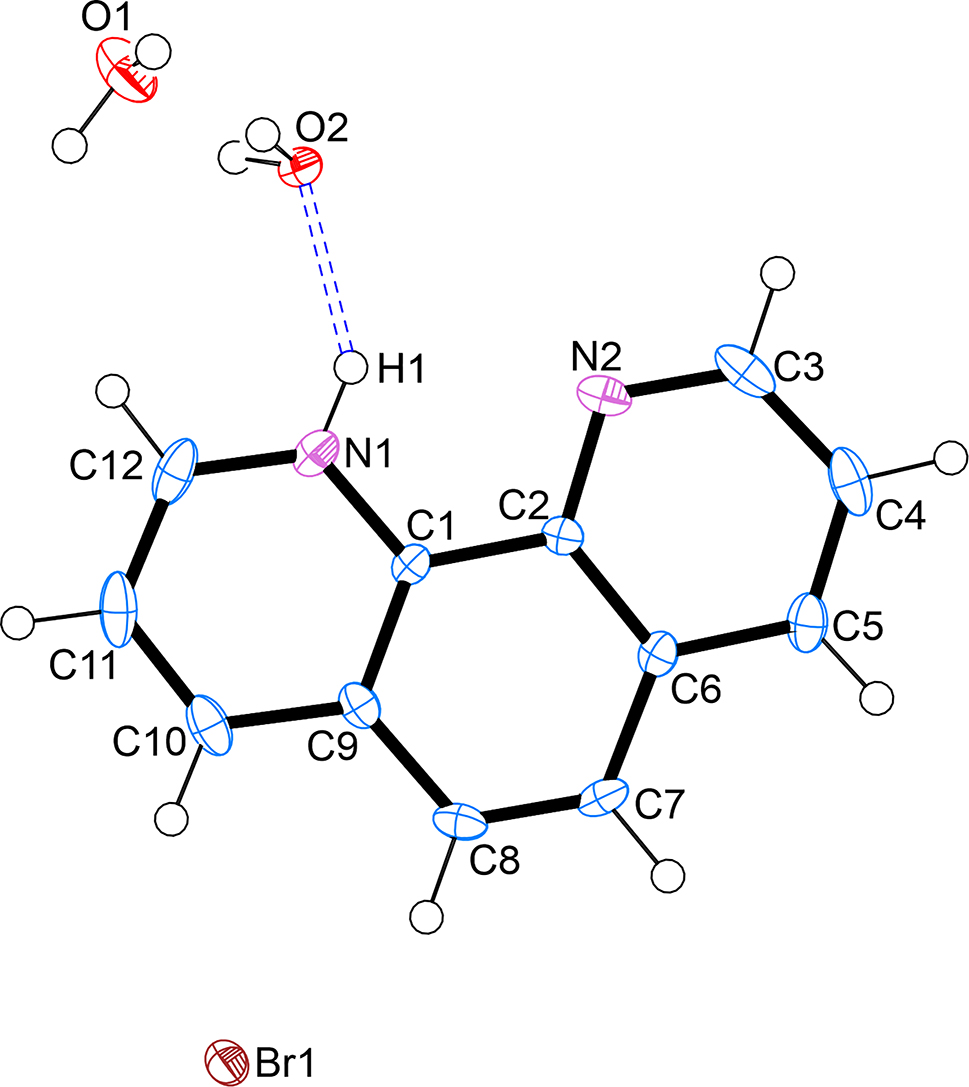
The asymmetric unit of the title crystal structure contains one 1,10-phenanthrolin-1-ium cation, one bromide anion and two free water molecules. One of the two water molecules is disordered over two positions with equal site occupancies. The title compound is solvatomorphic as the 1,10-phenanthrolinium bromide monohydrate structure reported by Buttery and co-workers [9]. All geometric parameters are within the expected ranges [10] and a least-squares fit calculation comparing the phenanthrolinium ions in these two crystal structures (title structure and monohydrate [9]) shows the r.m.s. deviation of bond lengths to be 0.0174 Å [11].
The 1,10-phenanthrolinium bromide dihydrate reported here however, crystallizes in the P
-
Author contributions: All the authors have accepted responsibility for the entire content of this submitted manuscript and approved submission.
-
Research funding: None declared.
-
Conflict of interest statement: The authors declare no conflicts of interest regarding this article.
References
1. COSMO, V1.61. Software for the {CCD} Detector Systems for Determining Data Collection Parameters; Bruker AXS Inc.: Madison, Wisconsin, USA, 2009.Search in Google Scholar
2. Bruker. Saint and Sadabs; Bruker AXS Inc.: Madison, Wisconsin, USA, 2004.Search in Google Scholar
3. Sheldrick, G. M. A short history of Shelx. Acta Crystallogr. 2008, A64, 112–122; https://doi.org/10.1107/s0108767307043930.Search in Google Scholar PubMed
4. Sheldrick, G. M. Crystal structure refinement with Shelxl. Acta Crystallogr. 2015, C71, 3–8; https://doi.org/10.1107/s2053229614024218.Search in Google Scholar PubMed PubMed Central
5. Dolomanov, O. V., Bourhis, L. J., Gildea, R. J., Howard, J. A. K., Puschmann, H. Olex2: a complete structure solution, refinement and analysis program. J. Appl. Crystallogr. 2009, 42, 339–341; https://doi.org/10.1107/s0021889808042726.Search in Google Scholar
6. Mörtel, M., Lindner, T., Scheurer, A., Heinemann, F. W., Khusniyarov, M. M. Phenanthroline-based molecular switches for prospective chemical grafting: a synthetic strategy and its application to spin-crossover complexes. Inorg. Chem. 2020, 59, 2659–2666; https://doi.org/10.1021/acs.inorgchem.9b01424.Search in Google Scholar PubMed
7. Wang, Z., Reibenspies, J., Motekaitis, R. J., Martell, A. E. Unusual stabilities of 6,6′-bis(aminomethyl)-2,2-bipyridyl chelates of transition-metal ions and crystal structures of the ligand and its copper(II) and nickel(II) complexes. J. Chem. Soc., Dalton Trans. 1995, 1511–1518; https://doi.org/10.1039/dt9950001511.Search in Google Scholar
8. Thanippuli Arachchi, D. H., Wijesekera, G. I. P., De Costa, M. D. P., Senthilnithy, R. Amino and chloro derivatives of 1,10-phenanthroline as turn-off fluorescence sensors for selective and sensitive detection of Fe(II). J. Photochem. Photobiol. A 2020, 402, 112805; https://doi.org/10.1016/j.jphotochem.2020.112805.Search in Google Scholar
9. Buttery, J. H., Effendy, Junk, P. C., Mutrofin, S., Skelton, B. W., Whitaker, C. R., White, A. H. The structural systematics of protonation of some important nitrogen-base ligands. IV. Some ethane-1,2-diaminium univalent anion salt/1,10-phenanthroline (hydrate) arrays. Z. Anorg. Allg. Chem. 2006, 632, 1326–1339; https://doi.org/10.1002/zaac.200500498.Search in Google Scholar
10. Thevenet, G., Toffoli, P., Rodier, N., Ceolin, R. Structure cristalline du chlorhydrate et du bromhydrate d’o-phénanthroline, C12H8N2…HX…H2O (X = Cl ou Br). Acta Crystallogr. 1977, B33, 2526–2529; https://doi.org/10.1107/s0567740877008838.Search in Google Scholar
11. Rohlíček, J., Skořepová, E. CrystalCMP: automatic comparison of molecular structures. J. Appl. Crystallogr. 2020, 53, 841–847; https://doi.org/10.1107/s1600576720003787.Search in Google Scholar
© 2023 the author(s), published by De Gruyter, Berlin/Boston
This work is licensed under the Creative Commons Attribution 4.0 International License.
Articles in the same Issue
- Frontmatter
- New Crystal Structures
- The crystal structure of (N-([1,1′:4′,1″-terphenyl]-4,4′-diethyl)-2-(bis(pyridin-2-ylmethyl)amino)acetamide-κ4N,N,N″, O)tri(nitrato-kO, O′) samarium(III) - methanol - acetonitrile (1/1/1), C40H39SmN8O14
- The crystal structure of 6,6′-(((2-(dimethylamino)ethyl)azanediyl)bis(methylene))bis(2-chloro-4-methyl phenolate-κ4N,N′,O,O′)-(pyridine-2,6-dicarboxylato-N,O,O′)-titanium(IV), C27H27Cl2N3O6Ti
- N′-[(1E)-(4–Fluorophenyl)methylidene]adamantane-1-carbohydrazide, C18H21FN2O
- Crystal structure of 4-bromo-3-nitro-1H-pyrazole-5-carboxylic acid monohydrate, C4H2N3BrO4·H2O
- Crystal structure of dipyridine-k1N-tris(2,2,6,6-tetramethyl-5-oxohept-3-en-3-olato-k2O,O′)dysprosium(III), DyC43H67O6N2
- Crystal structure of cyclo[tetraiodido-bis{μ2-1-[(benzotriazol-1-yl)methyl]-1-H-1,3-(2-isopropyl-imidazol)-k2N:N}dicadmiun(II)], C26H30N10Cd2I4
- The crystal structure of tert-butyl (E)-3-(2-(benzylideneamino)phenyl)-1H-indole-1-carboxylate, C26H24N2O2
- The crystal structure of 4-(3-carboxy-1-cyclopropyl-6-fluoro-8-methoxy-4-oxo-1,4- dihydroquinolin-7-yl)-2-methylpiperazin-1-ium 2,5-dihydroxybenzoate methanol solvate, C27H32FN3O9
- Crystal structure of (μ2-1-(4,4′-bipyridine-κ2N:N′)-bis[diaqua-(4-iodopyridine-2,6-dicarboxylato-κ3O,N,O′)–cobalt(II)], C24H20Co2I2N4O12
- The crystal structure of dimethyl 4,4′-(10,20-diphenylporphyrin-5,15-diyl)dibenzoate dichloromethane solvate, C49H36N4O4Cl2
- (E)-2-((E)-4-(2,6,6-trimethylcyclohex-1-en-1-yl)but-3-en-2-ylidene)hydrazine-1-carbothioamide C14H23N3S1
- The crystal structure of [1-(4-(trifluoromethyl)phenyl)-3,4-dihydroquinolin-2(1H)-one], C16H12F3NO
- Crystal structure of (E)-2-amino-N′-((3-hydroxy-5-(hydroxymethyl)-2-methylpyridin-4-yl)methylene)benzohydrazide – dimethylformamide – water (1/1/2), C15H16N4O3·C3H7NO·2H2O
- Crystal structure of 3-(4-bromophenyl)-5-methyl-1H-pyrazole, C10H9BrN2
- Crystal structure of 1,10-phenanthrolinium bromide dihydrate, C12H9N2Br
- Crystal structure of N-(4′-chloro-[1,1′-biphenyl]-2-yl)formamide, C13H10ClNO
- The crystal structure of nitroterephthalic acid, C8H5NO6
- Crystal structure of (2-((4-bromo-2,6-dichlorophenyl)amino)phenyl) (morpholino)methanone, C17H15BrCl2N2O2
- Crystal structure of tetraaqua-bis(ethanol-κO)-tetrakis(μ2-trifluoroacetate-κ2O:O′)-bis(trifluoroacetate-κ2O)digadolinium(III) Gd2C16H20O18F18
- The crystal structure of dimethyl 4,4′-[10,20-bis(2,6-difluorophenyl)porphyrin-5,15-diyl]dibenzoate chloroform solvate, C50H32Cl6F4N4O4
- The crystal structure of N,N′-((nitroazanediyl)bis(methylene))diacetamide, C6H12O4N4
- The crystal structure of [bis(2,2′-bipyridine-6-carboxylato-κ3N,N,O)magnesium(II)]dihydrate, C22H18N4O6Mg
- Crystal structure of poly[diaqua-(bis(μ2-1,4-bis(imidazol-1-ylmethyl)benzene)-κ2N,N′] cobalt(II)-tetraqua-bis(1,4-bis(imidazol-1-ylmethyl)benzene)-κ1N)-cobalt(II) di(2,5-thiophenedicarboxylate) dihydrate, C68H76Co2N16O16S2
- Crystal structure of poly[chlorido-μ2-chlorido-(μ2-1-[(2-ethyl-4-methyl-1H-imidazol-1-yl)methyl]-1H-benzotriazole-κN:N’)cadmium(II)], C13H15CdN5Cl2
- The crystal structure of (4-hydroxybenzenesulfonate)-k1O-6,6′-((1E,1′E)- (ethane-1,2-diylbis(azaneylylidene))bis(methaneylylidene)) bis(2-methoxyphenol)-κ2N,N,μ2O,O,κ2O, O)-(methanol)-cobalt(II) sodium(I), C25H27CoN2NaO9S
- Crystal structure of (1-methyl-3-(trifluoromethyl)-1H-pyrazol-4-yl)(4-((2-methyl-6-(trifluoromethyl)pyrimidin-4-yl)amino)piperidin-1-yl)methanone, C17H18F6N6O
- Crystal structure of bis{[(cyclohexylimino)(phenylimino)-l5-(methyl)diethylazane-κ2N:N′]-(ethyl)-zinc(II)]}, C38H62N6Zn2
- Crystal structure of 2-[(4-bromobenzyl)thio]-5-(5-bromothiophen-2-yl)-1,3,4-oxadiazole, C13H8Br2N2OS2
- Crystal structure of 10-methoxy-7,11b,12,13-tetrahydro-6H-pyrazino [2′,3′:5,6]pyrazino[2,1-a]isoquinoline, C15H16N4O
- The crystal structure of 1-propyl-2-nitro-imidazole oxide, C6H9N3O3
- The crystal structure of 3-nitrobenzene-1,2-dicarboxylic acid–2-ethoxybenzamide (1/1), C17H16N2O8
- The structure of RUB-1, (C8H16N)6[B6Si48O108], a boron containing levyne-type zeolite, occluding N-methyl-quinuclidinium in the cage-like pores
- The crystal structure of diaqua-(naphthalene-4,5-dicarboxylate-1,8-dicarboxylic anhydride-κ1O)-(4′-(4-(1H-benzimidazolyl-1-yl)phenyl)-2,2′:6′,2″-terpyridine-κ3N,N′,N″)–manganese(II) dihydrate, C42H27MnN5O9·2H2O
- Crystal structure of 6,6′-((1E,1′E)-hydrazine-1,2-diylidenebis(methanylylidene))bis (3-(3-bromopropoxy)phenol), C20H22Br2N2O4
- The crystal structure of 3-(2-hydroxyphenyl)-4-phenyl-6-(p-tolyl)-2H-pyran-2-one, C24H18O3
- Crystal structure of bis(μ2-2-(1,5-dimethyl–3-oxo-2-phenyl-2,3-dihydro-1H-pyrazol-4-yl)imino)methyl)phenolato-κ4O:O,N,O′)-(nitrato-κ2O,O′)dicobalt(II), C36H32Co2N8O4
- Synthesis and crystal structure of (3E,5S,10S,13S,14S,17Z)-17-ethylidene-10,13-dimethylhexadecahydro-3H-cyclopenta[α] phenanthren-3-one O-(4-fluorobenzoyl) oxime, C28H36FNO2
- The crystal structure of 4-aminiumbiphenyl benzenesulfonate, C18H17NO3S
- Synthesis and crystal structure of 1-(7-hydroxy-3-(4-hydroxy-3-nitrophenyl)-4-oxo-4H-chromen-8-yl)-N,N-dimethylmethanaminiumnitrate, C18H17N3O9
- Crystal structure of N-(Ar)-N′-(Ar′)-formamidine, C14H12Br2N2O
- The crystal structure of 4-(2,4-dichlorophenyl)-2-(4-fluorophenyl)-5-methyl-1H-imidazole, C16H11Cl2FN2
- Crystal structure of 1-(4–chlorophenyl)-4-benzoyl-3-methyl-1H-pyrazol-5-ol, C17H13ClN2O2
- The crystal structure of 5-amino-1-methyl-4-nitroimidazole, C4H6O2N4
- Crystal structure of 1,3-diisopropyl-4,5-dimethylimidazol-2-ylidene-N,N′-bis(1,3-bis(2,6-diisopropylphenyl)-1,3-dihydro-2H-1,3,2-diazaborol-2-yl)-l2-germenediamine, C63H94B2GeN8
- The crystal structure of (bromido, chlorido)-tricarbonyl-(5,5′-dimethyl-2,2′-bipyridine)-rhenium(I), C15H12Br0.2Cl0.8N2O3Re1
- Crystal structure of [N(E),N′(E)]-N,N′-(1,4-phenylenedimethylidyne)bis-3,5-bis(propan-2-yl)-1H-pyrazol-4-amine, C26H36N6
- The crystal structure of poly[2-(4-carboxypyridin-3-yl)terephthalpoly[diaqua-(μ4-2-(6-carboxylatopyridin-3-yl)terephthalato-κ5O,N:O′:O″,O‴)]) cadmium(II)] dihydrate, C28H20Cd3N2O16
- Crystal structure of [tetraaqua-bis((3-carboxy-5-(pyridin-4-yl)benzoate-κ1N)cobalt(II)] tetrahydrate, C26H32CoN2O16
- Crystal structure of bis(μ2-azido-κ2N:N)-tetrakis(azido-κ1N)-tetrakis(1,10-phenanthroline-κ2N,N′)dibismuth(III), C48H32N26Bi2
- Crystal structure of (Z)-N-(4-(4-(4-((4,5,6-trimethoxy-3-oxobenzofuran-2(3H)-ylidene)methyl)phenoxy)butoxy)phenyl)acetamide, C30H31NO8
- Crystal structure of poly[diaqua-(μ2-1,3-di(1H-imidazol-1-yl)propane-κ2N:N′)-bis(μ2-5-carboxybenzene-1,3-dicarboxylato-O,O′:O″)-aqua-di-zinc dihydrate solvate], C27H28N4O16Zn2
- Crystal structure of 2-(3,5,5-trimethylcyclohex-2-en-1-ylidene)malononitrile, C12H14N2
- Crystal structure of chlorido-(5-nitro-2-phenylpyridine-κ2N,C)-[(methylsulfinyl)methane-κ1S]platinum(II), C13H13ClN2O3PtS
- The crystal structure of the co-crystal 1,4-dioxane–4,6-bis(nitroimino)-1,3,5-triazinan-2-one(2/1), C11H19N7O9
- Crystal structure of [N(E),N′(E)]-N,N′-(1,4-phenylenedimethylidyne)bis-3,5-dimethyl-1H-pyrazol-4-amine di-methanol solvate, C18H20N6·2(CH3OH)
- Crystal structure of catena-poly[bis(μ2-azido-k2N:N′)-(nitrato-K2N:N′)-bis(1,10-phenanthroline-K2N:N′)samarium(III)], C24H16N11O3Sm
- Crystal structure of (Z)-2-(4-((5-bromopentyl)oxy)benzylidene)-4,5,6-trimethoxybenzofuran-3(2H)-one, C23H25BrO6
- Crystal structure of bis(3,5-dimethyl-1H-pyrazol-4-ammonium) tetrafluoroterephthate, 2[C5H10N3][C8F4O4]
- Crystal structure of 2-amino-4-(2-fluoro-4-(trifluoromethyl)phenyl)-9-methoxy-1,4,5,6-tetrahydrobenzo[h]quinazolin-3-ium chloride, C20H18ClF4N3O
- Crystal structure of 6-(pyridin-3-yl)-1,3,5-triazine-2,4-diamine-sebacic acid (2/1), C13H17N6O2