
Crystal structure of langite from Mine du Pradet (France)
-
Christian Paulsen


Abstract
Cu4(OH)6SO4·2H2O, monoclinic, Pc (no. 7), a = 7.1026(2) Å, b = 6.0143(1) Å, c = 11.1645(3) Å, β = 90.1103(8)°, V = 476.91(2) Å3, Z = 2, R gt (F 2) = 0.0206, wR ref (F 2) = 0.0563, T = 100 K.
The structure is shown in the figure. Table 1 contains crystallographic data and Table 2 contains the list of the atoms including atomic coordinates and displacement parameters.
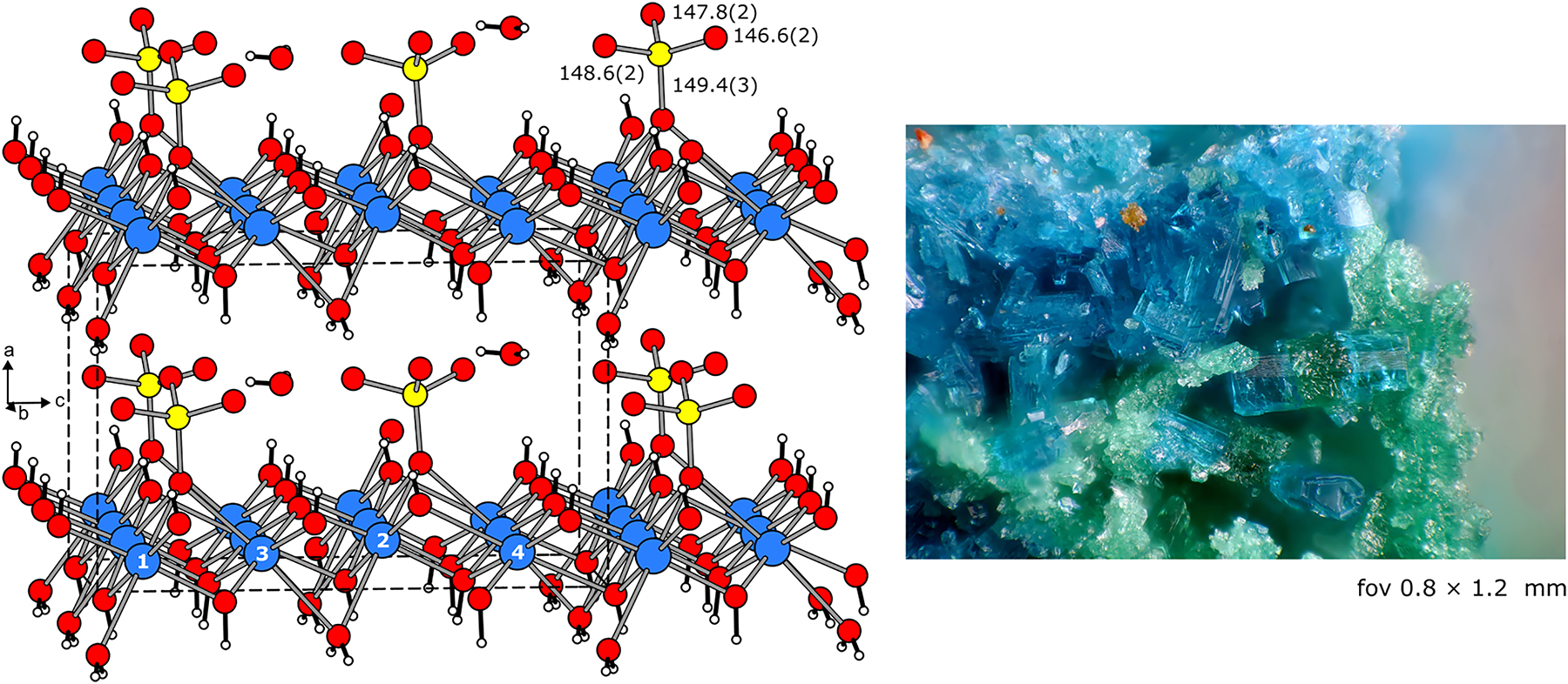
The crystal structure of Cu4(OH)6SO4·2H2O is shown in the left-hand part of the figure, emphasizing the condensed CuO6 octahedra, the sulfate groups as well as the hydroxide units and coordinating water molecules. Atom designations and relevant interatomic distances are indicated. The mineral specimen from Cap Garonne used for the study is presented at the right.
Data collection and handling.
| Crystal: | Blue plate |
| Size: | 30 × 40 × 55 μm |
| Wavelength: | Mo Kα radiation (0.71073 Å) |
| μ: | 9.10 mm−1 |
| Diffractometer, scan mode: | Bruker D8 Venture |
| θ max, completeness: | 32.1°, >99 % |
| N(hkl)measured, N(hkl)unique, R int: | 8815, 3244, 0.016 |
| Criterion for I obs, N(hkl)gt: | I obs > 2σ(I obs), 3147 |
| N(param)refined: | 185 |
| Programs: | X-Area [1], JANA2020 [2], SUPERFLIP [3, 4] |
Fractional atomic coordinates and isotropic or equivalent isotropic displacement parameters (Å2).
| Atom | x | y | z | U iso*/U eq |
|---|---|---|---|---|
| Cu1 | 0.1417 (3) | 0.50212 (3) | 0.0607 (2) | 0.00498 (7) |
| Cu2 | 0.1480 (3) | 0.00724 (3) | 0.0586 (2) | 0.00482 (9) |
| Cu3 | 0.1372 (3) | 0.25601 (3) | 0.3077 (2) | 0.00486 (7) |
| Cu4 | 0.1325 (3) | 0.75481 (3) | 0.3086 (2) | 0.00468 (9) |
| S1 | 0.5634 (2) | 0.31480 (7) | 0.14010 (12) | 0.00626 (9) |
| O1 | 0.2528 (3) | 0.49939 (18) | 0.39736 (18) | 0.0058 (3) |
| O2 | 0.2519 (3) | 0.00503 (18) | 0.39525 (18) | 0.0055 (3) |
| O3 | 0.0227 (3) | 0.01044 (19) | 0.21762 (19) | 0.0059 (3) |
| O4 | 0 | 0.25383 (19) | 0 | 0.0053 (3) |
| O5 | 0.2810 (3) | 0.24515 (17) | 0.61778 (17) | 0.0056 (3) |
| O6 | 0.0148 (3) | 0.50640 (19) | 0.22111 (18) | 0.0057 (3) |
| O7 | 0.8799 (3) | 0.2423 (2) | 0.45876 (18) | 0.0093 (3) |
| O8 | 0.6241 (3) | 0.0711 (2) | 0.86428 (17) | 0.0127 (4) |
| O9 | 0.3572 (3) | 0.26850 (18) | 0.14817 (17) | 0.0065 (3) |
| O10 | 0.6020 (3) | 0.4330 (2) | 0.02589 (16) | 0.0110 (3) |
| O11 | 0.6635 (3) | 0.0994 (2) | 0.13828 (16) | 0.0108 (3) |
| O12 | 0.6248 (3) | 0.4511 (2) | 0.24177 (17) | 0.0119 (3) |
| H1 | 0.349 (5) | 0.504 (4) | 0.403 (3) | 0.007* |
| H2 | 0.348 (5) | 0.999 (4) | 0.398 (3) | 0.0066* |
| H3 | 0.898 (4) | 0.031 (4) | 0.207 (2) | 0.0071* |
| H4 | 0.892 (4) | 0.251 (3) | 0.015 (2) | 0.0063* |
| H5 | 0.371 (4) | 0.248 (3) | 0.604 (3) | 0.0067* |
| H6 | 0.881 (4) | 0.489 (3) | 0.224 (2) | 0.0069* |
| H7a | 0.813 (4) | 0.334 (4) | 0.473 (2) | 0.0112* |
| H7b | 0.823 (4) | 0.159 (4) | 0.449 (2) | 0.0112* |
| H8a | 0.632 (4) | 0.067 (4) | 0.804 (2) | 0.0153* |
| H8ba | 0.636 (5) | 0.199 (6) | 0.881 (3) | 0.0153* |
-
aOccupancy: 0.75 (5).
1 Source of material
Langite has been originally observed on pillars 79 of the Fond de Mine and 25 of the Galerie du Mirror de Faille in the north part (Mine du Nord) of Mine du Pradet, Cap Garonne (France). Transparent blue platelets (see Figure) for the present investigation were selected from a specimen which was collected from the mine ceiling in the Salle des Racines (root vault). The langite crystals were accompanied/intergrown by greenish brochantite crystals (Cu4(OH)6SO4).
2 Experimental details
The Cu4(OH)6SO4·2H2O crystals were carefully broken out of the sample using a medical cannula as lever. The quality of the crystals was first examined by Laue photographs (Buerger camera, white Mo radiation, image plate detection system). Single crystal X-ray diffraction data was collected at 100 K on a Bruker D8 Venture single-crystal diffractometer, equipped with a MoKα microfocus source and a CCD detection system (Photon 100 CMOS). A numerical absorption correction was applied. The starting atomic parameters were deduced with the charge-flipping algorithm [3] implemented in Superflip [4] and the structure was refined on F 2 with the Jana2020 software package [2]. For the final refinement we used the standardized setting listed in the Pearson database [5].
3 Comment
Langite, Cu4(OH)6SO4·2H2O, is one of the rare minerals in Mine du Pradet at Cap Garonne in France [6]. This basic copper sulfate was first discovered in the mine by Sarp [7]; however, no structural characterization was performed. Langite originating from pillars 25 and 79 was later confirmed in 2009 and 2011 by the Jean Wyart association (https://www.amis-mineraux.fr/partenaires.html) with the help of EDX analyses.
In the present study we initially collected data at room temperature (the refinement of the non-hydrogen positions is deposited under CCDC-2301061) but could not reliably refine the hydrogen positions. The crystal chemical discussion thus relies on the 100 K diffraction data. The positions of the non-hydrogen atoms fully agree with the refinements of the langite structure from crystals originating from different deposits [8], [9], [10], [11], [12], [13], [14], [15], [16], [17].
The langite structure (see Figure) contains four crystallographically independent copper atoms, which all have the typical 4 + 2 oxygen coordination as a consequence of Jahn–Teller distortion. The Cu–O distances range from 190.5 to 201.7 pm for the CuO4 square planes and from 229.5 to 266.7 pm for the apical oxygen atoms. These elongated CuO6 octahedra condense to layers via six common edges in a distorted rock salt-type arrangement (similar to the layers of condensed octahedra in the CdI2 structure). Consequently, temperature dependent magnetic susceptibility studies indicated a quasi-two-dimensional spin-1/2 substructure that is subjected to magnetic frustration. Langite shows antiferromagnetic ordering below the Néel temperature of ca. 5.7 K [17].
The slightly distorted sulfate tetrahedra (146.6–149.4 pm S–O; 108.0–111.0° O–S–O) condense with the copper-oxygen layer via a common oxygen atom, however, solely on one side of the layer. All sulfate tetrahedra point in the -a direction, thus the non-centrosymmetric space group symmetry. The water entities in the langite structure have different crystal chemical function. Part of them coordinates to copper and is member of the condensed layer. The remaining ones are typical crystal water molecules and thus part of the hydrogen bonding network that holds the layers together.
Acknowledgments
We thank Dipl.-Ing. J. Kösters for the intensity data collections.
-
Conflict of interest statement: The authors declare no conflicts of interest regarding this article.
-
Author contributions: All the authors have accepted responsibility for the entire content of this submitted manuscript and approved submission.
References
1. X-Area. The STOE Single Crystal Diffraction Software Package; STOE & Cie GmbH: Darmstadt, Germany.Suche in Google Scholar
2. Petřìček, V., Palatinus, L., Plásil, J., Dušek, M. JANA2020 – a new version of the crystallographic computing system Jana. Z. Kristallogr. 2023, 238, 271–282; https://doi.org/10.1515/zkri-2023-0005.Suche in Google Scholar
3. Palatinus, L. The charge-flipping algorithm in crystallography. Acta Crystallogr. 2013, B69, 1–16; https://doi.org/10.1107/s0108768112051361.Suche in Google Scholar
4. Palatinus, L., Chapuis, G. SUPERFLIP – a computer program for the solution of crystal structures by charge flipping in arbitrary dimensions. J. Appl. Crystallogr. 2007, 40, 786–790; https://doi.org/10.1107/s0021889807029238.Suche in Google Scholar
5. Villars, P., Cenzual, K. Pearson’s Crystal Data: Crystal Structure Database for Inorganic Compounds (Release 2022/23); ASM International®: Materials Park, Ohio (USA), 2022.Suche in Google Scholar
6. Favreau, G., Galéa-Clolus, V. Cap Garonne, Association des Amis de la Mine de Cap Garonne (AAMCG)-Association Française de Microminéralogie (AFM); Couleur et Impression Les Arcades: Castelnau-Le-Lez, France, 2014.Suche in Google Scholar
7. Sarp, H. Private communication, 1990.Suche in Google Scholar
8. Pierrot, T., Sainfeld, P. Sur la langite des Vosges. Bull. Soc. Fr. Minér. Cristallogr. 1958, 81, 257–260; https://doi.org/10.3406/bulmi.1958.5281.Suche in Google Scholar
9. Williams, S. A. A new occurrence of langite. Am. Mineral. 1964, 49, 1143–1145.Suche in Google Scholar
10. Wappler, G. Zur Kristallstruktur von Langit, Cu4[(OH)6/SO4]·H2O. Ber. Dt. Ges. Geol. Wiss. B Miner. Lagerstättenforsch. 1971, 16, 175–203.Suche in Google Scholar
11. Pulou, R., Sempère, R., Tournemire, R. Découverte de la langite dans l’Aveyron. Bull. Soc. Fr. Minèr. Cristallogr. 1978, 101, 577–578; https://doi.org/10.3406/bulmi.1978.7235.Suche in Google Scholar
12. Ridkosil, T., Povondra, P. The relation between posnjakite and langite. Neues Jahrb. Mineral. Monatsh. 1982, 1, 16–28.Suche in Google Scholar
13. Galy, J., Jaud, J., Pulou, R., Sempère, R. Structure crystalline de la langite, Cu4[SO4(OH)6H2O]·H2O. Bull. Minéral. 1984, 107, 641–648; https://doi.org/10.3406/bulmi.1984.7808.Suche in Google Scholar
14. Gentsch, M., Weber, K. Structure of langite, Cu4[(OH)6|SO4]·2H2O. Acta Crystallogr. 1984, C40, 1309–1311.10.1107/S0108270184007769Suche in Google Scholar
15. Minčeva-Stefanova, J., Kostov, I. Morphology vs. structure of langite and posnjakite. Compt. Rend. Acad. Bulg. Sci. 2002, 55, 57–60.Suche in Google Scholar
16. Frost, R. L., Williams, P. A., Martens, W., Leverett, P., Kloprogge, J. T. Raman spectroscopy of basic copper (II) and some complex copper (II) sulfate minerals: implications for hydrogen bonding. Am. Mineral. 2004, 89, 1130–1137; https://doi.org/10.2138/am-2004-0726.Suche in Google Scholar
17. Lebernegg, S., Tsirlin, A. A., Janson, O., Redhammer, G. J., Rosner, H. Interplay of magnetic sublattices in langite Cu4(OH)6SO4·2H2O. New J. Phys. 2016, 18, 033020; https://doi.org/10.1088/1367-2630/18/3/033020.Suche in Google Scholar
© 2023 the author(s), published by De Gruyter, Berlin/Boston
This work is licensed under the Creative Commons Attribution 4.0 International License.
Artikel in diesem Heft
- Frontmatter
- New Crystal Structures
- Crystal structure of poly[diaqua-(μ4-3,3′-di(1H-1,2,4-triazol-1-yl)-[1,1′-biphenyl]-4,4′-dicarboxylate-N:N′:O:O′)cadmium(II)], C18H14N6O6Cd
- Crystal structure of (8R,8′S,13S,13′R)-8,8′-bis(hydroxymethyl)-9,9′,10,10′-tetramethoxy-5,5′,6,6′,8,8′,13,13′-octahydro-[13,13′-bi[1,3]dioxolo[4,5-g]isoquinolino[3,2-a]isoquinoline]-7,7′-diium chloride-methanol (1/2), C46H58N2O14Cl2
- The crystal structure of 8-methoxy-2,2-diphenyl-tosyl-1,2-dihydro-2λ4,3λ4-[1,3,2]diazaborolo[4,5,1-ig]quinoline, C29H25BN2O3S
- Crystal structure of aqua-(5,5,7,12,12,14-hexamethyl-1,4,8,11-tetraazacyclotetradecane-κ4N,N′,N″,N‴)copper(II) 5-carboxyisophthalate tetrahydrate, C25H50N4CuO11
- The crystal structure of 1-(naphthalen-2-ylsulfonyl)-2,2-diphenyl-1,2-dihydro-2λ4,3λ4-[1,3,2]diazaborolo[4,5,1-ij]quinoline, C31H23BN2O2S
- Crystal structure of iodido-(η6-benzene) (1-(pyridin-2-yl)-N-(p-fluoro-methanamine)-κ2N,Nʹ)ruthenium(II) hexaflourophosphate, (C18H15F7IN2RuP)
- The crystal structure of 1-(3-oxo-1-phenyl-3-(p-tolyl) propylidene)-1,3-dihydro-2H-inden-2-one, C25H20O2
- Crystal structure of tricyclohexyl[4-(4H-1,2,4-triazol-4-yl)-benzoato-κO]tin(IV), C27H39N3O2Sn
- Crystal structure of [triaqua-(8-carboxymethoxy-quinoline-2-carboxylate-κ4N,O,O,O)cadmium(II)]monohydrate, C12H15NO9Cd
- Crystal structure of ethyl 2-((4-(3,5-dimethylisoxazol-4-yl)-2,6-difluorophenyl)amino)benzoate, C20H18F2N2O3
- The crystal structure of 2-(hydroxymethyl)-2-(4H-1,2,4-triazol-4-yl)propane-1,3-diol, C6H11N3O3
- The crystal structure of 1,2-bis(2,4-dinitrophenyl) hydrazine, C12H8N6O8
- Crystal structure of 1-(2,6-dichloro-4-(3,5-dimethylisoxazol-4-yl)phenyl)-1,2-dihydro-4H-benzo[d][1,3]oxazin-4-one, C19H14Cl2N2O3
- The crystal structure of 5-amino-5-oxo-4-(1-oxo-4-(2-oxopyrrolidin-1-yl)isoindolin-2-yl)pentanoic acid, C17H19N3O5
- Crystal structure of N2,N6-bis(2-(((Z)-5-bromo-2-hydroxybenzylidene)amino) phenyl)pyridine-2,6-dicarboxamide, C33H23Br2N5O4
- The crystal structure of (E)-2-methoxy-6-(((5-methyl-1,3,4-thiadiazol-2-yl)imino)methyl)phenol, C11H11N3O2S
- The crystal structure of 3-((tert-butyldiphenylsilyl)methyl)-5,5-diphenyl-6-(p-tolyl) tetrahydro-2H-pyran-2-one, C41H42O2Si
- Crystal structure of 9-fluoro-4-(6-methoxypyridin-3-yl)-5,6-dihydrobenzo[h]quinazolin-2-amine, C18H15FN4O
- The crystal structure of 2-bromo-5-(4-cyanophenoxy)benzyl 1-methyl-1,2,5,6-tetrahydropyridine-3-carboxylate, C21H19BrN2O3
- Crystal structure of 3,3′-(1,4-phenylenebis(methylene))bis(1-isopropyl-1H-imidazol-3-ium) bis(hexafluorophosphate(V)), C10H14F6N2P
- The crystal structure of 2,2-di(thiophen-3-yl)-1-tosyl-1,2-dihydro-2λ4,3λ4-[1,3,2]diazaborolo[4,5,1-ig]quinoline, C24H19BN2O2S3
- Crystal structure of 5-bromo-1-(2-iodobenzoyl)-1H-indole-3-carbaldehyde, C16H9BrINO2
- The crystal structure of monocarbonyl-2-carboxypyridinato-κ2N,O-triphenylphosphine-rhodium(I) acetonitrile solvate, C26H20.50N1.50O3PRh
- Crystal structure of dichlorido-tetrakis(1-(2,4-dichlorophenyl)-4,4-dimethyl-2-(1,2,4-triazol-1-yl)pent-1-en-3-ol-κ1N)manganese(II), C60H68O4N12Cl10Mn
- Crystal structure of 3-(tert-butyldiphenylsilyl)-1-(2,6-dichlorophenyl)-2,2-diphenylpropan-1-ol, C37H36Cl2OSi
- Crystal structure of langite from Mine du Pradet (France)
- The crystal structure of 5′-(furan-2-yl)-3′-((4-methylphenyl)sulfonamido)-3′,4′,5′,6′-tetrahydro-[1,1′:3′,1″-terphenyl]-4′-carboxylic acid, C30H27NO5S
- Synthesis and crystal structure of bis{2-(((4-acetophenone)imino)methyl)-4-fluorophenolato-κ2N,O}zinc(II), C30H22F2N2O4Zn
- The crystal structure of poly[(tripyridine-κ3N,N′,N″) μ3-(pyridine-3,4-dicarboxylate-κ3N:O:O′) manganese(II)], C22H22N4O8Mn
- The crystal structure of (E)-4-chloro-N′-(1-(4-hydroxyphenyl)propylidene)benzohydrazide, C16H15ClN2O2
- Synthesis and crystal structure of bis{2-(tert-butyl)-6-((E)-((4-((E)-1-(methoxyimino) ethyl)phenyl)imino)methyl)phenolato-κ2N,O}cobalt(II), C40H46CoN4O4
- Crystal structure of tetraaqua-[(1-(carboxymethyl)-1H-pyrazole-3-carboxylato-κ2N,O)cobalt(II)], C6H12CoN2O8
- (6R,7S)-2,3,13-trimethoxy-6,7-dimethyl-5,6,7,8-tetrahydrobenzo[3′,4′]cycloocta [1′,2′:4,5]benzo[1,2-d][1,3]dioxol-1-ol, C22H26O6
- Crystal structure of 2-((2,6-dichloro-4-(3,5-dimethylisoxazol-4-yl)phenyl)amino)benzoic acid, C18H14Cl2N2O3
- Crystal structure of (5aS,6aS,8aR,9R,11aS, 11bS,13R,13aS)-1,1,8a,11a-tetramethyl-9-((S)-1-((S)-5-methyl-6-oxo-3,6-dihydro-2H-pyran-2-yl)ethyl)-3-oxo-1,7,8,8a,9,10,11,11a,11b,12,13,13a-dodecahydro-3H,6H-cyclopenta[5,6]cyclopropa[1,8a]naphtho[2,1-c]oxepin-13-yl acetate, C32H44O6
- Crystal structure of catena-poly[triaqua-(μ2-1-(4-carboxylatophenyl)-4-oxo-1,4-dihydropyridazine-3-carboxylato-O,O′:O″)cobalt(II)], C12H12N2O8Co
- Crystal structure of 3-[(furan-2-ylmethyl)-amino]-2-(2,3,4,5-tetrafluoro-benzoyl)-acrylic acid ethyl ester, C17H13F4NO4
- Crystal structure of methyl 4-(2-ethoxy-2-oxoethoxy)-3-methoxybenzoate, C13H16O6
- Crystal structure of 4-bromo-2-(4-chlorophenyl)-1-methyl-5-(trifluoromethyl)-1H-pyrrole-3-carbonitrile, C13H7BrClF3N2
- The crystal structure of triaqua-(8-carboxymethoxy-quinoline-2-carboxylate-κ3N,O,O)nickel(II) monohydrate, C12H15NO9Ni
- Crystal structure of dihydroxy(2,4,6-triisopro-pylphenyl)telluronium trifluoromethanesulfonate, C16H25F3O5STe
- The crystal structure of 1-(carboxymethyl)-1H-imidazole 3-oxide
- The crystal structure of 1,3,5-tris(dibromomethyl)benzene, C9H6Br6
- Crystal structure of (Z)-3-(4-methoxyphenyl)-4-(5-methyl-1-phenyl-1H-1,2,3-triazol-4-yl)-N-phenylthiazol-2(3H)-imine, C25H21N5OS
- Crystal structure of (Z)-3-(3-(4-hydroxyphenyl)-2-(phenylimino)-2,3-dihydrothiazol-4-yl)-2H-chromen-2-one, C24H16N2O3S