Ferrite/curcumin hybrid nanocomposite formulation: Physicochemical characterization, anticancer activity, and apoptotic and cell cycle analyses in skin cancer cells
-
Yousef A. Bin Jardan
 ,
Mahmoud Zaki El-Readi
,
Mahmoud Zaki El-Readi

Abstract
Skin cancer requires effective treatment due to its high incidence and mortality. Curcumin’s anti-tumor effects are restricted by its lower bioavailability, dose dependency, and poor skin permeability. Hybrid curcumin molecules with spinal metal ferrite and biogenic silver/silver chloride (Ag/AgCl) nanoparticles could be a promising approach for the efficient delivery of curcumin to cancer cells. AgClM0.5Fe2O4 and AgClM0.5Fe2O4/curcumin hybrid nanocomposites were synthesized via chemical co-precipitation and wet impregnation techniques. Furthermore, they were characterized using X-ray diffraction (XRD), Fourier transform infrared spectroscopy (FT-IR), energy-dispersive X-ray spectroscopy (EDX), transmission electron microscopy (TEM), dynamic light scattering (DLS), and vibrating sample magnetometry (VSM). The cytotoxicity, apoptosis, and cell cycling of hybrid nanocomposites were evaluated on human epidermoid skin carcinoma (A-431) and human skin fibroblast (HSF) cell lines. Results showed successful curcumin loading, high physical stability, and enhanced magnetic properties of the hybrid nanocomposites. Cytotoxicity assays revealed selective toxicity against both A-431 and HSF cell lines with IC₅₀ values of 26.83 and 34.83 µg/ml for AgClCd0.5Fe2O4 and AgClMg0.5Fe2O4/curcumin, respectively. AgClCd0.5Fe2O4/curcumin exhibited greater apoptosis induction (14.52% late apoptosis, 8.53% necrosis) compared to AgClMg0.5Fe2O4/curcumin (10.20% late apoptosis, 6.07% necrosis). In addition, cell cycle analysis showed a significant increase in the sub-G1 phase, with AgClCd0.5Fe2O4/curcumin demonstrating superior efficacy. In conclusion, hybrid nanocomposites triggered cell cycle arrest and apoptosis, with AgClCd0.5Fe₂O₄/curcumin being the most potent. Their lower toxicity compared to doxorubicin suggests their potential application in skin cancer treatment.
1 Introduction
Cancer is the largest contributing factor to the death of people globally and a serious global health concern [1]. About 20 million cases of cancer were reported in 2022, and the disease claimed the lives of nearly 9.7 million people worldwide, according to the American Cancer Society’s report on cancer facts and trends. An estimated 35 million cases of cancer are expected to occur by 2050 [2]. Skin cancer is the fifth most reported cancer globally, making it one of the most prevalent cancers. Non-melanoma skin cancer accounts for 1,234,533 cases worldwide and 69,416 deaths annually, according to the latest figures [3]. According to the American Academy of Dermatology, skin cancer may strike at least one in five Americans [4]. Two types of skin cancer are known: melanoma and non-melanoma, and melanoma accounts for the majority of skin cancer deaths [5]. Excision biopsy, chemotherapy, radiation, immunotherapy, electrodesiccation, and photodynamic therapy are examples of conventional treatment options that can result in several unwanted adverse effects that affect the quality of life of patients and general health. Resistance to commonly used anti-cancer drugs, e.g., cisplatin [6], doxorubicin [7], and paclitaxel [8], is considered a problem with using chemotherapy. This resistance can occur through drug efflux, target alteration, and improved DNA repair. Hence, innovative approaches have been investigated to overcome these obstacles [9].
Nanosized ferrites are inexpensive and are widely utilized in various applications, including electronics, biomedicine, and environmental remediation [10]. Ferrites are inorganic substances that are mostly made up of Fe3+ cations, O2− anions, and other cations like transition metals and earth metals (alkaline and rare earth metal elements). Ferrites are classified as spinel, garnet, ortho-, and hexaferrites based on their chemical compositions and structural characteristics. The most common variety of these is spinel ferrites [11]. Because of their specific magnetic qualities and multi-metal composition, spinel ferrite nanoparticles (NPs) are particularly useful in biomedicines and nanomedicines. Biologically, these NPs are generally considered biocompatible and non-toxic when synthesized properly, making them suitable for medical applications [12]. Typically, spinel ferrite NPs are synthesized using methods like co-precipitation, sol–gel, hydrothermal, and microwave-assisted synthesis, which provide precise control over particle size, composition, and surface characteristics, critical for tuning their biological responses [13]. In comparison to the other metallic NPs, silver/silver chloride Ag/AgCl NPs, which have a high surface area-to-volume ratio and a narrow plasmon resonance, have demonstrated exceptional biological properties [14,15,16]. Biogenic Ag/AgCl NPs have been demonstrated in numerous studies to have an anti-tumor effect against different cancer cell lines both in vitro and in mice models, as well as their ability to trigger apoptosis in embryonic zebrafish [17,18,19,20]. Interestingly, biogenic silver is much more toxic to cancer cells than to healthy cells [21]. Curcumin has been investigated for its anticancer properties by initiating apoptosis and inhibiting cancer cell growth through specific signaling pathways [22,23]. Despite its beneficial applications, its use is limited due to low solubility and bioavailability, rapid metabolism, and poor skin permeation [24,25]. Therefore, developing an effective formulation able to deliver curcumin to its target site in adequate amounts is crucial.
The concept of a hybrid curcumin molecule involves integrating or combining curcumin with specific materials [26]. Such diverse conjugations enhance the selectivity, biological activities, stability, targeted delivery, cellular uptake, and inherent properties of curcumin, which significantly improve its overall anti-cancer efficacy [13]. In addition, this hybrid system can overcome the limitations of free curcumin, such as poor solubility, rapid metabolism, and low bioavailability.
Therefore, the novelty of this study lies in the preparation of a hybrid curcumin metal ferrite nanocomposite system with certain transition metals, AgClM0.5Fe2O4/curcumin hybrid nanocomposites, where M represents calcium, cadmium, copper, or magnesium (Ca2+, Cd2+, Cu2+, or Mg2+), followed by studying the anticancer activity and biological mechanism of these materials in skin cancer cell lines. No reports on the application of these nanocomposites as anti-cancer agents for skin cancer currently exist.
The rationale for selecting specific transition metals (e.g., Cd2+, Ca2+, Cu2+, Mg2+) in the AgClM0.5Fe2O4/curcumin hybrid nanocomposite is because of their unique physicochemical properties, biological compatibility, and synergistic anticancer mechanisms. Furthermore, combining these metals with curcumin offers a dual metal-phytochemical approach to enhance cytotoxicity through reactive oxygen species (ROS) amplification, cell cycle disruption, and apoptotic induction [27,28,29]. This strategic selection balances therapeutic efficacy with biocompatibility, addressing gaps in prior research that overlooked the comparative advantages of these metals in ferrite–curcumin systems.
Basically, the study includes synthesizing AgClM0.5Fe2O4/curcumin hybrid nanocomposites with different transition metal dopants in the samples using chemical co-precipitation and wet impregnation techniques. This is followed by physicochemical characterization of the prepared nanocomposite using different techniques like X-ray diffraction (XRD), Fourier transform infrared (FT-IR)spectroscopy, energy-dispersive X-ray spectroscopy (EDX), transmission electron microscopy (TEM), dynamic light scattering (DLS), vibrating sample magnetometry (VSM), and their surface charges. A cytotoxicity study of the prepared materials was performed using two types of skin cell lines: human epidermoid skin carcinoma (A-431) and human skin fibroblasts (HSFs). Apoptosis induction and cell cycle studies were also investigated for nanocomposites with lower IC50 values in the human epidermoid skin carcinoma cell lines using flow cytometry and compared with doxorubicin (Dox).
2 Materials and methods
2.1 Materials
Silver nitrate (AgNO3, Assay: 99%, Sigma-Aldrich, UK), anhydrous ferric chloride (FeCl3, Assay: 98%, Alpha Chemicals, Egypt), copper nitrate hydrate (Cu(NO3)2·3H2O, Assay: 98%, Laboratory Chemicals), anhydrous calcium chloride (CaCl2, Assay: 98%, TopChem), cadmium nitrate tetrahydrate (Cd(NO3)2·4H2O, Assay: 99%, Alpha Chemical,. Cairo, Egypt), magnesium chloride hexahydrate (MgCl2·6H2O, Assay: 98%), and sodium hydroxide (NaOH, Assay: 99%, Alpha Chemicals, Assiut, Egypt). Curcumin (98%) and doxorubicin (99%) were purchased from Sigma-Aldrich, Co., UK.
2.2 Synthesis of AgClM0.5Fe2O4 nanocomposites
AgClM0.5Fe2O4 nanocomposites (M = Ca, Cd, Cu, or Mg) were synthesized via a chemical co-precipitation method [30,31,32]. As an example, for the synthesis of 2 g of AgClCu0.5Fe2O4, 0.97 g of AgNO3, 1.85 g of FeCl3, and 0.69 g of Cu(NO3)2·3H2O were dissolved in 200 ml of distilled water to form a mixture solution. The solution was continuously stirred at 300 rpm using a magnetic stirrer (HS-20, LK LAB-Korea) at 80°C for 30 min. Separately, a 3 M sodium hydroxide (NaOH) solution was prepared and used as the precipitating agent. The pH of the mixture was adjusted to 12.0 by gradually adding NaOH solution. The resulting precipitate was maintained at 80°C under continuous stirring for 2 h. Then, it was cooled to room temperature, and separated using a centrifuge (10,000 rpm, BT20 bench top high-speed centrifuge, China), washed twice with distilled water, and then with ethanol. The filtered product was dried at 80°C in an oven (Binder ED53 oven, Germany) for 24 h, ground into fine powder using a mortar, and then calcined at 600°C for another 4 h in a muffle furnace (Nabertherm L-401H1TN, Germany) under a static air atmosphere. The above-mentioned steps were also used for the synthesis of AgClCa0.5Fe2O4, AgClCd0.5Fe2O4, and AgClMg0.5Fe2O4. For the synthesis of AgClCa0.5Fe2O4, 1.00 g of AgNO3, 1.91 g of FeCl3, and 0.33 g of CaCl2 were used. However, for AgClCd0.5Fe2O4, 0.91 g of AgNO3, 1.73 g of FeCl3, and 0.82 g of Cd(NO3)2·4H2O were used. For the synthesis of AgClMg0.5Fe2O4, 1.03 g of AgNO3, 1.96 g of FeCl3, and 0.61 g of MgCl2·6H2O were used.
2.3 Synthesis of AgClM0.5Fe2O4/curcumin nanocomposites
The wet impregnation method was used to synthesize AgClM0.5Fe2O4/curcumin (M = Ca, Cd, Cu, or Mg) [33]. For example, to synthesize the AgClCu0.5Fe2O4/curcumin nanocomposite (50% AgClCu0.5Fe2O4, 50% curcumin), 0.5 g of each component was mixed in 5 ml of distilled water and stirred at 300 rpm for 5 min at room temperature. The obtained mixture was further dried at 60°C for 24 h. The resulting dried nanocomposite powders were ground in a mortar to obtain a homogeneous powder and ensure that all AgClCu0.5Fe2O4 NPs adhered to the surface of curcumin.
2.4 Characterization techniques
Crystallinity and structural properties of the nanocomposites were analyzed using an X-ray diffractometer (PW 2103, Philips, Netherlands) with CuKα radiation (λ = 1.5406 Å), collecting diffraction data over a 2θ range of 4–80° at a scanning rate of 5°/min. Functional groups in the powdered nanocomposites were analyzed using a Nicolet 6700 FT-IR spectrometer in the transmittance mode across a wavelength range of 400–4,000 cm−1. JEOL JSM-IT200 scanning electron microscopy coupled with EDX analysis was performed to determine the chemical composition of the nanocomposites. Surface morphologies were observed with a JEOL transmission electron microscope (TEM, Model JSM-5400 LV, JEOL, Tokyo, Japan). The zeta potential of the nanocomposites was measured using a Malvern Zetasizer Nano ZEN 3600 [34]. To analyze the magnetic properties of the prepared nanocomposites, a VSM (Lakeshore, 7410 s, India) is used.
2.5 Cell culture and cytotoxicity assay
Two types of cell lines were used in this study: A-431 and HSF cell lines. The investigated cell lines were chosen as A-431 cell lines to overexpress the epidermal growth factor receptor (EGFR), making it a commonly used model for the pathophysiology and therapeutic approach studies of skin cancer [35]. HSF serves as a standard model for normal skin cells, providing a baseline for determining the selective cytotoxicity. By comparing cancerous and non-cancerous cells in skin cancers, it is easier to determine the nanocomposites’ safety and specificity. The used cell lines were purchased from Nawah Scientific Inc. (Mokatam, Cairo, Egypt). Initially, cells were maintained in Dulbecco’s modified Eagle’s medium (DMEM) containing streptomycin and penicillin at 100 mg/ml and 100 units/ml, respectively. In addition, 10% fetal bovine serum was heat-inactivated under humidified conditions of 5% (v/v) CO2 at 37°C.
The SRB assay method was utilized to determine cell viability. Briefly, a cell suspension (5 × 10³ cells) of an approximate 100 μL was seeded in 96-well plates and then incubated for 24 h in complete media. Further, different nanocomposite concentrations in the used medium were prepared and added to cells with a volume of 100 μL [36,37]. After NP exposure, cells were fixed by changing the media with 150 μL of 10% trichloroacetic acid (TCA) and incubated at 4°C for approximately 1 h. Then, the TCA solution was removed from the cells, followed by washing them five times with distilled water. Sulforhodamine B (SRB, 70 μL) solution (0.4% w/v) was then added, and cells were left in a dark place at room temperature for 10 min. The well plates were washed three times with 1% acetic acid and left to dry in air overnight. The protein-bound SRB stain was dissolved by adding 150 μL of Tris(hydroxymethyl)aminomethane (TRIS) (10 mM), and the absorbance was measured using a microplate reader at λmax of 540 nm (Infinite F50 Microplate Reader, TECAN, Switzerland).
2.6 Cell apoptosis
Cell populations undergoing apoptosis and necrosis are assessed utilizing the Annexin V conjugated with FITC (fluorescein isothiocyanate), a fluorescent dye, Annexin V-FITC, Apoptosis Detection Kit (Abcam Inc., Cambridge Science Park, Cambridge, UK) in conjunction with dual fluorescence channel flow cytometry. After the treatment with AgClCd0.5Fe2O4 and AgClMg0.5Fe2O4 for 48 h, 105 cells are collected using trypsin, followed by washing twice with ice-cold PBS with a pH of 7.4 and a final concentration of (137 mM NaCl, 10 mM Phosphate, and 2.7 mM KCl). Cells are incubated with 0.5 ml of Annexin V-FITC/PI solution for 30 min in the dark place at room temperature. After staining the cells, they are injected into the ACEA Novocyte™ flow cytometer (ACEA Biosciences Inc., San Diego, CA, USA). Cells were analyzed for FITC and PI fluorescent signals utilizing the FL1 and FL2 signal detectors, respectively (λ ex/em 488/530 nm for FITC and λ ex/em 535/617 nm for PI). Each tested sample undergoes the acquisition of 12,000 events, with positive FITC and/or PI cells quantified through quadrant analysis and computed using ACEA NovoExpress™ software (ACEA Biosciences Inc., San Diego, CA, USA) [38,39].
2.7 Cell cycle assay
The tested cell lines were treated with the tested nanomaterials (AgClCd0.5Fe2O4 and AgClMg0.5Fe2O4) for 48 h. Then, cells (105 cells) were harvested via trypsinization, followed by washing two times with ice-cold phosphate-buffered saline (PBS) at pH 7.4. Cells were resuspended in 2 ml of ice-cold ethanol at a concentration of 60%, followed by incubation at 4°C for 1 h to affect their fixation. Two additional washing steps were also performed in the fixed cells with PBS (pH 7.4) and re-suspended in 1 ml of PBS containing 50 µg/ml RNAase A and 10 µg/ml propidium iodide (PI). Cells are assessed for DNA content after their incubation for 20 min in the dark at 37°C, using flow cytometry and an FL2 (λ ex/em 535/617 nm) signal detector (ACEA Novocyte™ flow cytometer, ACEA Biosciences Inc., San Diego, CA, USA). Each sample had 12,000 events acquired. Cell cycle distributions were determined utilizing ACEA NovoExpress™ software (ACEA Biosciences Inc., San Diego, CA, USA) [40,41].
2.8 Statistical analysis
Statistical analysis was performed via the GraphPad Prism Version 9 (GraphPad Software, San Diego, CA, USA). The results are presented using the mean standard deviation (SD). The comparison was made using ANOVA statistical tests with Turkey’s HSD post-hoc test, a multiple comparison between positive and negative control groups. Results are statistically significant when the p-value is less than 0.05.
3 Results and discussion
Certain transition metals were chosen in this study to prepare hybrid curcumin metal ferrite nanocomposite systems. Ca2+ was incorporated for its role in dysregulated calcium signaling pathways, a vulnerability in skin cancer cells, thereby enhancing chemosensitization [42,43]. Although Cd2+ is toxic in bulk, its trace doping was explored for its potent mitochondrial disruption and apoptosis-inducing capabilities, which are leveraged at controlled concentrations to target cancer cells selectively. [44,45]. Cu2+ was chosen for its redox-active nature, which amplifies ROS generation, thereby enhancing curcumin’s pro-apoptotic effects [27,46]. Mg2+ was included due to its biocompatibility and ability to stabilize the NP structure while modulating cell cycle arrest (e.g., G2/M phase), a critical mechanism for skin cancer suppression [28,47]. These metals were prioritized over alternatives (e.g., Zn, Ni, Co) due to their superior ionic radius compatibility with Fe2+ (0.78 Å) and Fe3+ (0.64 Å) in the spinel ferrite lattice (Cu2+: 0.73 Å; Mg2+: 0.72 Å), ensuring minimal lattice distortion and optimized magnetic/electronic properties [48]. In contrast, larger ions like Cd2+ (0.95 Å) and Ca2+ (0.99 Å) introduce controlled strain, which modifies catalytic and therapeutic effects without compromising stability [48].
3.1 Characterization of hybrid nanocomposites
AgClM0.5Fe2O4 nanocomposites were synthesized via a chemical co-precipitation method. This method is commonly favored for synthesizing spinel ferrites because of its simplicity and cost-effectiveness. It allows for control over particle size and morphology, ensures high compositional homogeneity, and facilitates adjustments to reaction parameters such as pH to achieve desired properties. This versatility makes it an effective technique for producing tailored spinel ferrite materials with consistent characteristics [49]. AgClM0.5Fe2O4 nanocomposites (50% by wt) were loaded on the surface of curcumin using the wet impregnation technique. The wet impregnation method provides numerous advantages, such as simplicity, versatility, cheap, effective control over particle size and distribution, capability to deposit active materials onto high surface area supports, and its suitability for large-scale production [50].
The XRD patterns of pure curcumin, AgClCa0.5Fe2O4/curcumin, AgClCd0.5Fe2O4/curcumin, AgClCu0.5Fe2O4/curcumin, and AgClMg0.5Fe2O4/curcumin nanocomposites are shown in Figure 1. Pure curcumin displayed reflections at 2θ values of 17°, 19.3°, 21.9°, 23.8°, 26.5°, and 28.1° [51]. All the prepared nanocomposites showed diffraction peaks at 2θ values of 27.8°, 32.2°, 46.2°, 54.8°, 57.4°, and 76.7°, which are attributed to the (111), (200), (220), (311), (222), and (420) planes, respectively, of cubic AgCl (JCPDS card no. 00-006-0480). In addition, AgClCa0.5Fe2O4/curcumin showed also reflections at 2θ values of 24.1°, 33.2°, 35.6°, 40.9°, 49.5°, 54.1°, 62.5°, 64.2°, and 72.1°, respectively, which correspond to the (012), (104), (110), (113), (024), (116), (214), (300), and (119) planes for rhombohedral Fe2O3 (JCPDS card no. 01-084-0306). Moreover, the peaks at 2θ values of 38.1° and 44.3° are related to cubic zero-valent Ag (JCPDS card no. 01-087-0717) and the peak at 67.4° belongs to CaFe2O4 (JCPDS card no. 00-019-0219). The XRD pattern of the AgClCd0.5Fe2O4/curcumin nanocomposite showed that, besides the AgCl peaks, additional peaks at 2θ values of 24.4°, 35.5°, and 49.4° are ascribed to rhombohedral Fe2O3 (JCPDS card no. 01-079-1741). In addition, the peaks at 28.9°, 34.1°, 51.4°, 60.2°, 67.4°, and 70.9° are attributed to cubic CdFe2O4 (JCPDS card no. 00-022-1063). Moreover, the peak at 38° is related to cubic zero-valent Ag (JCPDS card no. 01-087-0597) and the peak at 74.5° belongs to cubic AgCl (JCPDS card no. 00-002-0848). The AgClCu0.5Fe2O4/curcumin nanocomposite showed reflections at 2θ values of 24.1°, 33.2°, 40.9°, 49.5°, 54.1°, and 64°, which are assigned to rhombohedral Fe2O3 (JCPDS card no. 00-033-0664). Additionally, the peaks at 35.7° and 62.5° are assigned to CuFe2O4 (JCPDS card no. 01-077-0010). Moreover, the reflections at 38.8° and 67.5° are ascribed to monoclinic CuO (JCPDS card no. 00-045-0937). The AgClMg0.5Fe2O4/curcumin nanocomposite exhibited signals at 2θ values of 24.2°, 33.2°, 40.7°, 49.5°, 54.1°, and 64.1° are assigned to rhombohedral Fe2O3 (JCPDS card no. 01-073-2234). In addition, the peaks at 35.6°, 62.5°, and 74.5° are related to cubic MgFe2O4 (JCPDS card no. 00-001-1120). Moreover, the peak at 38.2° is ascribed to cubic zero-valent Ag (JCPDS card no. 01-087-0717) and the signal at 67.4° is related to cubic AgCl (JCPDS card no. 00-031-1238). The average crystallite sizes for the prepared nanocomposites were estimated using the Scherrer equation [33,52,53] and were found to be 11.1, 23.5, 20.3, 26.9, and 30.3 nm for pure curcumin, AgClCa0.5Fe2O4/curcumin, AgClCd0.5Fe2O4/curcumin, AgClCu0.5Fe2O4/curcumin, and AgClMg0.5Fe2O4/curcumin nanocomposites, respectively. The relative crystallinity (RC) of pure curcumin and the prepared nanocomposites was estimated using the following equation:
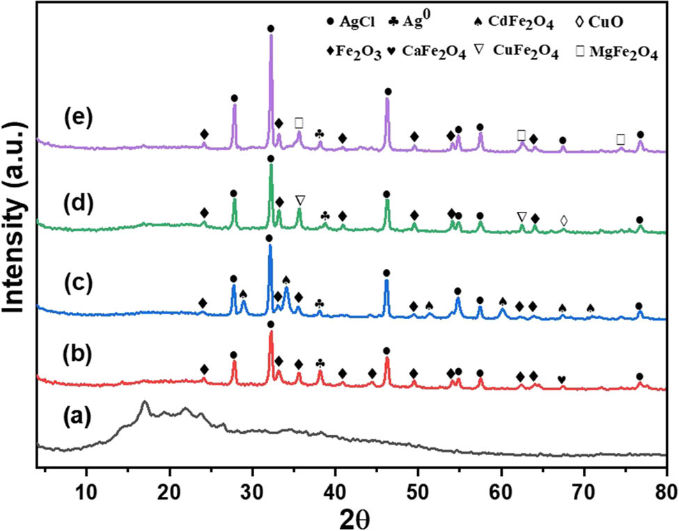
XRD patterns of (a) pure curcumin, (b) AgClCa0.5Fe2O4/curcumin, (c) AgClCd0.5Fe2O4/curcumin, (d) AgClCu0.5Fe2O4/curcumin, and (e) AgClMg0.5Fe2O4/curcumin hybrid nanocomposites.
It was found that the RC takes the following order: curcumin (59.9%) < AgClMg0.5Fe2O4/curcumin (67.5%) < AgClCu0.5Fe2O4/curcumin (72.3%) < AgClCa0.5Fe2O4/curcumin (74.8%) < AgClCd0.5Fe2O4/curcumin (82.2%).
FT-IR spectroscopy is a powerful tool for understanding and optimizing material surfaces by identifying and quantifying functional groups crucial to their performance and application. Figure 2 shows the FT-IR spectra of pure curcumin, AgClCa0.5Fe2O4/curcumin, AgClCd0.5Fe2O4/curcumin, AgClCu0.5Fe2O4/curcumin, and AgClMg0.5Fe2O4/curcumin nanocomposites.
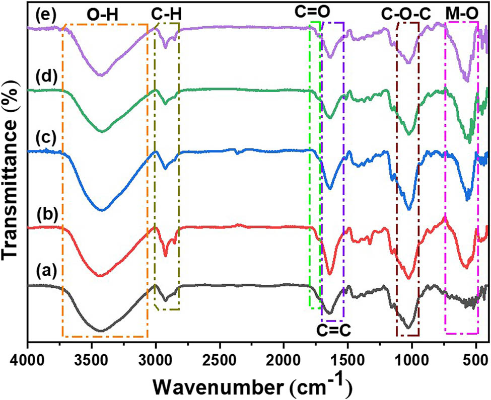
FT-IR spectra of (a) pure curcumin, (b) AgClCa0.5Fe2O4/curcumin, (c) AgClCd0.5Fe2O4/curcumin, (d) AgClCu0.5Fe2O4/curcumin, and (e) AgClMg0.5Fe2O4/curcumin hybrid nanocomposites.
The curcumin spectrum showed FT-IR bands at 3,417 cm−1 (phenolic hydroxyl (–OH) groups), 2,924 cm−1 (aliphatic C–H groups), 2,857 cm−1 (aliphatic C–H groups), 1,737 cm−1 (C═O stretching), 1,640 cm−1 (C═C stretching [54,55], 1,515 cm−1 (C═O and C═C vibrations) [54], 1,455 cm−1 (aromatic C–H groups), 1,372 cm−1 (aromatic C–H groups), 1,160 cm−1 (C–O–C stretching for methoxy groups), 1,078 cm−1 (C–O stretching mode), 1,031 cm−1 (C–O–C stretching vibrations), and 520 cm−1 (aromatic C–C–C out of plane bending) [55]. The FT-IR spectra of the nanocomposites revealed, in addition to the characteristic bands of curcumin, additional bands in the range of 670–430 cm−1, corresponding to metal–oxygen vibrations. These findings confirm the successful loading of AgClM0.5Fe2O4 onto the surface of curcumin. The FT-IR spectra of the nanocomposites also show distinct vibrational bands corresponding to various bonding modes and structural features in the region below 1,000 cm−1. In all samples, a band around 855 cm−1 (843 cm−1 for AgClMg0.5Fe2O4/curcumin) is attributed to the Fe–O–H bending mode [56]. The Fe–O, Fe–O–Fe, and O–Fe–O vibrations appear in the range of 568–573 cm−1 for AgClCa0.5Fe2O4/curcumin, AgClCd0.5Fe2O4/curcumin, and AgClCu0.5Fe2O4/curcumin, while AgClMg0.5Fe2O4/curcumin shows this band at 574 cm−1 [57]. The Fe–O bond at tetrahedral sites is observed at 533–555 cm−1, with a prominent band at 555 cm−1 for AgClCd0.5Fe2O4/curcumin [58], while the octahedral Fe–O bond is highlighted at 545 cm−1 for AgClCd0.5Fe2O4/curcumin. The Fe3+–O bond vibration is detected in all samples at 549–565 cm−1. Additionally, the intrinsic stretching vibration of metal cations in tetrahedral regions appears at 526–533 cm−1. The bands in the range of 452–462 cm−1 are assigned to oxygen stretching vibrations at the B sites [58], while Fe–O bending and stretching vibrations are observed at 446–452 cm−1 and 432–438 cm−1, respectively [59,60]. Unique bands include the Cu–O vibration at 476 cm−1 for AgClCu0.5Fe2O4/curcumin [61] and the Mg–O vibration at 410 cm−1 for AgClMg0.5Fe2O4/curcumin [59]. Lattice vibrations of Ca–O are also identified at 426 cm−1 for AgClCa0.5Fe2O4/curcumin [62]. Overall, these findings reflect the various bonding environments, structural properties of the nanocomposites, and the successful synthesis of these nanocomposites.
To determine the elemental composition of the AgClM0.5Fe2O4/curcumin nanocomposites, EDX analysis was performed. The EDX spectra of the synthesized composites are shown in Figure 3. The EDX spectra of the synthesized AgClM0.5Fe₂O₄/curcumin nanocomposites confirmed the presence of key elements. Specifically, the spectrum of AgClCa0.5Fe₂O₄/curcumin (Figure 3a) indicates the presence of C, O, Cl, Ca, Fe, and Ag. For AgClCd0.5Fe₂O₄/curcumin (Figure 3b), the detected elements include C, O, Cl, Cd, Fe, and Ag. Similarly, AgClCu0.5Fe₂O₄/curcumin (Figure 3c) shows the presence of C, O, Cl, Cu, Fe, and Ag, while AgClMg0.5Fe₂O₄/curcumin (Figure 3d) reveals C, O, Cl, Mg, Fe, and Ag. These findings confirm the successful synthesis of the respective nanocomposites. As summarized in Table 1, the AgClCa0.5Fe2O4/curcumin nanocomposite contains 35.03% carbon (C), 50.79% oxygen (O), 2.19% chlorine (Cl), 1.94% calcium (Ca), 5.35% iron (Fe), and 4.70% silver (Ag). The AgClCd0.5Fe2O4/curcumin nanocomposite consists of 25.80% C, 38.17% O, 3.91% Cl, 13.12% cadmium (Cd), 11.45% Fe, and 7.55% Ag. The AgClCu0.5Fe2O4/curcumin nanocomposite contains 38.29% C, 43.02% O, 2.16% Cl, 2.27% copper (Cu), 3.48% Fe, and 5.78% Ag, and the AgClMg0.5Fe2O4/curcumin nanocomposite shows 15.50% C, 36.87% O, 2.19% Cl, 6.96% magnesium (Mg), 19.68% Fe, and 18.80% Ag.
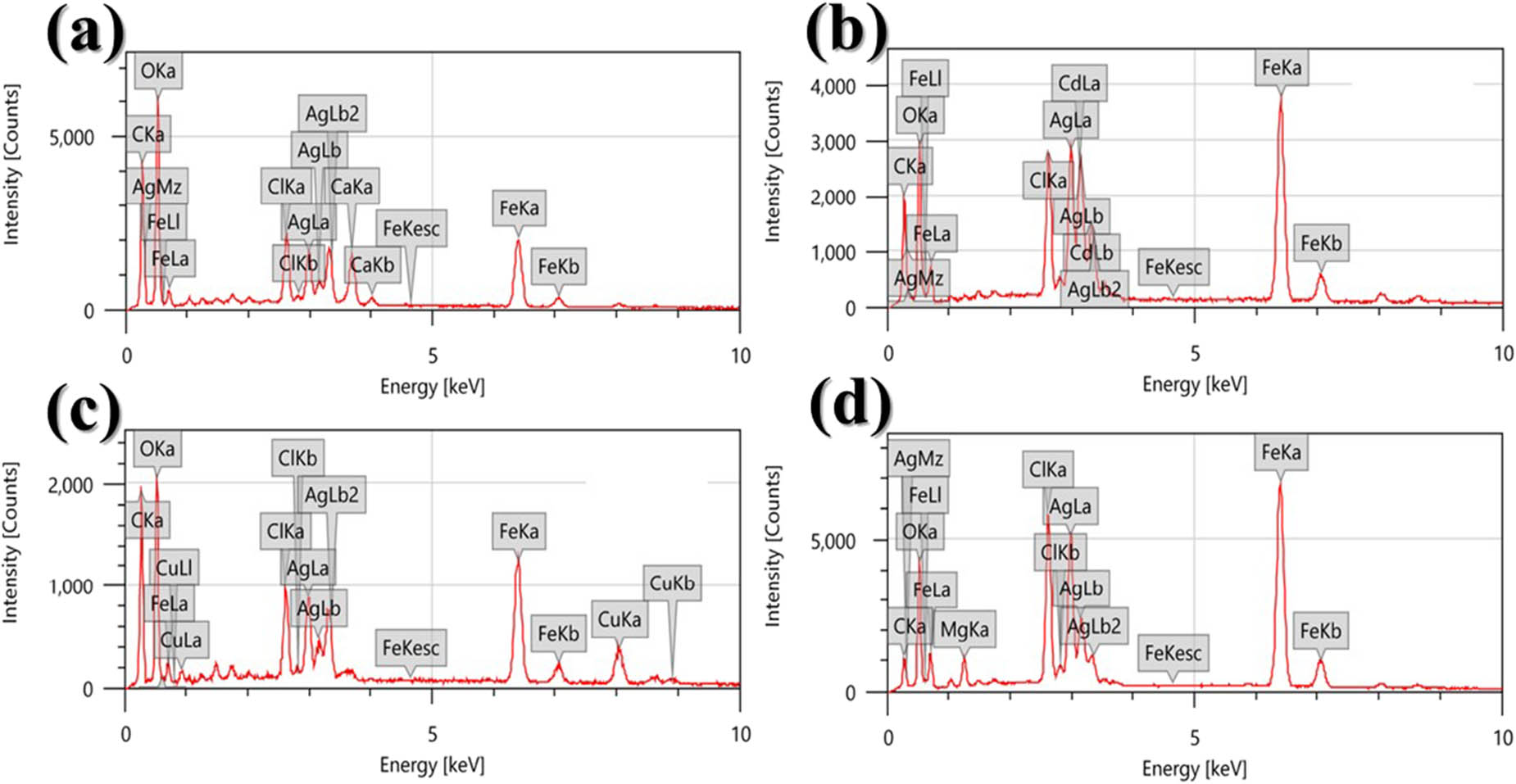
EDX spectra of (a) AgClCa0.5Fe2O4/curcumin, (b) AgClCd0.5Fe2O4/curcumin, (c) AgClCu0.5Fe2O4/curcumin, and (d) AgClMg0.5Fe2O4/curcumin hybrid nanocomposites.
Percentages of elemental composition in the AgClM0.5Fe2O4/curcumin hybrid nanocomposites
| Sample | Element | Mass (%) | Atomic (%) |
|---|---|---|---|
| AgClCa0.5Fe2O4/curcumin | C | 35.03 | 45.99 |
| O | 50.79 | 50.07 | |
| Cl | 2.190 | 0.970 | |
| Ca | 1.940 | 0.760 | |
| Fe | 5.350 | 1.510 | |
| Ag | 4.700 | 0.690 | |
| AgClCd0.5Fe2O4/curcumin | C | 25.80 | 42.51 |
| O | 38.17 | 47.23 | |
| Cl | 3.910 | 2.180 | |
| Cd | 13.12 | 4.650 | |
| Fe | 11.45 | 2.100 | |
| Ag | 7.550 | 1.330 | |
| AgClCu0.5Fe2O4/curcumin | C | 38.29 | 51.61 |
| O | 43.02 | 43.54 | |
| Cl | 2.160 | 0.980 | |
| Cu | 7.270 | 2.110 | |
| Fe | 3.480 | 0.890 | |
| Ag | 5.780 | 0.870 | |
| AgClMg0.5Fe2O4/curcumin | C | 15.50 | 29.28 |
| O | 36.87 | 52.27 | |
| Cl | 2.190 | 2.050 | |
| Mg | 6.960 | 4.450 | |
| Fe | 19.68 | 8.000 | |
| Ag | 18.80 | 3.950 |
TEM analysis provides unparalleled high-resolution imaging and detailed structural, chemical, and morphological information at the atomic scale, making it essential for advanced research in materials science, biology, and nanotechnology. Figure 4 shows the TEM micrographs of AgClCd0.5Fe2O4/curcumin, AgClMg0.5Fe2O4/curcumin, and their particle size distribution (nanocomposites with lower IC50 values). As shown in Figure 4a, the AgClCd0.5Fe2O4/curcumin nanocomposite has connected semi-spherical particles with an average diameter of 62.0 nm (Figure 4b). The TEM image of the AgClMg0.5Fe2O4/curcumin nanocomposite (Figure 4c) revealed agglomerate particles, semi-spherical in shape with an average NP diameter of 114 nm, as shown in Figure 4d. It was observed that the AgClCd0.5Fe2O4/curcumin nanocomposite, with an average particle size of 62.0 nm, was more aggregated than the AgClMg0.5Fe2O4/curcumin nanocomposite, which had an average particle size of 114 nm, which can be justified based on the well-established relationship between the NP size and aggregation behavior. Smaller NPs generally exhibit a significantly higher surface-to-volume ratio, leading to increased surface energy and stronger van der Waals forces, which drive the particles to aggregate to reduce the overall system energy [33,63]. The impact of the observed particle aggregation on the biological properties is investigated in the cytotoxicity study.
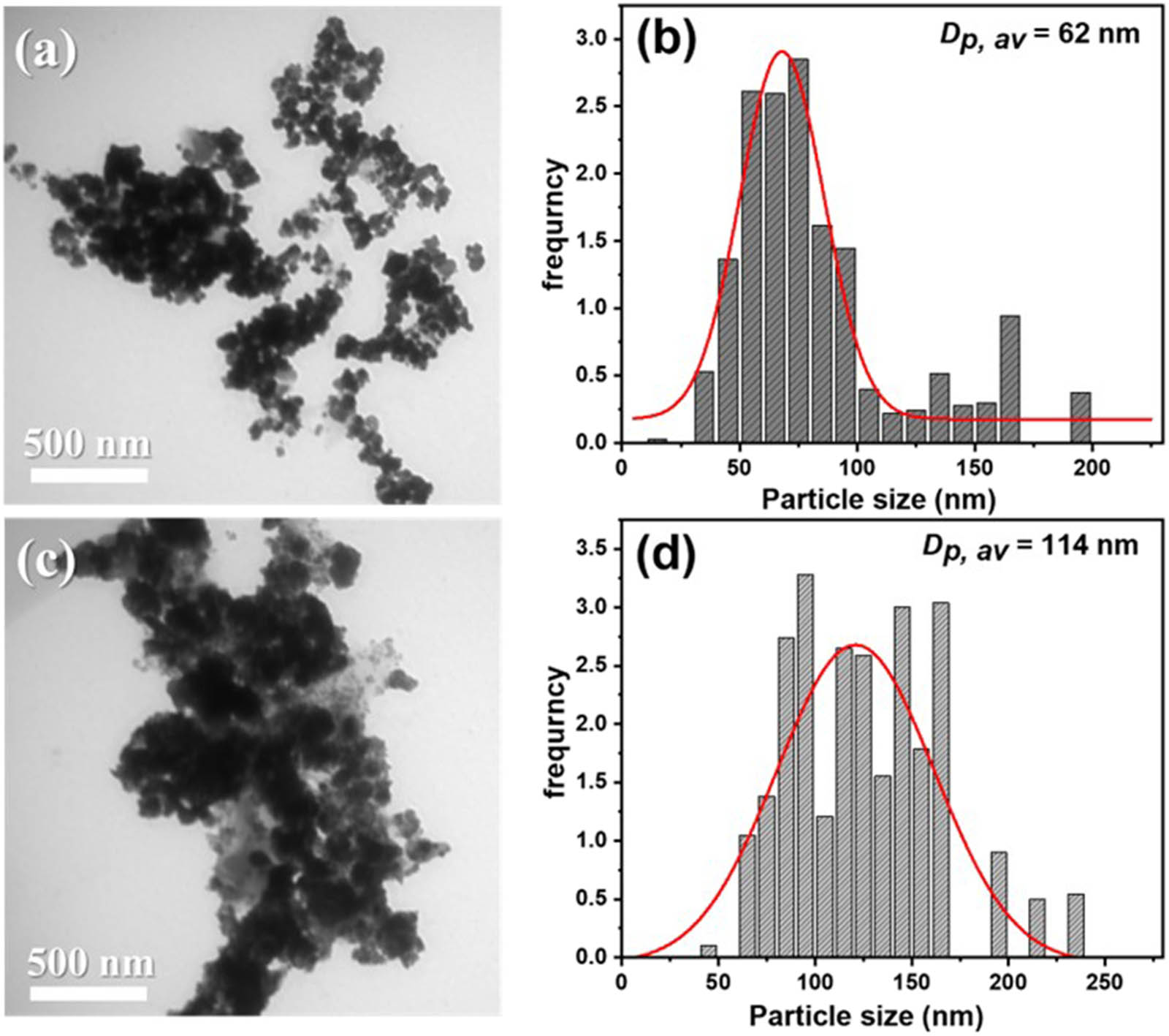
TEM images and particle size distribution curves of AgClCd0.5Fe2O4/curcumin (a) and (b) and AgClMg0.5 Fe2O4/curcumin (c) and (d) nanocomposites.
The stability of the AgClCd0.5Fe2O4/curcumin and AgClMg0.5Fe2O4/curcumin nanocomposites (hybrid nanocomposites with lower IC50 values) was assessed by measuring their zeta potential using Malvern Zetasizer nano ZEN 3600 (Malvern, LO, UK). The zeta potential, which indicates the stability of colloidal dispersions and reflects the surface charge of the nanocomposites, was found to be −28.1 ± 0.5 and −27.7 ± 0.9 mV for AgClCd0.5Fe2O4/curcumin and AgClMg0.5Fe2O4/curcumin nanocomposites, respectively, as shown in Figure 5. These values demonstrate the good physical stability of the synthesized curcumin hybrid nanocomposites. According to a published report, zeta potential values that are higher than +25 mV or less than −25 mV are favored because they provide a significant repulsion force between the NPs, which inhibits aggregation [64]. The AgClMg0.5Fe2O4/curcumin nanocomposite has a lower zeta potential negative value (−36.8 mV) than that obtained from pure MgFe2O4 [65].
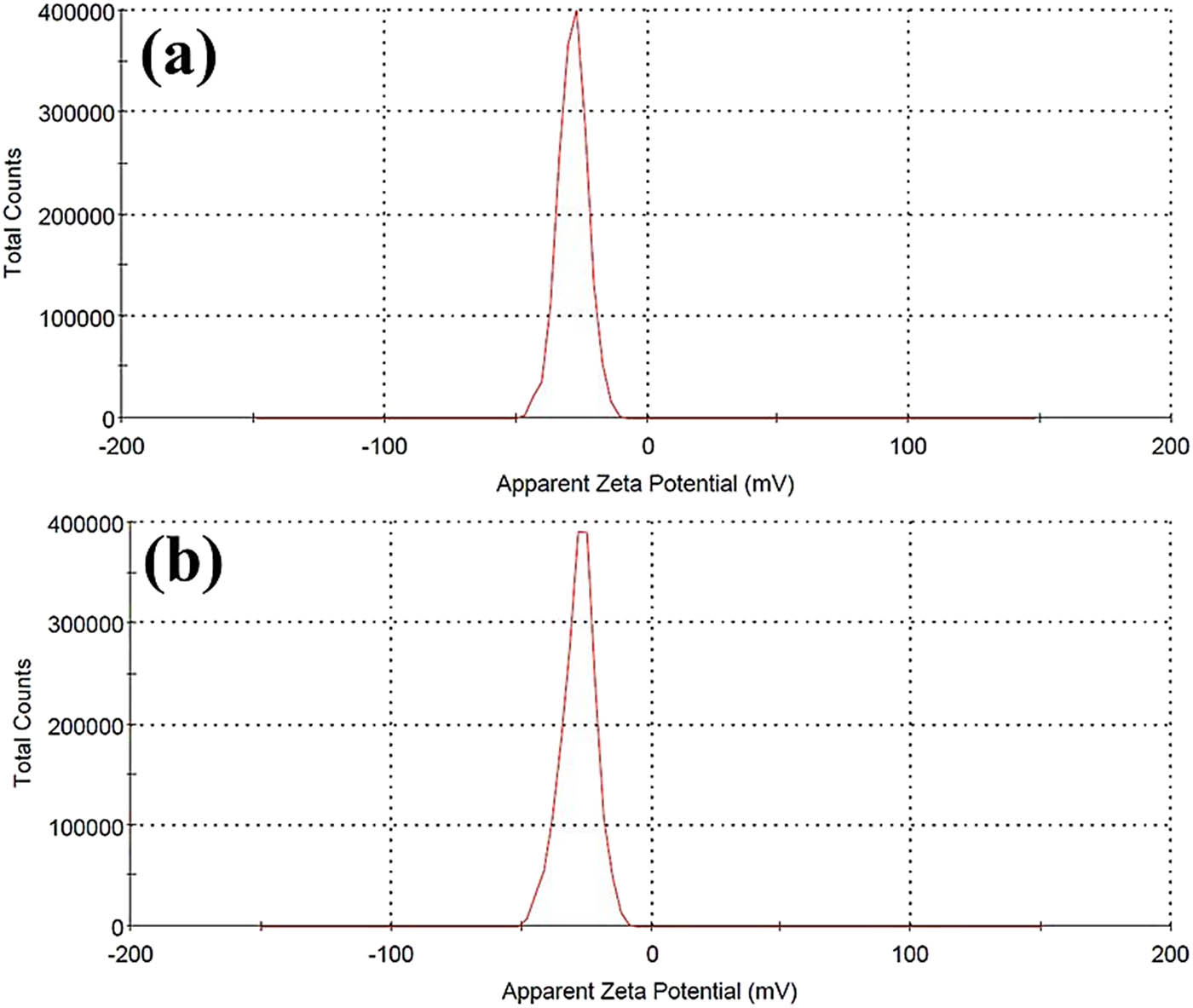
Zeta potential spectra of (a) AgClCd0.5Fe2O4/curcumin and (b) AgClMg0.5 Fe2O4/curcumin nanocomposites.
Figure 6 presents the hysteresis loop measured at room temperature for the prepared nanocomposites. The figure shows that the AgClCa0.5Fe2O4/curcumin and AgClCd0.5Fe2O4/curcumin nanocomposites exhibit a paramagnetic behavior because of the linear increase with the applied magnetic field. The AgClCu0.5Fe2O4/curcumin and AgClMg0.5Fe2O4/curcumin nanocomposites showed hysteresis loops with S-shaped response without a clear hysteresis loop, indicating superparamagnetic behavior. The obtained values, including saturation magnetization (Mₛ), coercivity (Hₒ), retentivity (Mᵣ), and the squareness ratio (M r/M s) of the synthesized hybrid nanocomposites, are calculated and listed in Table 2. The saturation magnetization according to an analysis of the data that was obtained follows the order curcumin AgClMg0.5Fe2O4 > AgClCu0.5Fe2O4 > AgClCd0.5Fe2O4 > AgClCa0.5Fe2O4/curcumin. Differences in the magnetic moment, ionic radius, super exchange contact strength, and structural stability decrease the saturation magnetization in the latter order. According to reports, a single-domain magnetic structure is indicated by a squareness ratio (Mᵣ/Mₛ) of ≥0.5, whereas the construction of a multidomain structure is suggested by a ratio of <0.5 [66]. All of the prepared nanocomposites have a multidomain structure, as shown in Table 2, and their squareness ratios ranged from 0.0061 to 0.1557. Owing to the observed magnetic properties of these materials, they are used to improve contrast for magnetic resonance imaging (MRI) and X-ray computed tomography (CT) imaging, which are both useful for detecting cancer cells. Additionally, they offer a powerful platform for cancer treatment guided by multimodal imaging [67].
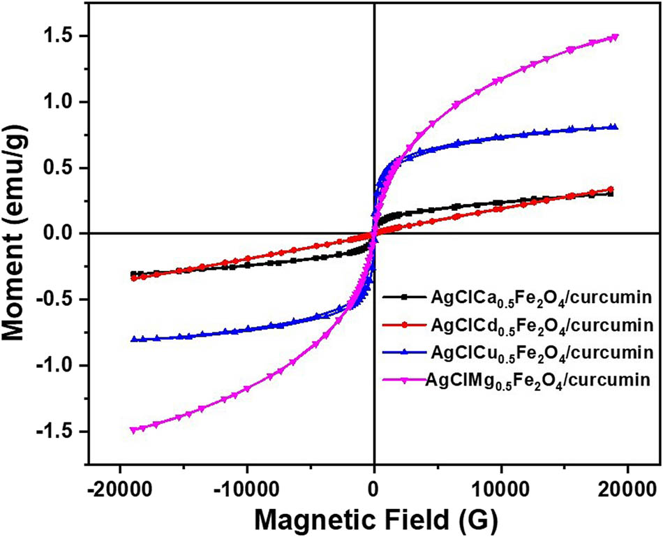
VSM spectra of the prepared nanocomposites.
Magnetic parameters, including saturation magnetization (M s in emu/g), coercivity (H c in G), remanent magnetization (M r in emu/g), and squareness ratio (M r/M s), of the prepared nanocomposites
| Samples | Magnetization Mₛ (emu/g) | Coercivity H c (G) | Retentivity Mᵣ (emu/g) | Squareness ratio (M r/M s) |
|---|---|---|---|---|
| AgClCa0.5Fe2O4/curcumin | 0.30482 | 6.6082 | 0.0025 | 0.0081 |
| AgClCd0.5Fe2O4/curcumin | 0.33774 | 52.030 | 0.00205 | 0.0061 |
| AgClCu0.5Fe2O4/curcumin | 0.80560 | 113.51 | 0.12548 | 0.1557 |
| AgClMg0.5Fe2O4/curcumin | 1.4909 | 24.799 | 0.01467 | 0.0098 |
3.2 Cytotoxicity study
The anticancer potential of the prepared spinal ferrite/curcumin hybrid nanocomposite systems was evaluated in normal and cancer skin cell lines, HSF and A-431, compared to free curcumin and Dox as a positive control anti-tumor. The study aimed to assess the IC50 values of the prepared curcumin hybrid nanocomposite and Dox on HSF and A-431 cell lines. Concentration-dependent plots for each of the cell lines, along with the respective IC50 values and statistical tests, are provided in the Supporting Information. These additional details provide a complete analysis of the nanocomposites’ cytotoxic effects (Figures S1 and S2). The data revealed a significant, dose-dependent reduction in the viability of A-431 cells upon treatment with the nanocomposites, while HSF cells exhibit minimal sensitivity under similar conditions. This differential cytotoxicity underscores the selective efficacy of the prepared hybrid curcumin nanocomposites against cancerous cells. The IC50 value, which is the concentration of a substance necessary to inhibit 50% of cell growth, is an important metric. Table 3 and Figure 7 present the IC50 values of the prepared compounds. Among the metal hybrid nanocomposites, AgClMg0.5Fe2O4/curcumin and AgClCd0.5Fe2O4/curcumin exhibited remarkable anticancer potential against the A-431 cell line, with IC50 values of 34.83 and 26.83 µg/ml, respectively. In contrast, AgClCu0.5Fe2O4/curcumin and AgClCa0.5Fe2O4/curcumin showed reduced efficacy, with IC50 values exceeding 100 µg/ml for the A-431 cell line. Particularly, the free curcumin at the tested concentrations revealed minimal anticancer activity and toxicity, with IC50 values exceeding 100 µg/ml for both cell line types.
Anticancer potential presented as IC50 (µg/ml) values of doxorubicin, curcumin, and the prepared doped ferrite/curcumin hybrid nanocomposite systems using HSF, human skin fibroblast, and A-431, human epidermoid skin carcinoma (skin/epidermis) cell lines
| Compound | IC50 (µg/ml) | |
|---|---|---|
| A-431 | HSF | |
| AgClCu0.5Fe2O4/curcumin | >100 | >100 |
| AgClCa0.5Fe2O4/curcumin | >100 | >100 |
| AgClMg0.5Fe2O4/curcumin | 34.83 | >100 |
| AgClCd0.5Fe2O4/curcumin | 26.83 | >100 |
| Curcumin | >100 | >100 |
| Dox | 0.15 | 2.85 |
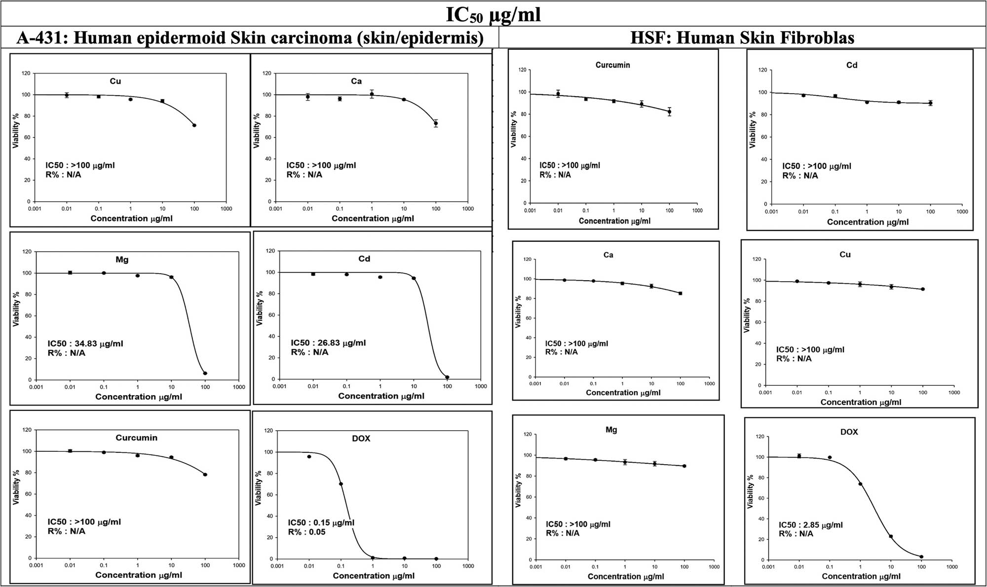
Percentage cell viability of Dox, curcumin, and the prepared doped ferrite/curcumin hybrid nanocomposites on HSF: Human skin fibroblast versus A-431: Human epidermoid skin carcinoma. Cu refers to (AgClCu0.5Fe2O4/curcumin), Mg (AgClMg0.5Fe2O4/curcumin), Ca (AgClCa0.5Fe2O4/curcumin), Cd (AgClCd0.5Fe2O4/curcumin), while Dox (doxorubicin) is used as a positive control.
Dox, a well-known anticancer medication, demonstrated a strong cytotoxic effect against the A-431 cell line (IC50 = 0.15 µg/ml). In addition, it also showed higher activity against the HSF cell line, with an IC50 value of 2.85 µg/ml, which proved their toxicity on normal cells at very low concentrations. These findings demonstrated the distinct sensitivity of malignant and non-cancerous skin cells to Dox, underlining the importance of tailored treatment options to reduce the negative effects on healthy cells [39]. The toxicity of Dox in the normal HSF cell line has also been reported previously [68,69]. Interestingly, all of the investigated nanocomposites showed a safe pattern on normal skin fibroblasts, as they showed IC50 values of >100 µg/ml, hence a better biocompatibility. Although TEM images delineated an observed particle aggregation, especially with AgClCd0.5Fe2O4/curcumin nanocomposite, than that of AgClMg0.5Fe2O4/curcumin nanocomposite, they have a small particle size of about 62.0 nm, which does not affect the biological behavior as depicted from the observed lower IC50. Many studies have shown that the NPs between 50 and 100 nm have high cellular internalization and absorption, as previously investigated [70].
The comparison of IC50 values between A-431 and HSF cells reveals important information about the selectivity and potential toxicity of the tested hybrid nanocomposites. The large variance in IC50 values for the investigated materials emphasizes the necessity of creating targeted medicines that specifically target cancer cells while causing minimal harm to healthy organs [71].
3.3 Cell apoptosis and cell cycle
The mechanisms underlying the potential toxicity of the prepared materials could be investigated by studying cell apoptosis and cell cycle, utilizing the flow cytometry assay.
3.4 Cell apoptosis
Results indicated notable differences in the cytotoxic effects of Dox and the nanocomposite hybrid treatments (AgClMg0.5Fe2O4/curcumin and AgClCd0.5Fe2O4/curcumin) in comparison to control untreated cells (CT). In the untreated control (CT) group, a high proportion of cells remained viable (95.61%), exhibiting minimal signs of apoptosis or necrosis, consistent with expectations for untreated cells. The low percentages of cells in early apoptosis (0.97%) and late apoptosis (3.06%) suggest a minimal induction of cell death under normal conditions. Our study demonstrated that AgClMg0.5Fe2O4/curcumin and AgClCd0.5Fe2O4/curcumin induce apoptosis and cause cycle arrest in A-431 cells. These effects are likely mediated through mechanisms like those observed with curcumin and its derivatives in various cancer models [72].
The administration of Dox showed a significant increase in necrosis (53.15%, P < 0.001), consistent with the compound’s strong cytotoxic effects, which cause considerable cellular damage and membrane disruption. Furthermore, a significant increase in late apoptosis (34.92%, P < 0.001) indicated that Dox effectively activates apoptotic pathways, presumably via DNA intercalation and topoisomerase II inhibition [73]. The diminished live cell population (11.56%) further corroborates the significant cytotoxicity of Dox. The initial apoptosis rate (0.37%, P < 0.001) was comparatively low, suggesting that cells predominantly advanced to late-stage apoptosis or necrosis following Dox treatment rather than undergoing an earlier apoptotic phase. AgClCd0.5Fe2O4/curcumin exhibited a moderate cytotoxic effect, with necrosis at 8.53% and late apoptosis at 14.52%, both significantly elevated in comparison to the untreated control cells (P < 0.001). This suggests that AgClCd0.5Fe2O4/curcumin can induce significant apoptosis, likely resulting from the combined effects of cadmium and curcumin in promoting oxidative stress and DNA damage [74]. The live cell population (76.19%, P < 0.001) was significantly higher than that of Dox, indicating that AgClCd0.5Fe2O4/curcumin exhibited lower overall toxicity, although they remain considerably more cytotoxic than the control group. Early apoptosis (0.75%) was marginally elevated compared to the control, indicating the commencement of apoptotic pathways.
AgClMg0.5Fe2O4/curcumin exhibited cytotoxicity, albeit to a lesser extent than AgClCd0.5Fe2O4/curcumin. The necrosis rate (6.07%) and late apoptosis (10.20%) were lower than those observed with AgClCd0.5Fe2O4/curcumin, yet remained significantly (P < 0.001) higher than the control group. The live cell percentage (83.03%) exceeded that of both Dox and AgClCd0.5Fe2O4/curcumin, indicating that AgClMg0.5Fe2O4/curcumin may exhibit lower toxicity, possibly attributable to variations in NP characteristics and cellular uptake mechanisms [75]. The early apoptosis rate of 0.70% was comparable to that observed with AgClCd0.5Fe2O4/curcumin, suggesting some activation of apoptosis pathways at an early stage, albeit to a lesser extent [76].
The results indicated that Dox produces the highest cytotoxicity, characterized by a substantial degree of necrosis and late apoptosis, which is in accordance with the cytotoxicity study. Both AgClCd0.5Fe2O4/curcumin and AgClMg0.5Fe2O4/curcumin exhibited promising apoptosis-inducing effects; however, at a lower level than Dox, with AgClCd0.5Fe2O4/curcumin demonstrating the highest potency among the two treatments. The relatively mild effect of AgClMg0.5Fe2O4/curcumin may indicate its potential for lower toxicity while still achieving effective cell death, suggesting it as a promising nanomaterial for further investigation in cancer therapies (Figure 8) [75].

Percentage of cells (mean ± SD) of living, early, and late apoptotic, along with dead cells, in non-treated control cells CT (a), doxorubicin Dox (b), AgClCd0.5Fe2O4/curcumin (c), AgClMg0.5Fe2O4/curcumin (d) treated A-431: Human epidermoid skin carcinoma for 48 h. The percentage of the cell of different living cells (e), early apoptosis (f), late apoptosis (g), and dead cells (h) (n = 3 biological replicates/group). Data were analyzed by one-way ANOVA with Tukey’s HSD post-hoc test; *P < 0.05, **P < 0.01, and ***P < 0.001 indicate the significant difference compared with CT, and #iP < 0.05, ## P < 0.01, and ### P < 0.001 indicate the significant difference compared with Dox.
3.5 Cell cycle
The sub-G1 phase, indicative of apoptosis through fragmented DNA, revealed a significant increase in apoptosis with treatments. Untreated cells exhibited baseline apoptosis (3.55%), while Dox treatment increased significantly (7.7-fold, P < 0.001), reflecting its cytotoxic effects via DNA intercalation and topoisomerase II inhibition. AgClCd0.5Fe2O4/curcumin showed the highest apoptosis (10-fold of control, P < 0.001), surpassing Dox, likely due to synergistic oxidative stress and DNA damage from cadmium and curcumin. AgClMg0.5Fe2O4/curcumin induced moderate apoptosis (6.2-fold of control, P < 0.001), lower than AgClCd0.5Fe2O4/curcumin but at the same time higher than Dox, suggesting reduced toxicity. In the G0/G1 phase, Dox caused a significant reduction (24.07%), consistent with its known efficacy to perform cell cycle arrest in S and G2/M phases. AgClCd0.5Fe2O4/curcumin induced a moderate decrease in G0/G1 (2-fold of Dox, P < 0.001), while AgClMg0.5Fe2O4/curcumin had a smaller reduction (2.02-fold of Dox, P < 0.001), suggesting partial cell cycle arrest. The S-phase analysis showed that Dox significantly reduced S-phase cells (7.13%), while AgClCd0.5Fe2O4/curcumin had little effect (13.03%), and AgClMg0.5Fe2O4/curcumin increased S-phase population (16.66%), indicating enhanced proliferation or delayed progression. Dox induced significant G1/M arrest (41.61%), while AgClCd0.5Fe2O4/curcumin and AgClMg0.5Fe2O4/curcumin caused mild-to-moderate G1/M arrest, with AgClMg0.5Fe2O4/curcumin exhibiting a slightly stronger effect, suggesting differences in bioactivity or uptake (Figure 9). These results are in agreement with the previous finding that the inhibition of A-431 cells in the G2/M phase of the cell cycle after treatment with curcumin. The increase in the number of cells in the G2/M phase corresponded with a decrease in the number of cells in the G0/G1 phase. The impact of curcumin on cell cycle progression has been previously reported in different types of cell lines [77,78]. Incorporation of Cd2+ and Mg2+ into curcumin in the prepared nanocomposites can synergistically improve the cell cycle arrest of curcumin [75]. Curcumin has been shown to induce apoptosis, possibly by causing the production of ROS, disruption of mitochondrial membrane potential, release of cytochrome c, and activation of caspase cascades. Curcumin also affects important signaling pathways, including the phosphoinositide 3-kinase/protein kinase B (PI3K/AKT) pathway, causing cell cycle arrest at the G2/M phase and apoptosis [79]. Hence, it is possible that the prepared curcumin hybrid nanocomposites could exert their cytotoxic effects through the same mechanisms. In addition, the presence of metal ions such as Mg2+ and Cd2+ may enhance these effects by regulating ROS production and apoptotic pathways. However, we are aware that extensive mechanistic studies should be pursued to elucidate the specific pathways mediating our nanocomposite cytotoxicity. Future research will focus on interrogating these mechanisms for a complete understanding of their anti-cancer potential.
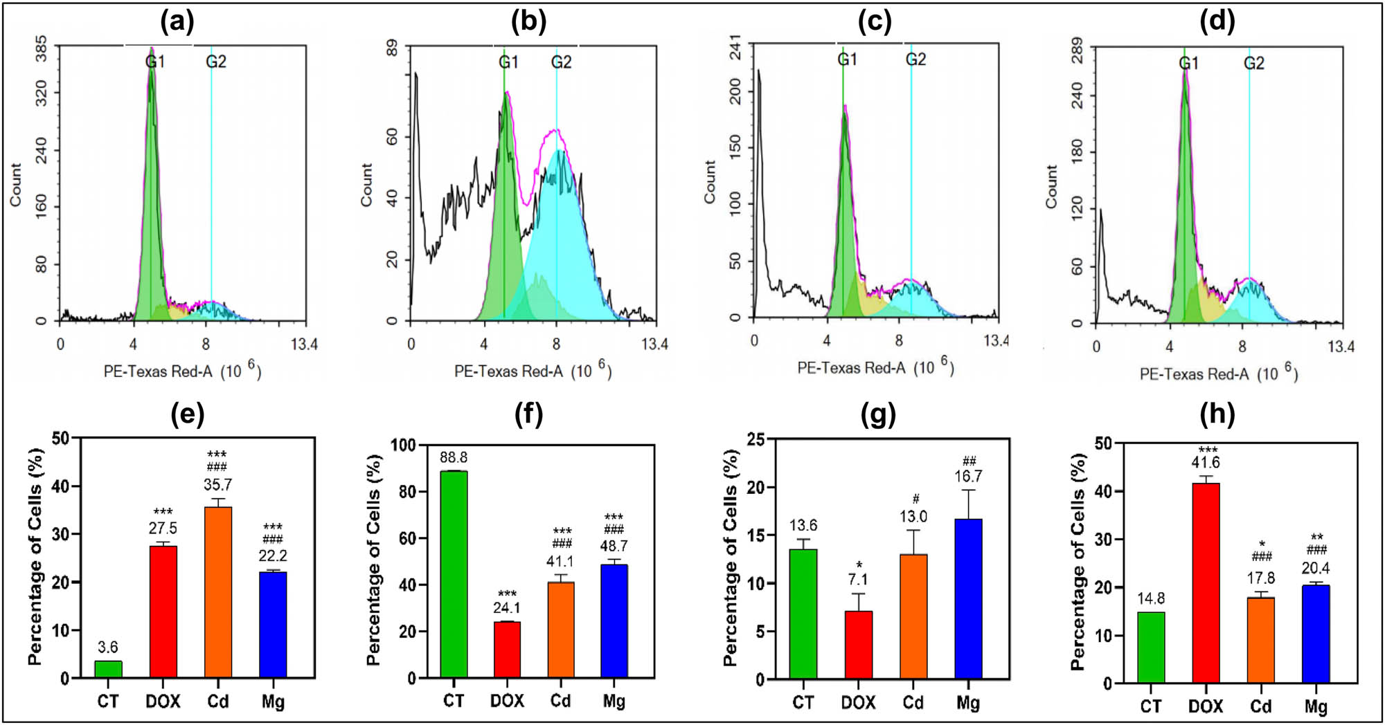
Percentage of cells (mean ± SD) in the different phases of the cell cycle in non-treated control cells CT (a), doxorubicin Dox (b), AgClCd0.5Fe2O4/curcumin (c), AgClMg0.5Fe2O4/curcumin (d), treated A-431: Human epidermoid skin carcinoma for 48 h. The percentage of the cell of different cell cycle sub-G1 (e), G0/G1 phase (f), S phase (g), and G2/M phase (h) (n = 3 biological replicates/group). Data were analyzed by one-way ANOVA with Tukey’s HSD post-hoc test; *P < 0.05, **P < 0.01, and ***P < 0.001 indicate the significant difference compared with CT, and # P < 0.05, ##iP < 0.01, and ### P < 0.001 indicate the significant difference compared with Dox.
The cell cycle and apoptosis data collectively highlight the differential cytotoxic effects of Dox, AgClCd0.5Fe2O4/curcumin, and AgClMg0.5Fe2O4/curcumin on the investigated cancer cells. Dox treatment caused significant apoptosis, with a marked increase in necrosis and late apoptosis, consistent with its established mechanism of inducing DNA damage and cell cycle arrest [80]. This resulted in a dramatic reduction in live cell populations, primarily through G1/S arrest and subsequent apoptosis. Both Cd and Mg-curcumin nanocomposites induced apoptosis as well, with AgClCd0.5Fe2O4/curcumin showing the most potent cytotoxic effects, surpassing Dox in late apoptosis and necrosis. This suggests that the combination of cadmium and curcumin in hybrid NPs may enhance the apoptotic response through synergistic oxidative stress and DNA damage. AgClMg0.5Fe2O4/curcumin exhibited a less pronounced cytotoxic effect, though still inducing apoptosis and cell cycle arrest, particularly in the S and G2/M phases. The lower overall toxicity of AgClMg0.5Fe2O4/curcumin compared to AgClCd0.5Fe2O4/curcumin nanocomposites suggests that they may offer a more controlled therapeutic option, with fewer adverse effects.
There are certain limitations of our study, such as the exclusive use of only two cell lines as skin models, which may not be capable of exhaustively exploring the variety of skin cancers. Future studies should be conducted to encompass different skin cancer models for enhancing the generalizability of the work conducted so far. Additionally, detailed mechanistic studies are required to determine the specific pathways through which oxidative stress and DNA damage caused the observed apoptotic effects of the prepared nanocomposites. Even though earlier reports have indicated possible tumor imaging applications of such nanocomposites with MRI and CT, it is vital to perform certain imaging studies to empirically verify such effects as well as determine their clinical implications.
4 Conclusion
The co-precipitation and wet impregnation techniques were used successfully to synthesize a hybrid curcumin metal ferrite nanocomposite system with certain transition metals, AgClM0.5Fe2O4 (M = Ca, Cd, Cu, Mg). XRD, FT-IR spectroscopy, and TEM confirmed the successful formation of the hybrid curcumin nanocomposites with varying degrees of crystallinity. The elemental composition of the prepared hybrid nanocomposites was analyzed using the EDX technique, and confirmed the presence of key elements. In addition, they demonstrated excellent magnetic properties, allowing them to be useful materials for tumor diagnosis using, e.g., MRI and X-ray CT imaging. AgClCd0.5Fe2O4/curcumin and AgClMg0.5Fe2O4/curcumin nanocomposites have particle sizes of 62.0 and 114.0 nm, respectively, and high zeta potential values, reflecting good physical stability. Furthermore, they demonstrated outstanding anticancer activity against A-431 skin cancer cell lines, with minimal cytotoxicity toward normal HSF cell lines in comparison to Dox, indicating a higher selectivity and biocompatibility. Flow cytometry findings demonstrated that while Dox remains the most effective in terms of apoptosis induction, AgClMg0.5Fe2O4/curcumin and AgClCd0.5Fe2O4/curcumin nanocomposites showed promising therapeutic potential, especially in terms of selectively targeting cancer cells with reduced toxicity, particularly AgClMg0.5Fe2O4/curcumin. The combination of apoptotic induction and cell cycle modulation highlights the potential of these materials for skin cancer treatment strategies. Finally, the findings in this research highlight the potential of curcumin-based hybrid nanocomposite metal ferrites as promising agents for skin cancer therapy. However, the molecular mechanisms of action should be investigated in future work to pave the way for clinical applications.
Acknowledgments
The authors would like to extend their sincere appreciation to the Ongoing Research Funding program (ORF-2025-457), King Saud University, Riyadh, Saudi Arabia, for funding this research work.
-
Funding information: This research work was funded by the Ongoing Research Funding program (ORF-2025-457), King Saud University, Riyadh, Saudi Arabia.
-
Author contributions: Yousef A. Bin Jardan: conceptualization, methodology, software, writing – original draft, and writing – review and editing. Ahmed A. H. Abdellatif: conceptualization, methodology, software, validation, writing – original draft, and writing – review and editing. Mahmoud Zaki El-Readi: conceptualization, methodology, software, and writing – review and editing. Mohamed Abdel El-Aal: conceptualization, methodology, writing – original draft, software, validation, and writing – review and editing. Hesham M. Tawfeek: conceptualization, methodology, writing – original draft, software, validation, and writing – review and editing. All authors have accepted responsibility for the entire content of this manuscript and approved its submission.
-
Conflict of interest: The authors state no conflict of interest.
-
Data availability statement: All data generated or analyzed during this study are included in this published article.
References
[1] Wu Z, Xia F, Lin R. Global burden of cancer and associated risk factors in 204 countries and territories, 1980–2021: A systematic analysis for the GBD 2021. J Hematol Oncol. 2024;17(1):119.10.1186/s13045-024-01640-8Suche in Google Scholar PubMed PubMed Central
[2] Society AC. American cancer society releases latest global cancer statistics; Cancer cases expected to rise to 35 million worldwide by 2050; 2024. https://pressroomcancerorg/GlobalCancerStatistics2024.Suche in Google Scholar
[3] Wang M, Gao X, Zhang L. Recent global patterns in skin cancer incidence, mortality, and prevalence. Chin Med J (Engl). 2025;138(2):185–92.10.1097/CM9.0000000000003416Suche in Google Scholar PubMed PubMed Central
[4] Siegel RL, Miller KD, Fuchs HE, Jemal A. Cancer statistics, 2022. CA Cancer J Clin. 2022;72(1):7–33.10.3322/caac.21708Suche in Google Scholar PubMed
[5] Rasheed N. Melanoma awareness programs and their impact on the life of Australian Queenslanders: A concise analysis. Int J Health Sci. 2024;18(1):1.Suche in Google Scholar
[6] Roy A, Basak NP, Banerjee S. Notch1 intracellular domain increases cytoplasmic EZH2 levels during early megakaryopoiesis. Cell Death Dis. 2012;3(8):e380.10.1038/cddis.2012.119Suche in Google Scholar PubMed PubMed Central
[7] Harris AL. Hypoxia–A key regulatory factor in tumour growth. Nat Rev Cancer. 2002;2(1):38–47.10.1038/nrc704Suche in Google Scholar PubMed
[8] Gerson SL. MGMT: Its role in cancer aetiology and cancer therapeutics. Nat Rev Cancer. 2004;4(4):296–307.10.1038/nrc1319Suche in Google Scholar PubMed
[9] Dostie B, Kromann L, Sørensen A. Overcoming obstacles to innovation: Can an educated workforce help? J Innov & Knowl. 2025;10(3):100707.10.1016/j.jik.2025.100707Suche in Google Scholar
[10] Alahmari F, Khan FA, Sozeri H, Sertkol M, Jaremko M. Electrospun Cu–Co ferrite nanofibers: synthesis, structure, optical and magnetic properties, and anti-cancer activity. RSC Adv. 2024;14(11):7540–50.10.1039/D3RA08087KSuche in Google Scholar
[11] Rafie SF, Abu-Zahra N, Sillanpää M. A comprehensive review of spinel ferrites and their magnetic composites as highly efficient adsorbents of rare earth elements. Emerg Contam. 2025;11(1):100429.10.1016/j.emcon.2024.100429Suche in Google Scholar
[12] Kefeni KK, Msagati TAM, Nkambule TT, Mamba BB. Spinel ferrite nanoparticles and nanocomposites for biomedical applications and their toxicity. Mater Sci Eng C Mater Biol Appl. 2020;107:110314.10.1016/j.msec.2019.110314Suche in Google Scholar PubMed
[13] Mokhosi SR, Mdlalose W, Nhlapo A, Singh M. Advances in the synthesis and application of magnetic ferrite nanoparticles for cancer therapy. Pharmaceutics. 2022;14(5):937.10.3390/pharmaceutics14050937Suche in Google Scholar PubMed PubMed Central
[14] Sultana N, Ruhul-Amin M, Hasan I, Kabir SR, Asaduzzaman AKM. Antibacterial, antioxidant, and anticancer effects of green synthesized silver/silver chloride nanoparticles using Spondias pinnata bark extract. Food Chem Adv. 2024;4:100709.10.1016/j.focha.2024.100709Suche in Google Scholar
[15] Abdellatif AAH, Rasheed Z, Alhowail AH, Alqasoumi A, Alsharidah M, Khan RA, et al. Silver citrate nanoparticles inhibit PMA-induced TNFalpha expression via deactivation of NF-kappaB activity in human cancer cell-lines, MCF-7. Int J Nanomed. 2020;15:8479–93.10.2147/IJN.S274098Suche in Google Scholar PubMed PubMed Central
[16] Al-Subaiyel A, Abdellatif AAH. Eco-friendly synthesis of silver nanoparticles by Trigonella foenum-graecum: formulations, characterizations, and application in wound healing. Drug Dev Ind Pharm. 2024;50(11):927–37.10.1080/03639045.2024.2431934Suche in Google Scholar PubMed
[17] Abdellatif AAH, Mostafa MAH, Konno H, Younis MA. Exploring the green synthesis of silver nanoparticles using natural extracts and their potential for cancer treatment. 3 Biotech. 2024;14(11):274.10.1007/s13205-024-04118-zSuche in Google Scholar PubMed PubMed Central
[18] Abdellatif AAH, Alsharidah M, Al Rugaie O, Tawfeek HM, Tolba NS. Silver nanoparticle-coated ethyl cellulose inhibits tumor necrosis factor-alpha of breast cancer cells. Drug Des Devel Ther. 2021;15:2035–46.10.2147/DDDT.S310760Suche in Google Scholar PubMed PubMed Central
[19] Kabir SR, Islam F, Asaduzzaman AKM. Biogenic silver/silver chloride nanoparticles inhibit human cancer cells proliferation in vitro and Ehrlich ascites carcinoma cells growth in vivo. Sci Rep. 2022;12(1):8909.10.1038/s41598-022-12974-zSuche in Google Scholar PubMed PubMed Central
[20] Kabir SR, Dai Z, Nurujjaman M, Cui X, Asaduzzaman AKM, Sun B, et al. Biogenic silver/silver chloride nanoparticles inhibit human glioblastoma stem cells growth in vitro and Ehrlich ascites carcinoma cell growth in vivo. J Cell Mol Med. 2020;24(22):13223–34.10.1111/jcmm.15934Suche in Google Scholar PubMed PubMed Central
[21] Abdellatif AAH, Alturki HNH, Tawfeek HM. Different cellulosic polymers for synthesizing silver nanoparticles with antioxidant and antibacterial activities. Sci Rep. 2021;11(1):84.10.1038/s41598-020-79834-6Suche in Google Scholar PubMed PubMed Central
[22] Mousa AM, Alhumaydhi FA, Abdellatif AAH, Abdulmonem WA, AlKhowailed MS, Alsagaby SA, et al. Curcumin and ustekinumab cotherapy alleviates induced psoriasis in rats through their antioxidant, anti-inflammatory, and antiproliferative effects. Cutan Ocul Toxicol. 2022;41(1):33–42.10.1080/15569527.2021.2003377Suche in Google Scholar PubMed
[23] Ratan C, Arian AM, Rajendran R, Jayakumar R, Masson M, Mangalathillam S. Nano-based formulations of curcumin: Elucidating the potential benefits and future prospects in skin cancer. Biomed Mater. 2023;18(5):052008.10.1088/1748-605X/acf0afSuche in Google Scholar PubMed
[24] Prasad S, Tyagi AK, Aggarwal BB. Recent developments in delivery, bioavailability, absorption and metabolism of curcumin: the golden pigment from golden spice. Cancer Res Treatment: Off J Korean Cancer Assoc. 2014;46(1):2–18.10.4143/crt.2014.46.1.2Suche in Google Scholar PubMed PubMed Central
[25] Burgos‐Morón E, Calderón‐Montaño JM, Salvador J, Robles A, López‐Lázaro M. The dark side of curcumin. Int J Cancer. 2010;126(7):1771–5.10.1002/ijc.24967Suche in Google Scholar PubMed
[26] Teiten MH, Dicato M, Diederich M. Hybrid curcumin compounds: A new strategy for cancer treatment. Molecules. 2014;19(12):20839–63.10.3390/molecules191220839Suche in Google Scholar PubMed PubMed Central
[27] Salehiabar M, Ghaffarlou M, Mohammadi A, Mousazadeh N, Rahimi H, Abhari F, et al. Targeted CuFe2O4 hybrid nanoradiosensitizers for synchronous chemoradiotherapy. J Control Rel. 2023;353:850–63.10.1016/j.jconrel.2022.12.004Suche in Google Scholar PubMed
[28] Du H, Akakuru OU, Yao C, Yang F, Wu A. Transition metal ion-doped ferrites nanoparticles for bioimaging and cancer therapy. Transl Oncol. 2022;15(1):101264.10.1016/j.tranon.2021.101264Suche in Google Scholar PubMed PubMed Central
[29] Ji P, Wang P, Chen H, Xu Y, Ge J, Tian Z, et al. Potential of copper and copper compounds for anticancer applications. Pharm (Basel). 2023;16(2):234.10.3390/ph16020234Suche in Google Scholar PubMed PubMed Central
[30] Elbeltagi S, Abdel Shakor AB, M. Alharbi H, Tawfeek HM, Aldosari BN, E. Eldin Z, et al. Synergistic effects of quercetin-loaded CoFe(2)O(4)@Liposomes regulate DNA damage and apoptosis in MCF-7 cancer cells: based on biophysical magnetic hyperthermia. Drug Dev Ind Pharm. 2024;50(6):561–75.10.1080/03639045.2024.2363231Suche in Google Scholar PubMed
[31] Elbeltagi S, Alfassam HE, Saeedi AM, Eldin ZE, Ibrahim EMM, Abdel Shakor Ab, et al. A novel quercetin-loaded NiFe2O4@Liposomes hybrid biocompatible as a potential chemotherapy/hyperthermia agent and cytotoxic effects on breast cancer cells. J Drug Delivery Sci Technol. 2024;91:105203.10.1016/j.jddst.2023.105203Suche in Google Scholar
[32] Elbeltagi S, Saeedi AM, Eldin ZE, Alfassam HE, Alharbi HM, Madkhali N, et al. Biosynthesis, characterization, magnetic hyperthermia, and in vitro toxicity evaluation of quercetin-loaded magnetoliposome lipid bilayer hybrid system on MCF-7 breast cancer. Biochim Biophys Acta (BBA) - Gen Subj. 2024;1868(3):130543.Suche in Google Scholar
[33] Essa RA, El-Aal MA, Sedky A, Abo Zeid EF, Amin S. ZnO NPs-modified biochar derived from banana peels for adsorptive removal of methylene blue from water. J Mol Struct. 2025;1321:139821.10.1016/j.molstruc.2024.139821Suche in Google Scholar
[34] Sadan M, Naem M, Tawfeek HM, Khodier MM, Zeitoun MM, El-Khodery S, et al. Can silver nanoparticles stabilized by Fenugreek (Trigonella foenm -graecum) improve tibial bone defects repair in rabbits? A preliminary study. Open Vet J. 2024;14(5):1281–93.10.5455/OVJ.2024.v14.i5.23Suche in Google Scholar PubMed PubMed Central
[35] Ashique S, Afzal O, Hussain A, Zeyaullah M, Altamimi MA, Mishra N, et al. It’s all about plant derived natural phytoconstituents and phytonanomedicine to control skin cancer. J Drug Delivery Sci Technol. 2023;84:104495.10.1016/j.jddst.2023.104495Suche in Google Scholar
[36] Allam RM, Al-Abd AM, Khedr A, Sharaf OA, Nofal SM, Khalifa AE, et al. Fingolimod interrupts the cross talk between estrogen metabolism and sphingolipid metabolism within prostate cancer cells. Toxicol Lett. 2018;291:77–85.10.1016/j.toxlet.2018.04.008Suche in Google Scholar PubMed
[37] Younis MA, Alsogaihi MA, Abdellatif AAH, Saleem I. Nanoformulations in the treatment of lung cancer: Current status and clinical potential. Drug Dev Ind Pharm. 2024;51:1–17.10.1080/03639045.2024.2437562Suche in Google Scholar PubMed
[38] Bouazzaoui A, Bogari NM, Al-Allaf FA, Ekram SN, Athar M, Dannoun A, et al. Anti-E. coli Immunoglobulin Yolk (IgY): Reduction of pathogen receptors and inflammation factors could be caused by decrease in E. coli load. Heliyon. 2023;9(3):e13876.10.1016/j.heliyon.2023.e13876Suche in Google Scholar PubMed PubMed Central
[39] El-Readi MZ, Abdulkarim MA, Abdellatif AAH, Elzubeir ME, Refaat B, Althubiti M, et al. Doxorubicin-sanguinarine nanoparticles: Formulation and evaluation of breast cancer cell apoptosis and cell cycle. Drug Dev Ind Pharm. 2024;1–15.10.1080/03639045.2024.2302557Suche in Google Scholar PubMed
[40] Abdellatif AAH, Tolba NS, Alsharidah M, Al Rugaie O, Bouazzaoui A, Saleem I, et al. PEG-4000 formed polymeric nanoparticles loaded with cetuximab downregulate p21 & stathmin-1 gene expression in cancer cell lines. Life Sci. 2022;295:120403.10.1016/j.lfs.2022.120403Suche in Google Scholar PubMed
[41] Aljohani ASM, Abdellatif AAH, Rasheed Z, Abdulmonem WA. Gold-nanoparticle-conjugated citrate inhibits tumor necrosis factor-alpha expression via suppression of nuclear factor kappa B (NF-kappaB) activation in breast cancer cells. J Biomed Nanotechnol. 2022;18(2):581–8.10.1166/jbn.2022.3266Suche in Google Scholar PubMed
[42] Kumari N, Pullaguri N, Rath SN, Bajaj A, Sahu V, Ealla KKR. Dysregulation of calcium homeostasis in cancer and its role in chemoresistance. Cancer Drug Resist. 2024;7:11.10.20517/cdr.2023.145Suche in Google Scholar PubMed PubMed Central
[43] Yin P, Li NF, Lei T, Liu L, Ouyang C. Effects of Ca on microstructure, mechanical and corrosion properties and biocompatibility of Mg-Zn-Ca alloys. J Mater Sci Mater Med. 2013;24(6):1365–73.10.1007/s10856-013-4856-ySuche in Google Scholar PubMed
[44] Branca JJV, Pacini A, Gulisano M, Taddei N, Fiorillo C, Becatti M. Cadmium-induced cytotoxicity: Effects on mitochondrial electron transport chain. Front Cell Dev Biol. 2020;8:604377.10.3389/fcell.2020.604377Suche in Google Scholar PubMed PubMed Central
[45] Branca JJV, Fiorillo C, Carrino D, Paternostro F, Taddei N, Gulisano M, et al. Cadmium-induced oxidative stress: Focus on the central nervous system. Antioxidants (Basel). 2020;9(6):492.10.3390/antiox9060492Suche in Google Scholar PubMed PubMed Central
[46] Komarnicka UK, Lesiów MK, Witwicki M, Bieńko A. The bright and dark sides of reactive oxygen species generated by copper–peptide complexes. Separations. 2022;9(3):73.10.3390/separations9030073Suche in Google Scholar
[47] Zhang T, Wang W, Liu J, Wang L, Tang Y, Wang K. A review on magnesium alloys for biomedical applications. Front Bioeng Biotechnol. 2022;10:953344.10.3389/fbioe.2022.953344Suche in Google Scholar PubMed PubMed Central
[48] Shannon RD. Revised effective ionic radii and systematic studies of interatomic distances in halides and chalcogenides. Acta Crystallogr Sect A. 1976;32(5):751–67.10.1107/S0567739476001551Suche in Google Scholar
[49] Soufi A, Hajjaoui H, Elmoubarki R, Abdennouri M, Qourzal S, Barka N. Spinel ferrites nanoparticles: Synthesis methods and application in heterogeneous Fenton oxidation of organic pollutants – A review. Appl Surf Sci Adv. 2021;6:100145.10.1016/j.apsadv.2021.100145Suche in Google Scholar
[50] Bensebaa F. Wet production methods. Nanoparticle technologies - From lab to market. Interface science and technology. Amsterdam: Academic Press; 2013. p. 85–146.10.1016/B978-0-12-369550-5.00002-1Suche in Google Scholar
[51] Abd El-Ghaffar S, Abd El-Aal M, Ewida RM, Hussein AA. Occurrence and molecular characterization of methicillin-resistant Staphylococcus aureus in marketable milk and soft cheese: Effect of curcumin and ginger nanoparticles on its survival. J Adv Vet Res. 2023;13(8):1593–9.Suche in Google Scholar
[52] Essa RA, Amin S, Sedky A, Zeid EFA, Abd El-Aal M. Efficient water purification: CuO-enhanced biochar from banana peels for removing Congo red dye. Env Sci Pollut Res Int. 2024;31(49):58889–904.10.1007/s11356-024-34929-9Suche in Google Scholar PubMed
[53] Aldosari BN, Abd El-Aal M, Abo Zeid EF, Faris TM, Aboelela A, Abdellatif AAH, et al. Synthesis and characterization of magnetic Ag-Fe(3)O(4)@polymer hybrid nanocomposite systems with promising antibacterial application. Drug Dev Ind Pharm. 2023;49(12):723–33.10.1080/03639045.2023.2277812Suche in Google Scholar PubMed
[54] Chen X, Zou LQ, Niu J, Liu W, Peng SF, Liu CM. The stability, sustained release and cellular antioxidant activity of curcumin nanoliposomes. Molecules. 2015;20(8):14293–311.10.3390/molecules200814293Suche in Google Scholar PubMed PubMed Central
[55] Mousa H, Abd El-Hay SS, El Sheikh R, Gouda AA, Abd El-Ghaffar S, Abd El-Aal M. Development of environmentally friendly catalyst Ag-ZnO@ cellulose acetate derived from discarded cigarette butts for reduction of organic dyes and its antibacterial applications. Int J Biol Macromol. 2024;258:128890.10.1016/j.ijbiomac.2023.128890Suche in Google Scholar PubMed
[56] Sulaiman NH, Ghazali MJ, Yunas J, Rajabi A, Majlis BY, Razali M. Synthesis and characterization of CaFe2O4 nanoparticles via co-precipitation and auto-combustion methods. Ceram Int. 2018;44(1):46–50.10.1016/j.ceramint.2017.08.203Suche in Google Scholar
[57] Elbeltagi S, Saeedi AM, Eldin ZE, Alfassam HE, Alharbi HM, Madkhali N, et al. Biosynthesis, characterization, magnetic hyperthermia, and in vitro toxicity evaluation of quercetin-loaded magnetoliposome lipid bilayer hybrid system on MCF-7 breast cancer. Biochim Biophys Acta Gen Subj. 2024;1868(3):130543.10.1016/j.bbagen.2023.130543Suche in Google Scholar PubMed
[58] El-Masry MM, Ramadan R. The effect of CoFe2O4, CuFe2O4 and Cu/CoFe2O4 nanoparticles on the optical properties and piezoelectric response of the PVDF polymer. Appl Phys A. 2022;128(2):110.10.1007/s00339-021-05238-6Suche in Google Scholar
[59] Gurav R, Surve SK, Babar S, Choudhari P, Patil D, More V, et al. Rust-derived Fe2O3 nanoparticles as a green catalyst for the one-pot synthesis of hydrazinyl thiazole derivatives. Org Biomol Chem. 2020;18(24):4575–82.10.1039/D0OB00109KSuche in Google Scholar
[60] Yassine R, Abdallah AM, Sayed Hassan R, Yaacoub N, Awad R, Bitar Z. Physical properties of nanosized (x)NiO/(1−x)CdFe2O4 composites. Ceram Int. 2022;48(10):14825–38.10.1016/j.ceramint.2022.02.019Suche in Google Scholar
[61] Meidanchi A, Ansari H. Copper spinel ferrite superparamagnetic nanoparticles as a novel radiotherapy enhancer effect in cancer treatment. J Clust Sci. 2020;32(3):657–63.10.1007/s10876-020-01832-5Suche in Google Scholar
[62] Imtiaz A, Farrukh MA, Khaleeq-ur-rahman M, Adnan R. Micelle-assisted synthesis of Al2O3·CaO nanocatalyst: Optical properties and their applications in photodegradation of 2,4,6-trinitrophenol. ScientificWorldJournal. 2013;2013:641420.10.1155/2013/641420Suche in Google Scholar PubMed PubMed Central
[63] Zhang W. Nanoparticle aggregation: Principles and modeling. Adv Exp Med Biol. 2014;811:19–43.10.1007/978-94-017-8739-0_2Suche in Google Scholar PubMed
[64] Tawfeek HM, Younis MA, Aldosari BN, Almurshedi AS, Abdelfattah A, Abdel-Aleem JA. Impact of the functional coating of silver nanoparticles on their in vivo performance and biosafety. Drug Dev Ind Pharm. 2023;49(5):349–56.10.1080/03639045.2023.2214207Suche in Google Scholar PubMed
[65] Borade RM, Kale SB, Khirade PP, Jadhav KM, Pawar RP. Solvent-free synthesis of 1, 4 dihydropyridines derivatives via Hantzsch reaction employing MgFe2O4 MNPs: An efficient and recyclable heterogeneous catalyst. J Inorg Organomet Polym Mater. 2023;34(3):1104–20.10.1007/s10904-023-02858-8Suche in Google Scholar
[66] Obiedallah FM, Abo Zeid EF, Abu-Sehly A-H, Aboraia AM, El-Ghaffar SA, El-Aal MA. Hydrothermal synthesis of CuWO4/Co3O4 nanocomposites for water remediation and antibacterial activity applications. Ceram Int. 2025;51(10):13478–92.10.1016/j.ceramint.2025.01.191Suche in Google Scholar
[67] Han L, Zhang Y, Zhang Y, Shu Y, Chen X-W, Wang J-H. A magnetic polypyrrole/iron oxide core/gold shell nanocomposite for multimodal imaging and photothermal cancer therapy. Talanta. 2017;171:32–8.10.1016/j.talanta.2017.04.056Suche in Google Scholar PubMed
[68] Netchareonsirisuk P, Puthong S, Dubas S, Palaga T, Komolpis K. Effect of capping agents on the cytotoxicity of silver nanoparticles in human normal and cancer skin cell lines. J Nanopart Res. 2016;18(11):322.10.1007/s11051-016-3624-6Suche in Google Scholar
[69] Abdellatif AAH, Aldosari BN, Al-Subaiyel A, Alhaddad A, Samman WA, Eleraky NE, et al. Transethosomal gel for the topical delivery of celecoxib: Formulation and estimation of skin cancer progression. Pharmaceutics. 2022;15(1):22.10.3390/pharmaceutics15010022Suche in Google Scholar PubMed PubMed Central
[70] Zhang W, Taheri-Ledari R, Ganjali F, Mirmohammadi SS, Qazi FS, Saeidirad M, et al. Effects of morphology and size of nanoscale drug carriers on cellular uptake and internalization process: A review. RSC Adv. 2023;13(1):80–114.10.1039/D2RA06888ESuche in Google Scholar PubMed PubMed Central
[71] Abdellatif AAH, Alsharidah M. Evaluation of the anticancer activity of Origanum Marjoram as a safe natural drink for daily use. Drug Dev Ind Pharm. 2023;49(9):572–9.10.1080/03639045.2023.2257796Suche in Google Scholar PubMed
[72] Zhou H, Ning Y, Zeng G, Zhou C, Ding X. Curcumin promotes cell cycle arrest and apoptosis of acute myeloid leukemia cells by inactivating AKT. Oncol Rep. 2021;45(4):11.10.3892/or.2021.7962Suche in Google Scholar PubMed PubMed Central
[73] Elfadadny A, Ragab RF, Hamada R, Al Jaouni SK, Fu J, Mousa SA, et al. Natural bioactive compounds-doxorubicin combinations targeting topoisomerase II-alpha: Anticancer efficacy and safety. 2023;461:116405.10.1016/j.taap.2023.116405Suche in Google Scholar PubMed
[74] Rahaiee S, Assadpour E, Esfanjani AF, Silva AS, Jafari SM. Application of nano/microencapsulated phenolic compounds against cancer. Adv Colloid Interface Sci. 2020;279:102153.10.1016/j.cis.2020.102153Suche in Google Scholar PubMed
[75] Prasad S, DuBourdieu D, Srivastava A, Kumar P, Lall R. Metal–curcumin complexes in therapeutics: An approach to enhance pharmacological effects of curcumin. Int J Mol Sci. 2021;22(13):7094.10.3390/ijms22137094Suche in Google Scholar PubMed PubMed Central
[76] Ahmad M, Taweel GMA, Hidayathulla S. Nano-composites chitosan-curcumin synergistically inhibits the oxidative stress induced by toxic metal cadmium. Int J Biol Macromol. 2018;108:591–7.10.1016/j.ijbiomac.2017.12.054Suche in Google Scholar PubMed
[77] Zhou Q-M, Wang X-F, Liu X-J, Zhang H, Lu Y-Y, Su S-B. Curcumin enhanced antiproliferative effect of mitomycin C in human breast cancer MCF-7 cells in vitro and in vivo. Acta Pharmacol Sin. 2011;32(11):1402–10.10.1038/aps.2011.97Suche in Google Scholar PubMed PubMed Central
[78] Blakemore LM, Boes C, Cordell R, Manson MM. Curcumin-induced mitotic arrest is characterized by spindle abnormalities, defects in chromosomal congression and DNA damage. Carcinogenesis. 2013;34(2):351–60.10.1093/carcin/bgs345Suche in Google Scholar PubMed PubMed Central
[79] Hu S, Xu Y, Meng L, Huang L, Sun H. Curcumin inhibits proliferation and promotes apoptosis of breast cancer cells. Exp Ther Med. 2018;16(2):1266–72.10.3892/etm.2018.6345Suche in Google Scholar PubMed PubMed Central
[80] Ali EM, Sonbul SN, Almuhaini EA, Al-Ghafari AB. Synergetic effect of doxorubicin and avenanthramide C on VDAC2/MTCH1 mitochondrial axis in breast cancer cells. Int J Health Sci. 2025;19(2):26.Suche in Google Scholar
© 2025 the author(s), published by De Gruyter
This work is licensed under the Creative Commons Attribution 4.0 International License.
Artikel in diesem Heft
- Research Articles
- MHD radiative mixed convective flow of a sodium alginate-based hybrid nanofluid over a convectively heated extending sheet with Joule heating
- Experimental study of mortar incorporating nano-magnetite on engineering performance and radiation shielding
- Multicriteria-based optimization and multi-variable non-linear regression analysis of concrete containing blends of nano date palm ash and eggshell powder as cementitious materials
- A promising Ag2S/poly-2-amino-1-mercaptobenzene open-top spherical core–shell nanocomposite for optoelectronic devices: A one-pot technique
- Biogenic synthesized selenium nanoparticles combined chitosan nanoparticles controlled lung cancer growth via ROS generation and mitochondrial damage pathway
- Fabrication of PDMS nano-mold by deposition casting method
- Stimulus-responsive gradient hydrogel micro-actuators fabricated by two-photon polymerization-based 4D printing
- Physical aspects of radiative Carreau nanofluid flow with motile microorganisms movement under yield stress via oblique penetrable wedge
- Effect of polar functional groups on the hydrophobicity of carbon nanotubes-bacterial cellulose nanocomposite
- Review in green synthesis mechanisms, application, and future prospects for Garcinia mangostana L. (mangosteen)-derived nanoparticles
- Entropy generation and heat transfer in nonlinear Buoyancy–driven Darcy–Forchheimer hybrid nanofluids with activation energy
- Green synthesis of silver nanoparticles using Ginkgo biloba seed extract: Evaluation of antioxidant, anticancer, antifungal, and antibacterial activities
- A numerical analysis of heat and mass transfer in water-based hybrid nanofluid flow containing copper and alumina nanoparticles over an extending sheet
- Investigating the behaviour of electro-magneto-hydrodynamic Carreau nanofluid flow with slip effects over a stretching cylinder
- Electrospun thermoplastic polyurethane/nano-Ag-coated clear aligners for the inhibition of Streptococcus mutans and oral biofilm
- Investigation of the optoelectronic properties of a novel polypyrrole-multi-well carbon nanotubes/titanium oxide/aluminum oxide/p-silicon heterojunction
- Novel photothermal magnetic Janus membranes suitable for solar water desalination
- Green synthesis of silver nanoparticles using Ageratum conyzoides for activated carbon compositing to prepare antimicrobial cotton fabric
- Activation energy and Coriolis force impact on three-dimensional dusty nanofluid flow containing gyrotactic microorganisms: Machine learning and numerical approach
- Machine learning analysis of thermo-bioconvection in a micropolar hybrid nanofluid-filled square cavity with oxytactic microorganisms
- Research and improvement of mechanical properties of cement nanocomposites for well cementing
- Thermal and stability analysis of silver–water nanofluid flow over unsteady stretching sheet under the influence of heat generation/absorption at the boundary
- Cobalt iron oxide-infused silicone nanocomposites: Magnetoactive materials for remote actuation and sensing
- Magnesium-reinforced PMMA composite scaffolds: Synthesis, characterization, and 3D printing via stereolithography
- Bayesian inference-based physics-informed neural network for performance study of hybrid nanofluids
- Numerical simulation of non-Newtonian hybrid nanofluid flow subject to a heterogeneous/homogeneous chemical reaction over a Riga surface
- Enhancing the superhydrophobicity, UV-resistance, and antifungal properties of natural wood surfaces via in situ formation of ZnO, TiO2, and SiO2 particles
- Synthesis and electrochemical characterization of iron oxide/poly(2-methylaniline) nanohybrids for supercapacitor application
- Impacts of double stratification on thermally radiative third-grade nanofluid flow on elongating cylinder with homogeneous/heterogeneous reactions by implementing machine learning approach
- Synthesis of Cu4O3 nanoparticles using pumpkin seed extract: Optimization, antimicrobial, and cytotoxicity studies
- Cationic charge influence on the magnetic response of the Fe3O4–[Me2+ 1−y Me3+ y (OH2)] y+(Co3 2−) y/2·mH2O hydrotalcite system
- Pressure sensing intelligent martial arts short soldier combat protection system based on conjugated polymer nanocomposite materials
- Magnetohydrodynamics heat transfer rate under inclined buoyancy force for nano and dusty fluids: Response surface optimization for the thermal transport
- Fly ash and nano-graphene enhanced stabilization of engine oil-contaminated soils
- Enhancing natural fiber-reinforced biopolymer composites with graphene nanoplatelets: Mechanical, morphological, and thermal properties
- Performance evaluation of dual-scale strengthened co-bonded single-lap joints using carbon nanotubes and Z-pins with ANN
- Computational works of blood flow with dust particles and partially ionized containing tiny particles on a moving wedge: Applications of nanotechnology
- Hybridization of biocomposites with oil palm cellulose nanofibrils/graphene nanoplatelets reinforcement in green epoxy: A study of physical, thermal, mechanical, and morphological properties
- Design and preparation of micro-nano dual-scale particle-reinforced Cu–Al–V alloy: Research on the aluminothermic reduction process
- Spectral quasi-linearization and response optimization on magnetohydrodynamic flow via stenosed artery with hybrid and ternary solid nanoparticles: Support vector machine learning
- Ferrite/curcumin hybrid nanocomposite formulation: Physicochemical characterization, anticancer activity, and apoptotic and cell cycle analyses in skin cancer cells
- Enhanced therapeutic efficacy of Tamoxifen against breast cancer using extra virgin olive oil-based nanoemulsion delivery system
- A titanium oxide- and silver-based hybrid nanofluid flow between two Riga walls that converge and diverge through a machine-learning approach
- Enhancing convective heat transfer mechanisms through the rheological analysis of Casson nanofluid flow towards a stagnation point over an electro-magnetized surface
- Intrinsic self-sensing cementitious composites with hybrid nanofillers exhibiting excellent piezoresistivity
- Research on mechanical properties and sulfate erosion resistance of nano-reinforced coal gangue based geopolymer concrete
- Review Articles
- A comprehensive review on hybrid plasmonic waveguides: Structures, applications, challenges, and future perspectives
- Nanoparticles in low-temperature preservation of biological systems of animal origin
- Fluorescent sulfur quantum dots for environmental monitoring
- Nanoscience systematic review methodology standardization
- Nanotechnology revolutionizing osteosarcoma treatment: Advances in targeted kinase inhibitors
- AFM: An important enabling technology for 2D materials and devices
- Carbon and 2D nanomaterial smart hydrogels for therapeutic applications
- Principles, applications and future prospects in photodegradation systems
- Do gold nanoparticles consistently benefit crop plants under both non-stressed and abiotic stress conditions?
- An updated overview of nanoparticle-induced cardiovascular toxicity
- Arginine as a promising amino acid for functionalized nanosystems: Innovations, challenges, and future directions
- Advancements in the use of cancer nanovaccines: Comprehensive insights with focus on lung and colon cancer
- Membrane-based biomimetic delivery systems for glioblastoma multiforme therapy
- The drug delivery systems based on nanoparticles for spinal cord injury repair
- Green synthesis, biomedical effects, and future trends of Ag/ZnO bimetallic nanoparticles: An update
- Application of magnesium and its compounds in biomaterials for nerve injury repair
- Micro/nanomotors in biomedicine: Construction and applications
- Hydrothermal synthesis of biomass-derived CQDs: Advances and applications
- Research progress in 3D bioprinting of skin: Challenges and opportunities
- Review on bio-selenium nanoparticles: Synthesis, protocols, and applications in biomedical processes
- Gold nanocrystals and nanorods functionalized with protein and polymeric ligands for environmental, energy storage, and diagnostic applications: A review
- An in-depth analysis of rotational and non-rotational piezoelectric energy harvesting beams: A comprehensive review
- Advancements in perovskite/CIGS tandem solar cells: Material synergies, device configurations, and economic viability for sustainable energy
- Deep learning in-depth analysis of crystal graph convolutional neural networks: A new era in materials discovery and its applications
- Review of recent nano TiO2 film coating methods, assessment techniques, and key problems for scaleup
- Antioxidant quantum dots for spinal cord injuries: A review on advancing neuroprotection and regeneration in neurological disorders
- Rise of polycatecholamine ultrathin films: From synthesis to smart applications
- Special Issue on Advanced Nanomaterials for Carbon Capture, Environment and Utilization for Energy Sustainability - Part III
- Efficiency optimization of quantum dot photovoltaic cell by solar thermophotovoltaic system
- Exploring the diverse nanomaterials employed in dental prosthesis and implant techniques: An overview
- Electrochemical investigation of bismuth-doped anode materials for low‑temperature solid oxide fuel cells with boosted voltage using a DC-DC voltage converter
- Synthesis of HfSe2 and CuHfSe2 crystalline materials using the chemical vapor transport method and their applications in supercapacitor energy storage devices
- Special Issue on Green Nanotechnology and Nano-materials for Environment Sustainability
- Influence of nano-silica and nano-ferrite particles on mechanical and durability of sustainable concrete: A review
- Surfaces and interfaces analysis on different carboxymethylation reaction time of anionic cellulose nanoparticles derived from oil palm biomass
- Processing and effective utilization of lignocellulosic biomass: Nanocellulose, nanolignin, and nanoxylan for wastewater treatment
- Retraction
- Retraction of “Aging assessment of silicone rubber materials under corona discharge accompanied by humidity and UV radiation”