Biogenic synthesized selenium nanoparticles combined chitosan nanoparticles controlled lung cancer growth via ROS generation and mitochondrial damage pathway
-
Rana I. Mahmood
 ,
Sabrean F. Jawad
,
Sabrean F. Jawad

Abstract
The green synthesis approach has drawn a lot of interest as an environmentally friendly and sustainable acceptable means of producing a diverse range of nanoparticles (NPs). This piece described a rapid approach for synthesizing selenium nanoparticles (SeNPs) with grape seed extract. A biologically active composition of selenium-chitosan nanoparticles (Se-chitosan NPs) has been prepared and characterized using, ultraviolet–visible, scanning electron microscopy, transmission electron microscopy, and zeta potential and size distribution experiments. To study the anticancer activity of prepared NP cytotoxicity (3-(4,5-dimethylthiazol-2-yl)-2,5-diphenyltetrazolium bromide) assay of chitosan nanoparticles (Chito-NPs), SeNPs were tested on two cancer cell lines: A549 and normal cell line (HK-2). In addition to a series of morphological changes, induction of apoptosis, reactive oxygen species (ROS) generation, and mitochondrial membrane potential. The results showed that the synthesized NPs were spherical with 55.285 and 30.9 nm, for SeNPs and Se-chitosan NPs, respectively. In the A549 cell line, SeNPs and Se-chitosan NPs exhibited dose-dependent cytotoxicity, with an IC50 for Chito-NPs of 24.09 µg/mL, whereas for SeNPs it was 18.56 µg/mL. Conversely, normal cell lines (MCF-10) were not significantly cytotoxically affected by SeNPs and Se-chitosan NPs. Additionally, SeNP and Se-chitosan NP treatment resulted in increased ROS generation and caused mitochondrial dysfunction. Based on ROS-mediated pathways, the results demonstrated that Chito-NPs, SeNPs, and Se-chitosan NPs cause apoptosis and death in A549 cells. As nanotherapeutics, Chito-NPs, SeNPs, and Se-chitosan NPs appear to offer a great deal of unrealized potential based on these findings. Further investigation is warranted and clinically significant to elucidate the specific therapeutic potential and safety of these NPs when applied in vivo. In this work, we show that exposure to SeNPs, Chito-NPs, and Se-chitosan NPs alters the human lung cancer cell line A549’s ROS route of signaling, thereby causing the induction of apoptosis.
1 Introduction
Nanotechnology and nanostructured materials have aided in the creation of cutting-edge materials with uses in nanomedicine, a branch of medicine [1,2]. It entails the catalytic production of medicinal medicines in nanoparticle (NP) form, which has a small particle size and high surface area [3,4]. Diverse samples obtained from the microorganism extracts, such as bacterial resources, micro and macro green algae, and botanical extracts, are employed in the natural production of NPs [5]. These samples are used as naturally occurring capping and reducing agents in the synthesis of metal and metal NP oxides in an environmentally sustainable manner. In contrast to the biosynthesis of NPs using fungi, bacteria, algae, and actinomycetes, green-produced NPs utilizing plant extract are more affordable and environmentally friendly. Increased reaction kinetics is the main benefit of green synthesis employing plant extract. Although medicinal plant components such as seeds, stems, roots and leaves are used to produce metal oxide nanoparticles (NPs), fruits are usually used because of their high phytochemical content [6].
Chitosan is a polysaccharide that is linear and abundant in nature, derived by chitin’s deacetylation; it has a unique set of functional properties due to many reactive groups that contain –OH and –NH2 [7,8]. It is well hydrophilic, biocompatible, biodegradable, nontoxic, nonimmunogenic, and low-cost polymer. Hence, it is intensively used in food, biotechnology, pharmacy, and agriculture [9]. Chitosan nanoparticles’ (Chito-NPs) advantages in drug delivery applications stem from their capacity to interact with other organic chemicals and go through enzymatic hydrolysis [10]. Many parameters can control the drug encapsulation and release the properties of Chito-NPs just liked molecular weight, size, potential of the surface, and stability [11]. Selenium (Se) is considered a necessary component for humans. It has two forms in nature: organic and inorganic selenium. The most common forms of inorganic selenium are sodium selenite and selenate which are usually used as components of dietary supplements [12]. Selenium nanoparticles (SeNPs) have a variety of shapes and sizes [13], the manufacture of drugs [14], analysis of DNA [15], nuclear magnetic resonance imaging [16], biosensors [17], environmental rehabilitation [18], pharmaceuticals [19], agricultural [20], commercial uses, and electronics [21]. Their small size and large surface area, SeNPs exhibit unique physical and chemical characteristics [22]. SeNPs have various purposes, especially in medicine, because of their therapeutic benefits, which include low toxicity, enhanced reactivity, minimal dosage requirements, and superior absorption when weighed against Se’s other oxidation states, including Se6+ and Sa4+ [23]. SeNPs are preferred over other forms of Se for biological activities because of their elevated biological activity and minimal toxicity [24]. Both chemical and inorganic methods can be used to create NPs [24,25]. Over the past few decades, interest in inorganic NPs for biomedical applications has grown. The interest in nanotoxicology, however, has grown over the past several years, and more information about the cytotoxic characteristics of inorganic NPs has been released [26]. Both humans and animals can use the biogenic NPs with little concern [27]. Because the microorganisms degrade selenites and selenates to nano-selenium through a detoxifying process, for the creation of distinct SeNPs, they are known as prospective biofactories [28]. Nada et al. produced SeNPs by using Bacillus cereus filtrate and increased its efficiency by gamma irradiation [29].
Chitosan and selenium together may improve the bioactivity, stability, and retention duration of selenium in the gastrointestinal tract. The primary determinants of the physicochemical characteristics of chitosan-based selenium composites are the chitosan’s molecular weight, concentration, and functional groups, as well as the conditions of manufacture. Additionally, it demonstrated the potential of chitosan-based selenium composites as Se supplements to improve the nutritional value of crops and animals for human consumption, as well as their hepatoprotective, antibacterial, anti-diabetic, and anticancer qualities [30]. Polyphenolic chemicals and secondary metabolites, which are abundant in grape seed extract (GSE), exhibit strong bactericidal qualities that work against both Gram-positive and Gram-negative bacteria as well as other infections [31]. The extract’s content, the proportion of phenols, and the kind of bacteria determine how well GSE inhibition works [32]. Additionally, GSE was examined for potential pharmacological and medically significant antioxidant, chemopreventive, cardioprotective, anti-inflammatory, and anticarcinogenic qualities [33]. In the end, non-biogenic methods render the NPs unfit for biomedical and dietary applications, whereas biogenic methods are secure, affordable, environmentally friendly, and nontoxic [34,35,36]. Using the human lung cancer cell line A549, we show in this research that exposure to SeNPs, Chito-NPs, and selenium nanoparticles combined chitosan nanoparticles (Se-chitosan NPs) modulates the reactive oxygen species (ROS) signaling pathway, which triggers the production of apoptosis.
2 Material and methods
2.1 Grape seed collection and preparation
The grape seeds were collected from Duhok City, Iraq. They were dried in the shade for 7–14 days and then ground into a fine powder using an electric mixer, followed by manual grinding. The active compounds are extracted by dispersing the powder (10 g) in deionized water (400 mL) for 150 min at 100°C. After obtaining a color solution, Whatman filter paper is used followed by centrifugation for further purification. The solution is stored in the fridge. The extraction process was carried out based on the works [37,38].
2.2 Preparation of SeNPs
The preparatory work was completed in compliance with some modifications [39,40,41]. A magnetic stirrer set at 600 rpm was used for 30 min to dissolve 2 g of sodium selenite (Na2SeO3) in deionized water (100 mL). Following full dissolution, 300 mL of GSE was combined with the precursor solution and then utilized a magnetic stirrer to mix for an hour. Subsequently, 50 mL of 10% hydrochloric acid (HCL) was pipetted into the mixture until a reddish-orange coloration occurred and a precipitate formed. After being separated using a centrifuge, the precipitate is repeatedly cleaned using ethanol and water. The precipitate was dried for 4 h at 100°C in an oven.
2.3 Chito-NP and Se-chitosan NP preparation
A solution of 0.33 g of citric acid (C₃H₂O₇) was dissolving the acid in deionized water (50 mL) using a magnetic stirrer for 30 min. Subsequently, 0.5 g chitosan was stirred with the acidic solution until fully dissolved. In parallel, 0.085 g of SeNPs were dispersed by magnetic stirring with 11 mL of deionized water for 30 min. To synthesize the nanocomposite, 1.5 mL of the SeNP dispersion, after being stirred for 70 min at 100°C, was combined with 25 mL of the chitosan solution. The resulting nanocomposite exhibited a distinct viscosity and a characteristic blood-red color.
2.4 Characterization of SeNPs
2.4.1 Ultraviolet–visible (UV–Vis) spectroscopy, transmission electron microscopy (TEM), and scanning electron microscopy/energy-dispersive X-ray spectroscopy (SEM–EDX)
UV–Vis (Analytik-Jena AG, Germany) and TEM (BRUKER Alpha, Germany) techniques were used to describe the characteristics of the SeNPs, Chito-NPs, and Se-chitosan NPs. EDS coupled with SEM to get the elemental composition with the shape of SeNPs, Chito-NPs, and Se-chitosan NPs. By using ultraviolet light and laminar airflow, the samples were sterilized. Following disinfection, the NPs were evenly carbon coated (JEOL-EC-32010CC) and used adhesive tape for carefully positioned on SEM stubs. They were then put in a sample chamber of SEM-EDS (JEOL JSM-IT 100, Japan) and scanned at various magnifications, ranging from 6,000 to 8,000 while being subjected to a voltage of 20 kV.
2.4.2 Particle size and zeta potential (ZP)
Zetasizer Nano Series (Malvern Version 7.02, Malvern Instruments Ltd., UK) was used to conduct ZP and size distribution experiments. To ascertain the ZP and particle size distribution, after dissolving the ingredients in deionized water, they were sonicated for 8 min. The dip cell kit’s cuvette was then filled with just over 0.5 mL of the fluid.
2.5 Cells and reagents
Lung cancer epithelial cells (A549 cells) and normal cells (MCF-10 cells) were purchased from (Sigma) (KPL). RPMI-1640, 3-(4,5-dimethylthiazol-2-yl)-2,5-diphenyltetrazolium bromide (MTT), fetal bovine serum, dimethyl sulfoxide (DMSO), trypsin–EDTA (Capricorn Scientific GmbH, Ebsdorfergrund, Germany), and Triton X-100 were all acquired from Sigma.
2.6 Cell lines culturing
PRMI-1640 (Sigma, USA) medium (Capricorn Scientific GmbH) containing 10% fetal bovine serum, 100 units/mL penicillin, and 100 g/mL streptomycin was used to culture A549 and MCF-10 cell lines. Trypsin–EDTA trypsinizes adherent monolayers that were cultivated at 37°C in an incubator with 5% CO2 for a short time. These cells undergo regular verification and testing.
2.7 Cytotoxicity assay
The MTT protocol was employed for cytotoxicity examination of the Chito-NPs, SeNPs, and Se-chitosan NPs. A549 and a density of 1 × 10⁴ MCF-10 cells per well were used to seed the cells into 96-well plates following an overnight culture. Following removing the growing medium and adding 200 µL of fresh medium with varying doses of SeNPs, Chito-NPs, and Se-chitosan NPs (12.5–200 µg/mL) for 72 h [42,43,44]. Following a 3-h wash with tepid water, the cells were stained with 3-(4,5-dimethylthiazol-2-yl)-2,5-diphenyltetrazolium bromide (MTT) solution at a concentration of 2 mg/mL (Invitrogen, Carlsbad, CA). Each well had its solution drained off before 100 µL of DMSO was added. A microplate reader was used to measure each sample’s absorbance at a wavelength of 492 nm [45,46]. The formula determined the rate at which cell growth was inhibited, or the cytotoxicity percentage:
where A represents the sample’s optical density while B is the control’s optical density [47].
2.8 Lactate dehydrogenase (LDH) release assay
This assay was carried out in compliance with the guidelines provided by the manufacturer for assessing toxicity in cultured cell lines. The cells were treated with Chito-NPs, SeNPs, and selenium-chitosan NPs for 24, 48, and 72 h. A 96-well plate was filled with the treated cells’ supernatant so that optical density at 490 nm could be used to evaluate LDH release characteristics [48].
2.9 Flow cytometry assay
A test of flow cytometry was used to quantify ROS amount produced by cells. A total of 106 of A549 cells seeded per well. The cells were treated with Chito-NPs, SeNPs, and Se-chitosan NPs for 10 h after being incubated for the entire night at IC50 concentrations of 24.09 and 18.56 µg/mL, respectively. Next, a 20 µM concentration of DCFH-DA ROS probe (Cat No. 35845, Sigma) was added to the fresh medium, and it incubated in the dark for an additional 30 min. A flow cytometer quantified the cells’ fluorescence intensity. Moreover, following treatment with Chito-NPs, SeNPs, and Se-chitosan NPs, lung cell line treatment was utilized to measure mitochondrial membrane potential (MMP) using a Rhodamine probe (Cat No. 83695, Sigma). The intensity of each cell’s fluorescence was measured by a flow cytometer and CyAn ADP (Beckman Coulter, CY20030) following pre-established procedures.
2.10 Acridine orange/ethidium bromide (AO/EtBr) staining
The A549 cells were plated in 12-well plates after being harvested. After being incubated for 24 h, the cells were exposed to Se-NPs, Se-chitosan NPs, and Chito-NPs for 24 h. After that, the cells were dyed for 2 min at 37°C with 10 µg/mL AO/EtBr so that the fluorescent microscope could detect them.
2.11 Apoptosis detection Annexin V/PI assay
Following treatment with Chito-NPs, SeNPs, and Se-chitosan NPs, the current study examined apoptosis in A549 lung cancer cells. The cells were gathered after a day and given two cold phosphate-buffered solution washes. After that, the cells were stained for 30 min using PI and Annexin V FITC. The labeled cells were examined using flow cytometry, which made it possible to determine whether apoptosis had occurred.
2.12 Statistical analysis
The information that is being displayed is based on three separate experiments. A mean and a standard deviation were used to represent the data. The significance of the differences was assessed using the two-tailed Student’s t-test. For the statistical analysis, GraphPad Prism was used (USA). A statistically significant result was obtained when p < 0.05 [44,49].
3 Results and discussion
3.1 Characterization
Successful NP production is shown by UV–Vis spectroscopy characterization. The UV–Vis method is one of the most important ways to ascertain whether the formation of NPs signifies the existence of metal surface plasmon resonance (SPR). The reaction solution was scanned between 300 and 800 nm in wavelength. Although there is no peak for Chito-NPs in the UV–Vis absorption spectrum, Figure 1 shows a noticeable peak at λ max ∼ 305 nm and λ max ∼ 290 nm, respectively, which establishes this formed material’s SPR has been induced Chito-NPs and SeNPs. The prepared NPs were shown in spherical forms, as shown in Figure 2, and were validated by TEM. The results of SEM images are shown in Figures 3–5(a). The SEM electron microscope offers great resolution at a low voltage so it is a good tool for imaging NPs. As has been documented in multiple investigations, the spherical and uniform shape of the produced SeNPs may be observed using the SEM technique [50,51]. The EDX analysis of SeNPs and Se-chitosan NPs exhibited that absorption peaks were the same (1.4), as shown in Figures 4 and 5(b). Using an electron microscope to scan the component elements, EDX analysis identifies their composition and concentrations [52,53]. Observed in the elemental analysis of free SeNPs and combined Se-chitosan NPs, reflected that absorption peaks were 1.4. The selenium element in the free SeNPs was associated with the highest peak. In various investigations, such as Fernández-Llamosas et al. [54], Srivastava and Mukhopadhyay [55], Cremonini et al. [56], Sharma et al. [57], and Dhanjal and Cameotra [58], the greatest peak associated with selenium production, found at 1.4, was investigated. The produced NPs’ spherical forms, as shown in Figure 4, were validated by TEM data.

UV–Vis spectra of prepared NPs.
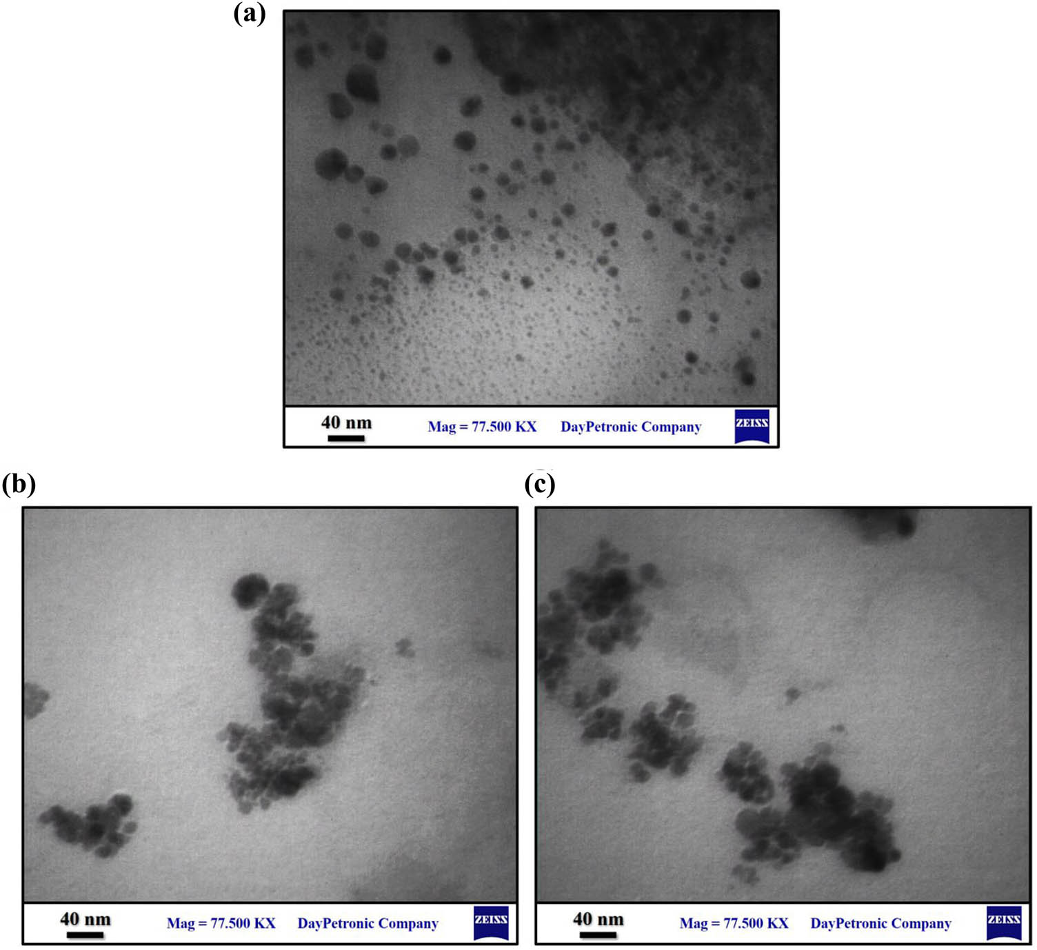
TEM images of chitosan NPs (a), SeNPs (b), and Se-chitosan NPs (c).
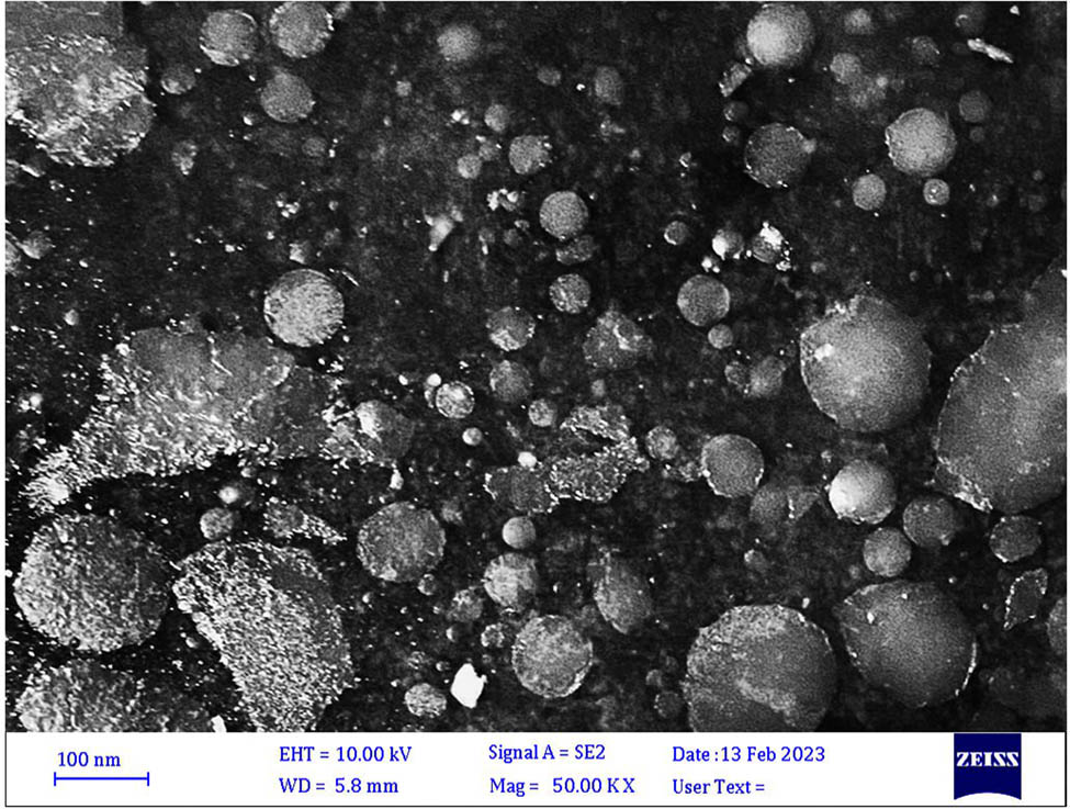
SEM–EDX of Chito-NPs.
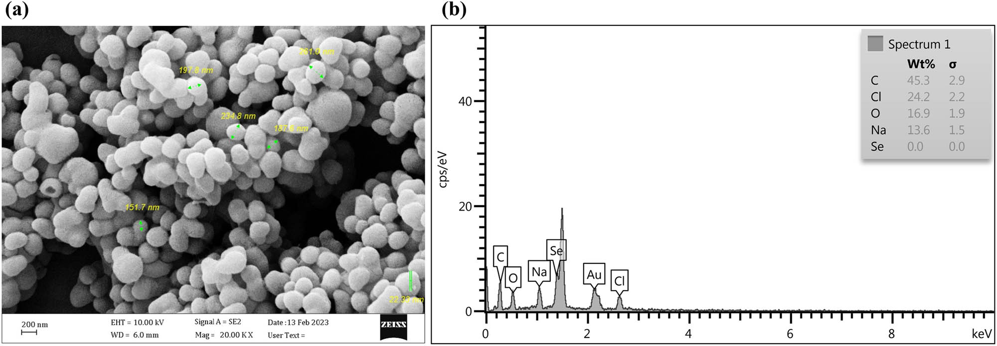
SEM–EDX of SeNPs. (a) SEM micrographs of SeNPs. (b) EDX analysis of SeNPs. SEM, scanning electron microscopy; EDX, energy-dispersive X-ray spectroscopy.
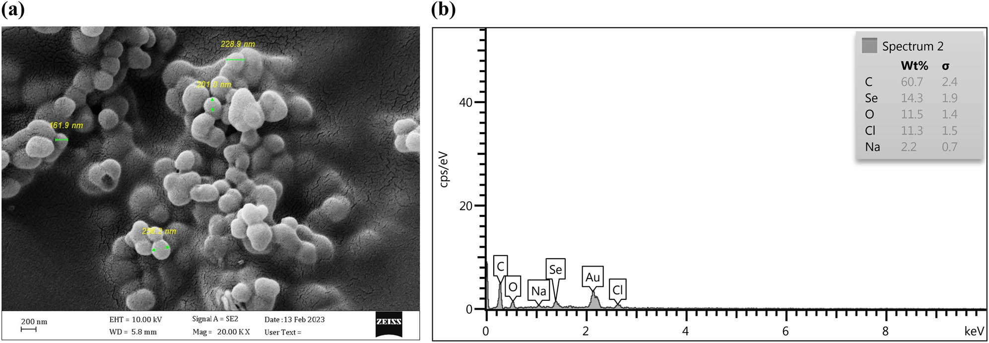
SEM–EDX of Se-chitosan NPs. (a) SEM micrographs of Se-chitosan NPs. (b) EDX analysis of Se-chitosan NPs. SEM, scanning electron microscopy; EDX, energy-dispersive X-ray spectroscopy.
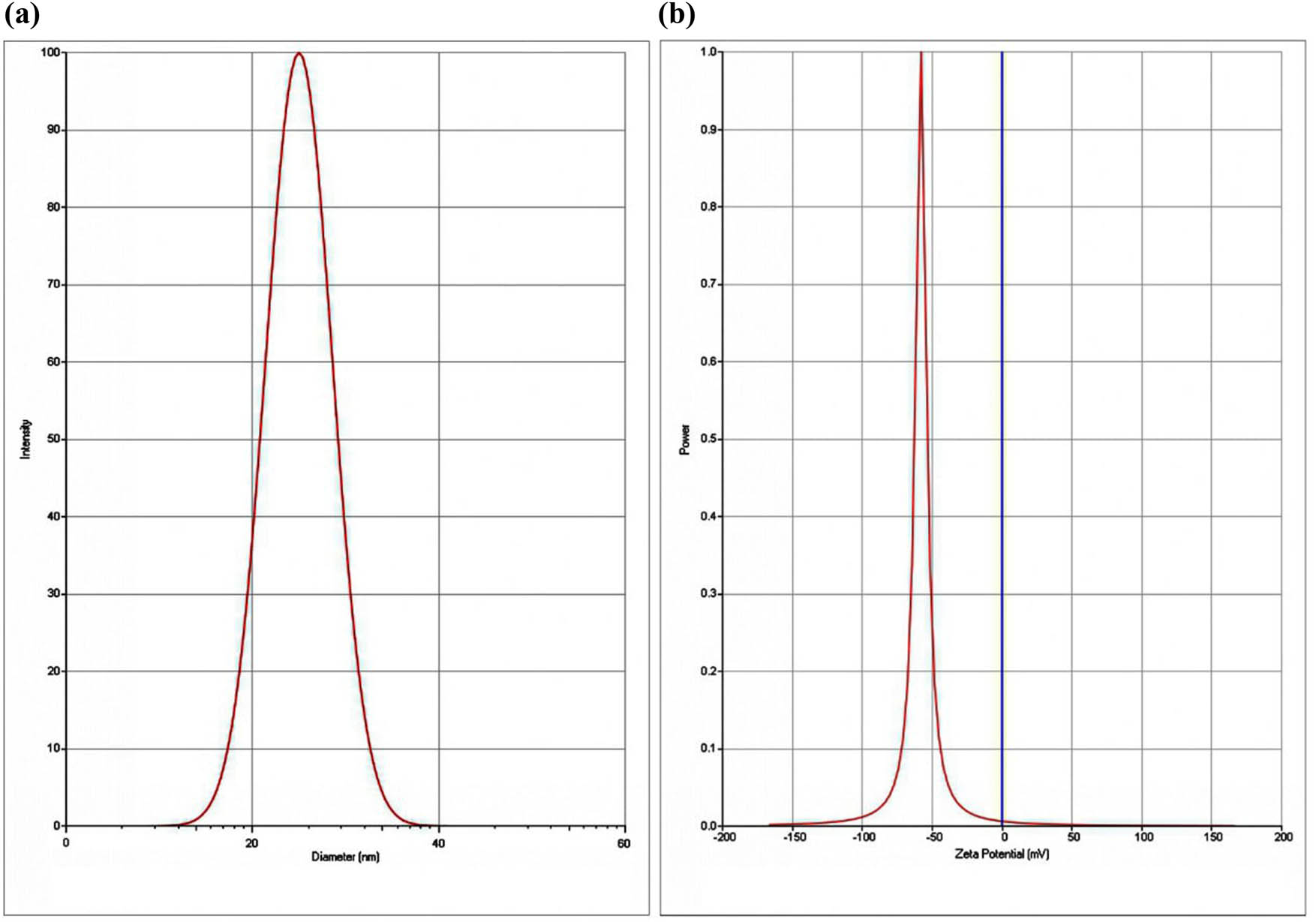
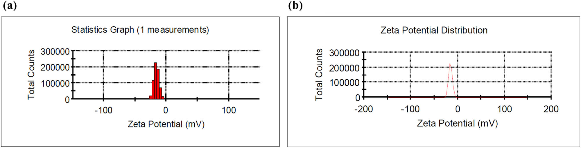
The produced NPs were spherical and had average sizes of 55.285 and 30.9 nm for SeNPs and Se-chitosan NPs, respectively. As shown in Table 1. The ZP of synthesized Chito-NPs, SeNPs, and Se-chitosan NPs is shown in Figures 6–8. NP’s ZP has been observed at varying rates in earlier research. For example, −7.7 mV was detected by Srivastava and Mukhopadhyay [59] and −28.8 mV was documented by Vekariya et al. [60]. In another study, the ZP was found to be −22.9 mV for synthetic SeNPs using fungus was −22.9 mV [61].
SeNPs and Se-chitosan NPs sizes and ZP. Three different experiments’ worth of data are presented in triplicate as ± standard deviation means
| Particles | Size (nm) | ZP (mV) |
|---|---|---|
| SeNPs | 55.285 | −15.4 ± 3.94 |
| Se-chitosan NPs | 30.9 | 13.8 ± 3.22 |
(a) Particle size distribution and (b) Chito-NPs’ ZP.
(a) Particle size distribution and (b) SeNPs’ ZP.
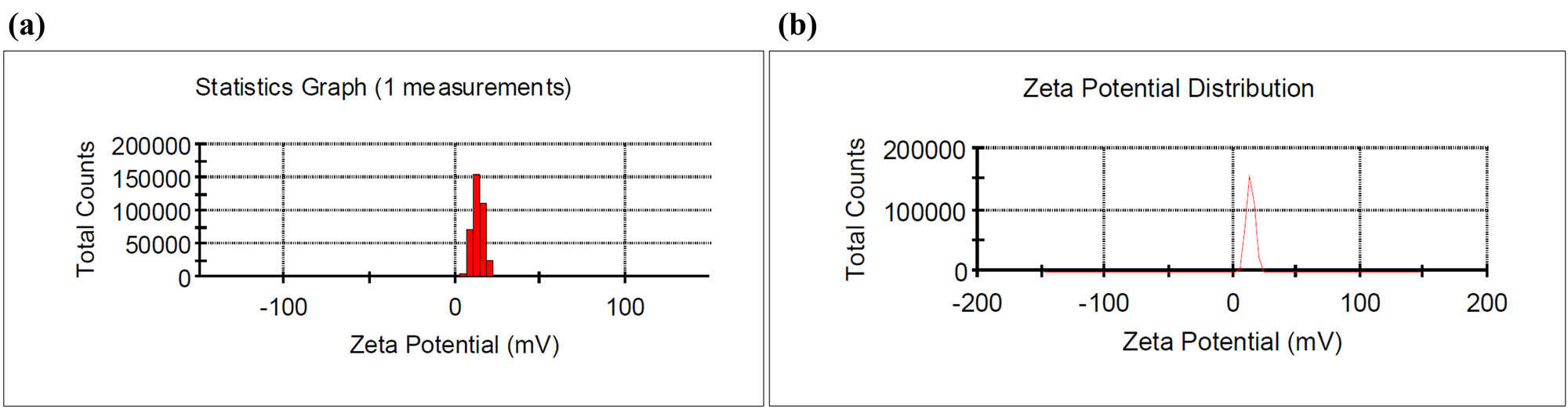
(a) Particle size distribution and (b) Se-chitosan NPs ZP.
3.2 Chito-NPs, SeNPs, and Se-chitosan NPs increase the LDH release
The enzyme LDH regulates the process by which lactate is converted to pyruvate, a process that needs cellular energy. LDH was used to evaluate the cytotoxic effects of Chito-NPs, SeNPs, and Se-chitosan NPs on lung cancer cell lines. Damage to the A549 cells subjected to Chito-NPs, SeNPs, and Se-chitosan NPs produces formazan from tetrazolium salt by releasing LDH from the cytoplasm. After lung cancer cells are handled with Chito-NPs, SeNPs, and Se-chitosan NPs, the proportion of released LDH in the suffering or dying cells is ascertained through measurement of the creation of formazan at a wavelength of 490 nm. The generation of formazan at a wavelength of 490 nm is measured to determine the percentage of LDH released in a lung carcinoma that is sick or dying. The information gathered suggests that Chito-NPs, SeNPs, and Se-chitosan NPs may be able to penetrate treated cells and induce vesicle development. The effect of concentration on the potential for LDH release by SeNPs, Chito-NPs, and Se-chitosan NPs is shown in Figure 9. The ability of Chito-NPs, SeNPs, and Se-chitosan NPs to enter cells as well as additional biological elements can lead to significant cellular disruption and induce the release of LDH. Concurrently, cells may absorb 100–200 nm NPs, which may trigger deleterious effects including genetic material mutations or DNA damage. The toxicity of NPs is probably related to processes that, by interfering with the antioxidant system, increase the body’s oxidative stress level [62]. Numerous membranes, such as those enclosing the mitochondria and the cell, have been damaged, which is caused by free radicals such as ROS. Because of this, the elements of cells that cause cell death, including proteins, lipids, fatty acids, and nucleic acids, interfere with the process of electronic information transfer. The cytotoxicity of A549 cells could perhaps be attributed to oxidative stress, which results in cellular disintegration. Furthermore, Chito-NPs, SeNPs, and Se-chitosan NPs may trigger concentration-dependent release of LDH, which could be related to cell membrane injury. The breakdown of cellular membranes, which releases cellular enzymes like LDH into the surrounding environment, could be connected to the LDH release that occurs in response to concentration-dependently to Chito-NPs, SeNPs, and Se-chitosan NPs.
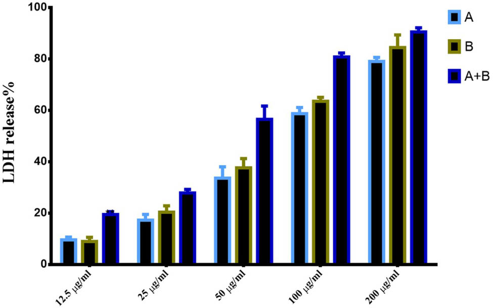
Prepared NPs induce LDH release in lung cancer cells. A = Chito-NPs, B = SeNPs, and A + B = Se-chitosan NPs.
3.3 Anticancer activity of NPs against lung cancer cells
Following a 72-h therapy course at different quantities of Chito-NPs, SeNPs, and Se-chitosan NPs (12.5, 25, 50, 100, and 200 g/mL), Chito-NPs, SeNPs, and Se-chitosan NPs were tested on lung cancer cells to see if they might stop growth suppression and proliferation. Chito-NPs, SeNPs, and Se-chitosan NPs reduced the cell viability that was dependent on dose, as cleared in Figure 10 (left panel). The inhibitory concentration (IC50) for Chito-NPs was at a quantity of 24.09 µg/mL, whereas the quantity for SeNPs was 18.56 µg/mL following a 72 h treatment with high quantities of Se-chitosan NPs, the viability of A549 cells decreased to almost 10%. This study’s findings indicated that the Chito-NPs and SeNPs caused cell death, and this inhibitory effect was improved by combining these two NPs as showed in the Se-chitosan NPs treated cells. In addition, acridine orange and ethidium bromide were used in a dual staining technique to analyze the nuclear morphology of the treated cells. The criterion for classifying apoptotic cells was DNA damage. Within the framework of this investigation, a look was also taken at how effective the Chito-NPs, SeNPs, and Se-chitosan NPs. To examine the different apoptotic features of the nuclear alterations, AO-EB staining was used. Following AO-EtBr staining, non-apoptotic cells displayed a green tone, while apoptotic cells displayed an orange or red coloration. As indicated in Figure 10 (right panel), there were significantly more apoptotic cells in the cells treated with Chito-NPs, SeNPs, and Se-chitosan NPs than in the untreated cells. These results were consistent with earlier research showing that Chito-NPs and SeNPs both exhibited apoptotic properties. According to a published study, SeNPs activated Ca2+ signals in various cancer cell lines. The stimulation of the process of apoptosis in cancer cell lines can be determined by their varying susceptibility to SeNPs. This can be achieved by processes of ER stress, modulation of the Ca2+ signaling system, and the initiation of several gene expression patterns that code for pro-apoptotic proteins [63]. Another investigation found that applying SeNPs to colon cancer cell lines caused immunogenic cell death is indicated by pro-apoptotic and immunogenic cell death markers. It also showed that these NPs could be an effective way to kill tumor cells indirectly by enhancing immunogenicity and causing apoptosis [64]. Also, according to a study, the combination of radiation and nano-Se inhibits the multiplication of lung cancer cells, hence acting as an anti-cancer agent [65]. Likewise, Shen and colleagues found that chitosan oligosaccharide (COS) in vitro reduced the quantity of S phase cells, inhibited cell proliferation, and slowed down the DNA synthesis rate due to an increase in p21 with cyclin A and CDK-2 decrease. COS impeded the growth of tumors by inhibiting the metastatic associated protein (MMP-9) in Lewis lung carcinoma (LLC) cells [66]. It may be possible to enhance the anticancer and nanotherapeutic effects of Chito-NPs on various human cancer cell lines [67]. On the other hand, the results showed a low cytotoxic effect against normal cell lines as indicated in Figure 11.
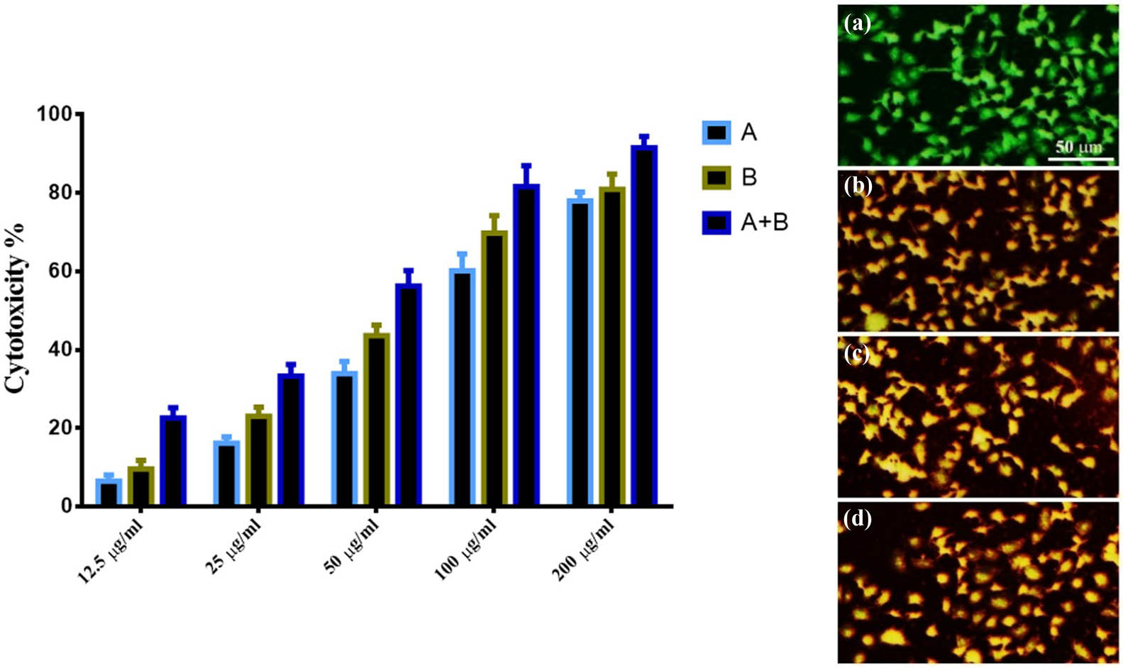
Anti-proliferative activity of prepared NPs against lung cancer cells. The right panel represented the cytotoxicity of prepared NPs against lung cancer cells. A = Chito-NPs, B = SeNPs, and A + B = Se-chitosan NPs. The left panel represented AO/EtBr double staining assay. A = control untreated cells, B = Chito-NPs, C = SeNPs, and D = Se-chitosan NPs.

Anti-proliferative activity of prepared NPs against normal breast cell line (MCF-10). A = Chito-NPs, B = SeNPs, and A + B = Se-chitosan NPs.
According to the flow cytometry data, the cells in quadrant Q3 that were going through apoptosis had Annexin V labels on them. Dot plots of A549 cells treated with SeNPs, Chito-NPs, and Se-chitosan NPs for 24 h at an IC50 concentration are displayed in Figure 12. While the majority of cells in the control A549 (97.5%) were viable and not undergoing apoptosis, the A549 treated with Chito-NPs, SeNPs, and Se-chitosan NPs showed a rise in apoptotic cells and a fall in viable cells. In the A549 control group, 0.56% of the cells were apoptotic. In contrast, the proportion rose to 86.5, 90.3, and 95.1% in A549 cells administered with Chito-NPs, SeNPs, and Se-chitosan NPs, respectively. Furthermore, the study’s findings have shown that Chito-NPs, SeNPs, and Se-chitosan NPs have no harmful effects on the typical cell line of the human lung, as clear in Figure 11. There are multiple possible ways that carbon nanotubes (CNTs) could damage lung cancer cells. One explanation for this is that they function as oxidative triggers, which encourage DNA damage and inflammation [68]. Our study’s findings demonstrated that the viability of A549 cells was considerably decreased when they were treated with Chito-NPs, SeNPs, and Se-chitosan NPs. Large quantities of ZnO-Fe3O4 composite magnetic NPs were shown to be fatal for the human cell line for breast cancer MDA-MB-231, but that is not for typical mouse fibroblasts (NIH 3T3), according to research by Bisht et al. [69]. The typical mouse fibroblast does not exhibit this impact. According to a prior study, metal NP cytotoxicity may be brought on by the generation of free radicals and oxygen species that are reactive (ROS) [70]. It has also been observed that elevated ROS levels can cause apoptosis by activating FOXO3a, a protein that can improve apoptosis signaling by encouraging the production of pro-apoptotic mitochondria-targeting protein members of the Bcl2 family [71]. The idea that high ROS can trigger apoptosis is supported by this finding. This finding was made possible by the activation of FOXO3a, which happens when ROS levels are too high. According to the ROS assay results, the presence of ZnO/CNT@Fe3O4 significantly increased the K562 cell line’s ROS generation amount. Observing these outcomes was interesting. Notably, current studies have shown that blocking the pathway of NF-kB with bortezomib, a well-known proteasome inhibitor increased the susceptibility of K562 cells to the deadly effects of ZnO/CNT@Fe3O4. This finding supports a theory that ZnO/CNT@Fe3O4-induced reduction of K562 cell sensitivity is most likely caused by nuclear factor-̙B pathway activation.
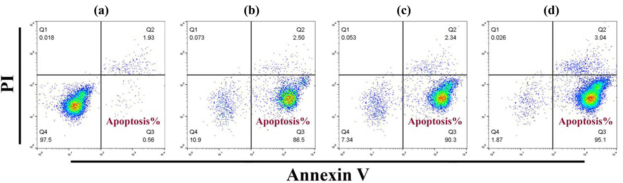
Prepared NPs induce apoptosis in A549 cells using Annexin V assay. a = control cells, b = Chito-NPs, c = SeNPs, and d = Se-chitosan NPs.
3.4 Chito-NPs, SeNPs, and Se-chitosan NPs disorder oxidative balance in A549 cells
Cells treated with SeNPs, Chito-NPs, and Se-chitosan NPs showed a significant increase in ROS production. Compared to the control cells, the ROS-induced fluorescence signal was observed to be higher. The current work examined the accumulation of ROS within cell lines of lung cancer following counseling by Chito-NPs, SeNPs, and Se-chitosan NPs. After treatment with Chito-NPs, SeNPs, and Se-chitosan NPs, there was a noticeable rise in ROS in the cells. A DCFH-DA probe was used to measure the levels of ROS, as illustrated in Figure 13 (upper panel). The level of ROS was increased in the lung cancer cells after treatment with Chito-NPs, SeNPs, and Se-chitosan NPs [72]. We looked at the potential effects of therapy with Chito-NPs, SeNPs, and Se-chitosan NPs on mitochondrial function. The presence of rhodamine dye, which shows current-dependent accumulation in the mitochondria, allowed for the detection of the loss of the MMP. The results of this study showed, as shown in Figure 13 (lower panel), that after exposure to Chito-NPs, SeNPs, and Se-chitosan NPs at dosages IC50 for 24 h, The proportion of cells with a depolarized mitochondrial membrane rose dramatically, and the effect of Se-chitosan NPs was the highest among all groups. Selenium is contained in the selenoproteins and seleno-enzymes structure, which can inhibit ROS and, as a result, oxidative damage development [73]. The majority of the pharmaceutical components that are currently available come from natural products. Despite advancements, creating bioactive molecules and medications from natural products has proven difficult, partly due to the issue of large-scale sequestration and mechanistic comprehension. As the field of cancer, treatment has advanced significantly and the usage of advanced technology has increased [74]. The cytoskeleton, cell growth and proliferation, the cell cycle, inflammation, angiogenesis, cell signaling, intrinsic apoptosis, and reducing chemoresistance are among the multitargeted functions of phenolics. Because the normal ovarian cell lineage can handle phenolic acids well, they are effective prophylactic agents against ovarian cancer. However, cancer treatment may benefit from the nonflavonoids’ antioxidant properties [75]. In recently published study [76], successfully created lipid nanocarriers using materials that the USFDA has designated as generally recognized as safe to address drug-related issues. This study aimed to assess the therapeutic efficacy of 6-o-stearoyl ascorbic acid nanostructured lipid carriers applied to CRT against mice suffering from colitis produced by dextran sodium sulphate. In addition to inhibiting the production of ROS during hypoxia and schemia/reoxygenation, selenium compounds stimulate mitochondrial biogenesis, which raises within-cell ATP and Ca2+ equilibrium levels with increased persistence of cells in the zone of penumbra [77,78]. The biogenic MgO NPs demonstrated their therapeutic potential against MDA-MB-231 cells by increasing cytotoxicity, inducing apoptosis, enhancing ROS production, promoting cell adhesion, and inhibiting cellular migration in a dose-dependent manner [79]. The results by Alserihi et al. [80] show that EGCG and EGCG NPs are effective anticancer agents in 3D spheroids of PCa cell lines. Following treatment with EGCG and EGCG NPs, the spheroid diameters of the cell lines under study were considerably decreased. EGCG and EGCG NPs caused ROS in PC3 cells but not in 22Rv1 cells, which may indicate that receptor-mediated antigens like PSMA are not involved. The MMP in 22Rv1 and PC3 spheroids treated with EGCG or EGCG NP did not significantly change. SeNPs have both prooxidant and antioxidant qualities, depending on the dosage. It has been demonstrated that a 12 μM concentration of SeNPs increases the cells’ antioxidant capacity, while a 24 μM concentration of SeNPs decreases the cells’ antioxidant capacity [81]. SeNPs induce ROS overproduction inside cancerous cells because of the acidic condition of pH and the imbalance of redox, which compromises mitochondrial integrity and causes ER stress. Numerous biochemical pathways, including NFκB, MAPK/Erk, Wnt/βcatenin, PI3K/Akt/mTOR, and the pathways of apoptotic, are activated as a result, leading to cellular stress [82].
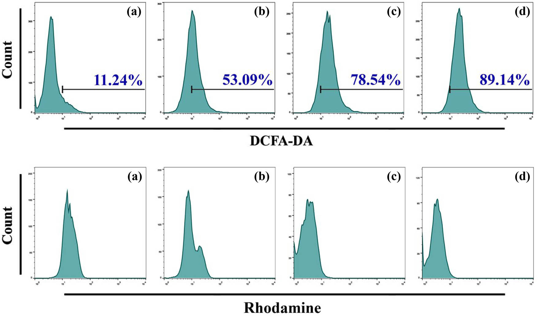
Chito-NPs, SeNPs, and Se-chitosan NPs cause mitochondrial malfunction and the production of ROS in A549 cells. (a) Untreated cells in control. (b) Cells subjected to Chito-NPs. (c) Cells exposed to SeNPs. (d) Cells treated to Se-chitosan NPs.
4 Conclusions
The pharmacological potential of SeNPs and Chito-NPs is well known, and its nanoformulation is expected to have major therapeutic benefits, especially in the fight against cancer. In this work, we examined the anticancer potential of SeNPs and Se-chitosan NPs that were biogenically produced against lung cancer cells (A549). Many biological assessments were estimated, including cytotoxicity, cellular morphology, apoptosis induction, ROS generation, and MMP. The biogenic NPs demonstrated their therapeutic potential against A549 cells by increasing cytotoxicity, inducing apoptosis, and enhancing ROS generation and mitochondrial dysfunction. The outcomes of the in vitro experiments cleared the ROS-mediated pathways used by Chito-NPs, SeNPs, and Se-chitosan NPs to trigger death in A549 cells. These results also imply that a lot of promise remains untapped. Further investigation is warranted and clinically significant to clarify a safety with comprehensive of the potential therapeutic of these NPs once applied in vivo. In the current work, we establish that exposing cells to SeNPs, Chito-NPs, and Se-chitosan NPs alters the human lung cancer cell line A549’s ROS signaling pathway, causing the induction of apoptosis. The given results may contribute to the creation of more potent medications for the treatment of lung cancer in humans.
Acknowledgments
The authors enjoy feeling their profound gratitude to the University of Technology-Iraq. The authors would like to extend their sincere appreciation to the Researchers Supporting Project Number (RSPD2025R986), King Saud University, Riyadh, Saudi Arabia.
-
Funding information: The authors state no funding involved.
-
Author contributions: All authors have accepted responsibility for the entire content of this manuscript and approved its submission.
-
Conflict of interest: The authors state no conflict of interest.
-
Data availability statement: All data generated or analyzed during this study are included in this published article.
References
[1] Tabrez S, Khan AU, Mirza AA, Suhail M, Jabir NR, Zughaibi TA, et al. Biosynthesis of copper oxide nanoparticles and its therapeutic efficacy against colon cancer. Nanotechnol Rev. 2022 Mar;11(1):1322–31.10.1515/ntrev-2022-0081Suche in Google Scholar
[2] Tabrez S, Khan AU, Hoque M, Suhail M, Khan MI, Zughaibi TA. Investigating the anticancer efficacy of biogenic synthesized MgONPs: An in vitro analysis. Front Chem. 2022 Sep;10:970193.10.3389/fchem.2022.970193Suche in Google Scholar PubMed PubMed Central
[3] Gowd V, Ahmad A, Tarique M, Suhail M, Zughaibi TA, Tabrez S, et al. Advancement of cancer immunotherapy using nanoparticles-based nanomedicine. Semin Cancer Biol. 2022;S1044-579X(22)00081-5. 10.1016/j.semcancer.2022.03.026.Suche in Google Scholar PubMed
[4] Alserihi RF, Mohammed MR, Kaleem M, Khan MI, Sechi M, Sanna V, et al. Development of (−)-epigallocatechin-3-gallate-loaded folate receptor-targeted nanoparticles for prostate cancer treatment. Nanotechnol Rev. 2021 Dec;11(1):298–311.10.1515/ntrev-2022-0013Suche in Google Scholar
[5] Alafaleq NO, Zughaibi TA, Jabir NR, Khan AU, Khan MS, Tabrez S. Biogenic synthesis of Cu-Mn bimetallic nanoparticles using pumpkin seeds extract and their characterization and anticancer efficacy. Nanomaterials. 2023 Mar;13(7):1201.10.3390/nano13071201Suche in Google Scholar PubMed PubMed Central
[6] Kadhim AA, Abbas NR, Kadhum HH, Albukhaty S, Jabir MS, Naji AM, et al. Investigating the effects of biogenic zinc oxide nanoparticles produced using papaver somniferum extract on oxidative stress, cytotoxicity, and the induction of apoptosis in the THP-1 cell line. Biol Trace Elem Res. 2023;201(10):4697–709.10.1007/s12011-023-03574-7Suche in Google Scholar PubMed
[7] Chen W, Yue L, Jiang Q, Liu X, Xia W. Synthesis of varisized chitosan-selenium nanocomposites through heating treatment and evaluation of their antioxidant properties. Int J Biol macromolecules. 2018 Jul;114:751–8.10.1016/j.ijbiomac.2018.03.108Suche in Google Scholar PubMed
[8] Kołodyńska D, Hałas P, Franus M, Hubicki Z. Zeolite properties improvement by chitosan modification – Sorption studies. J Ind Eng Chem. 2017 Aug;52:187–96.10.1016/j.jiec.2017.03.043Suche in Google Scholar
[9] Salar S, Mehrnejad F, Sajedi RH, Arough JM. Chitosan nanoparticles-trypsin interactions: Bio-physicochemical and molecular dynamics simulation studies. Int J Biol Macromolecules. 2017 Oct;103:902–9.10.1016/j.ijbiomac.2017.05.140Suche in Google Scholar PubMed
[10] Cao H, Xiao J, Liu H. Enhanced oxidase-like activity of selenium nanoparticles stabilized by chitosan and application in a facile colorimetric assay for mercury (II). Biochem Eng J. 2019 Dec;152:107384.10.1016/j.bej.2019.107384Suche in Google Scholar
[11] Song X, Chen Y, Zhao G, Sun H, Che H, Leng X. Effect of molecular weight of chitosan and its oligosaccharides on antitumor activities of chitosan-selenium nanoparticles. Carbohydr Polym. 2020 Mar;231:115689.10.1016/j.carbpol.2019.115689Suche in Google Scholar PubMed
[12] Rayman MP. Selenium and human health. Lancet. 2012;379(9822):1256–68.10.1016/S0140-6736(11)61452-9Suche in Google Scholar PubMed
[13] Sharma A, Goyal AK, Rath G. Recent advances in metal nanoparticles in cancer therapy. J Drug Target. 2018 Sep;26(8):617–32.10.1080/1061186X.2017.1400553Suche in Google Scholar PubMed
[14] Adeyemi OS, Sulaiman FA. Evaluation of metal nanoparticles for drug delivery systems. J Biomed Res. 2014 Dec;29(2):145.10.7555/JBR.28.20130096Suche in Google Scholar PubMed PubMed Central
[15] Kwon SJ, Bard AJ. DNA analysis by application of Pt nanoparticle electrochemical amplification with single label response. J Am Chem Soc. 2012 Jul;134(26):10777–9.10.1021/ja304074fSuche in Google Scholar PubMed
[16] Sun C, Lee JS, Zhang M. Magnetic nanoparticles in MR imaging and drug delivery. Adv Drug Delivery Rev. 2008 Aug;60(11):1252–65.10.1016/j.addr.2008.03.018Suche in Google Scholar PubMed PubMed Central
[17] Doria G, Conde J, Veigas B, Giestas L, Almeida C, Assunção M, et al. Noble metal nanoparticles for biosensing applications. Sensors. 2012 Feb;12(2):1657–87.10.3390/s120201657Suche in Google Scholar PubMed PubMed Central
[18] Vaseashta A, Vaclavikova M, Vaseashta S, Gallios G, Roy P, Pummakarnchana O. Nanostructures in environmental pollution detection, monitoring, and remediation. Sci Technol Adv Mater. 2007 Jan;8(1–2):47.10.1016/j.stam.2006.11.003Suche in Google Scholar
[19] Gadad AP, Kumar SV, Dandagi PM, Bolmol UB, Pallavi NP. Nanoparticles and their therapeutic applications in pharmacy. Int J Pharm Sci Nanotechnol. 2014 Aug;7(3):2515–6.10.37285/ijpsn.2014.7.3.2Suche in Google Scholar
[20] Khandelwal A, Joshi R. Synthesis of nanoparticles and their application in agriculture. Acta Sci Agric. 2018;2(3):10–3.Suche in Google Scholar
[21] Matsui I. Nanoparticles for electronic device applications: a brief review. J Chem Eng Jpn. 2005;38(8):535–46.10.1252/jcej.38.535Suche in Google Scholar
[22] Stroyuk AL, Raevskaya AE, Kuchmiy SY, Dzhagan VM, Zahn DR, Schulze S. Structural and optical characterization of colloidal Se nanoparticles prepared via the acidic decomposition of sodium selenosulfate. Colloids Surf A: Physicochem Eng Asp. 2008 May;320(1–3):169–74.10.1016/j.colsurfa.2008.01.055Suche in Google Scholar
[23] Lin W, Zhang J, Xu JF, Pi J. The advancing of selenium nanoparticles against infectious diseases. Front Pharmacology. 2021 Jul;12:682284.10.3389/fphar.2021.682284Suche in Google Scholar PubMed PubMed Central
[24] Maiyo F, Singh M. Selenium nanoparticles: Potential in cancer gene and drug delivery. Nanomedicine. 2017 May;12(9):1075–89.10.2217/nnm-2017-0024Suche in Google Scholar PubMed
[25] Mahmood RI, Abbass AK, Al-Saffar AZ, Al-Obaidi JR. An in vitro cytotoxicity of a novel pH-Sensitive lectin loaded-cockle shell-derived calcium carbonate nanoparticles against MCF-7 breast tumour cell. J Drug Delivery Sci Technol. 2021 Feb;61:102230.10.1016/j.jddst.2020.102230Suche in Google Scholar
[26] Soenen SJ, Rivera-Gil P, Montenegro JM, Parak WJ, De Smedt SC, Braeckmans K. Cellular toxicity of inorganic nanoparticles: common aspects and guidelines for improved nanotoxicity evaluation. Nano Today. 2011 Oct;6(5):446–65.10.1016/j.nantod.2011.08.001Suche in Google Scholar
[27] Alam H, Khatoon N, Khan MA, Husain SA, Saravanan M, Sardar M. Synthesis of selenium nanoparticles using probiotic bacteria Lactobacillus acidophilus and their enhanced antimicrobial activity against resistant bacteria. J Clust Sci. 2020 Sep;31:1003–11.10.1007/s10876-019-01705-6Suche in Google Scholar
[28] Husen A, Siddiqi KS. Plants and microbes assisted selenium nanoparticles: characterization and application. J Nanobiotechnology. 2014 Dec;12:1–10.10.1186/s12951-014-0028-6Suche in Google Scholar PubMed PubMed Central
[29] Nada HG, Ali HE, El-Behery RR, Shanab SM, Elshatoury EH. Nanoparticles biosynthesized by bacillus cereus filtrate and gamma rays enhancing Chlorella vulgaris biomass and lipid production. J Clust Sci. 2022;33:2055–68.10.1007/s10876-021-02122-4Suche in Google Scholar
[30] Chen W, Li X, Cheng H, Xia W. Chitosan-based selenium composites as potent Se supplements: Synthesis, beneficial health effects, and applications in food and agriculture. Trends Food Sci Technol. 2022 Nov 1;129:339–52.10.1016/j.tifs.2022.10.008Suche in Google Scholar
[31] Memar MY, Adibkia K, Farajnia S, Kafil HS, Yekani M, Alizadeh N, et al. The grape seed extract: a natural antimicrobial agent against different pathogens. Rev Res Med Microbiology. 2019 Jul;30(3):173–82.10.1097/MRM.0000000000000174Suche in Google Scholar
[32] Perumalla AV, Hettiarachchy NS. Green tea and grape seed extracts – Potential applications in food safety and quality. Food Res Int. 2011 May;44(4):827–39.10.1016/j.foodres.2011.01.022Suche in Google Scholar
[33] Mahmoud YI. Grape seed extract neutralizes the effects of Cerastes cerastes cerastes post-synaptic neurotoxin in mouse diaphragm. Micron. 2013 Jan;44:298–302.10.1016/j.micron.2012.07.007Suche in Google Scholar PubMed
[34] Wadhwani SA, Shedbalkar UU, Singh R, Chopade BA. Biogenic selenium nanoparticles: current status and future prospects. Appl Microbiol Biotechnol. 2016 Mar;100:2555–66.10.1007/s00253-016-7300-7Suche in Google Scholar PubMed
[35] Iranifam M, Fathinia M, Rad TS, Hanifehpour Y, Khataee AR, Joo SW. A novel selenium nanoparticles-enhanced chemiluminescence system for determination of dinitrobutylphenol. Talanta. 2013 Mar;107:263–9.10.1016/j.talanta.2012.12.043Suche in Google Scholar PubMed
[36] Mahmood RI, Abbass AK, Razali N, Al-Saffar AZ, Al-Obaidi JR. Protein profile of MCF-7 breast cancer cell line treated with lectin delivered by CaCO3NPs revealed changes in molecular chaperones, cytoskeleton, and membrane-associated proteins. Int J Biol Macromol. 2021 Aug;184:636–47.10.1016/j.ijbiomac.2021.06.144Suche in Google Scholar PubMed
[37] Rahmah MI, Saadoon NM, Mohasen AJ, Kamel RI, Fayad TA, Ibrahim NM. Double hydrothermal synthesis of iron oxide/silver oxide nanocomposites with antibacterial activity. J Mech Behav Mater. 2021 Jan;30(1):207–12.10.1515/jmbm-2021-0021Suche in Google Scholar
[38] Aziz WJ, Abid MA, Hussein EH. Biosynthesis of CuO nanoparticles and synergistic antibacterial activity using mint leaf extract. Mater Technol. 2020 Jul;35(8):447–51.10.1080/10667857.2019.1692163Suche in Google Scholar
[39] Shahabadi N, Zendehcheshm S, Khademi F. Selenium nanoparticles: Synthesis, in-vitro cytotoxicity, antioxidant activity and interaction studies with ct-DNA and HSA, HHb and Cyt c serum proteins. Biotechnol Rep. 2021 Jun;30:e00615.10.1016/j.btre.2021.e00615Suche in Google Scholar PubMed PubMed Central
[40] Shar AH, Lakhan MN, Wang J, Ahmed M, Alali KT, Ahmed R, Ali I, et al. Facile synthesis and characterization of selenium nanoparticles by the hydrothermal approach. Dig J Nanomater Biostruct. 2019 Oct;14:867–72.Suche in Google Scholar
[41] Ramamurthy CH, Sampath KS, Arunkumar P, Kumar MS, Sujatha V, Premkumar K, et al. Green synthesis and characterization of selenium nanoparticles and its augmented cytotoxicity with doxorubicin on cancer cells. Bioprocess Biosyst Eng. 2013 Aug;36:1131–9.10.1007/s00449-012-0867-1Suche in Google Scholar PubMed
[42] Jabir MS, Abood NA, Jawad MH, Öztürk K, Kadhim H, Albukhaty S, et al. Gold nanoparticles loaded TNF-α and CALNN peptide as a drug delivery system and promising therapeutic agent for breast cancer cells. Mater Technol. 2022 Dec;37(14):3152–66.10.1080/10667857.2022.2133073Suche in Google Scholar
[43] Al-Rahim AM, Mahmood RI, Mohammed MM, Omer D. In vitro evaluation of antioxidant and cytotoxic activity of folate-methotrexate conjugated to bovine serum albumin nanoparticles against MCF-7, HepG2, and PC3 cell lines. Gene Rep. 2022 Dec;29:101666.10.1016/j.genrep.2022.101666Suche in Google Scholar
[44] Mohammed-Salih HS, Ghazi A, Mahmood RI, Al‐Qazzaz HH, Supian FL, Al-Obaidi JR, et al. Enhancing orthodontic treatment control with fish scale-derived hydroxyapatite nanoparticles: Insights from an animal model study. Saudi Dental J. 2024 Aug;36(8):1128–34.10.1016/j.sdentj.2024.06.007Suche in Google Scholar PubMed PubMed Central
[45] Mohammed SA, Khashan KS, Jabir MS, Abdulameer FA, Sulaiman GM, Al-Omar MS, et al. Copper oxide nanoparticle‐decorated carbon nanoparticle composite colloidal preparation through laser ablation for antimicrobial and antiproliferative actions against breast cancer cell line, MCF‐7. BioMed Res Int. 2022;2022(1):9863616.10.1155/2022/9863616Suche in Google Scholar PubMed PubMed Central
[46] Hadi NA, Mahmood RI, Al-Saffar AZ. Evaluation of antioxidant enzyme activity in doxorubicin treated breast cancer patients in Iraq: a molecular and cytotoxic study. Gene Rep. 2021 Sep;24:101285.10.1016/j.genrep.2021.101285Suche in Google Scholar
[47] Abbas ZS, Sulaiman GM, Jabir MS, Mohammed SA, Khan RA, Mohammed HA, et al. Galangin/β-cyclodextrin inclusion complex as a drug-delivery system for improved solubility and biocompatibility in breast cancer treatment. Molecules. 2022 Jul;27(14):4521.10.3390/molecules27144521Suche in Google Scholar PubMed PubMed Central
[48] Farhana A, Lappin SL. Biochemistry, lactate dehydrogenase. InStatPearls [internet]. Treasure Island FL: StatPearls Publishing; 2023 May.Suche in Google Scholar
[49] Khashan KS, Jabir MS, Abdulameer FA. Carbon Nanoparticles decorated with cupric oxide Nanoparticles prepared by laser ablation in liquid as an antibacterial therapeutic agent. Mater Res Express. 2018 Mar;5(3):035003.10.1088/2053-1591/aab0edSuche in Google Scholar
[50] Kazemi M, Akbari A, Soleimanpour S, Feizi N, Darroudi M. The role of green reducing agents in gelatin-based synthesis of colloidal selenium nanoparticles and investigation of their antimycobacterial and photocatalytic properties. J Clust Sci. 2019 May;30:767–5.10.1007/s10876-019-01537-4Suche in Google Scholar
[51] Shakir M, Mohammed-Salih HS, Hussein FH, Al-Obaidi JR, Supian FL. Innovation of nano-hydrogels loaded with amelogenin peptide and hydroxyapatite nano-particles for remineralisation of artificially induced white spot lesions. J Drug Delivery Sci Technol. 2024 Sep;99:105986.10.1016/j.jddst.2024.105986Suche in Google Scholar
[52] Sengar M, Saxena S, Satsangee S, Jain R. Silver nanoparticles decorated functionalized multiwalled carbon nanotubes modified screen printed sensor for the voltammetric determination of butorphanol. J Appl Organomet Chem. 2021;1:95–108.Suche in Google Scholar
[53] Khosravanian A, Moslehipour A, Ashrafian H. A review on bioimaging, biosensing, and drug delivery systems based on graphene quantum dots. Prog Chem Biochem Res. 2021;4:44.Suche in Google Scholar
[54] Fernández-Llamosas H, Castro L, Blázquez ML, Díaz E, Carmona M. Speeding up bioproduction of selenium nanoparticles by using Vibrio natriegens as microbial factory. Sci Rep. 2017 Nov;7(1):16046.10.1038/s41598-017-16252-1Suche in Google Scholar PubMed PubMed Central
[55] Srivastava N, Mukhopadhyay M. Biosynthesis and structural characterization of selenium nanoparticles using Gliocladium roseum. J Clust Sci. 2015 Sep;26:1473–82.10.1007/s10876-014-0833-ySuche in Google Scholar
[56] Cremonini E, Zonaro E, Donini M, Lampis S, Boaretti M, Dusi S, et al. Biogenic selenium nanoparticles: characterization, antimicrobial activity and effects on human dendritic cells and fibroblasts. Microb Biotechnol. 2016 Nov;9(6):758–71.10.1111/1751-7915.12374Suche in Google Scholar PubMed PubMed Central
[57] Sharma G, Sharma AR, Bhavesh R, Park J, Ganbold B, Nam JS, et al. Biomolecule-mediated synthesis of selenium nanoparticles using dried Vitis vinifera (raisin) extract. Molecules. 2014 Feb;19(3):2761–70.10.3390/molecules19032761Suche in Google Scholar PubMed PubMed Central
[58] Dhanjal S, Cameotra SS. Aerobic biogenesis of selenium nanospheres by Bacillus cereus isolated from coalmine soil. Microb Cell Factories. 2010 Dec;9:1–11.10.1186/1475-2859-9-52Suche in Google Scholar PubMed PubMed Central
[59] Srivastava N, Mukhopadhyay M. Green synthesis and structural characterization of selenium nanoparticles and assessment of their antimicrobial property. Bioprocess Biosyst Eng. 2015 Sep;38:1723–30.10.1007/s00449-015-1413-8Suche in Google Scholar PubMed
[60] Vekariya KK, Kaur J, Tikoo K. ERα signaling imparts chemotherapeutic selectivity to selenium nanoparticles in breast cancer. Nanomed: Nanotechnol Biol Med. 2012 Oct;8(7):1125–32.10.1016/j.nano.2011.12.003Suche in Google Scholar PubMed
[61] Zare B, Babaie S, Setayesh N, Shahverdi AR. Isolation and characterization of a fungus for extracellular synthesis of small selenium nanoparticles. Nanomed J. 2013 Oct;1(1):13–9.Suche in Google Scholar
[62] Samrot AV, Ram Singh SP, Deenadhayalan R, Rajesh VV, Padmanaban S, Radhakrishnan K. Nanoparticles, a double-edged sword with oxidant as well as antioxidant properties – A review. Oxygen. 2022 Nov;2(4):591–604.10.3390/oxygen2040039Suche in Google Scholar
[63] Varlamova EG, Goltyaev MV, Mal’tseva VN, Turovsky EA, Sarimov RM, Simakin AV, et al. Mechanisms of the cytotoxic effect of selenium nanoparticles in different human cancer cell lines. Int J Mol Sci. 2021 Jul;22(15):7798.10.3390/ijms22157798Suche in Google Scholar PubMed PubMed Central
[64] Spyridopoulou K, Aindelis G, Pappa A, Chlichlia K. Anticancer activity of biogenic selenium nanoparticles: apoptotic and immunogenic cell death markers in colon cancer cells. Cancers. 2021 Oct;13(21):5335.10.3390/cancers13215335Suche in Google Scholar PubMed PubMed Central
[65] Tian J, Wei X, Zhang W, Xu A. Effects of selenium nanoparticles combined with radiotherapy on lung cancer cells. Front Bioeng Biotechnol. 2020 Nov;8:598997.10.3389/fbioe.2020.598997Suche in Google Scholar PubMed PubMed Central
[66] Shen KT, Chen MH, Chan HY, Jeng JH, Wang YJ. Inhibitory effects of chitooligosaccharides on tumor growth and metastasis. Food Chem Toxicol. 2009 Aug;47(8):1864–71.10.1016/j.fct.2009.04.044Suche in Google Scholar PubMed
[67] Ramadan MA, Sharaky M, Faid AH. Ionic gelation synthesis, characterization and cytotoxic evaluation of chitosan nanoparticles on different types of human cancer cell models. Egypt J Chem. 2022 Feb;65(2):153–9.Suche in Google Scholar
[68] Cinat D, Coppes RP, Barazzuol L. DNA damage-induced inflammatory microenvironment and adult stem cell response. Front Cell Dev Biol. 2021 Oct;9:729136.10.3389/fcell.2021.729136Suche in Google Scholar PubMed PubMed Central
[69] Bisht G, Rayamajhi S, Kc B, Paudel SN, Karna D, Shrestha BG. Synthesis, characterization, and study of in vitro cytotoxicity of ZnO-Fe3O4 magnetic composite nanoparticles in human breast cancer cell line (MDA-MB-231) and mouse fibroblast (NIH 3T3). Nanoscale Res Lett. 2016;11:537.10.1186/s11671-016-1734-9Suche in Google Scholar PubMed PubMed Central
[70] Kessler A, Hedberg J, Blomberg E, Odnevall I. Reactive oxygen species formed by metal and metal oxide nanoparticles in physiological media – a review of reactions of importance to nanotoxicity and proposal for categorization. Nanomaterials. 2022 Jun;12(11):1922.10.3390/nano12111922Suche in Google Scholar PubMed PubMed Central
[71] Tan J, Xiang Y, Xiong Y, Zhang Y, Qiao B, Zhang H. Crebanine induces ROS-dependent apoptosis in human hepatocellular carcinoma cells via the AKT/FoxO3a signaling pathway. Front Pharmacology. 2023 Feb;14:1069093.10.3389/fphar.2023.1069093Suche in Google Scholar PubMed PubMed Central
[72] Di Stefano A, Frosali S, Leonini A, Ettorre A, Priora R, Di Simplicio FC, et al. GSH depletion, protein S-glutathionylation and mitochondrial transmembrane potential hyperpolarization are early events in initiation of cell death induced by a mixture of isothiazolinones in HL60 cells. Biochim Biophys Acta (BBA)-Molecular Cell Res. 2006 Feb;1763(2):214–25.10.1016/j.bbamcr.2005.12.012Suche in Google Scholar PubMed
[73] Lee SH, Choi BY, Kho AR, Jeong JH, Hong DK, Lee SH, et al. Protective effects of protocatechuic acid on seizure-induced neuronal death. Int J Mol Sci. 2018 Jan;19(1):187.10.3390/ijms19010187Suche in Google Scholar PubMed PubMed Central
[74] Muhammad N, Usmani D, Tarique M, Naz H, Ashraf M, Raliya R, et al. The role of natural products and their multitargeted approach to treat solid cancer. Cells. 2022 Jul;11(14):2209.10.3390/cells11142209Suche in Google Scholar PubMed PubMed Central
[75] Nazam N, Jabir NR, Ahmad I, Alharthy SA, Khan MS, Ayub R, et al. Phenolic acids-mediated regulation of molecular targets in Ovarian Cancer: current understanding and future perspectives. Pharmaceuticals. 2023 Feb;16(2):274.10.3390/ph16020274Suche in Google Scholar PubMed PubMed Central
[76] Mishra RK, Ahmad A, Kumar A, Ali A, Jori C, Tabrez S, et al. Cortisone-loaded stearoyl ascorbic acid based nanostructured lipid carriers alleviate inflammatory changes in DSS-induced colitis. Biomater Adv. 2023 May;148:213383.10.1016/j.bioadv.2023.213383Suche in Google Scholar PubMed
[77] Mehta SL, Kumari S, Mendelev N, Li PA. Selenium preserves mitochondrial function, stimulates mitochondrial biogenesis, and reduces infarct volume after focal cerebral ischemia. BMC Neurosci. 2012 Dec;13:1–12.10.1186/1471-2202-13-79Suche in Google Scholar PubMed PubMed Central
[78] Shultz SR, Wright DK, Zheng P, Stuchbery R, Liu SJ, Sashindranath M, et al. Sodium selenate reduces hyperphosphorylated tau and improves outcomes after traumatic brain injury. Brain. 2015 May;138(5):1297–313.10.1093/brain/awv053Suche in Google Scholar PubMed PubMed Central
[79] Khan MR, Alafaleq NO, Ramu AK, Alhosaini K, Khan MS, Zughaibi TA, et al. Evaluation of biogenically synthesized MgO NPs anticancer activity against breast cancer cells. Saudi. J Biol Sci. 2024 Jan;31(1):103874.10.1016/j.sjbs.2023.103874Suche in Google Scholar PubMed PubMed Central
[80] Alserihi RF, Mohammed MR, Kaleem M, Khan MI, Sechi M, Zughaibi TA, et al. Comparative efficacy of epigallocatechin gallate and its nano-formulation in prostate cancer 3D spheroids model. J King Saud Univ-Sci. 2023 May;35(4):102627.10.1016/j.jksus.2023.102627Suche in Google Scholar
[81] Wang H, He Y, Liu L, Tao W, Wang G, Sun W, et al. Prooxidation and cytotoxicity of selenium nanoparticles at nonlethal level in sprague‐dawley rats and buffalo rat liver cells. Oxid Med Cell Longev. 2020;2020(1):7680276.10.1155/2020/7680276Suche in Google Scholar PubMed PubMed Central
[82] Kondaparthi P, Flora SJ, Naqvi S. Selenium nanoparticles: An insight on its Pro-oxidant and antioxidant properties. Front Nanosci Nanotechnol. 2019 Dec;6(1):5.10.15761/FNN.1000189Suche in Google Scholar
© 2025 the author(s), published by De Gruyter
This work is licensed under the Creative Commons Attribution 4.0 International License.
Artikel in diesem Heft
- Research Articles
- MHD radiative mixed convective flow of a sodium alginate-based hybrid nanofluid over a convectively heated extending sheet with Joule heating
- Experimental study of mortar incorporating nano-magnetite on engineering performance and radiation shielding
- Multicriteria-based optimization and multi-variable non-linear regression analysis of concrete containing blends of nano date palm ash and eggshell powder as cementitious materials
- A promising Ag2S/poly-2-amino-1-mercaptobenzene open-top spherical core–shell nanocomposite for optoelectronic devices: A one-pot technique
- Biogenic synthesized selenium nanoparticles combined chitosan nanoparticles controlled lung cancer growth via ROS generation and mitochondrial damage pathway
- Fabrication of PDMS nano-mold by deposition casting method
- Stimulus-responsive gradient hydrogel micro-actuators fabricated by two-photon polymerization-based 4D printing
- Physical aspects of radiative Carreau nanofluid flow with motile microorganisms movement under yield stress via oblique penetrable wedge
- Effect of polar functional groups on the hydrophobicity of carbon nanotubes-bacterial cellulose nanocomposite
- Review in green synthesis mechanisms, application, and future prospects for Garcinia mangostana L. (mangosteen)-derived nanoparticles
- Entropy generation and heat transfer in nonlinear Buoyancy–driven Darcy–Forchheimer hybrid nanofluids with activation energy
- Green synthesis of silver nanoparticles using Ginkgo biloba seed extract: Evaluation of antioxidant, anticancer, antifungal, and antibacterial activities
- A numerical analysis of heat and mass transfer in water-based hybrid nanofluid flow containing copper and alumina nanoparticles over an extending sheet
- Investigating the behaviour of electro-magneto-hydrodynamic Carreau nanofluid flow with slip effects over a stretching cylinder
- Electrospun thermoplastic polyurethane/nano-Ag-coated clear aligners for the inhibition of Streptococcus mutans and oral biofilm
- Investigation of the optoelectronic properties of a novel polypyrrole-multi-well carbon nanotubes/titanium oxide/aluminum oxide/p-silicon heterojunction
- Novel photothermal magnetic Janus membranes suitable for solar water desalination
- Green synthesis of silver nanoparticles using Ageratum conyzoides for activated carbon compositing to prepare antimicrobial cotton fabric
- Activation energy and Coriolis force impact on three-dimensional dusty nanofluid flow containing gyrotactic microorganisms: Machine learning and numerical approach
- Machine learning analysis of thermo-bioconvection in a micropolar hybrid nanofluid-filled square cavity with oxytactic microorganisms
- Research and improvement of mechanical properties of cement nanocomposites for well cementing
- Thermal and stability analysis of silver–water nanofluid flow over unsteady stretching sheet under the influence of heat generation/absorption at the boundary
- Cobalt iron oxide-infused silicone nanocomposites: Magnetoactive materials for remote actuation and sensing
- Magnesium-reinforced PMMA composite scaffolds: Synthesis, characterization, and 3D printing via stereolithography
- Bayesian inference-based physics-informed neural network for performance study of hybrid nanofluids
- Numerical simulation of non-Newtonian hybrid nanofluid flow subject to a heterogeneous/homogeneous chemical reaction over a Riga surface
- Enhancing the superhydrophobicity, UV-resistance, and antifungal properties of natural wood surfaces via in situ formation of ZnO, TiO2, and SiO2 particles
- Synthesis and electrochemical characterization of iron oxide/poly(2-methylaniline) nanohybrids for supercapacitor application
- Impacts of double stratification on thermally radiative third-grade nanofluid flow on elongating cylinder with homogeneous/heterogeneous reactions by implementing machine learning approach
- Synthesis of Cu4O3 nanoparticles using pumpkin seed extract: Optimization, antimicrobial, and cytotoxicity studies
- Cationic charge influence on the magnetic response of the Fe3O4–[Me2+ 1−y Me3+ y (OH2)] y+(Co3 2−) y/2·mH2O hydrotalcite system
- Pressure sensing intelligent martial arts short soldier combat protection system based on conjugated polymer nanocomposite materials
- Magnetohydrodynamics heat transfer rate under inclined buoyancy force for nano and dusty fluids: Response surface optimization for the thermal transport
- Fly ash and nano-graphene enhanced stabilization of engine oil-contaminated soils
- Enhancing natural fiber-reinforced biopolymer composites with graphene nanoplatelets: Mechanical, morphological, and thermal properties
- Performance evaluation of dual-scale strengthened co-bonded single-lap joints using carbon nanotubes and Z-pins with ANN
- Computational works of blood flow with dust particles and partially ionized containing tiny particles on a moving wedge: Applications of nanotechnology
- Hybridization of biocomposites with oil palm cellulose nanofibrils/graphene nanoplatelets reinforcement in green epoxy: A study of physical, thermal, mechanical, and morphological properties
- Design and preparation of micro-nano dual-scale particle-reinforced Cu–Al–V alloy: Research on the aluminothermic reduction process
- Spectral quasi-linearization and response optimization on magnetohydrodynamic flow via stenosed artery with hybrid and ternary solid nanoparticles: Support vector machine learning
- Ferrite/curcumin hybrid nanocomposite formulation: Physicochemical characterization, anticancer activity, and apoptotic and cell cycle analyses in skin cancer cells
- Enhanced therapeutic efficacy of Tamoxifen against breast cancer using extra virgin olive oil-based nanoemulsion delivery system
- A titanium oxide- and silver-based hybrid nanofluid flow between two Riga walls that converge and diverge through a machine-learning approach
- Enhancing convective heat transfer mechanisms through the rheological analysis of Casson nanofluid flow towards a stagnation point over an electro-magnetized surface
- Intrinsic self-sensing cementitious composites with hybrid nanofillers exhibiting excellent piezoresistivity
- Research on mechanical properties and sulfate erosion resistance of nano-reinforced coal gangue based geopolymer concrete
- Review Articles
- A comprehensive review on hybrid plasmonic waveguides: Structures, applications, challenges, and future perspectives
- Nanoparticles in low-temperature preservation of biological systems of animal origin
- Fluorescent sulfur quantum dots for environmental monitoring
- Nanoscience systematic review methodology standardization
- Nanotechnology revolutionizing osteosarcoma treatment: Advances in targeted kinase inhibitors
- AFM: An important enabling technology for 2D materials and devices
- Carbon and 2D nanomaterial smart hydrogels for therapeutic applications
- Principles, applications and future prospects in photodegradation systems
- Do gold nanoparticles consistently benefit crop plants under both non-stressed and abiotic stress conditions?
- An updated overview of nanoparticle-induced cardiovascular toxicity
- Arginine as a promising amino acid for functionalized nanosystems: Innovations, challenges, and future directions
- Advancements in the use of cancer nanovaccines: Comprehensive insights with focus on lung and colon cancer
- Membrane-based biomimetic delivery systems for glioblastoma multiforme therapy
- The drug delivery systems based on nanoparticles for spinal cord injury repair
- Green synthesis, biomedical effects, and future trends of Ag/ZnO bimetallic nanoparticles: An update
- Application of magnesium and its compounds in biomaterials for nerve injury repair
- Micro/nanomotors in biomedicine: Construction and applications
- Hydrothermal synthesis of biomass-derived CQDs: Advances and applications
- Research progress in 3D bioprinting of skin: Challenges and opportunities
- Review on bio-selenium nanoparticles: Synthesis, protocols, and applications in biomedical processes
- Gold nanocrystals and nanorods functionalized with protein and polymeric ligands for environmental, energy storage, and diagnostic applications: A review
- An in-depth analysis of rotational and non-rotational piezoelectric energy harvesting beams: A comprehensive review
- Advancements in perovskite/CIGS tandem solar cells: Material synergies, device configurations, and economic viability for sustainable energy
- Deep learning in-depth analysis of crystal graph convolutional neural networks: A new era in materials discovery and its applications
- Review of recent nano TiO2 film coating methods, assessment techniques, and key problems for scaleup
- Antioxidant quantum dots for spinal cord injuries: A review on advancing neuroprotection and regeneration in neurological disorders
- Rise of polycatecholamine ultrathin films: From synthesis to smart applications
- Special Issue on Advanced Nanomaterials for Carbon Capture, Environment and Utilization for Energy Sustainability - Part III
- Efficiency optimization of quantum dot photovoltaic cell by solar thermophotovoltaic system
- Exploring the diverse nanomaterials employed in dental prosthesis and implant techniques: An overview
- Electrochemical investigation of bismuth-doped anode materials for low‑temperature solid oxide fuel cells with boosted voltage using a DC-DC voltage converter
- Synthesis of HfSe2 and CuHfSe2 crystalline materials using the chemical vapor transport method and their applications in supercapacitor energy storage devices
- Special Issue on Green Nanotechnology and Nano-materials for Environment Sustainability
- Influence of nano-silica and nano-ferrite particles on mechanical and durability of sustainable concrete: A review
- Surfaces and interfaces analysis on different carboxymethylation reaction time of anionic cellulose nanoparticles derived from oil palm biomass
- Processing and effective utilization of lignocellulosic biomass: Nanocellulose, nanolignin, and nanoxylan for wastewater treatment
- Retraction
- Retraction of “Aging assessment of silicone rubber materials under corona discharge accompanied by humidity and UV radiation”