
Crystal structure of bis(μ2-1,5-bis[(E)-1-(2-hydroxyphenyl)ethylidene] thiocarbonohydrazide)-bis(dimethylformamide)-dizinc(II) dimethylformamide solvate, C40H46N10O6S2Zn2⋅C3H7NO
-
Miljan Bigović
 and
Željko K. Jaćimović
and
Željko K. Jaćimović


Abstract
C40H46N10O6S2Zn2⋅C3H7NO, orthorhombic, Pbca (no. 61), a = 15.2562(4) Å, b = 20.5310(6) Å, c = 29.7848(6) Å, V = 9329.3(4) Å3, Z = 8, R gt (F) = 0.0576, wR ref (F2) = 0.0870, T = 180 K.
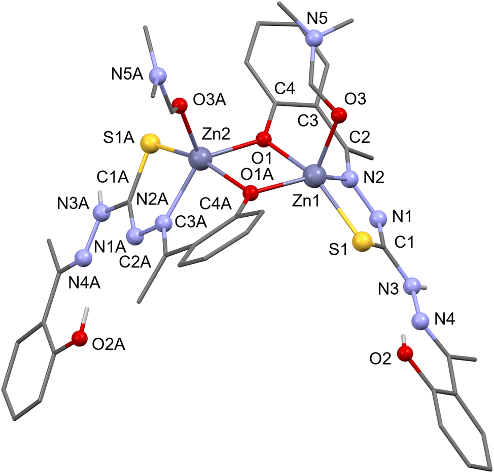
The molecular structure is shown in the figure. Table 1 contains the crystallographic data and the list of the atoms including atomic coordinates and displacement parameters can be found in the cif-file attached to this article.
Data collection and handling.
| Crystal: | Clear light colourless prism |
| Size: | 0.29 × 0.15 × 0.05 mm |
| Wavelength: | Mo Kα radiation (0.71073 Å) |
| μ: | 1.18 mm−1 |
| Diffractometer, scan mode: | Bruker APEX2, φ and ω scans |
| θmax, completeness: | 29.4°, 100 % |
| N(hkl)measured, N(hkl)unique, Rint: | 42005, 11020, 0.074 |
| Criterion for Iobs, N(hkl)gt: | Iobs > 2 σ(Iobs), 6508 |
| N(param)refined: | 632 |
| Programs: | Bruker, 1 SIR2014, 2 SHELX, 3 Mercury, 4 WinGX 5 |
1 Source of materials
Synthesis of ligand (H 2 L): A mixture of thiocarbohydrazide (0.4 g) and o-hydroxyacetophenone (0.92 ml) in ethanol was heated under reflux for 3 h in the presence of one drop of concentrated hydrochloric acid. The reaction mixture was allowed to cool overnight. The white precipitate was filtered off and purified by recrystallization from absolute ethanol. Pale-yellow powder-like substance was obtained (yield 0.72 g, 67.4 %). Synthesis of complex [Zn2(μ–L)2(DMF)2]⋅DMF: To a hot solution of zinc(II) acetate dihydrate (0.25 mmol; 54.8 mg) in dimethylformamide was added the hot solution of L (0.5 mmol, 112 mg). After 5 days the mixture was filtered, yielding yellow-orange crystals (yield: 0.194 g, 95.44 %).
2 Experimental details
Dimethylformamide (DMF) bonded to Zn1 was refined by applying rigid-body restraint (RIGU) on the atoms O3, C18, and N5. Oxygen and carbon atoms of the DMF ligand bonded to Zn2 are disordered. To model the disorder, SIMU and SADI restraints were used. Occupancies of O3A, C18A/O3B,C18B were refined to 0.65/0.35. Hydrogen atoms bonded to O were located in difference Fourier map. Remaining hydrogen atoms were placed at calculated positions. All hydrogen atoms were refined using riding model.
3 Comment
Thiocarbohydrazones are important in various biological processes, including catalytic reactions 6 and anticancer activity. 7 We reported previously the crystal structure of the title ligand 1,5-bis[(E)-1-(2-hydroxyphenyl)ethylidene]thiocarbonohydrazide (H2L) as a dimethylsulfoxide solvate. 8 Due to versatile coordination capabilities thiocarbohydrazones can form complexes of varying nuclearity. 9 In many cases biological activity of thiocarbohydrazones is influenced by their variable coordination modes towards metal atoms. 10 The crystal structures of the title ligand H2L coordinated to Mo 11 and Sn 12 , 13 have been reported previously. Here, we report the synthesis and crystal structure of the Zn(II) complex with the H2L.
The asymmetric unit consist of dimeric Zn(II) complex molecule and dimethylformamide solvent. The figure depicts the complex molecule and atom labeling scheme. Due to clarity hydrogen atoms bonded to C and some C labels have been omitted from the Figure. Both metal atoms are five-coordinated in a distorted square-pyramidal environment. The base of the square-pyramid for both zinc atoms is formed by the sulfur, imine nitrogen and two bridging phenolate oxygen atoms. The vertices of the coordination polyhedra are occupied by the dimethylformamide (DMF). The Addison distortion index τ 14 is 0.25 for Zn1 and 0.43 for Zn2. DMF ligand bonded to Zn1 is disordered. Only one of the disordered DMF positions is shown in the Figure and is considered for the calculation of Adisson distortion index. Comparison of the bond lengths in ligand L with the uncoordinated molecule H2L 15 indicate lengthening of the bonds involving coordinated atoms (C1A–S1A = 1.73/1.74 Å, N2A–N1A = 1.40/1.39 Å, vs. C–N = 1.68 Å, N–N = 1.37 Å in uncoordinated ligand). In both ligands L, the hydroxy group is hydrogen bonded to a double bonded N atom, generating a six-membered ring. Geometry of these interactions is O2–H52⋯N4 = 1.88(4) Å/148(5)° and O2A–H51⋯N4A = 1.80(4) Å/142(4)°. Same interaction is observed in this ligand in the crystal structures where it is uncoordinated 16 or coordinated. 11 , 12 , 13 The complex molecules are arranged into chains directed along the a-axis. This arrangement is associated with intermolecular C–H⋯π interaction (C13A–H19⋯O2 i = 2.46 Å/156°); (symmetry code: i = 1/2 + x, 1/2 − y, 1 − z). There are no other significant intermolecular contacts between the molecules in the chain. The DMF solvent is situated between the molecular chains and bonded to complex molecule through the hydrogen bond, N3A–H53⋯O4 = 2.24(4) Å/177(4)°. The solvent molecule is not involved in significant intermolecular contacts between the molecular chains.
References
1. Bruker. Apex2, Saint and Sadabs; Bruker AXS Inc.: Madison, Wisconsin, USA, 2009.Search in Google Scholar
2. Burla, M. C.; Caliandro, R.; Carrozzini, B.; Cascarano, G. L.; Cuocci, C.; Giacovazzo, C.; Mallamo, M.; Mazzone, A.; Polidori, G. Crystal Structure Determination and Refinement via SIR2014. J. Appl. Cryst. 2015, C48, 306–309. https://doi.org/10.1107/S1600576715001132.Search in Google Scholar
3. Sheldrick, G. M. Crystal Structure Refinement with Shelxl. Acta Crystallogr. 2014, C71, 3–8. https://doi.org/10.1107/S2053229614024218.Search in Google Scholar PubMed PubMed Central
4. Macrae, C. F.; Edgington, P. R.; McCabe, P.; Pidcock, E.; Shields, G. P.; Taylor, R.; van der Streek, T.; van de Streek, J. Mercury: Visualization and Analysis of Crystal Structures. J. Appl. Crystallogr. 2020, 39, 453–457. https://doi.org/10.1107/S002188980600731X.Search in Google Scholar
5. Farrugia, L. J. WinGXand ORTEP for Windows: An Update. J. Appl. Crystallogr. 2012, 45, 849–854. https://doi.org/10.1107/S0021889812029111.Search in Google Scholar
6. Lateef, L.; Manoj, E.; Prathapachandra Kurup, M. R. Synthesis and Spectral Characterization of Zinc(II) Complexes of N(4)-substituted Thiosemicarbazone Derived from Salicylaldehyde: Structural Study of a Novel –OH Free Zn(II) Complex. Polyhedron 2007, 26, 4107–4113. https://doi.org/10.1016/j.poly.2007.05.023.Search in Google Scholar
7. Saswati, L. S.; Mohanty, M.; Banerjee, A.; Biswal, S.; Horn, A.Jr.; Schenk, G.; Brzezinski, K.; Sinn, E.; Reuter, H.; Dinda, R. Polynuclear Zinc(II) Complexes of Thiosemicarbazone: Synthesis, X-ray Structure and Biological Evaluation. J. Inorg. Biochem. 2020, 203, 110908–110921. https://doi.org/10.1016/j.jinorgbio.2019.110908.Search in Google Scholar PubMed
8. Bigović’, M.; Kaludjerovic, M.; Shova, S.; Tomić’, Z. D.; Jaćimović, Ž. K. Crystal Structure of 1,5-bis[(E)-1-(2-hydroxyphenyl)ethylidene] Thiocarbonohydrazide Dimethyl Sulfoxide Monosolvate, C17H18N4O2S.C2H6OS. Z. Kristallogr. N. Cryst. Struct. 2024, 239, 941–943. https://doi.org/10.1515/ncrs-2024–0244.10.1515/ncrs-2024-0244Search in Google Scholar
9. Dragancea, D.; Arion, V. B.; Shova, S.; Rentschler, E.; Gerbeleu, N. V. Azine-Bridged Octanuclear Copper(II) Complexes Assembled with a One-Stranded Ditopic Thiocarbohydrazone Ligand. Angew. Chem. Int. Ed. 2005, 44/b, 7938–7942. https://doi.org/10.1002/anie.200501807.Search in Google Scholar PubMed
10. Wang, J.; Wang, Y.-T.; Fang, Y.; Lu, Y.-L.; Li, M.-X. Tin Thiocarbonohydrazone Complexes: Synthesis, Crystal Structures and Biological Evaluation. Toxicol. Res. 2019, 8, 862–867. https://doi.org/10.1039/c9tx00109c.Search in Google Scholar PubMed PubMed Central
11. Rana, A.; Dinda, R.; Sengupta, P.; Ghosh, S.; Falvello, L. R. Synthesis, Characterisation and Crystal Structure of cis-dioxomolybdenum(VI) Complexes of Some Potentially Pentadentate but Functionally Tridentate (ONS) Donor Ligands. Polyhedron 2002, 21, 1023–1030. https://doi.org/10.1016/S0277–5387(02)00913–0.10.1016/S0277-5387(02)00913-0Search in Google Scholar
12. Chee, D. N. A.; Affan, M. A.; Ahmad, F. B.; Asaruddin, M. R.; Sam, N.; Salam, M. A.; Ismail, A.; Tan, S. H. Synthesis, Characterization, and Antibacterial Activity of organotin(IV) Complexes with 2-hydroxyacetophenone Thiocarbohydrazone. J. Coord. Chem. 2011, 64, 4191–4200. https://doi.org/10.1080/00958972.2011.631532.Search in Google Scholar
13. Affan, M. A.; Dayang, N. A.; Chee, C.; Assima, Z.; Ng, S. W. Di-n-butyl(1-[1-(2-hydroxyphenyl)ethylidene]-5-[1-(2-oxidophenyl)ethylidene] thio-carbazonato-k3O5,N5,S)tin(IV). Acta Cryst. 2010, E66, m618–m619. https://doi.org/10.1107/S1600536810016016.Search in Google Scholar PubMed PubMed Central
14. Addison, A. W.; Rao, T. N.; Reedijk, J.; van Rijn, J.; Verschoor, G. C. Synthesis, Structure, and Spectroscopic Properties of copper(II) Compounds Containing nitrogen-sul[hur Donor Ligands; the Crystal and Molecular Structure of aqua[1,7-bis(N-methylbenzimidazol-2′-yl)-2,6-dithiaheptane]copper(II) Perchlorate. J. Chem. Soc. Dalton Trans. 1984, 1349–1356. https://doi.org/10.1039/DT9840001349.Search in Google Scholar
15. Affan, M. A.; Chee, D. N. A.; Ahmad, F. B.; Tiekink, E. R. T. 1,5-Bis[(E)-1-(2-hydroxyphenyl)ethylidene]thiocarbonohydrazide Monohydrate. Acta Cryst. 2010, E66, o555. https://doi.org/10.1107/S1600536810004241.Search in Google Scholar PubMed PubMed Central
16. Schmitt, B.; Gerber, T.; Hosten, E.; Betz, R. A Monoclinic Polymorph of (1E,5E)-1,5-bis(2-hydroxybenzilidene) Thiocarbonohydrazide. Acta Cryst. 2011, E67, o2206–o2207; https://doi.org/10.1107/S1600536811030340.Search in Google Scholar PubMed PubMed Central
© 2025 the author(s), published by De Gruyter, Berlin/Boston
This work is licensed under the Creative Commons Attribution 4.0 International License.
Articles in the same Issue
- Frontmatter
- New Crystal Structures
- Crystal structure of (S)-N-(10-((2,2-dimethoxyethyl)amino)-1,2,3-trimethoxy-9-oxo-5,6,7,9-tetrahydrobenzo[a]heptalen-7-yl)acetamide, C25H32N2O7
- The crystal structure of 6,6′-difluoro-3,3′-dimethyl-5,5′-di(10H-phenoxazin-10-yl)- [1,1′-biphenyl]-2,2′-dicarbonitrile, C40H24F2N4O2
- Crystal structure of poly[(di-ethylenediamine-κ2N,N′)cadmium(II) tetradedocyloxidohexavanadate] (V4+/V5+ = 2/1), C4H16CdN4O14V6
- The crystal structure of poly[bis(dimethylformamide-κ1N)-(μ4-2′,3,3″,5′-tetrakis(trifluoromethyl)-[1,1′:4′,1″-terphenyl]-4,4″-dicarboxylato-κ4 O,O′: O″,O‴)dicadmium(II)], C27H15CdF12NO5
- Crystal structure of bis(μ2-ferrocenylcarboxylato-O,O′)-(μ3-oxido-κ3O:O:O)-bis(μ2-salicyladoximato-κ2N,O,O′)-(μ2-isopropoxo)-tris(isopropoxy-κ1O trititanium(IV)), C48H55N2O13Fe2Ti3
- Crystal structure of 3-(diethylamino)-7,9,11-trimethyl-8-phenyl-6H,13H-12λ4,13λ4-chromeno[3′,4′:4,5]pyrrolo[1,2-c]pyrrolo[2,1-f][1,3,2]diazaborinin-6-one, C28H26BF2N3O2
- The crystal structure of catena-poly[aqua-μ2-2-nitro-benzene-1,3-dicarboxylato-κ2O,O′)-(1,10-phenanthroline-κ2N,N′)-zinc(II)], C20H13N3O7Zn
- Crystal structure of poly[diaqua-{μ3-1-(3-carboxylatophenyl)-4-oxo-1,4-dihydropyridazine-3-carboxylato-κ4O,O′:O′′:O′′′′}manganese(II)] hydrate
- Crystal structure of N′-((1-hydroxycyclohexyl)(phenyl)methyl)-2-methoxybenzohydrazide methanol solvate, C22H28N2O4
- The cocrystal of caffeic acid — progesterone — water (1/2/1), C51H70O9
- Crystal structure of (((oxido(quinolin-6-yl)methoxy)triphenyl-λ5-stibanyl)oxy)(quinolin-7-yl)methanolate
- Crystal structure of [(E)-6′-(diethylamino)-2-(2-(((E)-pyren-1-ylmethylene)amino)ethyl)-4′-(2-((E)-1,3,3-trimethylindolin-2-ylidene)ethylidene)-1′,2′,3′,4′-tetrahydrospiro[isoindoline-1,9′-xanthen]-3-one]-methanol, solvate C57H56N4O3
- The crystal structure of 1-(acridin-9-yl)pyrrolidine-2,5-dione, C17H22N2O2
- Crystal structure of N-(4-acetylphenyl)-2-(6-methoxynaphthalen-2-yl)propanamide, C22H21NO3
- The crystal structure of 5,10,15,20-tetrakis(4-(1H-1,2,4-triazol-1-yl)phenyl)porphyrin, C52H34N16
- Crystal structure of hexacarbonyl-μ2-[phenylmethanedithiolato-κ4S:S,S′:S′]diiron (Fe–Fe) C13H6Fe2O6S2
- Crystal structure of diiodo-bis(1-((2-propyl-1H-benzo[d]imidazol-1-yl)methyl)-1H-benzo[d][1,2,3]triazole-κ1N)cadmium(II), C34H34CdI2N10
- Crystal structure of (E)-(3-(3-bromophenyl)acryloyl)ferrocene, C19H15BrFeO
- Crystal structure of catena-poly(μ2-6-chloropyridine-2-carboxylato-κ3N,O:O′)(6-chloropyridine-2-carboxylato-κ2O,N)copper(II), C12H6Cl2N2O4Cu
- Crystal structure of poly[diaqua-μ3-(5-(3,5-dicarboxy-2,4,6-trimethylbenzyl)-2,4,6-trimethylisophthalato)-κ6O,O′:O″,O‴:O‴′,O‴″) terbium(III)-monohydrate], C23H28TbO12
- Crystal structure of (E)-2-(((5-chloro-3-methyl-1-phenyl-1H-pyrazol-4-yl)methylene)amino)-3′,6′-dihydroxyspiro[isoindoline-1,9′-xanthen]-3-one – ethanol (1/2), C35H33ClN4O6
- The crystal structure of 3-(5-amino-3-phenylisoxazol-4-yl)-4-chloro-3-hydroxyindolin-2-one, C17H12ClN3O3
- The crystal structure of dimethylammonium 4-[2-(4-fluorophenyl)-4, 5-diphenyl-1H-imidazol-1-yl]benzenesulfonate, C29H26FN3O3S
- Crystal structure of (R)-2-ammonio-3-((5-carboxypentyl)thio)propanoate
- Crystal structure of 4-cyclohexyl-5-(thiophen-2-yl)-2,4-dihydro-3H-1,2,4-triazole-3-thione, C12H15N3S2
- The crystal structure of 4,6-bis(dimethylamino)-2-fluoroisophthalonitrile, C12H13FN4
- Hydrogen bonding in the crystal structure of nicotin-1,1′-dium tetrabromidomanganate(II)
- The crystal structure of bis(2-bromobenzyl)(2-((2-oxybenzylidene)amino)-4-methylpentanoato-κ3N, O,O′)tin(IV), C27H27Br2NO3Sn
- Crystal structure of (E)-(3-(p-tolyl)acryloyl)ferrocene, C20H18FeO
- Crystal structure of (E)-7-fluoro-2-((5-(4-methylpiperazin-1-yl)pyridin-2-yl)methylene)-3,4-dihydronaphthalen-1(2H)-one, C21H22FN3O
- Crystal structure of (E)-7-methoxy-2-((5-(4-methylpiperazin-1-yl)pyridin-2-yl)methylene)-3,4-dihydronaphthalen-1(2H)-one, C22H25N3O2
- The crystal structure of poly(bis(μ2-1,3,5-tri(1H-imidazol-1-yl)benzene-κ2N:N′)-(μ2-2,3,5,6-tetrafluoroterephthalato-κ2O:O′)-manganese(II), C38H24F4N12O4Mn
- Crystal structure of (3,4-dimethoxybenzyl)triphenylphosphonium bromide ethanol solvate, C29H32BrO3P
- Crystal structure of tetraethylammonium hydrogencarbonate – (diaminomethylene)thiourea – water (2/1/3)
- Crystal structure of N, N-Dimethyl-N′-tosylformimidamide, C10H14N2O2S
- The crystal structure of ethyl 2-methyl-5-oxo-4-(2-methoxyphenyl)-1,4,5,6,7,8-hexahydroquinoline-3-carboxylate, C20H23N2O4
- Crystal structure of bis(μ2-1,5-bis[(E)-1-(2-hydroxyphenyl)ethylidene] thiocarbonohydrazide)-bis(dimethylformamide)-dizinc(II) dimethylformamide solvate, C40H46N10O6S2Zn2⋅C3H7NO
- Crystal structure of azido-κ1N{hydridotris(3-tert-butyl-5-methylpyrazol-1-yl)borato-κ3N,N′,N″}copper(II), C24H40BCuN9
- The crystal structure of fac-tricarbonyl(1,10-phenanthroline-κ2N,N′)-(azido-κ1N)rhenium(I), C15H8N5O3Re
- Crystal structure of 4-((triphenylphosphonio)methyl)pyridin-1-ium tetrachloridozincate(II), C24H22Cl4NPZn