Ultrafast optical modulation of vibrational strong coupling in ReCl(CO)3(2,2-bipyridine)
-
Liying Chen
 ,
Alexander M. McKillop
,
Ashley P. Fidler
und
Marissa L. Weichman
,
Alexander M. McKillop
,
Ashley P. Fidler
und
Marissa L. Weichman


Abstract
Polaritons – hybrid light-matter states formed from the strong coupling of a bright molecular transition with a confined photonic mode – may offer new opportunities for optical control of molecular behavior. Vibrational strong coupling (VSC) has been reported to impact ground-state chemical reactivity, but its influence on electronic excited-state dynamics remains unexplored. Here, we take a first step towards excited-state VSC by demonstrating optical modulation of the ReCl(CO)3(bpy), (bpy = 2,2-bipyridine) complex under VSC using femtosecond ultraviolet (UV)-pump/infrared (IR)-probe spectroscopy. We establish ground-state VSC of ReCl(CO)3(bpy) in a microfluidic Fabry-Pérot cavity equipped with indium tin oxide (ITO)-coated mirrors. ITO is effectively dichroic as it is reflective in the IR and transmissive in the UV-visible and therefore minimizes optical interference. Excitation with UV pump light drives ReCl(CO)3(bpy) into a manifold of electronic excited states that subsequently undergo non-radiative relaxation dynamics. We probe the transient response of the strongly-coupled system in the mid-IR, observing both Rabi contraction and cavity-filtered excited-state absorption signatures. We reconstruct the intrinsic response of intracavity molecules from the transient cavity transmission spectra to enable quantitative comparison with extracavity control experiments. We report no changes in the excited-state dynamics of ReCl(CO)3(bpy) under ground-state VSC. However, we do observe significant amplification of transient vibrational signals due to classical cavity-enhanced optical effects. This effort lays the groundwork to pursue direct excited-state VSC aimed at modulating photochemical reactivity.
1 Introduction
The strong interaction of molecular vibrations with confined electromagnetic fields to form hybrid light-matter states known as polaritons has emerged as a potential new route for photonic control of molecular behavior [1], [2], [3], [4], [5], [6], [7], [8]. The emergence of vibrational polaritons appears to correlate with altered ground-state reactivity and vibrational energy redistribution [9], [10], [11], [12], [13], though many open questions remain surrounding the mechanisms and reproducibility of these effects [4], [14], [15], [16]. In any event, the early work in this field has stimulated broad interest in understanding how and when vibrational strong coupling (VSC) modulates molecular behavior. In this work, we take a first step towards examining how VSC may impact electronic excited-state trajectories by optically exciting the ReCl(CO)3(bpy), (bpy = 2,2-bipyridine) metal carbonyl complex under VSC in a microfluidic Fabry-Pérot (FP) optical cavity (Figure 1(A) and (B)).

Experimental scheme for ultrafast UV-pump/IR-probe spectroscopy of ReCl(CO)3(bpy) in free space and under vibrational strong coupling (VSC) in a microfluidic Fabry-Pérot cavity. (A) Excitation of extracavity ReCl(CO)3(bpy) with a 390 nm UV pump pulse initiates excited-state dynamics, which are subsequently probed with a broadband mid-IR probe pulse centered near 2,000 cm−1. The schematic energy-level diagram illustrates how photoexcitation drives ReCl(CO)3(bpy) from its S0 electronic ground state to the singlet metal-to-ligand charge transfer (1MLCT) manifold, after which the system undergoes intersystem crossing to the triplet (3MLCT) manifold, and ultimately vibrational cooling and relaxation back to the ground state. (B) Scheme for spectroscopy of ReCl(CO)3(bpy) under VSC of a carbonyl stretching mode in the S0 ground state, which generates upper and lower polariton states (UP, LP). Excitation with a UV pump pulse again initiates excited-state dynamics, while a broadband mid-IR probe pulse records subsequent transient changes to the cavity transmission spectrum in the carbonyl stretching region. (C) Infrared absorption spectrum of ReCl(CO)3(bpy) in solution in DMSO in the carbonyl stretching region. The a′ symmetric carbonyl stretch near 2,018 cm−1 (marked with a purple arrow) is targeted for resonant VSC in this work. (D) UV-visible absorption spectrum of ReCl(CO)3(bpy) in solution in DMSO. The 390 nm pump used here excites the red shoulder of the broad 1MLCT band.
Transient vibrational motions are sensitive reporters of excited-state dynamics [17], [18] and can even influence photochemical outcomes [19], [20], [21]. Mode-selective infrared (IR) excitation of excited-state vibrations has accordingly been used to modulate charge transfer in donor-bridge-acceptor complexes [22], [23], [24], [25]. Such mode-selective excitation strategies face well-known challenges, however: competition with intramolecular vibrational relaxation and energy dissipation limits the scope of control over reaction outcomes. Achieving broadly-applicable vibrational modulation of excited-state dynamics remains an open research goal, motivating the search for new approaches, including cavity coupling. A particularly interesting possibility is the idea of engineering VSC in electronic excited states, where a cavity mode couples resonantly to a vibrational transition in the electronically excited manifold. While excited-state VSC has been proposed theoretically [26] and excited-state processes have been studied under ground-state VSC [27], actual experimental implementation of excited-state VSC remains elusive.
Achieving excited-state VSC requires a molecular candidate with: (a) a sufficiently strong IR-active vibrational mode to achieve VSC in the electronic excited state; (b) an optically bright electronic transition which can be pumped to populate the excited state; (c) distinct vibrational frequencies in the electronic ground and excited states to ensure resonant cavity-coupling only upon electronic excitation; and (d) sufficient population in the excited state to achieve collective excited-state VSC, even transiently. This last criterion is challenging to establish experimentally. Reaching the collective strong coupling regime relies on achieving a sufficiently large Rabi splitting, ℏΩ R , which scales with N 1/2, where N is the number of coupled molecules [2], [8].
Here, we identify the ReCl(CO)3(bpy) complex as a target molecule that appears to meet criteria (a)–(c) above. ReCl(CO)3(bpy) exhibits strong and well-characterized absorption features in both the mid-IR and the UV-visible (Figure 1(C) and (D)). The lowest electronic band lies between 350 and 400 nm (Figure 1(D)) and corresponds to excitation from the S0 electronic ground state to the manifold of singlet metal-to-ligand charge transfer (1MLCT) excited states [28], [29]. As discussed by Šrut et al. [28], the 1MLCT band is dominated by the S1, S2, and S3 electronic states, with S2 expected to be the brightest state that will be initially populated by optical pumping. Excitation into the 1MLCT manifold leads to rapid intersystem crossing into the triplet 3MLCT manifold on a 1–2 ps timescale, followed by electronic relaxation, vibrational cooling, and solvent reorganization on 5–10 ps timescales (Figure 1(A)). Prior UV-pump/IR-probe experiments performed on ReCl(CO)3(bpy) report significant blue-shifting of its three carbonyl stretching modes upon optical excitation [28], [30], [31], [32], [33]. For instance, the strongest-absorbing a′ carbonyl symmetric stretch lies at 2,018 cm−1 in the S0 ground state (Figure 1(C)) and appears at >2,060 cm−1 in the excited-state manifold [28], [30], [31], [32], [33]. The intensity of this symmetric carbonyl stretching mode, its large frequency shift upon photoexcitation, and its relative lack of spectral congestion make it a compelling target for modulation of ground-state VSC with UV excitation, as we demonstrate here, and perhaps eventually excited-state VSC.
Here, we perform ultrafast UV-pump/IR-probe spectroscopy on ReCl(CO)3(bpy) in dimethyl sulfoxide (DMSO) solution embedded in a microfluidic FP optical cavity. We demonstrate VSC of the a′ symmetric carbonyl stretching mode of ReCl(CO)3(bpy) in the S0 electronic ground state and modulate the collective coupling strength via optical pumping of population into the excited-state manifold, as shown schematically in Figure 1(B). This result represents both a first step towards excited-state VSC and a new platform for cavity-mediated nonlinear optics. This system also provides a means to test whether ground-state VSC has any influence on excited-state dynamics. We ultimately find no statistically-significant change in the excited-state dynamics of ReCl(CO)3(bpy) under ground-state VSC, as is perhaps expected given that the excited-state vibrations are both weakly and off-resonantly cavity-coupled in the current experiments.
Three important technical considerations underpin this work. First, we use indium tin oxide (ITO)-coated optics as dichroic cavity mirrors to enable UV-pump/IR-probe experiments with minimal optical artifacts. While the dichroic properties of ITO are well-known [34], its use as a mirror coating is not yet widespread for VSC. Secondly, we introduce a spectral reconstruction algorithm that allows us to extract the dynamics of strongly-coupled intracavity molecules, yielding observables that can be compared directly against extracavity controls. Transient spectroscopy of polaritonic systems can be a minefield of optical artifacts and interference effects that must be properly accounted for during analysis [35], [36], [37]. Our approach here is to reconstruct the intrinsic response of intracavity molecules by fitting transient cavity data to the analytical classical optics expression for transmission through an FP cavity. Finally, our measurements reveal a pronounced cavity-mediated enhancement of transient pump-probe signals. This finding can be attributed to classical cavity-enhancement effects, e.g., the extreme sensitivity of the FP cavity transmission spectrum to minute changes in intracavity absorption.
Our results demonstrate the feasibility of optical modulation of VSC, tracking excited-state vibrational dynamics inside microfluidic cavity structures, and quantitative comparison of molecular dynamics across extracavity and intracavity experiments. We also provide a foundation for future efforts to achieve excited-state VSC, which would enable tests of cavity-modification of energy dissipation, charge transport, and photochemistry on excited-state surfaces.
2 Methods and materials
The ultrafast UV-pump/IR-probe spectrometer used herein represents a minor modification to the apparatus we have described previously [38], [39]. The system is based on a Ti:sapphire amplifier (Astrella, Coherent) which delivers 7 mJ, 60 fs pulses of 800 nm light at a 1 kHz repetition rate. UV pump pulses are generated using a tunable optical parametric amplifier (OPA, OPerA Solo with FH/SHSF options, Light Conversion), pumped with 2.5 mJ of the 800 nm fundamental to produce 46 µJ per pulse at 390 nm. Mid-IR probe pulses are produced using a second OPA (OPerA Solo with NDFG option, Light Conversion), pumped with 2.5 mJ of 800 nm fundamental light to produce 30 µJ pulses centered at 2,040 cm−1 (4,900 nm) with a full-width at half-maximum (fwhm) bandwidth of 250 cm−1.
We delay the mid-IR probe relative to the UV pump by up to 2.2 ns using a motorized stage (DL325, Newport). The pump and probe beams are spatially overlapped at the sample with typical beam diameters of 200 µm. The probe strikes the sample at normal incidence while the pump is incident at a crossing angle of 7°. All measurements reported herein employ a magic-angle polarization scheme between pump and probe. Mid-IR probe light transmitted through the sample is spectrally dispersed using a diffraction grating and collected on a HgCdTe array detector (2DMCT, PhaseTech) operating in shot-to-shot mode, with the pump beam modulated at 500 Hz using a mechanical chopper (MC2000, Thorlabs). We calculate differential signals as ΔOD = −log10[I T,t /I T,0], where I T,t represents the intensity of probe light transmitted by the sample at time t after the pump pulse is incident and I T,0 represents the “pump-off” probe transmission spectrum acquired when the pump beam is blocked. We establish an instrument response function of 120 fs, comparable to that reported in our prior work [38]. Data is recorded using home-built LabVIEW programs and processed in MATLAB.
We prepare saturated (>40 mM) solutions of ReCl(CO)3(bpy) (99 %, Strem Chemicals) in DMSO (≥99.9 %, Sigma-Aldrich). We choose DMSO as solvent because it features high solubility for ReCl(CO)3(bpy) and has been used in prior ultrafast studies of this complex, facilitating direct comparison with established literature [28], [31]. ReCl(CO)3(bpy):DMSO solutions are flowed through a demountable microfluidic cell (TFC-M13-3, Harrick Scientific) using a peristaltic pump (Masterflex) to ensure continuous sample refreshment. For extracavity measurements, we assemble the flow cell with CaF2 windows spaced with a 25 µm polytetrafluoroethylene (PTFE) spacer. For intracavity experiments, we fit the same flow cell with ITO-coated CaF2 substrates (Colorado Concept Coatings) that serve as dichroic cavity mirrors. The ITO mirrors consist of a 20–30 nm SiO2 base layer to adhere the coating to the substrate, a 150 nm (15 Ω/sq.) ITO layer, and a 50 nm SiO2 protective overcoat. The microfluidic cell assembly and ITO mirrors are illustrated in Figure 2(A).
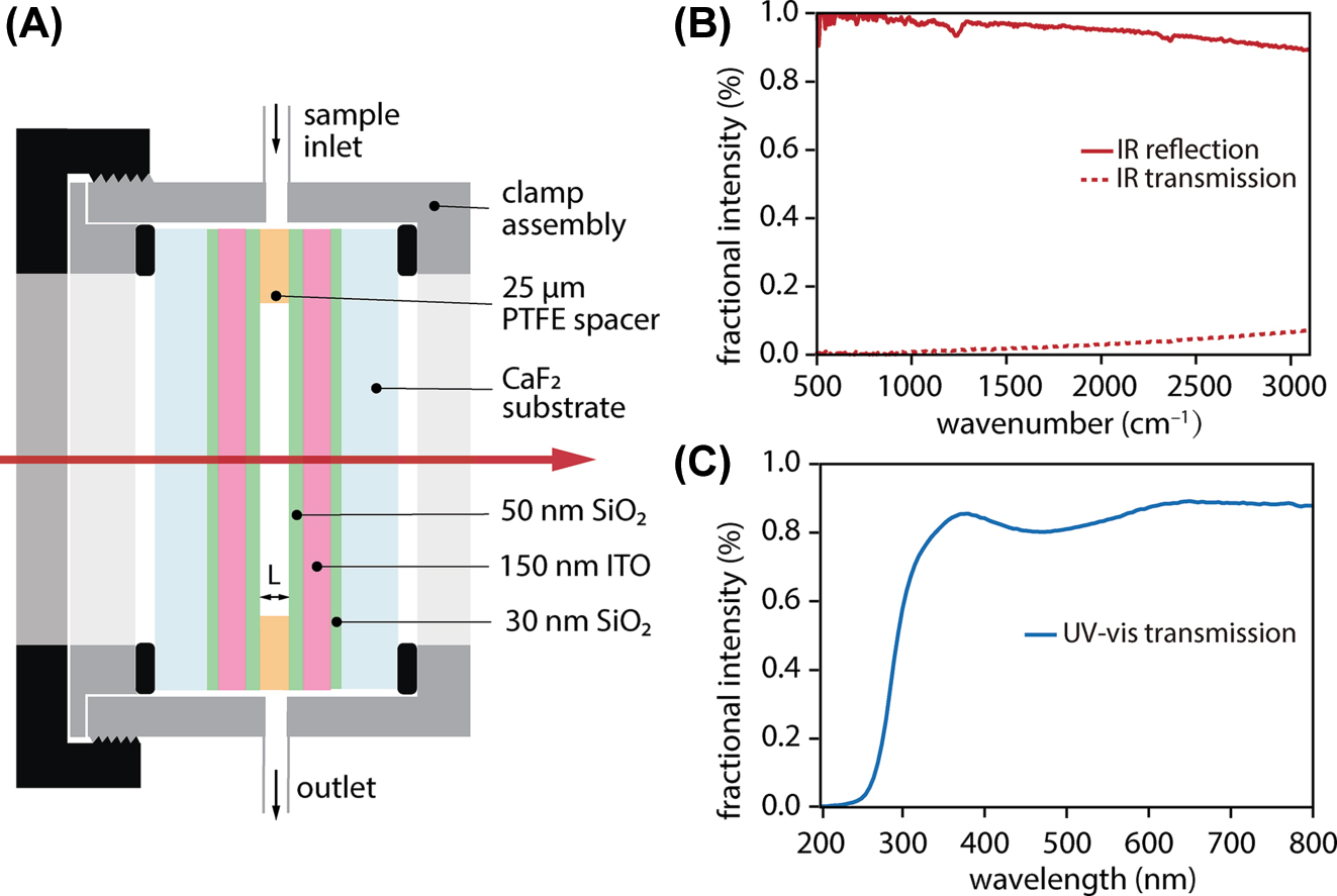
Microfluidic Fabry-Pérot cavity with dichroic ITO mirrors used for vibrational strong coupling and UV-pump/IR-probe experiments. (A) Schematic of microfluidic cavity assembled from two ITO-coated CaF2 substrates separated by a 25 µm polytetrafluoroethylene (PTFE) spacer. (B) Reflectivity and transmission spectra of a single ITO mirror confirming high reflectivity across the mid-IR. (C) Transmission spectrum of a single ITO mirror demonstrating high broadband transmittance in the UV-visible.
We use a commercial Fourier transform spectrometer (Nicolet iS50 FT-IR, Thermo Scientific) and UV-visible spectrometer (Cary 60 UV–Vis Spectrophotometer, Agilent) to characterize the broadband absorption spectra of extracavity solutions (Figure 1(C) and (D)), the reflectivity and transmission spectra of the ITO mirrors (Figure 2(B) and (C)), and the transmission spectra of empty FP cavities constructed from these ITO mirrors (Figure 3(A)). All other transmission spectra of strongly-coupled cavity devices shown herein are acquired using our ultrafast mid-IR beamline; these spectra are comparatively narrowband but the small spot size afforded by the collimated laser beam minimizes the impacts of cavity non-uniformity on transmission measurements [16]. We take advantage of deviations from perfect mirror parallelism to access distinct cavity detuning conditions by translating the cavity in the plane of the mirrors.

Characterization of ITO Fabry-Pérot cavities. (A) Transmission spectrum of a representative empty cavity formed by two ITO mirrors separated by a 25 µm PTFE spacer. (B) Cavity transmission spectrum of ReCl(CO)3(bpy) in DMSO under strongly-coupled conditions in a 25 µm microfluidic cavity (solid light red trace) plotted against the bare molecular absorption spectrum in the region near the a′ symmetric carbonyl stretch of ReCl(CO)3(bpy) (blue trace). A simulated strongly-coupled transmission spectrum is also provided (red dotted trace) using the classical expression for the transmission spectrum of a Fabry-Pérot cavity (Eq. (1)).
3 Results and discussion
We first characterize the static optical properties of our dichroic ITO cavities and confirm that we can achieve VSC in solution-phase ReCl(CO)3(bpy) samples in Section 3.1. We then review the ultrafast excited-state dynamics measured for extracavity ReCl(CO)3(bpy) with UV-pump/IR-probe spectroscopy in Section 3.2. In Section 3.3, we detail UV-pump/IR-probe cavity transmission measurements performed for ReCl(CO)3(bpy) under VSC, highlighting the distinct observables between intracavity measurements and extracavity controls. Finally, in Section 3.4 we introduce and apply a spectral reconstruction algorithm to recover the response of intracavity molecules independent of cavity filtering and directly compare the extracted intracavity dynamics against extracavity benchmarks.
3.1 Vibrational strong coupling of ReCl(CO)3(bpy) in ITO cavities
We begin by characterizing the optical properties of the dichroic ITO mirrors and the resulting FP microcavities constructed from these mirrors. ITO is chosen as a mirror coating to provide sufficient reflectivity to achieve VSC in the mid-IR, while maintaining high transparency in the UV for efficient photoexcitation of the intracavity sample. Figure 2(B) plots the reflection and transmission spectra of a single ITO mirror across the mid-IR. The mirror reflectivity exceeds 90 % in the carbonyl stretching region near 2,000 cm−1, which is of interest for VSC of ReCl(CO)3(bpy). Meanwhile, Figure 2(C) plots the transmission spectrum of a single ITO mirror across the UV-visible. The transmission exceeds 80 % in the 350–400 nm region where the excited-state 1MLCT band of ReCl(CO)3(bpy) lies. The mid-IR transmission spectrum of a pair of ITO mirrors assembled into an empty 25 µm FP cavity is shown in Figure 3(A). The ITO cavities used herein feature typical free spectral ranges of 150 cm−1 and empty-cavity mode linewidths of 15–18 cm−1 fwhm.
We next confirm that ReCl(CO)3(bpy) can be vibrationally strongly-coupled in such a cavity. To achieve resonant VSC, ReCl(CO)3(bpy):DMSO solution is injected into a 25 µm FP cavity and a cavity mode is tuned into resonance with the desired molecular band by compressing the microfluidic clamp assembly to slightly change the cavity length. We couple to longitudinal FP cavity modes of typical mode order m ∼ 13–15. Figure 3(B) shows the static transmission spectrum of a representative cavity strongly-coupled to the a′ carbonyl symmetric stretching band of ReCl(CO)3(bpy) at 2,018 cm−1 (solid light red trace) plotted against the extracavity ReCl(CO)3(bpy) absorption spectrum (blue trace). A simulated cavity transmission spectrum produced using the classical FP cavity expression discussed below in Section 3.4 is also plotted in Figure 3(B) (red dotted trace). We observe a clear splitting of the cavity transmission peak into upper and lower vibrational polaritons (UP, LP). This system achieves a typical Rabi splitting of 24–30 cm−1 which exceeds the linewidths of both the bare cavity mode (∼15 cm−1 fwhm) and molecular absorption line (∼10 cm−1 fwhm).
3.2 Extracavity excited-state vibrational dynamics
We now report on the ultrafast UV-pump/IR-probe spectroscopy of extracavity ReCl(CO)3(bpy) to establish a baseline for its excited-state vibrational dynamics and benchmark against prior literature. We excite the system at 390 nm and record transient signals in the carbonyl stretching region near 2,000 cm−1 (Figure 1(A)). Representative extracavity experimental data are presented in the left-hand column of Figure 4. The free-space static absorption spectrum of ReCl(CO)3(bpy) is reproduced in Figure 4(A), zoomed in on the a′ symmetric carbonyl stretch at 2,018 cm−1. Transient UV-pump/IR-probe spectra are presented in Figure 4(D) as a function of wavenumber and pump-probe delay. Spectral lineouts at selected time delays are plotted in Figure 4(G).

Ultrafast UV-pump/IR-probe spectroscopy of ReCl(CO)3(bpy) in DMSO. Extracavity control data is presented in the left-hand column and intracavity data collected under resonant VSC conditions is presented in the central column. The right-hand column presents the molecular response extracted from intracavity data, which can be compared directly to extracavity data. (A) Steady-state extracavity IR absorption spectrum of ReCl(CO)3(bpy) in DMSO showing the a′ symmetric carbonyl stretching mode near 2,018 cm−1. (B) Static (pump-off) transmission spectrum of strongly-coupled cavity filled with ReCl(CO)3(bpy) in DMSO, exhibiting upper and lower polariton (UP, LP) features with a Rabi splitting of ∼24 cm−1. (C) Representative pump-off intracavity absorption spectrum for ReCl(CO)3(bpy), α 0(ν), modeled with a single Lorentzian lineshape and fit to the pump-off cavity transmission spectrum in the first step of the spectral reconstruction algorithm. (D–F) Ultrafast UV-pump/IR-probe spectra plotted as a function of delay time and wavenumber showing (D) the differential absorption of an extracavity sample, (E) the differential cavity transmission of an intracavity sample under resonant VSC, and (F) the intracavity molecular response, Δα t (ν), reconstructed from the data in panel (E). (G–I) Spectral lineouts at selected pump-probe delays of transient data acquired for (G) the extracavity sample, (H) the intracavity sample, and (I) the reconstructed intracavity molecular response.
The transient UV-pump/IR-probe spectra are dominated by a bleach feature centered near 2,018 cm−1 that appears within 1.5 ps (Figure 4(D) and (G)). We attribute this feature to the ground-state bleach (GSB) of the a′ carbonyl stretching mode in the S0 electronic ground state as population is transferred to the 1MLCT manifold [27], [28], [29], [30], [31], [32]. Concurrently, an excited-state absorption (ESA) band appears, initially centered at 2,040 cm−1 and blue-shifting past 2,060 cm−1 within 5–10 ps. This ESA feature is associated with the symmetric carbonyl stretching mode in the excited state manifold. The spectral shifting of the ESA feature is consistent with structural and electronic relaxation and solvent reorganization in the manifold of 3MLCT states [27], [28], [29], [30], [31], [32]. Both GSB and ESA signals decay gradually for time delays >100 ps, consistent with nonradiative relaxation and repopulation of the S0 electronic ground state.
To quantify the observed extracavity dynamics, we take temporal lineouts at 2,018 cm−1 for the GSB and 2,060 cm−1 for the ESA. We then perform exponential fits to extract the short-term rise and long-term decay time constants for the GSB feature and the long-time decay constant for the ESA feature. To capture the frequency shifting of the ESA feature, we fit the ESA peak center for traces with time delay <50 ps using a Lorentzian and perform an exponential fit to the peak center as a function of time. More details and representative fits are presented in Section S1 of the Supplementary Material. Fitted extracavity time constants from individual experiments are laid out in Table S1, and values averaged across all experiments are summarized in Table 1. All extracavity observations are in agreement with prior studies of the excited-state vibrational dynamics of ReCl(CO)3(bpy) [27], [28], [29], [30], [31], [32] and provide a baseline for comparison with intracavity behavior.
Time constants for the ground-state bleach (GSB) rise and decay and excited-state absorption (ESA) decay and shift from UV-pump/IR-probe experiments on ReCl(CO)3(bpy) in DMSO. Values are reported for extracavity control samples as well as intracavity samples under resonant VSC of the a′ symmetric carbonyl stretch at 2,018 cm−1 and detuned intracavity control samples whose dynamics have been reconstructed with the procedure detailed in Section 3.4. Error bars represent standard deviations arising from averaging time constants from the individual datasets presented in Tables S1–S3 in Section S4 of the Supplementary Material.
| Dynamic process | Extracavity | Intracavity, resonant VSC | Intracavity, detuned |
|---|---|---|---|
| GSB rise (ps) from lineout at 2,018 cm−1 | 1.4 ± 0.3 | 1.6 ± 0.4 | 1.6 ± 0.3 |
| GSB decay (ps) from lineout at 2,018 cm−1 | 150 ± 10 | 170 ± 20 | 150 ± 40 |
| ESA decay (ps) from lineout at 2,060 cm−1 | 120 ± 20 | 150 ± 30 | 130 ± 30 |
| ESA shift (ps) from Lorentzian fit | 6.1 ± 0.7 | 5.2 ± 1.2 | 6.0 ± 1.6 |
3.3 Transient cavity transmission spectra
We now present UV-pump/IR-probe measurements of ReCl(CO)3(bpy) under VSC. Representative experimental data are presented in the central column of Figure 4. ReCl(CO)3(bpy) is prepared under resonant VSC of its a′ symmetric carbonyl stretch at 2,018 cm−1, as described in Section 3.1. Figure 4(B) reproduces the static strongly-coupled cavity transmission spectrum from Figure 3(B), acquired without any UV excitation. We excite the system at 390 nm through the high-transmission region of the ITO cavity mirrors and record transient cavity transmission signals in the carbonyl stretching region near 2,000 cm−1 (Figure 1(B)). Transient UV-pump/IR-probe cavity transmission spectra are presented in Figure 4(E) as a function of wavenumber and pump-probe delay, and spectral lineouts at selected time delays are plotted in Figure 4(H).
The raw transient cavity transmission spectra in the central column of Figure 4 are, at first glance, markedly different than the extracavity transient absorption spectra in the left-hand column. Transient pump-induced changes in the intracavity complex refractive index alter the interference conditions that govern where cavity modes appear and how much light they transmit [37]. Instead of distinct GSB and ESA peaks, the cavity transmission signals are dominated by derivative-like lineshapes centered at the polariton frequencies whose amplitudes and positions evolve as the system relaxes. These derivative lineshapes are due to the well-known pump-induced inward-shifting of polariton modes known as Rabi contraction [6], [35]. Rabi contraction results simply from a reduction in the collective coupling strength as molecular population is transiently driven out of the lower state of the cavity-coupled transition. The timescales for the appearance and decay of these Rabi contraction features are therefore similar to those of the extracavity GSB dynamics.
The ESA dynamics are slightly more difficult to see in the transient intracavity data. We know from the extracavity results (Section 3.2) that the ESA of the carbonyl mode of interest blueshifts from ∼2,040 cm−1 past 2,060 cm−1 in the first 5–10 ps following pump excitation. This ESA feature therefore passes through the spectral region of the UP formed from strong cavity coupling of the ground-state carbonyl vibration, leading to a transient reduction in cavity transmission in the UP region. The ESA therefore manifests as increased ΔOD in the UP region (near 2,040 cm−1) as compared to the LP region (near 2,000 cm−1) in the early time traces in Figure 4(H). This overlap of the ESA with the ground-state UP is precisely what will allow us to reconstruct the intracavity ESA dynamics (see Section 3.4 below). At the same time, the ESA/UP overlap may ultimately present a challenge for achieving excited-state VSC in this system, which may well require a larger shift between ground and excited state vibrational frequencies to avoid spectral congestion.
In any event, this qualitative discussion of transient cavity transmission signals is insufficient for accurate extraction of the dynamics of the intracavity molecules. Transient cavity spectra are more complex than transient absorption spectra because they bake in the response of the changing optical interference conditions along with the dynamics of the intracavity molecules. It is therefore not sound practice to take temporal lineouts of differential cavity transmission traces to analyze population dynamics as one would for a GSB or ESA feature; the positive and negative lobes of, e.g., the derivative lineshapes in Figure 4(H) simply do not correlate directly with state populations. Changes in cavity transmission at a given frequency arise from both shifting positions of cavity modes as well as changes in intracavity absorption at that frequency. For instance, a temporal lineout of the transient cavity transmission data at the 2,040 cm−1 UP frequency will feature overlapping dynamics from both the Rabi contraction and absorption from the shifting ESA feature. One must be thoughtful about disentangling these effects. We therefore turn to fitting the transient cavity transmission spectra to reconstruct the intracavity molecular response, an observable that can be compared directly to the extracavity control measurements.
3.4 Spectral reconstruction of the intracavity molecular response
We now introduce the spectral reconstruction algorithm that we use to extract the intrinsic intracavity molecular response from transient cavity transmission spectra. This procedure is depicted in Figure 5 and consists of two steps performed for the transient cavity transmission spectrum acquired at each time delay: (a) fitting the pump-off cavity transmission spectrum and (b) fitting the differential cavity transmission spectrum. In each of these steps, we rely on the analytical expression for the frequency-dependent fractional transmission of light, I T (ν)/I 0, through a Fabry-Pérot cavity composed of two identical mirrors [8], [37], [40], [41]:

Spectral reconstruction workflow used to extract intracavity molecular dynamics from strongly-coupled cavity transmission data. (A) In step 1, we fit the pump-off transmission spectrum for the cavity-coupled system, I T,0(ν). We represent the pump-off intracavity molecular absorption coefficient, α 0(ν), as a single Lorentzian function with amplitude c 0, central frequency ν 0, and fwhm linewidth γ 0. We perform a nonlinear fit between simulated and experimental pump-off spectra by floating c 0, ν 0, and γ 0, along with the cavity length L, mirror reflectivity R, and mirror transmission T. (B) In step 2, we fit the differential cavity transmission spectrum acquired at time t, ΔI T,t (ν). The time-dependent intracavity absorption coefficient, α t (ν) = α 0(ν) + Δα t (ν), is constructed by taking the transient absorption Δα t (ν) as a sum of three Lorentzian functions with amplitudes {c n }, center frequencies {ν n }, and fwhm widths {γ n }. We use α t (ν), along with the parameters L, R, and T optimized in step 1, to generate the pump-on cavity transmission spectrum for a given pump-probe time delay, I T,t (ν). The differential cavity transmission spectrum ΔI T,t (ν) is then constructed from I T,t (ν) and I T,0(ν). A second nonlinear optimization loop refines both the transient Lorentzian parameters ({c n }, {ν n }, and {γ n }) and the pump-off Lorentzian parameters (c 0, ν 0, and γ 0) to best reproduce the experimental differential cavity transmission spectrum. This process enables extraction of Δα t (ν), which encodes the intrinsic dynamics of the intracavity molecules.
Here ν is frequency, T and R are the transmission and reflection intensity coefficients for each mirror, α(ν) and n(ν) are the frequency-dependent absorption coefficient and refractive index of the intracavity medium, L is the cavity length, and c is the speed of light.
In the first step of the routine (Figure 5(A)), we construct the pump-off (unperturbed) transmission spectrum for the cavity-coupled ReCl(CO)3(bpy) system. Here, we work only in the narrow spectral region around the a′ symmetric carbonyl stretch. We define the unperturbed frequency-dependent absorption coefficient of intracavity molecules, α 0(ν), as a single Lorentzian peak with amplitude c 0, center frequency ν 0, and fwhm linewidth γ 0. An initial guess for α 0(ν) is generated by fitting the extracavity static absorption spectrum to constrain c 0, ν 0, and γ 0. From this initial guess for α 0(ν), we compute the corresponding initial refractive index n 0(ν) via the Kramers–Kronig relation. Initial guesses for T and R are taken from experimental measurements of the ITO mirrors and the cavity length L is initially taken to be 25 μm. We plug these initial values for α 0(ν), n 0(ν), T, R, and L into Eq. (1) to generate an initial guess for the pump-off transmission spectrum I T,0(ν). We then use a non-linear least-squares optimization (fmincon method in MATLAB) to refine the values of c 0, ν 0, γ 0, T, R, and L by minimizing the residual between the simulated I T,0(ν) and the experimental pump-off cavity transmission spectrum. This procedure is repeated for the pump-off spectrum acquired at each time delay to define the reference point for subsequent fitting of the transient differential cavity spectra at each time delay.
In the second step of the routine (Figure 5(B)), we construct the differential transmission spectrum for the cavity-coupled system at time t following optical pumping. We define the time-dependent intracavity absorption coefficient α t (ν) = α 0(ν) + Δα t (ν), where the initial guess for α 0(ν) is obtained from step 1, and Δα t (ν) captures the transient absorption of intracavity molecules at time t. For ReCl(CO)3(bpy), we construct Δα t (ν) as a sum of three Lorentzian components to capture the central GSB of the a′ symmetric carbonyl stretch and two ESA features, one on either side of the GSB. We find that adding additional Lorentzian components does not significantly improve the fitting results or alter the resulting extracted dynamics in this system.
We generate initial guesses for the amplitudes {c n }, center frequencies {ν n }, and fwhm linewidths {γ n } (n = 1–3) of the Lorentzian components of Δα t (ν) by fitting the extracavity transient data at the same time delay t. We compute α t (ν) from the initial guesses for α 0(ν) and Δα t (ν), then use the Kramers–Kronig relation to obtain the corresponding time-dependent refractive index n t (ν). We simulate an initial guess for I T,t (ν), the pump-on cavity transmission spectrum at time t, by plugging our guesses for α t (ν) and n t (ν) into Eq. (1), along with the values of T, R, and L obtained in step 1. Finally, an initial guess for the differential cavity transmission spectrum is calculated according to ΔI T,t (ν) = −log10[I T,t (ν)/I T,0(ν)], using the initial guess for I T,t (ν) and the fitted pump-off transmission spectrum I T,0(ν) from step 1. An iterative optimization loop (again implemented with the MATLAB fmincon method) subsequently refines the values of c 0, ν 0, γ 0, {c n }, {ν n }, and {γ n } until the simulated ΔI T,t (ν) converges with the experimental differential cavity transmission spectrum. Note that we allow the values of c 0, ν 0, and γ 0 determined in step 1 to float in step 2, which impact both I T,t (ν) and I T,0(ν) in the calculation of ΔI T,t (ν). The fitted values for c 0, ν 0, and γ 0 found in step 2 ultimately do not deviate substantially from those found in step 1. We have also experimented with refining the mirror transmission and reflection coefficients (T, R) and cavity length L again in step 2, but find that floating these parameters does not noticeably improve the fits. We therefore leave the T, R, and L parameters fixed at the values determined in step 1.
The central outcome of this fitting routine is to determine the transient intracavity absorption coefficient, Δα t (ν). Importantly, Δα t (ν) disentangles the time-dependent molecular response from the obscuring optical filtering of the cavity and can be directly compared to extracavity transient absorption data. To demonstrate this, we plot representative reconstructed intracavity transient absorption spectra for ReCl(CO)3(bpy) in the right-hand column of Figure 4, obtained from fitting the intracavity data in the central column of Figure 4. The α 0(ν) absorption coefficient used to fit the pump-off spectrum is shown in Figure 4(C). The reconstructed Δα t (ν) spectra are plotted in Figure 4(F) as a function of wavenumber and pump-probe delay, and lineouts at selected time delays are plotted in Figure 4(I).
The reconstructed intracavity molecular response under VSC (Figure 4(F) and (I)) closely resembles the behavior observed in the extracavity data (Figure 4(D) and (G)), recovering both the GSB and ESA features. To quantify the dynamics of these features, we take temporal lineouts from the reconstructed Δα t (ν) spectra and perform exponential fits to extract rise and decay time constants just as we do for the extracavity data. Details are provided in Section S1 of the Supplementary Material. Extracted time constants from all experiments performed under resonant VSC conditions are laid out in Table S2 and values averaged across all experiments are summarized in Table 1. The spectral reconstruction approach also robustly extracts intracavity molecular dynamics in cavities detuned from resonance, as presented in Section S2 of the Supplementary Material. Extracted time constants from all experiments performed in detuned cavities are laid out in Table S3, and average values are again summarized in Table 1. All time constants measured under both resonant VSC and in detuned cavities are found to be statistically indistinguishable from the extracavity results, confirming that ground-state vibrational cavity-coupling does not detectably alter the excited-state dynamics of ReCl(CO)3(bpy).
We close this section by noting that the spectral reconstruction approach introduced here is complementary to other methods used in the polariton spectroscopy literature. A more common strategy is “forward construction” of transient cavity transmission data by considering optical cavity filtering of extracavity transient absorption data; the work of Lüttgens et al. [42] is a good example. In Section S3 of the Supplementary Material, we demonstrate that we can indeed accurately simulate transient cavity transmission data by processing extracavity transient absorption data using Eq. (1). This provides further validation that extracavity and intracavity ReCl(CO)3(bpy) molecules feature indistinguishable dynamics. That said, our reconstruction algorithm provides a cleaner, more interpretable means to compare extracavity and intracavity dynamics, as the evolution of features in transient absorption spectra are easier to interpret and quantify than transient cavity transmission spectra, per our discussion in Section 3.3.
4 Conclusions
The central outcome of this work is the optical modulation of vibrational strong coupling in intracavity ReCl(CO)3(bpy) molecules via excitation with UV light. The carbonyl stretching region of ReCl(CO)3(bpy) proves an apt system for this aim, as the excited-state vibrational modes are blue-shifted from the ground-state modes, yielding well-separated GSB and ESA features in the transient spectra.
We introduce a spectral reconstruction protocol to extract the intrinsic dynamics of intracavity molecules from transient cavity transmission spectra. We expect that this will be an important tool for the spectroscopy of polaritonic systems moving forward. By parameterizing both static and transient intracavity absorption coefficients in terms of Lorentzian components, then propagating these quantities through the FP expression for cavity transmission, we obtain a clear picture of the intracavity molecular response that circumvents cavity-mediated optical filtering effects. The time constants we extract for the excited-state vibrational dynamics of ReCl(CO)3(bpy) are consistent across all intracavity and extracavity experiments within experimental error. This finding underscores the quantitative performance of our reconstruction method and confirms that ground-state VSC does not alter excited-state dynamics in this system. While we only showcase reconstruction of the transient UV-pump/IR-probe spectra of ReCl(CO)3(bpy) here, we believe that this approach can be easily generalized for quantitative studies of the dynamics of a range of molecular systems under cavity strong coupling.
Our spectral reconstruction protocol highlights the fact that cavity coupling can yield a significant enhancement of transient signals. In the UV-pump/IR-probe measurements of ReCl(CO)3(bpy) embedded in ITO FP cavities, we observe differential cavity transmission signals as large as 15–20 ΔmOD that arise from transient changes in the reconstructed intracavity molecular absorption coefficient of only ∼5 ΔmOD. We therefore quote a roughly 3–4-fold cavity-enhancement of transient signals in the cavities used here. This signal increase is consistent across the measurements performed both under resonant VSC (comparing the peak transient signal excursion in Figure 4(E) and (H) to those in Figure 4(F) and (I)) as well as under detuned intracavity conditions (comparing the peak transient signal excursion in Figure S5(E) and (H) to those in Figure S5(F) and (I)). We ascribe this signal enhancement to classical optical cavity-enhanced effects, which we have discussed in more detail elsewhere [37]. Put simply, optical cavities are extremely sensitive to the complex refractive index of the intracavity medium, and their transmission spectra can accordingly amplify tiny changes in intracavity absorption. We anticipate that this cavity-mediated amplification of transient signals may be broadly useful for improving detection limits in condensed-phase spectroscopy, as has already been explored in the context of ultrafast gas-phase dynamics [43].
This effort also represents a first step towards potential implementations of excited-state VSC and polariton-mediated IR-UV photonic transduction. Per the recent proposal of Waverly et al. [26], polaritonic IR-UV transduction requires cavity-coupling of a vibrational mode that is both IR and Franck–Condon active; the a′ carbonyl stretch of ReCl(CO)3(bpy) targeted for VSC here is of the right symmetry to meet these conditions. However, based on our findings here, the excited-state vibrational absorption bands of ReCl(CO)3(bpy) may ultimately prove too weak – and perhaps too close in frequency to the corresponding ground-state bands – for excited-state VSC. It is worth continuing the search for molecular candidates that feature stronger IR transition dipoles and more brightly absorbing optical transitions, or that can be prepared in denser samples. If such systems can be identified, it will be of great interest to extend the ultrafast platform we introduce here to examine excited-state dynamics under VSC and test theoretical predictions for how cavity-coupling of excited-state vibrations might impact photophysics.
Supplementary Material
See the Supplementary Material for temporal fits of ground-state bleach and excited-state absorption dynamics from extracavity and reconstructed intracavity datasets; control measurements under detuned conditions; representative forward-construction of transient cavity transmission spectra from extracavity data; and reported time constants from independent experimental datasets.
Funding source: Basic Energy Sciences
Award Identifier / Grant number: DE-SC0022948
Acknowledgments
We thank Professor Haochuan Mao and Professor Wei Xiong for helpful discussion regarding ITO mirror design.
-
Research funding: This work was supported by the US Department of Energy, Office of Science, Basic Energy Sciences, CPIMS Program under Early Career Research Program award DE-SC0022948.
-
Author contributions: All authors have accepted responsibility for the entire content of this manuscript and consented to its submission to the journal, reviewed all the results and approved the final version of the manuscript. LC conceived of, designed, and conducted the experiments, analysed the data, and drafted the original manuscript. AMM and APF supported in data acquisition, data analysis, and validation, and contributed to revising and editing the manuscript. MLW conceptualized and supervised the project, validated the findings, and revised and edited the manuscript.
-
Conflict of interest: Authors state no conflicts of interest.
-
Data availability: The datasets generated and/or analyzed during the current study are available from the corresponding author upon reasonable request.
References
[1] R. F. Ribeiro, L. A. Martínez-Martínez, M. Du, J. Campos-Gonzalez-Angulo, and J. Yuen-Zhou, “Polariton chemistry: controlling molecular dynamics with optical cavities,” Chem. Sci., vol. 9, no. 30, pp. 6325–6339, 2018. https://doi.org/10.1039/C8SC01043A.Suche in Google Scholar
[2] F. Herrera and J. Owrutsky, “Molecular polaritons for controlling chemistry with quantum optics,” J. Chem. Phys., vol. 152, no. 10, 2020, Art. no. 100902. https://doi.org/10.1063/1.5136320.Suche in Google Scholar PubMed
[3] F. J. Garcia-Vidal, C. Ciuti, and T. W. Ebbesen, “Manipulating matter by strong coupling to vacuum fields,” Science, vol. 373, no. 6551, p. eabd0336, 2021. https://doi.org/10.1126/science.abd0336.Suche in Google Scholar PubMed
[4] B. S. Simpkins, A. D. Dunkelberger, and J. C. Owrutsky, “Mode-specific chemistry through vibrational strong coupling (or a wish come true),” J. Phys. Chem. C, vol. 125, no. 35, pp. 19081–19087, 2021. https://doi.org/10.1021/acs.jpcc.1c05362.Suche in Google Scholar
[5] D. S. Wang and S. F. Yelin, “A roadmap toward the theory of vibrational polariton chemistry,” ACS Photonics, vol. 8, no. 10, pp. 2818–2826, 2021. https://doi.org/10.1021/acsphotonics.1c01028.Suche in Google Scholar
[6] A. D. Dunkelberger, B. S. Simpkins, I. Vurgaftman, and J. C. Owrutsky, “Vibration-cavity polariton chemistry and dynamics,” Annu. Rev. Phys. Chem., vol. 73, no. 1, pp. 429–451, 2022. https://doi.org/10.1146/annurev-physchem-082620-014627.Suche in Google Scholar PubMed
[7] W. Xiong, “Molecular vibrational polariton dynamics: what can polaritons do?” Acc. Chem. Res., vol. 56, no. 7, pp. 776–786, 2023. https://doi.org/10.1021/acs.accounts.2c00796.Suche in Google Scholar PubMed PubMed Central
[8] B. S. Simpkins, A. D. Dunkelberger, and I. Vurgaftman, “Control, modulation, and analytical descriptions of vibrational strong coupling,” Chem. Rev., vol. 123, no. 8, pp. 5020–5048, 2023. https://doi.org/10.1021/acs.chemrev.2c00774.Suche in Google Scholar PubMed
[9] A. Thomas et al.., “Tilting a ground-state reactivity landscape by vibrational strong coupling,” Science, vol. 363, no. 6427, pp. 615–619, 2019. https://doi.org/10.1126/science.aau7742.Suche in Google Scholar PubMed
[10] K. Nagarajan, A. Thomas, and T. W. Ebbesen, “Chemistry under vibrational strong coupling,” J. Am. Chem. Soc., vol. 143, no. 41, pp. 16877–16889, 2021. https://doi.org/10.1021/jacs.1c07420.Suche in Google Scholar PubMed
[11] B. Xiang et al.., “Intermolecular vibrational energy transfer enabled by microcavity strong light–matter coupling,” Science, vol. 368, no. 6491, pp. 665–667, 2020. https://doi.org/10.1126/science.aba3544.Suche in Google Scholar PubMed
[12] T.-T. Chen, M. Du, Z. Yang, J. Yuen-Zhou, and W. Xiong, “Cavity-enabled enhancement of ultrafast intramolecular vibrational redistribution over pseudorotation,” Science, vol. 378, no. 6621, pp. 790–794, 2022. https://doi.org/10.1126/science.add0276.Suche in Google Scholar PubMed
[13] W. Ahn, J. F. Triana, F. Recabal, F. Herrera, and B. S. Simpkins, “Modification of ground-state chemical reactivity via light–matter coherence in infrared cavities,” Science, vol. 380, no. 6650, pp. 1165–1168, 2023. https://doi.org/10.1126/science.ade7147.Suche in Google Scholar PubMed
[14] M. V. Imperatore, J. B. Asbury, and N. C. Giebink, “Reproducibility of cavity-enhanced chemical reaction rates in the vibrational strong coupling regime,” J. Chem. Phys., vol. 154, no. 19, 2021, Art. no. 191103. https://doi.org/10.1063/5.0046307.Suche in Google Scholar PubMed
[15] G. D. Wiesehan and W. Xiong, “Negligible rate enhancement from reported cooperative vibrational strong coupling catalysis,” J. Chem. Phys., vol. 155, no. 24, 2021, Art. no. 241103. https://doi.org/10.1063/5.0077549.Suche in Google Scholar PubMed
[16] M. A. Michon and B. S. Simpkins, “Impact of cavity length non-uniformity on reaction rate extraction in strong coupling experiments,” J. Am. Chem. Soc., vol. 146, no. 44, pp. 30596–30606, 2024. https://doi.org/10.1021/jacs.4c12269.Suche in Google Scholar PubMed
[17] E. T. J. Nibbering, H. Fidder, and E. Pines, “Ultrafast chemistry: using time-resolved vibrational spectroscopy for interrogation of structural dynamics,” Annu. Rev. Phys. Chem., vol. 56, no. 1, pp. 337–367, 2005. https://doi.org/10.1146/annurev.physchem.56.092503.141314.Suche in Google Scholar PubMed
[18] J. N. Schrauben, K. L. Dillman, W. F. Beck, and J. K. McCusker, “Vibrational coherence in the excited state dynamics of Cr(acac)3 : probing the reaction coordinate for ultrafast intersystem crossing,” Chem. Sci., vol. 1, no. 3, pp. 405–410, 2010. https://doi.org/10.1039/C0SC00262C.Suche in Google Scholar
[19] F. F. Crim, “Vibrationally mediated photodissociation: exploring excited-state surfaces and controlling decomposition pathways,” Annu. Rev. Phys. Chem., vol. 44, no. 1, pp. 397–428, 1993. https://doi.org/10.1146/annurev.pc.44.100193.002145.Suche in Google Scholar
[20] Y. Huang, C. T. Rettner, D. J. Auerbach, and A. M. Wodtke, “Vibrational promotion of electron transfer,” Science, vol. 290, no. 5489, pp. 111–114, 2000. https://doi.org/10.1126/science.290.5489.111.Suche in Google Scholar PubMed
[21] J. Repp, P. Liljeroth, and G. Meyer, “Coherent electron–nuclear coupling in oligothiophene molecular wires,” Nat. Phys., vol. 6, no. 12, pp. 975–979, 2010. https://doi.org/10.1038/nphys1802.Suche in Google Scholar
[22] D. Davis, M. C. Toroker, S. Speiser, and U. Peskin, “On the effect of nuclear bridge modes on donor – acceptor electronic coupling in donor – bridge – acceptor molecules,” Chem. Phys., vol. 358, no. 1, pp. 45–51, 2009. https://doi.org/10.1016/j.chemphys.2008.12.016.Suche in Google Scholar
[23] Z. Lin et al.., “Modulating unimolecular charge transfer by exciting bridge vibrations,” J. Am. Chem. Soc., vol. 131, no. 50, pp. 18060–18062, 2009. https://doi.org/10.1021/ja907041t.Suche in Google Scholar PubMed
[24] M. Delor et al.., “Toward control of electron transfer in donor-acceptor molecules by bond-specific infrared excitation,” Science, vol. 346, no. 6216, pp. 1492–1495, 2014. https://doi.org/10.1126/science.1259995.Suche in Google Scholar PubMed
[25] M. Delor et al.., “Directing the path of light-induced electron transfer at a molecular fork using vibrational excitation,” Nat. Chem., vol. 9, no. 11, pp. 1099–1104, 2017. https://doi.org/10.1038/nchem.2793.Suche in Google Scholar PubMed
[26] C. K. Terry Weatherly, J. Provazza, E. A. Weiss, and R. Tempelaar, “Theory predicts UV/vis-to-IR photonic down conversion mediated by excited state vibrational polaritons,” Nat. Commun., vol. 14, no. 1, p. 4804, 2023. https://doi.org/10.1038/s41467-023-40400-z.Suche in Google Scholar PubMed PubMed Central
[27] M. K. Mahato, K. S. Mony, H. Baliyan, S. Biswas, and A. Thomas, “Exploring excited state proton transfer in thin films under vibrational strong coupling,” Angew. Chem., Int. Ed., vol. 64, no. 5, 2025, Art. no. e202424247. https://doi.org/10.1002/anie.202424247.Suche in Google Scholar PubMed
[28] A. Šrut et al.., “Nonadiabatic excited-state dynamics of ReCl(CO)3(bpy) in two different solvents,” Phys. Chem. Chem. Phys., vol. 24, no. 42, pp. 25864–25877, 2022. https://doi.org/10.1039/D2CP02981B.Suche in Google Scholar PubMed
[29] A. El Nahhas et al.., “Ultrafast excited-state dynamics of [Re(L)(CO)3 (bpy)]n complexes: involvement of the solvent,” J. Phys. Chem. A, vol. 114, no. 22, pp. 6361–6369, 2010. https://doi.org/10.1021/jp101999m.Suche in Google Scholar PubMed
[30] Y. Wang, J. B. Asbury, and T. Lian, “Ultrafast excited-state dynamics of Re(CO)3Cl(dcbpy) in solution and on nanocrystalline TiO2 and ZrO2 thin films,” J. Phys. Chem. A, vol. 104, no. 18, pp. 4291–4299, 2000. https://doi.org/10.1021/jp9936648.Suche in Google Scholar
[31] J. Bredenbeck, J. Helbing, and P. Hamm, “Labeling vibrations by light: ultrafast transient 2D-IR spectroscopy tracks vibrational modes during photoinduced charge transfer,” J. Am. Chem. Soc., vol. 126, no. 4, pp. 990–991, 2004. https://doi.org/10.1021/ja0380190.Suche in Google Scholar PubMed
[32] A. El Nahhas et al.., “Ultrafast excited-state dynamics of rhenium(I) photosensitizers [Re(Cl)(CO)3(N,N)] and [Re(imidazole)(CO)3(N,N)]+: diimine effects,” Inorg. Chem., vol. 50, no. 7, pp. 2932–2943, 2011. https://doi.org/10.1021/ic102324p.Suche in Google Scholar PubMed
[33] H. Kvapilová, A. Vlček, V. Barone, M. Biczysko, and S. Záliš, “Anharmonicity effects in IR spectra of [Re(X)(CO)3 (α-diimine)] (α-diimine = 2,2′-bipyridine or pyridylimidazo[1,5- a ]pyridine; X = Cl or NCS) complexes in ground and excited electronic states,” J. Phys. Chem. A, vol. 119, no. 40, pp. 10137–10146, 2015. https://doi.org/10.1021/acs.jpca.5b07585.Suche in Google Scholar PubMed
[34] T. Bauer, J. S. Kolb, T. Löffler, E. Mohler, H. G. Roskos, and U. C. Pernisz, “Indium – tin – oxide-coated glass as dichroic mirror for far-infrared electromagnetic radiation,” J. Appl. Phys., vol. 92, no. 4, pp. 2210–2212, 2002. https://doi.org/10.1063/1.1493650.Suche in Google Scholar
[35] S. Renken et al.., “Untargeted effects in organic exciton–polariton transient spectroscopy: a cautionary tale,” J. Chem. Phys., vol. 155, no. 15, 2021, Art. no. 154701. https://doi.org/10.1063/5.0063173.Suche in Google Scholar PubMed
[36] C. G. Pyles, B. S. Simpkins, I. Vurgaftman, J. C. Owrutsky, and A. D. Dunkelberger, “Revisiting cavity-coupled 2DIR: a classical approach implicates reservoir modes,” J. Chem. Phys., vol. 161, no. 23, 2024, Art. no. 234202. https://doi.org/10.1063/5.0239301.Suche in Google Scholar PubMed
[37] A. M. McKillop and M. L. Weichman, “A cavity-enhanced spectroscopist’s lens on molecular polaritons,” Chem. Phys. Rev., vol. 6, no. 3, 2025, Art. no. 031308. https://doi.org/10.1063/5.0282687.10.1063/5.0282687Suche in Google Scholar
[38] A. P. Fidler, L. Chen, A. M. McKillop, and M. L. Weichman, “Ultrafast dynamics of CN radical reactions with chloroform solvent under vibrational strong coupling,” J. Chem. Phys., vol. 159, no. 16, 2023, Art. no. 164302. https://doi.org/10.1063/5.0167410.Suche in Google Scholar PubMed
[39] L. Chen, A. P. Fidler, A. M. McKillop, and M. L. Weichman, “Exploring the impact of vibrational cavity coupling strength on ultrafast CN + c-C6H12 reaction dynamics,” Nanophotonics, vol. 13, no. 14, pp. 2591–2599, 2024. https://doi.org/10.1515/nanoph-2023-0747.Suche in Google Scholar PubMed PubMed Central
[40] Y. Zhu, D. J. Gauthier, S. E. Morin, Q. Wu, H. J. Carmichael, and T. W. Mossberg, “Vacuum Rabi splitting as a feature of linear-dispersion theory: analysis and experimental observations,” Phys. Rev. Lett., vol. 64, no. 21, pp. 2499–2502, 1990. https://doi.org/10.1103/PhysRevLett.64.2499.Suche in Google Scholar PubMed
[41] W. G. Nagourney, Quantum Electronics for Atomic Physics and Telecommunication, 2nd ed. Oxford, UK, Oxford University Press, 2014.10.1093/acprof:oso/9780199665488.001.0001Suche in Google Scholar
[42] J. M. Lüttgens, Z. Kuang, N. F. Zorn, T. Buckup, and J. Zaumseil, “Evidence for a polariton-mediated biexciton transition in single-walled carbon nanotubes,” ACS Photonics, vol. 9, no. 5, pp. 1567–1576, 2022. https://doi.org/10.1021/acsphotonics.1c01590.Suche in Google Scholar PubMed PubMed Central
[43] M. A. R. Reber, Y. Chen, and T. K. Allison, “Cavity-enhanced ultrafast spectroscopy: ultrafast meets ultrasensitive,” Optica, vol. 3, no. 3, pp. 311–317, 2016. https://doi.org/10.1364/OPTICA.3.000311.Suche in Google Scholar
Supplementary Material
This article contains supplementary material (https://doi.org/10.1515/nanoph-2025-0471).
© 2025 the author(s), published by De Gruyter, Berlin/Boston
This work is licensed under the Creative Commons Attribution 4.0 International License.
Artikel in diesem Heft
- Frontmatter
- Reviews
- Light-driven micro/nanobots
- Tunable BIC metamaterials with Dirac semimetals
- Large-scale silicon photonics switches for AI/ML interconnections based on a 300-mm CMOS pilot line
- Perspective
- Density-functional tight binding meets Maxwell: unraveling the mysteries of (strong) light–matter coupling efficiently
- Letters
- Broadband on-chip spectral sensing via directly integrated narrowband plasmonic filters for computational multispectral imaging
- Sub-100 nm manipulation of blue light over a large field of view using Si nanolens array
- Tunable bound states in the continuum through hybridization of 1D and 2D metasurfaces
- Integrated array of coupled exciton–polariton condensates
- Disentangling the absorption lineshape of methylene blue for nanocavity strong coupling
- Research Articles
- Demonstration of multiple-wavelength-band photonic integrated circuits using a silicon and silicon nitride 2.5D integration method
- Inverse-designed gyrotropic scatterers for non-reciprocal analog computing
- Highly sensitive broadband photodetector based on PtSe2 photothermal effect and fiber harmonic Vernier effect
- Online training and pruning of multi-wavelength photonic neural networks
- Robust transport of high-speed data in a topological valley Hall insulator
- Engineering super- and sub-radiant hybrid plasmons in a tunable graphene frame-heptamer metasurface
- Near-unity fueling light into a single plasmonic nanocavity
- Polarization-dependent gain characterization in x-cut LNOI erbium-doped waveguide amplifiers
- Intramodal stimulated Brillouin scattering in suspended AlN waveguides
- Single-shot Stokes polarimetry of plasmon-coupled single-molecule fluorescence
- Metastructure-enabled scalable multiple mode-order converters: conceptual design and demonstration in direct-access add/drop multiplexing systems
- High-sensitivity U-shaped biosensor for rabbit IgG detection based on PDA/AuNPs/PDA sandwich structure
- Deep-learning-based polarization-dependent switching metasurface in dual-band for optical communication
- A nonlocal metasurface for optical edge detection in the far-field
- Coexistence of weak and strong coupling in a photonic molecule through dissipative coupling to a quantum dot
- Mitigate the variation of energy band gap with electric field induced by quantum confinement Stark effect via a gradient quantum system for frequency-stable laser diodes
- Orthogonal canalized polaritons via coupling graphene plasmon and phonon polaritons of hBN metasurface
- Dual-polarization electromagnetic window simultaneously with extreme in-band angle-stability and out-of-band RCS reduction empowered by flip-coding metasurface
- Record-level, exceptionally broadband borophene-based absorber with near-perfect absorption: design and comparison with a graphene-based counterpart
- Generalized non-Hermitian Hamiltonian for guided resonances in photonic crystal slabs
- A 10× continuously zoomable metalens system with super-wide field of view and near-diffraction–limited resolution
- Continuously tunable broadband adiabatic coupler for programmable photonic processors
- Diffraction order-engineered polarization-dependent silicon nano-antennas metagrating for compact subtissue Mueller microscopy
- Lithography-free subwavelength metacoatings for high thermal radiation background camouflage empowered by deep neural network
- Multicolor nanoring arrays with uniform and decoupled scattering for augmented reality displays
- Permittivity-asymmetric qBIC metasurfaces for refractive index sensing
- Theory of dynamical superradiance in organic materials
- Second-harmonic generation in NbOI2-integrated silicon nitride microdisk resonators
- A comprehensive study of plasmonic mode hybridization in gold nanoparticle-over-mirror (NPoM) arrays
- Foundry-enabled wafer-scale characterization and modeling of silicon photonic DWDM links
- Rough Fabry–Perot cavity: a vastly multi-scale numerical problem
- Classification of quantum-spin-hall topological phase in 2D photonic continuous media using electromagnetic parameters
- Light-guided spectral sculpting in chiral azobenzene-doped cholesteric liquid crystals for reconfigurable narrowband unpolarized light sources
- Modelling Purcell enhancement of metasurfaces supporting quasi-bound states in the continuum
- Ultranarrow polaritonic cavities formed by one-dimensional junctions of two-dimensional in-plane heterostructures
- Bridging the scalability gap in van der Waals light guiding with high refractive index MoTe2
- Ultrafast optical modulation of vibrational strong coupling in ReCl(CO)3(2,2-bipyridine)
- Chirality-driven all-optical image differentiation
- Wafer-scale CMOS foundry silicon-on-insulator devices for integrated temporal pulse compression
- Monolithic temperature-insensitive high-Q Ta2O5 microdisk resonator
- Nanogap-enhanced terahertz suppression of superconductivity
- Large-gap cascaded Moiré metasurfaces enabling switchable bright-field and phase-contrast imaging compatible with coherent and incoherent light
- Synergistic enhancement of magneto-optical response in cobalt-based metasurfaces via plasmonic, lattice, and cavity modes
- Scalable unitary computing using time-parallelized photonic lattices
- Diffusion model-based inverse design of photonic crystals for customized refraction
- Wafer-scale integration of photonic integrated circuits and atomic vapor cells
- Optical see-through augmented reality via inverse-designed waveguide couplers
- One-dimensional dielectric grating structure for plasmonic coupling and routing
- MCP-enabled LLM for meta-optics inverse design: leveraging differentiable solver without LLM expertise
- Broadband variable beamsplitter made of a subwavelength-thick metamaterial
- Scaling-dependent tunability of spin-driven photocurrents in magnetic metamaterials
- AI-based analysis algorithm incorporating nanoscale structural variations and measurement-angle misalignment in spectroscopic ellipsometry