Disability and Care in Late Medieval Lund, Sweden: An Analysis of Trauma and Intersecting Identities, Aided by Photogrammetric Digitization and Visualization
-
 ,
,

Abstract
This article is a Bioarchaeology of Care and Disability focused analysis of an individual who suffered a severe fracture of the left knee in Late Medieval Lund, Scania (1300–1536 CE). We question the degree to which written sources from the period represent the reality of the disability experience, and to that end how identities intersect in the Medieval urban landscape. Following an index of care model methodology, we provide an interpretation of the pathological evidence within the archaeological and historical context of Medieval Lund. In this case, the individual received both short and long-term care, which included treatment of pain and inflammation, assistance with hygiene and nutrition, and the management of disability as a result of physical impairment for the remainder of the individual’s life. Their treatment in death with a prominent burial position close to the church, a symbol of upper social status during the Medieval period, demonstrates that the identity of disability in the past is a much more complex process than can be gleaned from the written sources. Our analysis also employed 3D online visualization and annotation solutions to aid in the analysis and dissemination of our results, making data more accessible to readers and researchers alike.
1 Introduction
1.1 Bioarchaeology of Care and Disability
The study of impairment, disability, and care in past populations can substantially contribute to the understanding of ancient lifeways. Both the Bioarchaeology of Impairment and disability (BoD) and the Bioarchaeology of Care (BoC) are relatively newly developed branches of social bioarchaeology. Analyses of care, impairment, and disability in past populations were conducted during the latter part of the last millennium (Finlay, 1999a), but these perspectives were still underrepresented in the wider discussions on social theory in bioarchaeology (Finlay, 1999b). However, in the ensuing decades, breakthroughs were made by Tilley and Oxenham (2011), followed by Tilley and Cameron (2014). These authors introduced a systematic analytical approach for studying ancient care, as well as an open-accessed web-based protocol, i.e., the Index of Care. The protocol includes four steps under which rigorous documentation of contextual and pathological traits are registered and interpreted. Theoretical and methodological advancements within the BoC were later published by Tilley (2015) and Tilley and Schrenk (2017), which cemented the BoC approach within social bioarchaeology.
The BoD and BoC have then been explored in a number of publications offering insights into disability and care provision related to e.g., infectious disease (Battles & Gilmour, 2022; Roberts, 2017; Solari et al., 2020), congenital pathologies (Domett et al., 2022; Málaga & Makowski, 2019; van Duijvenbode et al., 2015), and trauma (Bethard et al., 2021; Li et al., 2021; Tornberg & Jacobsson, 2018; Vlok et al., 2017). Most case studies are based on contextual analyses of hard tissue, but BoC approaches to mummified remains have been explored as well (Beckett & Conlogue, 2019; Brown & Wilson, 2019; Nystrom & Tilley, 2019; Tilley & Nystrom, 2019; Verostick et al., 2019; Zink et al., 2019). Although the BoC was originally oriented toward case-based analyses through osteobiographic approaches (Saul, 1972), there is a clear development in population studies regarding disability (Bohling et al., 2022) and care (Schrenk & Tremblay, 2022a; Tremblay Critcher, 2017; Wesp, 2017). Population-level analyses of care could significantly enhance the understanding of the complexity of care provision in past societies since it covers a wide range of pathological conditions with both short- and long-term needs of attendance from others (Schrenk & Tremblay, 2022b, p. 3).
Although skeletal assemblages in the Nordic countries are generally of good preservation status and well-documented, past care provision is vastly unexplored. Archaeologically, medical treatment and the professionalization of health care in the medieval period and renaissance has been explored through textual and archaeological sources by Bergqvist (2013), but the substantial scientific contribution remains relatively inaccessible to a broader community due to the language of the publication. Except for a study of care provision of a Neolithic individual suffering from severe skull trauma (Tornberg & Jacobsson, 2018) and an unpublished master’s thesis following a population approach to the BoD of Medieval Helsingborg (Wendt, 2023), no previous case or population level BoD or BoC analysis has been performed within the Nordic context.
In this article, we approach disability and care of an individual from the Trinitatis cemetery of Late Medieval Lund, southernmost Sweden (Figure 1), suffering from a severe dislocated fracture of the left knee. We contextualize our analysis of the individual’s trauma within Medieval Lund, combining written sources from the period and the archaeological record of Lund. The goal of this article is to go beyond our understanding of social norms from the texts alone and uncover the ways in which identities intersected in the past, particularly identities associated with disability. We have also employed 3D modelling as a documentation and online visualization and annotation tool, a powerful method of viewing and querying the traumatic injury and related skeletal changes that increases the readers ability to investigate the physical remains as described in our study. Also, the digitization of excavation records coupled with 3D visualization and annotation has the potential to provide new perspectives on the spatial analysis of cemeteries in relation to trauma in the fields of BoD and BoC research.
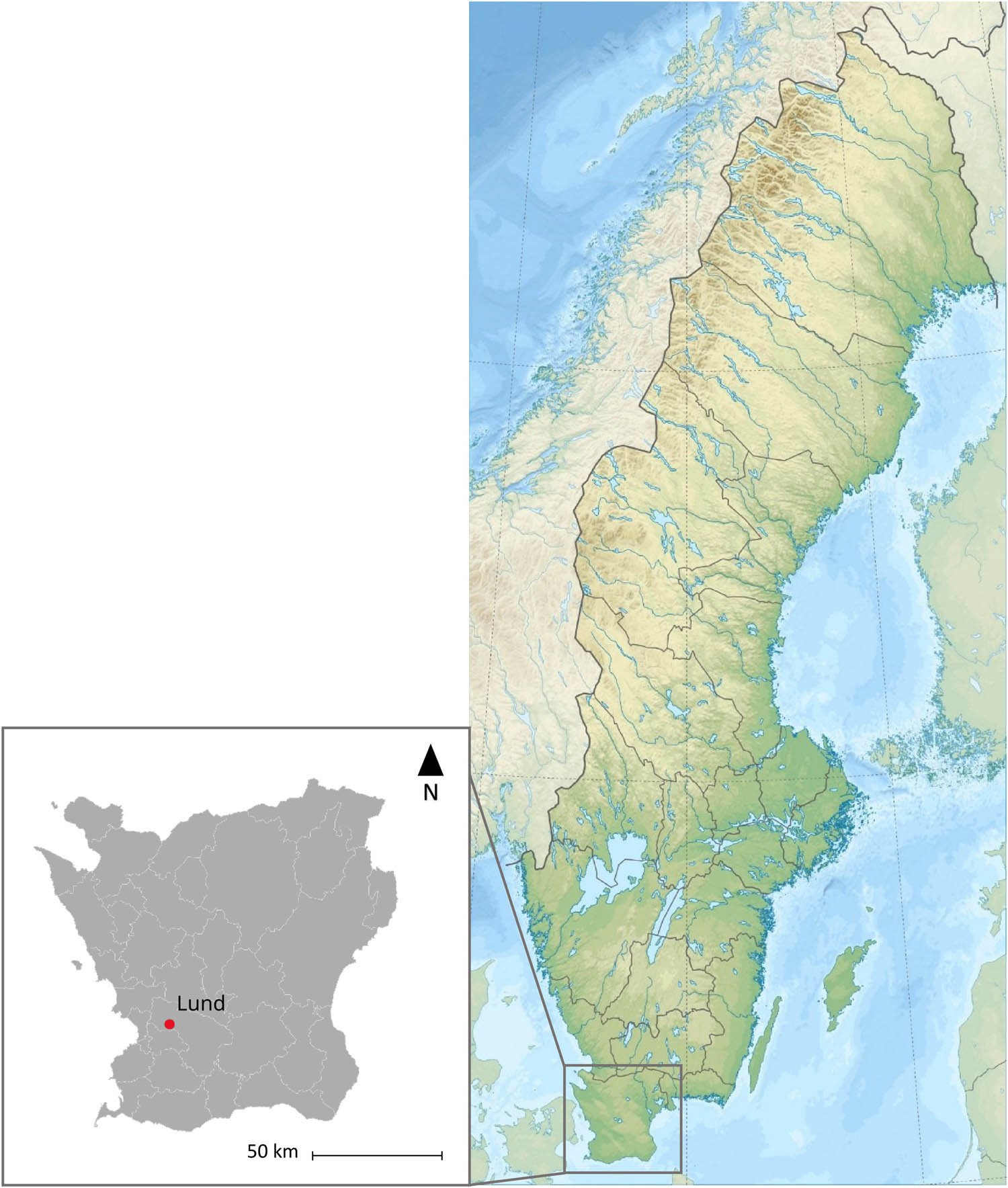
Map of Scania in Southern Sweden, Lund located in the Southwest of the Province. Created by Sandra Tornberg Fritz using ArcGIS.
1.2 Medieval Lund and the Trinitatis Cemetery
Established ca. 990 CE, throughout much of the Middle Ages Lund functioned as the seat of ecclesiastical and royal power in the Danish Kingdom. The character of the early settlement has been somewhat elusive, but most agree that during this time Lund consisted of a series of farm plots, perhaps connected to a royal estate, organized around two identified Christian churches and cemeteries. One of these is the Trinitatis stave church and cemetery that were the predecessor to the Romanesque stone church and cemetery in focus in this study. Andrén (1985) describes this as the “congested countryside” and in his book emphasizes the importance of the church in the process of urbanization during the Early Middle Ages in Denmark (990–1100 CE). Lund was no exception, as many as 27 religious institutions (e.g. churches, monasteries, and the cathedral) had been established in the town during the Middle Ages. The major urban restructuring took place during the eleventh and twelfth centuries; archaeological evidence from several quarters in the town shows how the earlier more loosely organized farm plots were redeveloped into smaller rectangular urban plots. At the centre of this was the market area of Stortorget, plots organized along the street with accompanying stalls facing into the square which was paved with wooden planks (Carelli, 2012, pp. 108–116, 234–236). Another signifier of Lund’s early economic importance was the royal mint located in town, with suggestions that coins were struck here as early as the late 10th century (Cinthio, 2018, pp. 30–32; Malmer, 1997). In the High Middle Ages (1100–1300 CE) many of the churches that had previously been built in wood were now being converted to Romanesque stone churches, perhaps in connection with the early establishment of the parishes (Arcini, 1999, p. 40). During this period Trinitatis also underwent rebuilding in connection with the establishment of the Premonstratensian monastery which was active from the mid-twelfth century until the early thirteenth century on the site (Cinthio, 1996).
Trinitatis church was a part of Drottens parish (Figure 2), first mentioned at the beginning of the fourteenth century (Carelli, 2012, p. 173). We can get an idea of the parishioners’ social status during the Late Middle Ages (1300–1536 CE) through the written record, referring to wealthy merchants and members of the town council, e.g. several mayors, councillors, and aldermen (Blomqvist, 1951, pp. 207–208, 276–277). These were influential figures in the politics and economy of a town that was searching for more autonomy during a very tumultuous period in the Danish Kingdom. It is also possible to say something about the status of the congregation by examining the organization and character of the graves at Trinitatis that date to the Late Middle Ages. Burials from this period were very plain, few grave goods (e.g. rosary beads, walking canes), and most were interred in earthen graves or a simple wooden coffin, however, in the same period there was a stark increase in the number of burials in the floor of the church. A total of 190 graves were located beneath the floor of Trinitatis, a small portion of these graves are pre-fourteenth century having been placed during the time of the Premonstratensian monastery, while most were placed during the Late Middle Ages (Arcini, 1999, p. 48). Securing such a position in the church would have come at quite a significant cost and is perhaps a sign that there were those in the society who were able to maintain their wealth even during a time of economic hardships related to the Late Medieval Crises (Cinthio, 2002, p. 198).
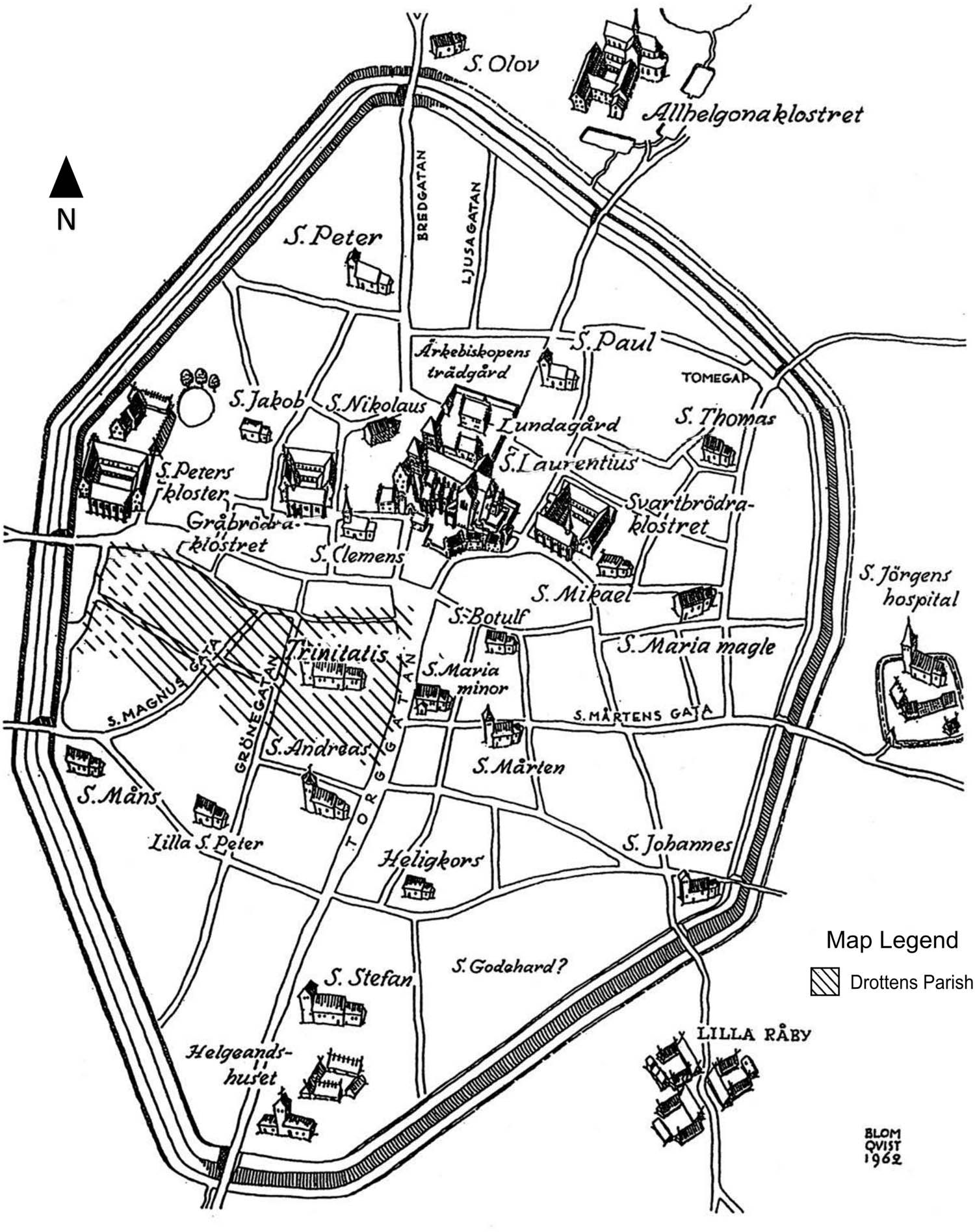
Map of Lund in the Late Medieval ca.1536 CE, Trinitatis church with Drottens parish marked. Reproduced with permission from Caroline Ahlström-Arcini (Arcini et al., 2016, p. 110), original Blomqvist (1963, p. 11) with later additions by Maria Cinthio (1999, p. 43), and Blair Nolan in this publication (map legend and arrow).
During the Late Middle Ages, the urban structure of the town was changing. The plots that had previously been divided in the eleventh century were now being amalgamated into larger urban farm plots, and there was an increased transfer of lands into the hands of the church and establishment of ecclesiastical residences in the town (Carelli, 2012, pp. 319, 336–338). Lund’s influence in regional trade was dwindling as several port towns and smaller markets were being established along the coasts of Scania, which had benefited from connections with the Hanseatic League across the Baltic and North Sea. Rapidly developing cities like Malmö and Copenhagen had pulled economic influence away from Lund, the royal mint moving from Lund to Malmö in the 1440s being another sign of this economic decline. Lund would however persist throughout the remainder of the Middle Ages as a centre for religion (Blomqvist, 1951). As for the fate of Trinitatis post-reformation, demolition of the church had already begun in 1537 and by 1544 little remained standing (Cinthio, 2002, p. 235). The stone material was taken for use in other buildings in Lund, leaving behind the foundations of the church which were preserved post-excavation for public visitation alongside a museum exhibit.
1.3 Medieval Scandinavian Perspective on Disability
Disability in Medieval Scandinavia was closely defined in relation to religious and cultural norms, which do not necessarily directly correspond to the severity of the impairment. The boundary between illness and trauma/wounds in medieval Scandinavia was not always clear, and one could be considered as being “ill in wounds,” the wounds being the cause of illness/disease (Bergqvist, 2013, p. 134). The religious view of bodily malfunction is complex and could include both views of bodily malfunction as a punishment from God, and as a divine test which required penance. The overarching idea was that bodily malfunction was the work of God, and the affected could be socially excluded because of their sinful souls (Bergqvist, 2013, p. 112). Conversely, the Church promoted and took part in alms for the sick and injured (Davis, 2014), and the monasteries were the main distributors of institutionalized medical care (Bergqvist, 2013, p. 238). This more blanket approach to disability and impairment is further nuanced by digging into the legal texts of the period.
The legal codes of Medieval Scania provide some perspective on the way that disability and physical impairment were viewed by wider society. The twelfth- to thirteenth-century Danish Scanian Law, including Archbishop Anders Sunesen’s Latin translation Liber Legis Scaniae, defined disability not only through the severity of impairment but gave significant weight to the visibility of injury. Injuries that could be concealed by hair, headwear, or other clothes were considered less disabling than unconcealable ones (Tamm & Vogt, 2016, p. 34; Tamm, 2018, p. 83). Injuries that were so severe that the (free-born) individual could no longer pursue labour were lawfully compensated with the same amount as if the individual was dead (mansbot) (Lawing, 2021, p. 54). The same compensation was granted for losing a nose, two hands or feet, tongue, both eyes, or male genitalia (Tamm, 2018, p. 83). Loss of the nose was particularly shameful since removal of the nose was a common punishment for theft (Tamm & Vogt, 2016, p. 34). The laws written in the following centuries would borrow some of the principles from these earlier texts.
The Lund town law, Lundarätten (Lund rights), of 1326 follows the previous Scanian legal texts in that visible injury was subject to higher fines and that loss of the hands, nose, or male genitalia required fines equal to those of the “mansbot.” The Lund rights mention specific cases in which a wound exposes bone, a fine for each piece of bone that “sticks in a bowl” would be exacted (Carelli, 2012, p. 229). The privileges granted to Lund in 1361 by King Valdemar Atterdag, while not as prescriptive in their treatment of injury do call for a heavy fine for serious woundings, and that beheading or amputation of the hand are possible forms of punishment for grievous crimes (Carelli, 2012, pp. 375–377). As such, injuries that were both physically impairing and clearly visible, were considered most severe. Deducing social norms regarding physical impairment and disability from primary and secondary sources on the religious and legal texts of the period is difficult because it presents an idealized perspective. We can enrich our understanding of disability and identity through detailed osteological analysis, and archaeological and historical contextualization.
2 Material and Methods
2.1 Material
The individual analysed in this study (grave number 2399) was exhumed from the Trinitatis cemetery during rescue excavations that took place from 1982 to 1984, one of the thousands that were recovered (Nilsson, 1985). The skeleton was in an overall good state of preservation, the elements which were not preserved or excavated being the rib bones and a few isolated foot and hand bones, which is likely due to recovery biases based on the experience of the excavators and current excavation practice. Dating of the Trinitatis church and cemetery and dating of the graves has been determined via several methods: dendrochronological dating of well-preserved wood from coffins and structural elements (Bartholin, 1976), stratigraphic position related to the building activities of the churches, and by the arm positions of the individuals in the graves (Cinthio, 2002; Kieffer-Olsen, 1993; Redin, 1976). Arm position and cemetery stratigraphy suggest that individual 2399 was buried in the Late Middle Ages (1300–1536 CE). The individual was buried on top of the foundation stones at the base of the western church tower. Cemetery organization during the Danish Middle Ages was associated with socio-economic status, and the most prominent burials were placed in the choir. The aim was to have a final resting place as close to the holy relics as possible (Iregren & Redin, 1995, pp. 21; Jonsson, 2009, p. 124). Burials beneath the roof gap of the church were also highly valued because of a cultural and religious belief that the rainwater dripping from the roof was holy (Jonsson, 2009, pp. 62). Individual 2399’s burial plot was in a place of prominence within the cemetery.
2.2 Methods
2.2.1 Sex, Age, and Stature Estimation
Morphological traits of the pelvis (Buikstra & Ubelaker, 1994; Phenice, 1969) and the skull (Buikstra & Ubelaker, 1994) were assessed for estimation of sex. Age-at-death was estimated with the Transition Analysis 3 (TA3) software (Richardson & Ousley, 2021). Traditional methods typically rely on a single feature or a small number of features for age estimation. Transition analysis on the other hand employs numerous traits from multiple locations on the skeleton to avoid preservation biases of different traits. The current build is the 0.8.5 version, accessible at https://www.statsmachine.net/software/TA3/. The team is still working with the method and removing features that are found not be strong indicators of age-at-death, but in its current state the software has the capacity to provide good estimates of age throughout adulthood and well into advanced age (Boldsen et al., 2021, pp. 126–128; Milner et al., 2021, pp. 148–151). Per the guidelines of the trait manual for TA3 (Milner et al., 2019), skeletal changes deemed to be anomalous due to pathological change or trauma were excluded from observation to avoid errors in age estimation, this relies on the experience of the observer to identify these anomalies.
Stature was estimated using the Sjøvold (1990) model, which uses the maximum length of the femur. The Sjøvold model is based on organic correlation from a wide range of populations, which neutralizes possible differences in bone lengths with ethnicity, a plausible bias with models based on linear regression.
2.2.2 Digitization and Visualization
Radiographic imaging was conducted at the Skåne University Hospital in Lund. All in-text figures of pathological lesions, 3D models, and additional photographs and radiographic images are available as supplementary data in the archaeological interactive report (AIR) database. Direct links to the models are available in the Appendix.
3D High-resolution data were gathered using range-based modelling and image-based modelling (Remondino & Campana, 2014; Remondino et al., 2018; Scopigno et al., 2011). The 3D scanning portion of the data acquisition was done using an Artec Space Spider scanner and Artec Eva Scanner for the large bones.
Once the bones were 3D modelled, they were integrated into AIR, which proved the most suitable system due to its capabilities in 3D model integration and exploration flexibility. AIR is a 3D-based online information system developed within DARKLab (Derudas et al., 2023) and is currently used as a documentation and data management system in several excavation projects at the Department of Archaeology and Ancient History at Lund University. AIR facilitates the documentation and management of archaeological excavations, allowing for the integration of 2D and 3D datasets. AIR allowed for the combination of 2D legacy data (maps and drawings) from the 1982 to 1984 excavation with the 3D models of the skeletal material produced 40 years later (https://omeka.ht.lu.se/s/care/page/home).
The studied skeletal elements were recorded into AIR as individual finds, with detailed information on their physical characteristics, retrieval status, and conservation using the find-recording sheets. Eight 3D models of individual skeletal elements were produced and registered into AIR, connected to the corresponding finds (see find-recording sheet of the right humerus and the linked 3D representation https://omeka.ht.lu.se/s/care/item/7422). Visualising the digital find recording sheet allows for the exploration and measurement of the digital copies. Thanks to the capabilities of AIR, we could leverage the available heterogeneous dataset more effectively. By merging legacy and 3D data, we created a comprehensive hybrid representation of the excavation. The excavation context was meticulously recorded in AIR using the available written documentation and hand-drawn 2D site plans. The information from the individual under study (context 2399) was meticulously recorded in the digital context sheet, specifically designed for the documentation of human skeletal remains (Figure 3). The hand-drawing burial map from the cemetery area investigated between 1982 and 1984 was scanned, converted into a textured plane, geo-referenced, and imported as a media element in the context sheet (https://omeka.ht.lu.se/s/care/item/7349).
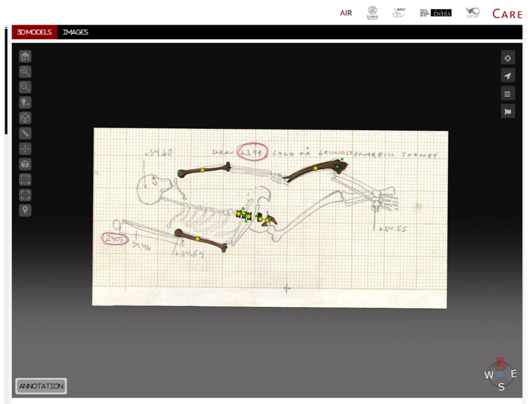
Integration of the 3D modelled remains into the legacy data from the context drawings of individual 2399’s grave in the excavation trench. Annotations represented by green orbs.
From an osteological point of view, it is crucial for osteologists to 3D annotate skeletal elements to visually highlight pathologies, the site of trauma, or other areas relevant to the osteological analysis and link information gathered to said annotations. We rendered 3D annotations as clickable polygons or points located at the area of interest and linked the related information as metadata (Figure 4). This way, the annotations become part of the online archive and can be queried and explored independently. The 3D visualisation of all available skeletal elements in context and the ability to explore related information in an efficient manner significantly supports osteological analysis. This provides a detailed spatial understanding of the individual’s remains and their position relative to other burials in the cemetery, the stratigraphy of the cemetery, and their position in relation to other archaeological features, thereby enhancing the accuracy and reliability of our findings.
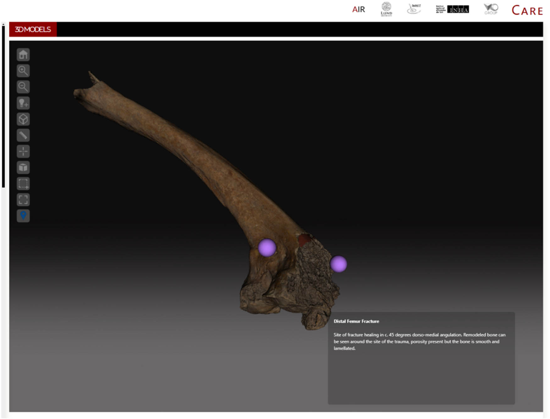
3D Modelled left femur, looking at the distal section. Annotations are visible as purple orbs, and clicking annotations reveal the attached metadata as shown.
3 Results: Osteological Observations and Differential Diagnosis
Individual 2399 was determined to be male with a calculated stature of 171.6 cm (Sjøvold, 1990). Age-at-death was estimated using Transition analysis 3 (TA3) and provided a most probable age of 30.5 years with a 95% confidence interval of 22.4–39.3 years. The standard error was measured at 8.2, and the correlation between the real age and predicted age was 0.902, with good overall preservation of traits for analysis.
Individual 2399 suffered an oblique ante-mortem fracture of the left distal femur (Figure 5), visible in radiographic images in AIR (S). A small pond fracture (c. 3 × 10 mm) was noted on the left frontal bone, but due to its small size and lack of related fracture or bony changes would likely have been of little impact to the individual and is not possible to link to the fractured knee. The knee fracture resulted in complete dislocation of the condyles which healed at a 45-degree angulation in a dorso-medial position. The position and dislocation suggest that the distal epiphysis was fused at the time of trauma and thus provides an earliest date of injury at approximately 20 years of age (Buikstra & Ubelaker, 1994; Scheuer & Black, 2000). The fracture pattern and angulation suggest high-force impact from the lateral side. High-energy trauma to the knee commonly results in open fractures and bacterial infection of soft and hard tissue (osteomyelitis or osteitis) (Polykandriotis et al., 2005). This type of injury is often seen in motor vehicle and horse-riding accidents today (Buechel, 2002) but in the context of Medieval Lund might be associated with a kick from a horse or a heavy object falling on the knee, such as stone or other materials during construction (Arcini, 1999, p. 149).
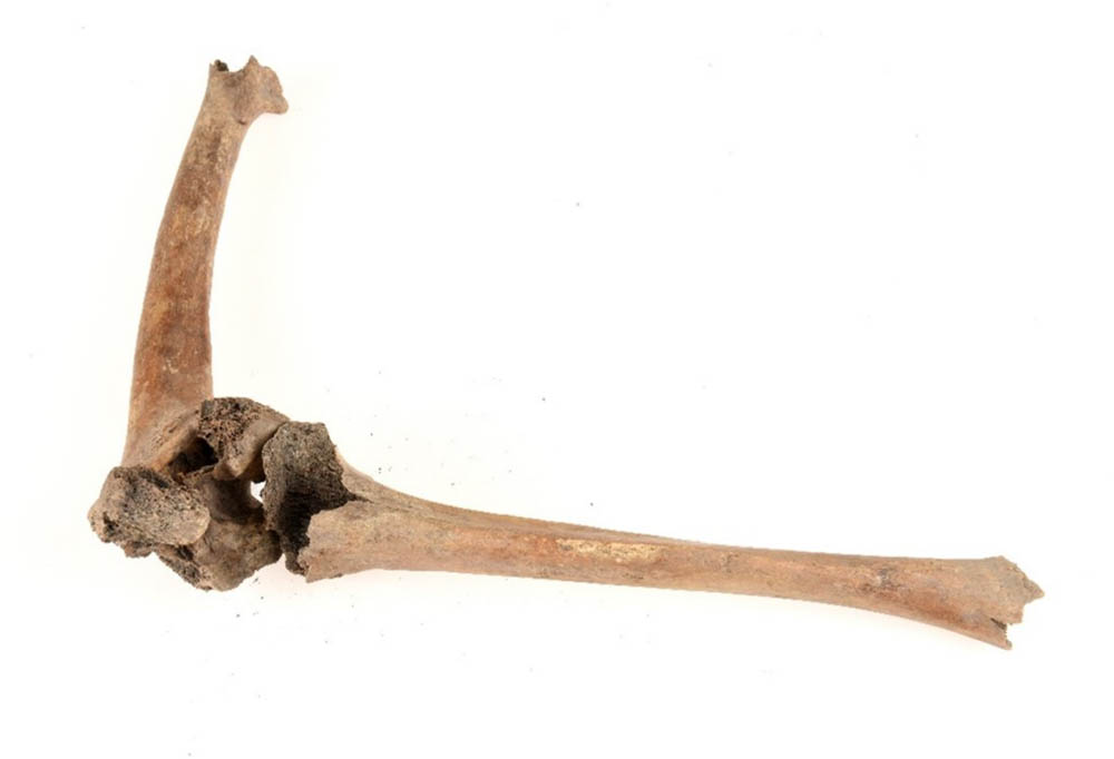
The femoral fracture in grave 2399 with the tibia repositioned to show the 45-degrees angulation. Photo: Nelly Hercberg, Cultural Museum in Lund.
The area of the trauma exhibited post-deposition and post-excavation damage, taphonomic changes exposing the trabecular bone in certain area, which is important to note as this might limit observation of certain features and was certainly considered when looking at the condition of the remodelled bone. Upon initial observation, it was clear that the distal portion of the femoral diaphysis exhibited significant swelling and new bone growth around the site of the trauma. A cloaca is visible in the intercondylar space, surrounded by remodelled bone (S:2). The new bone growth is well organized, presenting as smooth and lamellated with larger porosity. The articular surfaces of the femoral and tibial condyles both exhibit periosteal new bone growth that is porous but looks to be limited in its expansion beyond the articulation space.
The observation of severely remodelled bone around the site of the trauma, the presence of the cloaca, the limited expanse of the inflammatory response beyond the site of trauma, and no evidence of sequestrum, are in our estimation indicative of a localized post-traumatic exogenic osteomyelitic infection and in line with the criteria outlined by Roberts (2019, p. 297). Post traumatic osteomyelitic infection in the case of exposed fractures is very often poly-microbial (Waldron, 2021, p. 151), but in almost 90% of cases, Staphylococcus aureus is the causative agent (Roberts, 2019, p. 299). Misidentification of osteomyelitic infection as neoplasms of bone has been noted in both the clinical and palaeopathological literature (Gould et al., 2007; Huang et al., 2013; Salik et al., 2021; Suzuki, 1987; Tamarit et al., 2003); therefore, we took precaution to consider the possibility of trauma that was secondary to weakening of the bone in relation to tumorous growth. Macroscopically, new bone growth related to the trauma was not consistent with that of cancers such as osteosarcoma, the remodelled bone lacks the identifiable disorganized “sunburst” pattern. The radiographic images also showed no clear delineation of tumorous growth from that of the cortical bone of the femur. Analysis of radiographic images was done in consultation with orthopaedic surgeons at the University Hospital in Lund, who were also of the mind that there was no evidence of cancerous growth in the individual.
The individual exhibits ankylosis of the patellofemoral joint (Figure 6; S:1), which is a possible case of heterotopic ossification traumatica (stimulated soft tissue ossification post trauma) involving the quadriceps tendon insertion superiorly on the patella (Waldron, 2021, pp. 301–302). Severe trauma to the knee would have resulted in arthrofibrosis, the build-up of scar tissue around the joint during the inflammatory phase that causes prolonged stiffening and pain (Thompson et al., 2019; Usher et al., 2019). Arthrofibrosis with fusion of the patella is commonly seen in poorly treated severe distal femur fractures (Buechel, 2002). Periarticular trauma of this nature would also have very likely led to the spread of blood into the joint space that could influence new bone growth, perhaps part of the cause for ankylosis and some of the periosteal new bone on the surfaces of the femoral and tibial condyles (Waldron, 2021, p. 235).
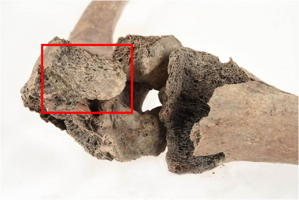
Close-up of the left knee with the fused patella visible to the left (red box). Photo: Nelly H Hercberg, Cultural Museum in Lund.
All lumbar vertebrae were affected by osteophyte formation, and there was a slight degree of wedging in L2 (S:3.2) and L3 (S:3.3). The degree of osteophyte formation is quite extensive for an individual that died around the age of 30 years, this could be indicative of manual labour earlier in life, but we believe there is a high likelihood that it is related to strain caused post-trauma. Bilateral entheseal changes (minor enthesophytes) at the insertion of the superior glenohumeral ligament on the greater tubercle of the humeral head, a substantially larger linea musculi solei on the right posterior tibia relative to the left side. Periosteal new bone formation was noted on the right tibial diaphysis (Figure 7), as well as dorsally on the right pubic bone at the insertion site for the adductor longus muscle (S:4).
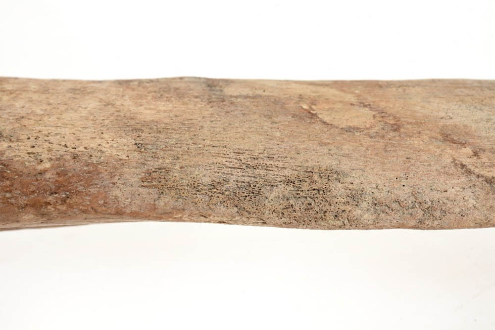
Active and healed periosteal new bone growth on the right tibial diaphysis. Photo: Nelly H Hercberg, Cultural Museum in Lund.
4 Discussion: Disability, Care, and Identity
4.1 Experienced Disability
The World Health Organization’s (WHO) International Classification of Functioning, Disability, and Health (ICF) (WHO, 2002), and World Disability Report (WHO, 2011), identify disability as encompassing impairment of bodily function, limitations on activity, and restrictions of one’s participation or involvement in various aspects of life. It is through understanding Disability as a “biopsychosocial” concept (WHO, 2002, p. 20) that we combine the medical and social understandings of disability in the present and past. In the case of individual 2399, we can try to understand how an impairing injury such as trauma to the knee joint impacted bodily function post-trauma and would have necessitated care or assistance.
Acutely, post-trauma, individual 2399 would have experienced a great deal of pain that would require management. Bed rest would have been required, in the short term, and during this time, mobility would have been limited. Individual 2399 would have needed the assistance of caregivers for daily functions during this acute phase and then other forms of physical assistance long-term. Based on the osteological analysis and differential diagnosis we believe there are clear signs that individual 2399 would have maintained a degree of mobility post-trauma with assistance. The degree of remodelling around the fracture site and the permanent angulation of the joint would have been prohibitive to mobility, possibly limiting the individual’s ability to participate in specific occupations post-trauma and requiring some degree of assistance through a mobility aid in navigating the urban context.
Osteological observations in the post-cranial skeleton support our assertion that individual 2399 was used some form of mobility aid long-term after the impairing injury. Enthesophyte formation on the proximal aspects of the humeri suggests there was an increased load placed on the shoulder joints, perhaps due to the use of crutches in supporting weight. The degree of osteophyte formation and wedging in the vertebral elements is not to be expected of an individual that would be in early adulthood at the time of death, and while manual labour may have been a factor we believe that it is more likely that unequal loading and strain post-trauma resulted in the observed changes. Periosteal new bone formation results from the inflammation of the periosteum, and one of the causes for this inflammation can be increased mechanical loading and strain placed on the skeletal element (Waldron, 2021, pp. 182–184). Perisoteal new bone growth on the right tibial diaphysis and enthesophyte formation ventrally on the surface of the right pubic bone would indicate that the right lower limb was under increased mechanical stress. The location of the bone formation on the pubic bone is at the insertion site of the adductor longus muscle, which functions in creating stability of the lower limb. These observations suggest that individual 2399 would have likely been living with limitations to daily physical activities from the time of their injury until the time of their death, but that this would have been managed through some form of mobility aid.
4.2 Constructing a Model of Care
Written sources and the archaeological record support that specialized health care was available in Medieval Lund (Bergqvist, 2013, pp. 134–139; 194–206). Numerous archaeological finds such as scalpels, surgical pliers, tweezers, and ointment jars all point toward a professionalized standard of care (Bergqvist, 2013, pp. 361–388). Professional health care was likely exclusive to higher social classes, but Bergqvist (2013, pp. 194) argues that some basic medical knowledge was present in most households. A deep knowledge of both surgical and medicinal treatments was available from the start of the Medieval period, invasive procedures were performed, and medicinal plants were used to treat and relieve symptoms and illnesses (Wallis, 2010, p. 134). Individual 2399, having experienced such a severe injury, would no doubt have required professional medical assistance.
During the acute phase of the injury, individual 2399 would have experienced a great deal of pain. The post-traumatic pain and inflammation would have been dealt with using a combination of bed rest and available pain relief, such as ointments made of lavender oil, opium, and alcohol (Bergqvist, 2013, pp. 277–287). Individual 2399 would have required assistance to maintain hygiene, nutrition, pain relief, along with cleaning and dressing of the wound. It is not likely that there were attempts to repose the fracture through traction treatment, a conclusion similarly reached by Arcini (1999, p. 150) when she noted the high frequency of misaligned femoral and tibial fractures in skeletal collections from Medieval Lund, even though this was a method already practiced in other parts of Europe during this time. Considering the risk of comorbidity associated with prolonged bed rest, such as respiratory and cardio-vascular complications, active care to reduce immobility must have been crucial (Jakobsen et al., 2021). Mobility aids would be required during both the acute and chronic phases, in the form of crutches or a leg stand. Impaired or injured limbs were sometimes bound for better coordination (Arcini, 2012, p. 431; Hernigou, 2013). The Arabic Andalusian physician Albucasis (d.1106) called for the leg to be bent at the knee and have the lower segment firmly fixed to the upper segment in a splint (Metzler, 2006, p. 113). It is plausible that binding of the leg would allow the patient to mobilize early, but it must be considered that pain in the joint would have been a limiting factor in recovery (Cunningham et al., 2022) and the degree of inflammation would have been prohibitive of such extreme binding, requiring a more tailored solution. The osteological evidence for the use of mobility aids has been covered previously, but this would have been an integral part of the treatment regimen in both the immediate and long-term.
Post-traumatic osteomyelitis would also have required its own course of treatment. Bergqvist (2013, pp. 134–138) comments that physicians in the Medieval period were not expected, nor advised, to treat such severe injuries as those afflicting the bone marrow (marvsår). Modern treatment for post-traumatic osteomyelitis requires a course of antibiotics, but prior to the antibiotic revolution care options would have been limited to drainage of infected site, incision, and abrasion to remove infected soft tissue or bone, or amputation of the affected limb (Forsgren & Kronvall, 1996, p. 226; Ryan et al., 2017). There was no observable osteological evidence of surgical intervention, neither treatment through abrasion and opening of the medullary cavity for drainage nor amputation of the limb. In the skeletal material from Trinitatis, there are two separate cases of amputation (Arcini, 1999, pp. 145–146), but this was a rare occurrence, and we do not know whether these cases were medical or legal in nature. Based on the artefactual evidence present that indicated the presence of knowledgeable physicians we can infer that individual 2399 would have undergone regular treatment for post-traumatic osteomyelitic infection and that the wound was likely opened to allow for the drainage of pus.
4.3 Intersecting Identities
Approximately 1.3 billion people worldwide experience significant disability, roughly 16% of the human population. Inequities in the experience of disability arise because of multiple factors including social structures (e.g. class dynamics), barriers within the health system, and varied living conditions that expose populations to increased risk of morbidity and mortality (WHO, 2022). These complex relationships existed in Medieval Scania as well, and analyses of patterns regarding morbidity, mortality, and identity in relation to societal norms can provide important insights into how living with impairment and restrictions to daily life would have affected individual 2399. We consider the case of individual 2399 within the specific context of Medieval Lund, relying on sources such as the Medieval legal texts from Scandinavia, the archaeological record, and the osteological data on trauma gathered from Lund.
Through our understanding of the Medieval Scanian Law, particularly in reference to modes of compensation and punishment for crimes, we can infer how society may have viewed individual 2399. Injuries that were both severely physically impairing and fully visible were compensated much more generously than impairments considered less impactful and/or possible to conceal (Carelli, 2012, pp. 227–230, 375–377; Tamm & Vogt, 2016; Tamm, 2018). Further, in cases of severe or recurrent offences, mutilation was commonly practiced. The texts depict an organized penal system in which a first offense was punished by a fine, a second by loss of hands and/or feet, and a third by loss of eyes, nose, ears, lips, and/or scalp. The fact that corporal punishment included intended impairment in the form of loss of limbs, sight, or hearing, gives glimpses into the societal and structural view of disability, making a distinction between physical and sensory impairment. It also provides an important understanding as to why compensation for intentional or workplace injuries was provided differently depending on the affected body part. The loss of, or severe injury to, body parts that could be mistaken as corporal punishment was considered more disabling than other impairments (Metzler, 2013, p. 38).
The frequency of impairing injuries present in the osteological record from Medieval Lund provides an important frame of reference for understanding the identity of disability and the availability of care in past populations. In societies where impairing injuries are common, disability is less likely to be considered deviant, and care is more likely to be provisioned. In these types of societies, pre-injury identities might be less altered, at least in some socio-economic classes where subsistence can be acquired without manual labour. No detailed analysis of trauma within a specific regional context in the Nordics existed until Arcini’s (1999) thesis on health in Lund during the Middle Ages provided such an examination. Arcini’s analysis of the Trinitatis cemetery, the same cemetery in which individual 2399 was interred, showed that 214 (9.4%) of the 2266 adults examined exhibited evidence of skeletal trauma and that the frequency of trauma in the population increased over time. The frequency of individuals affected by cranial and post-cranial trauma was significantly higher in the High (1100–1300 CE) and Late Middle Ages (1,300–1,536) than in the Early Middle Ages (990–1100 CE). Sex appeared to influence traumatic injury risk; 12.5% of males versus 9.3% of females exhibited evidence of trauma. Femoral fractures, specifically, were recorded in 21 individuals (11 males, 9 females, and 1 unsexed), and in terms of spread across the Middle Ages, they were found with the highest frequency in the periods c.990–1100 and c.1300–1536 (Arcini, 1999, p. 187). A similar supracondylar fracture impacting the knee joint was identified in a second individual, also a male in his early 30s at the time of death and in a grave dated to the period c.1300–1536 (Arcini, 1999, p. 141). Arcini attributed the upward trend in trauma, in part, to accidental injuries related to urban growth and the construction of churches and other building projects. Social status does appear to have influenced trauma patterns to some extent; individuals suffering from skull trauma were more likely to have prominent burial plots, while there was no discernible pattern in burial location associated with post-cranial trauma (Arcini, 1999, p. 146). Individual 2399’s proximity to the church, and his burial atop the foundation stones of the church’s western tower, indicates a position of upper socioeconomic status. Status likely influenced his experience of disability, in opposition to what might have been expected based on our understanding of societal norms as projected through legal and religious perspectives.
In an analysis of trauma in Medieval Sweden, Wendt (2023) found clear patterns between severe and visually unconcealable impairments and burial placement in Early Medieval Helsingborg. He interprets it as discriminatory attitudes and reduced status related to disability. He found that individuals with concealable impairments were placed in the choir or in the longhouse of the church, while individuals with visual impairments were grouped together and separated from other contemporaneous burials on the outskirts of the cemetery. Despite what would clearly have been a visual impairment, individual 2399 was buried very close to the church. Similarly, Bohling et al. (2022) performed a population-level analysis on burial patterns in relation to trauma in Late Anglo-Saxon England, in which they stressed the timing of physical impairment as a key factor in understanding disability and the identity of disability in the past. They make the distinction between physical impairments as being congenital (present from birth) or acquired during life – ante-mortem physical impairment could be classified as occurring at the end of life or short, medium, to long term, or definitively long-term before death. It was their finding amongst multiple churchyards that the treatment of burials in regard to physical impairment or disability, whether short term and related to end-of-life or long-term and having notable effects on bodily function, was variable. Often individuals are found interred in proximity to the church, in relatively high-status burials regardless of physical impairment. Late Anglo-Saxon England is of course not an identical context to Late Medieval Denmark, but this is another example of how the reality of lived experience cannot be interpreted in a vacuum.
Attitudes towards disability likely fluctuated throughout the Medieval period in Scania and would likely be context-specific as in the case of Lund, but what is clear is that individual 2399’s status within society was not wholly defined by disability. During the Late Medieval period, we know that several members of Drottens parish were wealthy merchants and members of the town council in Lund. In the Trinitatis cemetery, there were burials of exceptionally high status inside the church, members of the clergy or the wealthy that could afford such placement (Cinthio, 2002, p. 187), but members of the burgher class or others of socioeconomic significance would aim for a burial plot in the cemetery (Arcini, 1999, p. 46) as close to the church as possible. Individual 2399’s prominent burial place within the churchyard may indicate that they were a part of the burgher class, someone of socioeconomic importance, and this distinction may have superseded the identity of living with disability. Considering the biopsychosocial implications of disability, we might infer that individual 2399 occupied a position within society, either through familial connection or attained status during life, that provided both economic and social support post-trauma. A position which afforded him the opportunity to participate in society despite the physical impairment to bodily function and limitations to mobility. The church and legal stances on physical impairment and disability during this time then seem not to have had a considerable effect on this individual’s status within society, at least from what we can tell by our interpretation of the osteological, archaeological, and written sources. These identities, burgher and disabled, were in some way not at odds with one another but a part of the whole individual and coloured their lived experience within society.
5 Conclusions
This article is unique in its application of BoD and BoC principles, particularly the index of care method, within the Medieval Nordic context. The understanding of physical impairment, disability, and care in Medieval Scandinavia is heavily influenced by our readings and interpretations of the written sources, legal, and religious texts which become scarcer the further we go back into the Medieval period. Literary sources present us with an idealized framework for societal norms surrounding disability but expanding datasets into the archaeological and osteological realms brings to light a more complex discussion on identity in the past. An expectation of stigmatization and ostracization of those living with disability due to severe physical impairment hasn’t been validated in our study. The evidence presented for Individual 2399, the Trinitatis cemetery, and contemporary cemeteries in Medieval Northern Europe corroborate our interpretation that disability, as much as today, is only one facet of identity at the individual and population level. In the case of individual 2399, it is apparent that the experience of disability post-trauma was affected by their social status, evident through the acute treatment process, long-term care availability post-trauma, and their treatment in death with a prominent burial plot within the cemetery. Future research goals would be to expand this study to larger collections of material from both urban and rural cemeteries, examine various types of physical impairment, and look at its relation to the organization of cemeteries throughout the Medieval period.
In this study, we have also been explicit in our use of 3D visualization and documentation techniques, not only for the purposes of increased dissemination and visibility within the study but also in demonstrating the usefulness of these tools analytically. Detailed high-resolution photogrammetric models of the trauma are here demonstrated as a valuable tool in the fields of BoD and BoC research as readers are given the opportunity to manipulate and inspect the skeletal elements and site of traumatic injury for themselves, going beyond the more limited two-dimensional photographic component. Combining models with records from the excavation, both site drawings and excavation reports when available, allows us to bring forth legacy data that has in many cases been forgotten in the archives or seen as providing little value. The future applications of these methods as shown in the AIR system, developed in collaboration between the DarkLab at Lund University and the French National Institute of Art History, are on the cutting edge of digital archaeology methods and will be extremely valuable in the preservation and renewed exploration of past excavation records.
Acknowledgements
We are grateful to the staff at the Cultural Museum in Lund, especially Johanna Hermansson, Nina Davies, and Nelly Hercberg for all the help with access to the collections and assistance in photography. We also want to acknowledge Caroline Ahlström Arcini for engaging in discussions as well as providing burial data and giving permission for the republication of figures. We also want to thank Magnus Tägil M.D. at the University Hospital in Lund for assistance with radiographic imaging and discussions about diagnosis. We would like to extend our gratitude to Danilo Marco Campanaro for his assistance with the digitization of the Trinitatis excavation records, site georeferencing, and remodelling of 3D bone scans, and to Federico Nurra at INHA for the development of the annotation functionalities that are of such importance in this study and future research. We thank the reviewers for their insightful comments and thought-provoking questions, which have strengthened this article.
-
Funding information: The authors state no funding involved.
-
Author contributions: All authors accept responsibility for the content of the manuscript and approve its submission. BN, AT, and STF were responsible for the conceptualization of the study. BN, AT, and STF were responsible for the osteological analysis of the remains and bioarchaeological interpretation of findings through a bioarcheology of care and disability lens. PD and DD were responsible for the digitization of legacy excavation record data, 3D modelling of the human remains analysed in this study, and visualization of the models within the AIR database. BN, STF, AT, PD, and DD were all involved in writing the original draft; PD and DD aiding specifically in the preparation of the digital methods section and conclusions regarding applications of the digital methods. BN was responsible for writing the revised draft.
-
Conflict of interest: The authors state no conflict of interest.
-
Data availability statement: All data presented in this study are available in the Appendix and publicly accessible through the AIR database portal at https://omeka.ht.lu.se/s/care/page/home.
Below you will find direct links to the 3D models in the database AIR. The specific points associated with the care evaluation in this study are visible in the referenced models. The links are numbered according to their position in the article. S:1 and S:2 both link to the same model but reference separate features on the skeletal element.
S:1 Fusion of the right patella in the position of the patellar fossa related to the femur fracture: https://omeka.ht.lu.se/s/care/item/7424.
S:2 Cloaca visible intercondylarly on the distal left femur: https://omeka.ht.lu.se/s/care/item/7424.
S:3 Lumbar vertebra with osteophyte formation and lateral wedging. All lumbar vertebrae were affected by osteophyte formation.
3-1: 1st Lumbar https://omeka.ht.lu.se/s/care/item/7418.
3-2: 2nd Lumbar https://omeka.ht.lu.se/s/care/item/7419.
3-3: 3rd Lumbar https://omeka.ht.lu.se/s/care/item/7420.
3-5: 5th Lumbar https://omeka.ht.lu.se/s/care/item/7421.
S:4 Right pubic bone, with the location of periosteal new bone formation noted: https://omeka.ht.lu.se/s/care/item/7425.
Homepage with links to all modelled remains: https://omeka.ht.lu.se/s/care/page/home.
References
Andrén, A. (1985). Den urbana scenen: Städer och samhälle i det medeltida Danmark. Acta Archaeologica Lundensia. (Series in 8°Nr 13. Diss). Lund University.Search in Google Scholar
Arcini, C. (1999). Health and disease in early Lund, Osteo-pathologic studies of 3,305 individuals buried in the first cemetery area of Lund 990-1536. (Doctoral dissertation). Lund University.Search in Google Scholar
Arcini, C. (2012). Sjukdomar, hälsa och levnadsförhållanden i medeltidens Lund. In Carelli, P. Lunds historia: Staden och omlandet. 1 Medeltiden: En metropol växer fram. Lunds kommun.Search in Google Scholar
Arcini, C., Price, T. D., Cinthio, M., Drenzel, L., Andersson, M., Persson, B., Menander, H., Vretemark, M., Kjellström, A., Hedvall, R., & Tagesson, G. (2016). Living conditions in times of plague. In P. Lagerås (Ed.), Environment, Society and the Black Death: An interdisciplinary approach to the late-medieval crisis in Sweden (pp. 104–140). Oxbow Books.10.2307/j.ctvh1dr32.9Search in Google Scholar
Bartholin, T. S. (1976). Dendrokronologiske og vedanatomiske undersøgelser af træfundene. In A. W. Mårtensson (Ed.), Uppgrävt förflutet för PKbanken i Lund: En investering i arkeologi (pp. 145–169). Archaeologica Lundensia VII, Investigationes de Antiqvitatibus Urbis Lundae, Kulturen Lund, Malmö.Search in Google Scholar
Battles, H., & Gilmour, R. (2022). Beyond mortality: Survivors of epidemic infections and the bioarchaeology of impairment and disability. Bioarchaeology International, 6(1–2), 23–40.10.5744/bi.2021.0003Search in Google Scholar
Beckett, R. G., & Conlogue, G. J. (2019). The importance of pathophysiology to the understanding of functional limitations in the bioarchaeology of care approach. International Journal of Paleopathology, 25, 118–128.10.1016/j.ijpp.2018.06.006Search in Google Scholar
Bergqvist, J. (2013). Läkare och läkande: Läkekonstens professionalisering i Sverige under medeltid och renässans. [Doctoral dissertation]. Lunds Universitet.10.37852/oblu.114Search in Google Scholar
Bethard, J. D., Ainger, T. J., Gonciar, A., & Nyárádi, Z. (2021). Surviving (but not thriving) after cranial vault trauma: A case study from Transylvania. International Journal of Paleopathology, 34, 122–129.10.1016/j.ijpp.2021.06.006Search in Google Scholar
Blomqvist, R. (1951). Lunds historia 1, Medeltiden. LiberLaromedel/Gleerup.Search in Google Scholar
Blomqvist, R. (1963). Grundgrävning och Arkeologi. In R. Blomqvist & A. W. Mårtensson (Eds.), Thulegrävningen 1961 (pp. 9–15). Archaeologica Lundensia. Investigationes de Antiquitatibus Urbis Lundae II. Kulturen Lund.Search in Google Scholar
Bohling, S., Croucher, K., & Buckberry, J. (2022). The Bioarchaeology of Disability: A population-scale approach to investigating disability, physical impairment, and care in archaeological communities. International Journal of Paleopathology, 38, 76–94.10.1016/j.ijpp.2022.05.006Search in Google Scholar
Boldsen, J. L., Milner, G. R., & Ousley, S. D. (2022). Paleodemography: From archaeology and skeletal age estimation to life in the past. Yearbook Biological Anthropology, 178(Suppl. 74), 115–150. doi: 10.1002/ajpa.24462.Search in Google Scholar
Brown, E. L., & Wilson, A. S. (2019). Using evidence from hair and other soft tissues to infer the need for and receipt of health-related care provision. International Journal of Paleopathology, 25, 91–98.10.1016/j.ijpp.2018.08.008Search in Google Scholar
Buechel Sr, F. F. (2002). Knee arthroplasty in post-traumatic arthritis. The Journal of Arthroplasty, 17(4), 63–68.10.1054/arth.2002.32809Search in Google Scholar
Buikstra, J. E., & Ubelaker, D. H. (1994). Standards for data collection from human skeleton remains: Proceedings of a seminar at the Field Museum of Natural History, organized by Jonathan Haas. Arkansas Archaeological Survey.Search in Google Scholar
Carelli, P. (2012). Lunds historia: Staden och omlandet. 1, Medeltiden: En metropol växer fram (990–1536). Lunds kommun.Search in Google Scholar
Cinthio, M. (1996). Kyrkorna kring Kattesund: Ett rekonstruktionsförsök. Arkeologiska rapporter från Lund, 14. Kulturen.Search in Google Scholar
Cinthio, M. (1999). The archaeological context. In C. Arcini (Ed.), Health and Disease in Early Lund: Osteo-Pathologic Studies of 3,305 Individuals Buried in the First Cemetery Area of Lund 990–1536 (pp. 18-46). Archaeological Lundensia. [Doctoral Dissertation, Lund University].Search in Google Scholar
Cinthio, M. (2002). De första stadsborna: Medeltida gravar och människor i Lund. Brutus Östlings bokförlag Symposion.Search in Google Scholar
Cinthio, M. (2018). Lund från första början. In M. Cinthio & A. Ödman (Eds.), Vägar mot Lund. En antologi om stadens uppkomst, tidigaste utveckling och entreprenaden bakom de stora stenbyggnaderna. Historiska Media.Search in Google Scholar
Cunningham, D. J., Paniaugua, A. R., LaRose, M. A., DeLaura, I. F., Blatter, M. K., & Gage, M. J. (2022). Regional anesthesia does not decrease inpatient or outpatient opioid demand in distal femur fracture surgery. Archives of Orthopaedic and Trauma Surgery, 142, 1873–1883. doi: 10.1007/s00402-021-03892-2.Search in Google Scholar
Davis, A. J. (2014). The social and religious meanings of charity in medieval Europe. History Compass, 12(12), 935–950.10.1111/hic3.12207Search in Google Scholar
Derudas, P., Nurra, F., & Svensson, A. (2023). New AIR for the archaeological process? The use of 3D web semantic for publishing archaeological reports. Journal on Computing and Cultural Heritage, 16(3), 1–28. Article 57. doi: 10.1145/3594722.Search in Google Scholar
Domett, K., Colbert, A., & Chang, N. (2022). The impact of clubfoot: A holistic, paleopathological case study from Bronze Age Thailand using the bioarchaeology of care framework. Bioarchaeology International, 6(4), 274.10.5744/bi.2021.0022Search in Google Scholar
Finlay, N. (Ed.). (1999a). Disability and archaeology. Archaeological Review from Cambridge, 15(2).Search in Google Scholar
Finlay, N. (Ed.). (1999b). Disabling archaeology: An introduction. Archaeological Review from Cambridge, 15(2), 1–6.Search in Google Scholar
Forsgren, A., & Kronvall, G. (1996). Klinisk bakteriologi. Studentlitteratur.Search in Google Scholar
Gould, C. F., Ly, J. Q., Lattin, G. E., Beall, D. P., & Sutcliffe, J. B. (2007). Bone Tumor Mimics: Avoiding Misdiagnosis. Current Problems in Diagnostic Radiology, 36(3), 124–141. doi: 10.1067/j.cpradiol.2007.01.001.Search in Google Scholar
Hernigou, P. (2013). Crutch art painting in the Middle Ages as orthopaedic heritage (part II: The peg leg, the bent-knee peg and the beggar). International Orthopaedics (SICOT), 38, 1535–1542. doi: 10.1007/s00264-014-2278-1.Search in Google Scholar
Hjort Jakobsen, D., Høgdall, C., & Seibæk, L. (2021). Postoperative mobilisation as an indicator for the quality of surgical nursing care. British Journal of Nursing, 30(4), S4–S15. doi: 10.12968/bjon.2021.30.4.S4.Search in Google Scholar
Huang, P.-Y., Wu, P.-K., Chen, C.-F., Lee, F.-T., Wu, H.-T., Liu, C.-L., Chen, T.-H., & Chen, W.-M. (2013). Osteomyelitis of the femur mimicking bone tumors: a review of 10 cases. World Journal of Surgical Oncology, 11(1), 283. doi: 10.1186/1477-7819-11-283.Search in Google Scholar
Iregren, E., & Redin, L. (Ed.). (1995). I Adams barn -: En diskussion om etiska aspekter på museiförvaring och återbegravning av medeltida skelettmaterial. University of Lund, Institute of Archaeology.Search in Google Scholar
Jonsson, K. (2009). Practices for the living and the dead: Medieval and post-reformation burials in Scandinavia. [Doctoral dissertation]. Stockholms Universitet.Search in Google Scholar
Kieffer-Olsen, J. (1993). Grav og Gravskik i det Middelalderlige Danmark– 8 Kirkegårdsudgravninger. [Doctoral dissertation]. University of Aarhus.Search in Google Scholar
Lawing, S. B. (2021). Victims of Maiming in Sturlunga saga: Worse off Living than Dead? Mirator, 20(2), 54–72.Search in Google Scholar
Li, H., He, L., Gibbon, V. E., Xiao, X., & Wang, B. (2021). Individual centred social‐care approach: Using computer tomography to assess a traumatic brain injury in an Iron Age individual from China. International Journal of Osteoarchaeology, 31(1), 99–107.10.1002/oa.2928Search in Google Scholar
Málaga, M. R. P., & Makowski, K. (2019). Bioarchaeological evidence of care provided to a physically disabled individual from Pachacamac, Peru. International Journal of Paleopathology, 25, 139–149.10.1016/j.ijpp.2018.08.002Search in Google Scholar
Malmer, B. (1997). The Anglo-Scandinavian Coinage c.995-1020, Kungl. Vitterhets Historie och Antikvitets Akademien.Search in Google Scholar
Metzler, I. (2006). Disability in medieval Europe: Thinking about physical impairment in the high middle ages, c. 1100–c. 1400. Routledge.Search in Google Scholar
Metzler, I. (2013). A social history of disability in the Middle Ages: Cultural considerations of physical impairment. Routledge.10.4324/9780203371169Search in Google Scholar
Milner, G. R., Ousley, S. D., Boldsen, J. L., Getz, S. M., Weise, S., & Tarp, P. (2019). Transition Analysis 3 (TA3) Trait Manual (Public Distribution Version 1.0.).Search in Google Scholar
Milner, G. R., Boldsen, J. L., Ousley, S. D., Getz, S. M., Weise, S., & Tarp, P. (2021). Great expectations: The rise, fall, and resurrection of adult skeletal age estimation. In B. F. B. Algee-Hewitt & J. Kim (Eds.), Remodeling Forensic Skeletal Age (pp. 139–154). Academic Press.10.1016/B978-0-12-824370-1.00011-0Search in Google Scholar
Nilsson, T. (1985). Drottenskyrkan och dess föregångare: Nya arkeologiska rön i Lund. (pp. 173–182). Kulturens Årsbok.Search in Google Scholar
Nystrom, K. C., & Tilley, L. (2019). Mummy studies and the bioarchaeology of care. International Journal of Paleopathology, 25, 64–71.10.1016/j.ijpp.2018.06.004Search in Google Scholar
Phenice, T. W. (1969). A newly developed visual method of sexing the os pubis. American Journal of Physical Anthropology, 30(2), 297–301.10.1002/ajpa.1330300214Search in Google Scholar
Polykandriotis, E., Stangl, R., Hennig, H. H., Lennerz, J. K. M., Frank, W. M., Loos, M. D., & Horch, R. E. (2005). The composite vastus medialis–patellar complex osseomuscular flap as a salvage procedure after complex trauma of the knee – an anatomical study and clinical application. British Journal of Plastic Surgery, 58(5), 646–651.10.1016/j.bjps.2005.01.008Search in Google Scholar
Redin, L. (1976). Lagmanshejdan: Ett gravfält som spegling av sociala strukturer i Skanör. (Series in 4°; Vol. 10). Acta Archaeologica Lundensia.Search in Google Scholar
Remondino, F., & Campana, S. (2014). 3D recording and modelling in archaeology and cultural heritage. BAR International Series, 2598, 111–127.10.30861/9781407312309Search in Google Scholar
Remondino, F., Georgopoulos, A., Gonzalez-Aguilera, D., & Agrafiotis, P. (2018). Latest developments in reality-based 3D surveying and modelling. MDPI.Search in Google Scholar
Richardson, R., & Ousley, S. (2021). Transition Analysis 3, [computer program], https://statsmachine.net/software/TA3/.Search in Google Scholar
Roberts, C. (2017). Applying the ‘Index of Care’ to a person who experienced leprosy in late medieval Chichester, England. In L. Tilley & A. A. Schrenk (Eds.), New developments in the bioarchaeology of care: Further case studies and expanded theory (pp. 101–124). Springer International Publishing. doi: 10.1007/978-3-319-39901-0_6.Search in Google Scholar
Roberts, C. A. (2019). Chapter 10 - Infectious disease: Introduction, periostosis, periostitis, osteomyelitis, and septic arthritis. In J. E. Buikstra (Ed.), Ortner's Identification of Pathological Conditions in Human Skeletal Remains (Third Edition) (pp. 285–319). Academic Press. doi: 10.1016/B978-0-12-809738-0.00010-7.Search in Google Scholar
Ryan, S., Eward, W., Brigman, B., & Zura, R. (2017). Chronic osteomyelitis of the distal femur treated with resection and delayed endoprosthetic reconstruction: A report of three cases. Case Reports in Orthopedics, 2017(1), 1–6. doi:10.1155/2017/5141032.Search in Google Scholar
Salik, M., Mir, M. H., Philip, D., & Verma, S. (2021). Brodie's abscess: a diagnostic conundrum. Cureus, 13(7), Article e16426. doi: 10.7759/cureus.16426.Search in Google Scholar
Saul, F. P. (1972). The human skeletal remains of Altar de Sacrificios. An osteobiographic analysis. PAP. PEABODY MUS., 63(2), 3–123.Search in Google Scholar
Scheuer, L., & Black, S. M. (2000). Developmental juvenile osteology. Academic.10.1016/B978-012624000-9/50004-6Search in Google Scholar
Schrenk, A., & Tremblay, L. A. (Eds.). (2022a). Bioarchaeology of care through population-level analyses. University of Florida Press.Search in Google Scholar
Schrenk, A., & Tremblay, L. A. (2022b). New perspectives on past health care. Opportunities for bioarchaeological analyses of population-level health care in the past. In A. Schrenk & L. A. Tremblay (Eds.), Bioarchaeology of care through population-level analyses. University of Florida Press.10.2307/j.ctv2djhg55Search in Google Scholar
Scopigno, R., Callieri, M., Cignoni, P., Corsini, M., Dellepiane, M., Ponchio, F., & Ranzuglia, G. (2011). 3D models for cultural heritage: Beyond plain visualization. Computer, 44(7), 48–55.10.1109/MC.2011.196Search in Google Scholar
Sjøvold, T. (1990). Estimation of stature from long bones utilizing the line of organic correlation. Human Evolution, 5, 431–447. doi: 10.1007/BF02435593.Search in Google Scholar
Solari, A., da Silva, S. F., Pessis, A. M., Martin, G., & Guidon, N. (2020). Applying the Bioarchaeology of Care model to a severely diseased infant from the Middle Holocene, north‐eastern Brazil: A step further into research on past health‐related caregiving. International Journal of Osteoarchaeology, 30(4), 482–491.10.1002/oa.2876Search in Google Scholar
Suzuki, T. (1987). Paleopathological study on a case of osteosarcoma. American Journal of Physical Anthropology, 74(3), 309–318. doi: 10.1002/ajpa.1330740305.Search in Google Scholar
Tamarit, L. V., Herrerin, J., & Garralda, M. D. (2003). Exogenous versus hematogenous osteomyelitis of an-cient adult skeletal remains: differential diagnosis and histopathologic approach. Journal of Paleopathology, 15(2), 77–90.Search in Google Scholar
Tamm, D. (Ed.). (2018). The Liber legis Scaniae: The Latin text with introduction, translation and commentaries. Routledge, Taylor & Francis Group.10.4324/9781315114729Search in Google Scholar
Tamm, D., & Vogt, H. (2016). The Danish Medieval Laws: The laws of Scania, Zealand and Jutland. Routledge Taylor and Francis Group.10.4324/9781315646374Search in Google Scholar
Thompson, R., Novikov, D., Cizmic, Z., Feng, J. E., Fideler, K., Sayeed, Z., Meftah, M., Anoushiravani, A. A., & Schwarzkopf, R. (2019). Arthrofibrosis After Total Knee Arthroplasty: Pathophysiology, Diagnosis, and Management. Orthop Clin North Am, 50(3), 269–279. doi: 10.1016/j.ocl.2019.02.005.Search in Google Scholar
Tilley, L. (2015). Theory and practice in the bioarchaeology of care. Springer.10.1007/978-3-319-18860-7Search in Google Scholar
Tilley, L., & Cameron, T. (2014). Introducing the index of care: A web-based application supporting archaeological research into health-related care. International Journal of Paleopathology, 6, 5–9.10.1016/j.ijpp.2014.01.003Search in Google Scholar
Tilley, L., & Nystrom, K. (2019). A ‘cold case’ of care: Looking at old data from a new perspective in mummy research. International Journal of Paleopathology, 25, 72–81.10.1016/j.ijpp.2018.08.001Search in Google Scholar
Tilley, L., & Oxenham, M. F. (2011). Survival against the odds: Modeling the social implications of care provision to seriously disabled individuals. International Journal of Paleopathology, 1(1), 35–42.10.1016/j.ijpp.2011.02.003Search in Google Scholar
Tilley, L., & Schrenk, A. A. (Eds.). (2017). New developments in the bioarchaeology of care further case studies and expanded theory. Springer International Publishing.Search in Google Scholar
Tornberg, A., & Jacobsson, L. (2018). Care and consequences of traumatic brain injury in Neolithic Sweden: A case study of ante mortem skull trauma and brain injury addressed through the bioarchaeology of care. International Journal of Osteoarchaeology, 28(2), 188–198.10.1002/oa.2646Search in Google Scholar
Tremblay Critcher, L. A. (2017). An exploration of a modified bioarchaeology of care methodological approach for historic institutionalized populations. In L. Tilley & A. A. Schrenk (Eds.), New developments in the bioarchaeology of care: Further case studies and expanded theory (pp. 277–288). Springer International Publishing.10.1007/978-3-319-39901-0_14Search in Google Scholar
Usher, K. M., Zhu, S., Mavropalias, G., Carrino, J. A., Zhao, J., & Xu J. (2019). Pathological mechanisms and therapeutic outlooks for arthrofibrosis. Bone Research, 7(1), 9. doi: 10.1038/s41413-019-0047-x.Search in Google Scholar
van Duijvenbode, A., Herschensohn, O. J., & Morgan, M. E. (2015). A severe case of congenital aural atresia in pre-Columbian Venezuela. International Journal of Paleopathology, 9, 15–19.10.1016/j.ijpp.2014.11.002Search in Google Scholar
Verostick, K. A., Teixeira-Santos, I., Bryant Jr, V. M., & Reinhard, K. J. (2019). The Skiles Mummy: Care of a debilitated hunter-gatherer evidenced by coprolite studies and stable isotopic analysis of hair. International Journal of Paleopathology, 25, 82–90.10.1016/j.ijpp.2018.08.004Search in Google Scholar
Vlok, M., Paz, V., Crozier, R., & Oxenham, M. (2017). A new application of the bioarchaeology of care approach: A case study from the Metal Period, the Philippines. International Journal of Osteoarchaeology, 27(4), 662–671.10.1002/oa.2588Search in Google Scholar
Waldron, T. (2021). Palaeopathology (2nd ed.). Cambridge University Press.10.1017/9781108583961Search in Google Scholar
Wallis, F. (Ed.). (2010). Medieval medicine: A reader. University of Toronto Press.Search in Google Scholar
Wendt, H. (2023). The bioarchaeology of impairment and disability in medieval Helsingborg: - A population analysis of the skeletal material from the covenant of S:t Nicolai. (MA thesis). Lund University: Department of Archaeology and Ancient History. Available through: https://lup.lub.lu.se/student-papers/search/publication/9123990.Search in Google Scholar
Wesp, J. K. (2017). Caring for bodies or simply saving souls: The emergence of institutional care in Spanish Colonial America. In L. Tilley & A. A. Schrenk (Eds.), New developments in the bioarchaeology of care: Further case studies and expanded theory (pp. 253–276). Springer International Publishing.10.1007/978-3-319-39901-0_13Search in Google Scholar
World Health Organization. (2002). Towards a common language for functioning, disability and health. https://cdn.who.int/media/docs/default-source/classification/icf/icfbeginnersguide.pdf?sfvrsn=eead63d3_4&download=true (Last accessed 20230523).Search in Google Scholar
World Health Organization. (2011). World report on disability. World Health Organization.Search in Google Scholar
World Health Organization. (2022). Global report on health equity for persons with disabilities. Geneva: World Health Organization. Licence: CC BY-NC-SA 3.0 IGO.Search in Google Scholar
Zink, A., Samadelli, M., Gostner, P., & Piombino-Mascali, D. (2019). Possible evidence for care and treatment in the Tyrolean Iceman. International Journal of Paleopathology, 25, 110–117.10.1016/j.ijpp.2018.07.006Search in Google Scholar
© 2025 the author(s), published by De Gruyter
This work is licensed under the Creative Commons Attribution 4.0 International License.
Articles in the same Issue
- Research Articles
- Etched in Stone: The Kevermes Stone Stela From the Great Hungarian Plain
- Waste Around Longhouses: Taphonomy on LBK Settlement in Hlízov
- Raw Materials and Technological Choices: Case Study of Neolithic Black Pottery From the Middle Yangtze River Valley of China
- Disentangling Technological Traditions: Comparative Analysis of Chaînes Opératoires of Painted Pre-Hispanic Ceramics From Nariño, Colombia
- Ancestral Connections: Re-Evaluating Concepts of Superimpositioning and Vandalism in Rock Art Studies
- Disability and Care in Late Medieval Lund, Sweden: An Analysis of Trauma and Intersecting Identities, Aided by Photogrammetric Digitization and Visualization
- Assessing the Development in Open Access Publishing in Archaeology: A Case Study From Norway
- Decorated Standing Stones – The Hagbards Galge Monument in Southwest Sweden
- Geophysical Prospection of the South-Western Quarter of the Hellenistic Capital Artaxata in the Ararat Plain (Lusarat, Ararat Province, Armenia): The South-West Quarter, City Walls and an Early Christian Church
- Lessons From Ceramic Petrography: A Case of Technological Transfer During the Transition From Late to Inca Periods in Northwestern Argentina, Southern Andes
- An Experimental and Methodological Approach of Plant Fibres in Dental Calculus: The Case Study of the Early Neolithic Site of Cova del Pasteral (Girona, Spain)
- Bridging the Post-Excavation Gaps: Structured Guidance and Training for Post-Excavation in Archaeology
- Everyone Has to Start Somewhere: Democratisation of Digital Documentation and Visualisation in 3D
- The Bedrock of Rock Art: The Significance of Quartz Arenite as a Canvas for Rock Art in Central Sweden
- The Origin, Development and Decline of Lengyel Culture Figurative Finds
- New “Balkan Fashion” Developing Through the Neolithization Process: The Ceramic Annulets of Amzabegovo and Svinjarička Čuka
- From a Medieval Town to the Modern Fortress of Rosas (Girona-Spain). Combining Geophysics and Archaeological Excavation to Understand the Evolution of a Strategic Coastal Settlement
- Technical Transfers Between Chert Knappers: Investigating Gunflint Manufacture in the Eastern Egyptian Desert (Wadi Sannur, Northern Galala, Egypt)
- Early Neolithic Pottery Production in the Maltese Islands: Initiating a Għar Dalam and Skorba Pottery Fabric Classification
- Revealing the Origins: An Interdisciplinary Study Into the Provenance of Sacral Microarchitecture–The Unique Case of the Church Model From Žatec in Bohemia
- An Analogical and Analytical Approach to the Burçevi Monumental Tomb
- A Glimpse at Raw Material Economy and Production of Chipped Stones at the Neolithic (Starčevo) Site of Svinjarička Čuka, South Serbia
- Archaeological Lithotheques of Siliceous Rocks in Spain: First Diagnosis of the Lithotheque Thematic Network
- Mapping Changes in Settlement Number and Demography in the South of Israel from the Hellenistic to the Early Islamic Period
- Review Article
- Structural Measures Against the Risks of Flash Floods in Patara and Consequent Considerations Regarding the Location of the Oracle Sanctuary of Apollo
- Commentary Article
- A Framework for Archaeological Involvement with Human Genetic Data for European Prehistory
- Special Issue on Digital Religioscapes: Current Methodologies and Novelties in the Analysis of Sacr(aliz)ed Spaces, edited by Anaïs Lamesa, Asuman Lätzer-Lasar - Part II
- Goats and Goddesses. Digital Approach to the Religioscapes of Atargatis and Allat
- Conceiving Elements of Divinity: The Use of the Semantic Web for the Definition of Material Religiosity in the Levant During the Second Millennium BCE
- Deep Mapping the Asklepieion of Pergamon: Charting the Path Through Challenges, Choices, and Solutions
- Special Issue on Engaging the Public, Heritage and Educators through Material Culture Research, edited by Katherine Anne Wilson, Christina Antenhofer, & Thomas Pickles
- Inventories as Keys to Exploring Castles as Cultural Heritage
- Hohensalzburg Digital: Engaging the Public via a Local Time Machine Project
- Monastic Estates in the Wachau Region: Nodes of Exchange in Past and Present Days
- “Meitheal Adhmadóireachta” Exploring and Communicating Prehistoric Irish Woodcraft Through Remaking and Shared Experience
- Community, Public Archaeology, and Co-construction of Knowledge Through the Educational Project of a Rural Mountain School
- Valuing Material Cultural Heritage: Engaging Audience(s) Through Development-Led Archaeological Research
- Engaging the Public Through Prehistory: Experiences From an Inclusive Perspective
- Material Culture, the Public, and the Extraordinary – “Unloved” Museums Objects as the Tool to Fascinate
- Archaeologists on Social Media and Its Benefits for the Profession. The Results and Lessons Learnt from a Questionnaire
- Special Issue on Network Perspectives in the Archaeology of the Ancient Near East and Eastern Mediterranean, edited by Maria Gabriella Micale, Helen Dawson, & Antti A. Lahelma
- Network Perspectives in the Archaeology of the Ancient Near East and Eastern Mediterranean
- Networks of Pots: The Usage of Ceramics in Network Analysis in Mediterranean Archaeology
- Networks of Knowledge, Materials, and Practice in the Neolithic Zagros
- Weak Ties on Old Roads: Inscribed Stopping-Places and Complex Networks in the Eastern Desert of Graeco-Roman Egypt
- Mediterranean Trade Networks and the Diffusion and Syncretism of Art and Architecture Styles at Delos
- People and Things on the Move: Tracking Paths With Social Network Analysis
- Networks and the City: A Network Perspective on Procopius De Aed. I and the Building of Late Antique Constantinople