Scanning electrochemical microscopy methods (SECM) and ion-selective microelectrodes for corrosion studies
-
Ines Traxler
Ines Traxler is PhD student at CEST – Competence Centre for Electrochemical Surface Technology in Linz. Her research topic focuses on hydrogen embrittlement of zinc and zinc-alloy coated high strength steels.
Tanja D. Singewald is a PhD student at CEST – Competence Centre for Electrochemical Surface Technology in Linz. Her research topic is the interfacial delamination processes of polymer-coated galvanized steel surfaces.

 ,
Gabriela Schimo-Aichhorn
,
Gabriela Schimo-Aichhorn
Gabriela Schimo-Aichhorn studied chemical engineering at JKU Linz and is currently a post-doc researcher at CEST Linz. Her main research fields are corrosion and hydrogen in metals.
Sabine Hild is a university professor and head of the Institute of Polymer Science at JKU Linz. She did her PhD in physical chemistry at TU Clausthal (Germany) and received her habilitation in physics of macromolecular materials in Ulm (Germany). Her research focuses on the high-resolution physicochemical characterisation of polymer surfaces/interfaces under various conditions using scanning probe microscopy methods and confocal-Raman spectroscopy. As a council member of the Upper Austrian Council for Research and Technology, she contributes her experience to applied university research.
Markus Valtiner is a university professor at TU Wien. He did his PhD at the Max Planck Institute in Düsseldorf with Prof. Dr. Martin Stratmann and worked as a post-doc with Jacob Israelachvili at UC Santa Barbara (USA). He is an expert in surface analytics, and solid-liquid interfaces, (electrochemical) non-equilibrium processes and material degradation/corrosion. His expertise includes the development of operando methods to characterize material stability under reaction conditions and the characterization of the properties of complex solid-electrolyte interfaces.

Abstract
Over the last 30 years, scanning electrochemical microscopy (SECM) has become a fundamental technique in corrosion research. With its high spatial resolution and its ability to study local electrochemistry, it contributes essentially to the understanding of corrosion processes. By using selective micro- and nano-sensors, concentration profiles of different corrosion relevant species, from protons to metal ions, can be established. This review provides a comprehensive overview about SECM based techniques and discusses various types of microsensors, including materials selection and preparation techniques, and it provides extensive tables on redox-couples for specific corrosion research applications.
1 Introduction
Since the first description of SECM measurements in the late 1980s (Bard et al. 1989; Bard et al. 1990; Engstrom and Pharr 1989; Kwak and Bard 1989a) a continuous advancement of the measurement setup, more precisely of the ultra-micro electrode (UME) could be observed and new measuring principles evolved (Eckhard et al. 2006; Horrocks et al. 1993a; Lee et al. 1991a).
Starting as tool for the investigation of corrosion processes and surface modification (Denuault et al. 1995), SECM proved its utility in a broad range of research areas like studying the kinetics of electron transfer processes on gold substrates (Bae et al. 2017) or the investigation of carbon nanotube materials (Amemiya et al. 2016) to identify different stages of the malignancy of melanoma (Lin et al. 2016).
Figure 1 points out four exemplary applications of the SECM that illustrate the versatility of the method in corrosion research. Self-healing coatings were tested on a steel surface with the SECM after a scratch was made in the coating. Results showed that over time iron oxidation and the associated oxygen reduction decreased due to the self-healing properties of the coating (Figure 1a) (Pilbáth et al. 2012). The use of SECM is not limited to coated metals, as Fernández-Pérez et al. (2014) showed in their study. As Figure 1b demonstrate, SECM is an useful tool in the study of cut edge corrosion processes. The distributions of cathodic and anodic activities on the cut edge of galvanized carbon steel were imaged. The changes in this electrochemical activity at the cut edge were monitored over an extended time period. Furthermore, it can be seen in Figure 1c that SECM is also a suitable method to study the behaviour of corrosion inhibitors on different materials such as copper (Izquierdo et al., 2010). Another corrosion phenomenon often investigated with SECM is that of pitting corrosion as seen in Figure 1d (Yin et al., 2009).
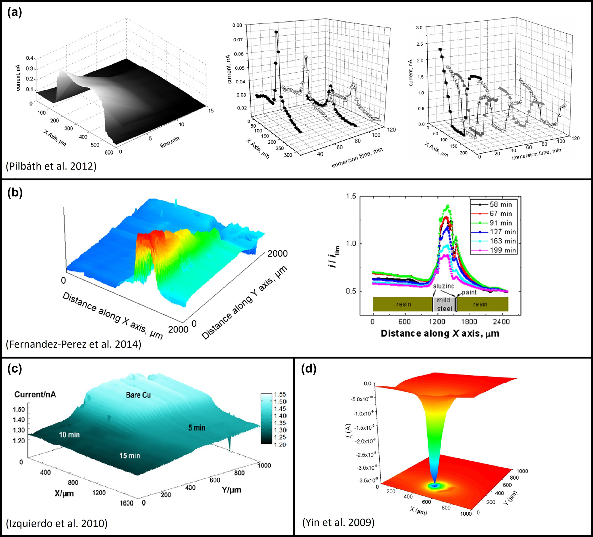
Summary of some of the applications of the SECM in corrosion research, where the self-healing properties of a coating (a), the mechanism of cut edge corrosion (b), corrosion inhibition effect (c) and pitting corrosion (d) are studied. Reproduced with permission from (a) Pilbáth et al. (2012), (b) Fernandez-Perez et al. (2014), (c) Izquierdo et al. (2010) and (d) Yin et al. (2009).
Based on the specific measuring methods and further developments of the SECM tip, in most cases an ultra-micro electrode (UME), a large number of scientific papers and books have been published in the last few years. Among these publications there are by now several review papers, giving a broad general overview about SECM and its application (Amemiya et al. 2008; Mirkin et al. 2011; Li et al. 2016; Zoski 2016). The majority of them addresses a specific field of application. For example, Eckhard and Schuhmann (2008) focused on alternating-current techniques for SECM (AC-SECM), whereas electroanalytical SECM measurement methods were summarized by Mirkin and Horrocks (2000). A review on biological SECM applications was published by Huang et al. (2018), whereas a relatively new review article was published by Polcari et al. (2016) giving an insight into applied experimental set-ups of different operational SECM methods. Several reviews on the use of SECM for corrosion studies were published over the last 15 years by Jensen and Tallman (2012), Keddam et al. (2018), Niu et al. (2009), Payne et al. (2017), and Pust et al. (2008). Regarding the fast development on SECM-based corrosion methods, this work gives an updated, comprehensive overview of the possibilities and advantages of SECM in the field of corrosion research.
The main focus of the first part of this paper is on the description of common SECM measuring modes that have been used for electrochemical analysis of corrosion processes. Subsequently, for each mode specific applications in corrosion science are listed. Based on this information, the second part focuses on UME sensors, which have been applied for corrosion research and their manufacturing process.
2 Instrumentation
The essential elements of a SECM setup are a (bi) potentiostat, to control and measure the current respectively the potential, the SECM tip (an UME tip or a selective sensor), a piezo positioner allowing an accurate three-dimensional navigation of the UME relative to the sample surface and a computer to control the system and gather the generated data as illustrated in Figure 2. The UME is of central importance for the whole measurement and is explained in detail in Section 4. The SECM tip and the substrate are components of the electrochemical cell that is complemented with a reference and counter electrode. As electrolyte usually a low-molar salt solution, in most cases containing additionally a certain redox mediator (RM), is used.
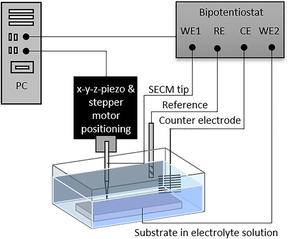
Schematic representation of a typical SECM setup and electrode connections (WE, working electrode, RE, reference electrode, CE, counter electrode).
3 Operational modes
Over time, many different measurement methods have been developed for the SECM to meet the growing demands of expanding research areas. Many of them are well suited for corrosion research and will be explained in detail. In Table 1, different selected SECM modes are shown schematically and explained briefly assuming an oxidation reaction to be occurring at the tip. Additionally to the modes listed in Table 1, there are several other modes, such as the direct mode, where the tip is used as counter electrode and which can be used e. g. to induce pitting on stainless steel samples (Eckhard et al. 2008). Moreover, a combination with atomic force microscopy is possible, allowing for correlating topographical information with electrochemical surface activity (Davoodi et al. 2007; Izquierdo et al. 2015; Izquierdo et al. 2016a; Velmurugan et al. 2017). In several articles, especially for localized metal deposition on the substrate, a potential was applied to the substrate (e. g. Malel et al. (2011), Radtke et al. (2006)). Additionally, potentiometric modes are frequently applied, which are described in Section 3.4.
Overview and description of selected, very common SECM operation modes.
| Mode | Scheme | Description |
|---|---|---|
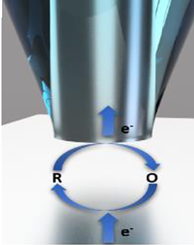
| Feedback (FB) in bulk solution |
|
A defined potential is applied to the tip, so that the RM in solution is oxidised and the resulting current is recorded. If the tip is located in bulk solution, the current can be calculated according to Eq. (2). |
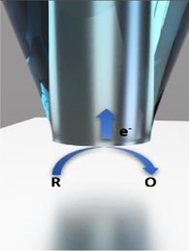
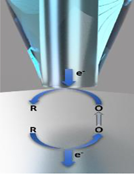
| Negative FB |
|
Application of a defined tip potential leads to an oxidation of the reduced species (R) in solution. Tip movement towards an insulating substrate area, leads to a hindered diffusion of the RM towards the electroactive surface of the tip and therefore, a decrease in current can be observed. |
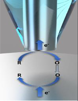
| Positive FB |
|
In case of approximating a conductive substrate area, which actively recovers the RM and re-supplies it to the redox cycle, an increase of current is observed. |
| Substrate generation tip collection (SG/TC) |
|
The redox species of interest is oxidised at the substrate by applying a certain potential. The oxidised species (O) diffuses towards the tip where it is reduced. The monitored current represents the local concentration of the redox species around active spots on a substrate. |
| Tip generation substrate collection (TG/SC) |
|
Inversion of the processes occurring in the SG/TC mode. |
| Redox competition (RC) |

|
A defined potential is applied to both UME and substrate, while the tip current is recorded. Above inactive surface areas the reduction of the redox species will only take place at the tip, while above active spots tip and substrate will compete and consequently a drop in the recorded current can be observed. |
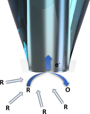
3.1 Feedback mode
The feedback (FB) mode was introduced as one of the first SECM measurement techniques and is still widely used in corrosion research and other research areas. When approaching a sample surface in FB mode, either a positive or a negative FB can be displayed depending on the properties of the sample surface. The basic mathematical model for a UME immersed in a weak electrolyte solution with an RM that approaches a conductive or non-conductive substrate was described by Kwak and Bard (1989b). Assuming that the disc-shaped UME is polarized, applying an appropriate potential, reduction or oxidation of the RM occurs as described in reaction (1):
For simplicity, reversible redox pairs are chosen as mediators, reacting in a fast, simple electron transfer process (Bard et al. 1989). Table 2 gives an overview of successfully used RMs in corrosion studies which also includes dissolved oxygen in the electrolyte solution as RM. A major advantage of oxygen as RM is that dissolved oxygen participates directly in the corrosion process under investigation by reduction to hydroxide ions on the cathodic side. However, oxygen as RM differs from the other listed RMs as this multi-electron reaction is irreversible and beside hydroxyl ions also hydrogen peroxide can be formed, which is relevant in corrosion studies due to its reactivity (Zhang et al. 2020). Although RM should not interfere with the electrochemically active system under investigation, added RM can sometimes affect the Nernst potential at the interface between the sample and the electrolyte. This can lead to difficulties in separating the interference from the added RM from the information obtained from the sample surface (Santana et al. 2010; Souto et al. 2009).
Redox mediators (RM) for different SECM operational modes and applications. ET indicates the potential applied to the SECM tip.
| RM-abbreviation | RM | Redox reaction | Solvent | E T (V vs. SHE) | Mode | Application | Source |
|---|---|---|---|---|---|---|---|
| HQ | Hydroquinone | H2Q → Q + 2H+ + 2 e− | Aqueous | 0.699 | G/C | Investigation of electron transfer from reduction and oxidation processes on Al and Al alloys (AA2024-T3) with different mediators (0.01 M) in near neutral Na2SO4 and acidic H2SO4 (pH 2–3) solution. | Jensen et al. (2008) |
| HQS | Hydroquinone sulphonate | H2QS → QS + 2H+ + 2e− | 0.789 | ||||
| Br− | Bromide | 2Br− → Br2 + 2e− | Aqueous | 0.844 | G/C | Local electrochemical activities on titanium foils with anodically grown TiO2 layer were investigated in a solution mixture of 0.05 M H2SO4 solution with as electrolyte and 1.0 M KBr as RM. | Casillas et al. (1993) |
| Acetonitrile | 0.199 | G/C | Electrically generated bromine on passivated titanium electrodes (Ti/TiO2) in 0.1 M KBr solution was analysed. The results were compared with other redox-active species such as Cl−, I−, Fe(CN)64− and Fe(CN)63− in H2O and nitrobenzene in acetonitrile. | Basame and White (1995) | |||
| Cl− | Chloride | 2Cl- → Cl2 + 2e− | Aqueous | 1.357 | G/C, RC | The local electrocatalytic activity of dimensionally stable anodes (DSA) for Cl2 evolution from brine was demonstrated and visualized. As electrolyte a 5 M NaCl solution (pH 2) was used. | (Zeradjanin et al. 2011; Zeradjanin et al. 2014) |
| The faradaic selectivity of the chlorine and oxygen evolution reaction on Ti-Ru-Ir mixed metal oxide surfaces (Ti–Ru–Ir-based DSA) was determined. | |||||||
| DMAFc+ | (Dimethylamino)methylferrocene | DMAFc+ → DMAFc2+ + e− | Aqueous | 0.554 | FB | The spatially heterogeneous cathodic activity on AA2024-T3 aluminium alloy samples in sodium borate solution (pH 8.25) containing DMAFc+ as RM was investigated. | Seegmiller and Buttry (2003) |
| H2 evolution over a scribed area of PANI and PMMA coated AA2024-T3 aluminium alloy was observed in two different electrolytes (DMAFc + as RM in 10 mM H2SO4 or 10 mM Na2SO4 solution, respectively). | Seegmiller et al. (2005) | ||||||
| The corrosion inhibition of zr(IV)-alkyl-or -aryl-phosphonate thin films deposited on AA2024-T3, which inhibit O2 reduction, was analysed in sodium borate solution (pH 8.25) with DMAFc + as RM. | Dufek and Buttry (2009) | ||||||
| Fe2+ | Ferrous ions | Fe2+ → Fe3+ + e− | Aqueous | 0.771 | G/C | The oxidation of Fe2+ emanating from metastable pits on 304 stainless steel was investigated in 0.1 M HCl solution. | (González-Garcĭ;a et al. 2004) |
| 0.799 | Detection of redox species involved in open-circuit corrosion processes. All measurements were carried out on iron sheets immersed in 0.1 M NaCl solution. | Bastos et al. (2004) | |||||
| 0.799 | The influence of oxygen on the degradation processes of polyurethane coated mild steel plates was studied in 0.1 M KCl solution. | González et al. (2011) | |||||
| FcMeOH | Ferrocene methanol | FcMeOH → [FcMeOH]+ + e− | Aqueous | 0.610 | FB | The surface kinetics of native oxide films on Ti6Al4V alloy were studied in 0.1 M KCl. | Pust et al. (2007a) |
| 0.607 | The local corrosion kinetics of uranium dioxide were determined with FcMeOH acting both as RM and cathodic oxidant. | He et al. (2010) | |||||
| 0.699 | In-situ detection of the swelling and the delamination of organic coatings when immersed in an aqueous electrolyte. | Souto et al. (2004) | |||||
| Investigation of the lixiviation through polymer coatings. | Duarte et al. (2012) | ||||||
| Investigation of the electrochemical reactivity of passive materials such as nitinol. | (Izquierdo et al. 2016b) | ||||||
| The study of horse-chestnut extract as green corrosion inhibitor tested on a bronze substrate. | Varvara et al. (2020) | ||||||
| 0.799 | Monitoring the loss of passivity on nitinol. | Asserghine et al. (2020) | |||||
| Studies on galvanic coupling effects and cytotoxic effects on implants. | Asserghine et al. (2021) | ||||||
| 0.707 | Adsorption of corrosion inhibitors on copper. | (Izquierdo et al. 2010; Izquierdo et al. 2012a) | |||||
| K4[Fe(CN)6] | Potassium hexacyanoferrate-(III)/(II) | [Fe(CN)6]4- → [Fe(CN)6]3- + e− | Aqueous | 1.1 | FB | The local hydrogen evolution on the surface of a corroding Fe–Si alloy was examined in deaerated 0.01 M HCl with 0.01 M K4[Fe(CN)6]. | Fushimi et al. (2007) |
| 0.610 | The surface kinetics of native oxide layer on biphasic alloy Ti6Al4V in 0.1 M KCl were investigated. | (Basame and White 1995; Pust et al. 2007b; Pust et al. 2007a) | |||||
| K3[Fe(CN)6] | 0.755 | The passivation of steel immersed in a simulated concrete pore solution as well as the kinetics of the passivation process were studied. | Torbati-Sarraf and Poursaee (2018) | ||||
| H2 | Hydrogen | H2 → 2 H+ + 2 e− | Aqueous | 0.155 | FB (substrate polarized) | The anomalous hydrogen evolution from anodically-polarized magnesium-based material monitored and the effect of aluminium was determined. Study of HER on Mg(OH)2 covered Mg surface. | (Filotás et al. 2020; Filotás et al. 2021; Salleh et al. 2015) |
| H2O2 | Hydrogen peroxide | H2O2 + 2 OH− → O2 + 2 H2O + 2e− | Aqueous | 0.449 | G/C | The influence of oxygen on degradation processes of polyurethane coated mild steel plates was investigated in 0.1 M KCl solution. | González et al. (2011) |
| I− | Iodide | 2I− → I2 + 2e− | Aqueous | 0.199 | G/C | Analysis of electrogenerated iodide on passivated titanium electrodes (Ti/TiO2). | Basame and White (1995) |
| 3I− → I3− + 2e− | 0.199 | G/C | Investigation of the oxidation of iodide and simultaneously oxide growth and metal dissolution on passivated tantalum electrodes (Ta/Ta2O5) in 0.1 M K2SO4 and 10 mM KI solution. | (Basame and White, 1999b; Basame and White, 1999a) | |||
| 0.799 | FB | The dissolution of MnS inclusions during initiation of pitting corrosion was studied for two different stainless steel grades (SS303 and SS304) in 0.1 M NaCl solution containing 10 mM KI. | Paik et al. (2000) | ||||
| 0.799 | FB | Pitting corrosion on Ni200 samples located near sulphide inclusions was studied 0.1 M NaCl solution containing 10 mM KI as RM. | Paik and Alkire (2001) | ||||
| [IrCl6]3− | Iridium chloride | [IrCl6]3− → [IrCl6]2− + e− | Aqueous | 1.21 | FB | The surface kinetics of the native oxide film on biphasic alloy Ti6Al4V was studied in 0.1 M KCl solution with 1 mmol K3[IrCl6]. | Pust et al. (2007a) |
| AQDS | Anthraquinone-2, 6-disulfonate | AQDS + 2H+ + 2e− → AHQDS | Aqueous | −0.181 | G/C | Investigation of electron transfer from reduction and oxidation processes on Al and Al alloys (AA2024-T3) with different mediators (0.01 M) in near neutral Na2SO4 and acidic H2SO4 (pH 2–3) solution. | Jensen et al. (2008) |
| NO2− | Nitrite ions | NO2− + H2O → NO3− + 2H+ + 2e− | Aqueous | +1.155 | AFM-SECM | The nucleation as well as the growth of pits at an Fe passive oxide layer was monitored. | Izquierdo et al. (2015) |
| FcCOOH | Ferrocene carboxylic acid | [FcCOOH]+ + e− → FcCOOH | Aqueous | 0.529 | FB | Surface reactivity and local corrosion processes on Nd13.5Fe79.5Si1B6 permanent magnets were monitored in a 0.1 M potassium phosphate buffer (pH 7) with 5 mM FcCOOH in presence and absence of 0.1 M KCl. | (Kranz et al. 1997; Malik and Kulesza 2007; Malik et al. 2009) |
| Fe3+ | Ferric ions | Fe3+ + e− → Fe2+ | Aqueous | +1.155 | AFM-SECM | The nucleation and growth of pits at an Fe passive oxide layer was monitored. | (Izquierdo et al. 2014a) |
| H+ | Hydrogen | 2H+ + 2e− → H2 | Aqueous | −0.50 | G/C | The corrosion behaviour of Mg alloy AZ31 was studied in a simulated biological fluid based on oxidation of H2, which was evolved during corrosion process. | Jamali et al. (2015) |
| 0.150 | H2 fluxes from corroding AM50 Mg alloy surfaces in 0.6 M NaCl solution were quantitatively detected. | Tefashe et al. (2014) | |||||
| TCA | Trichloroacetic acid | Cl3CCOO− + H2O + 2 e− → Cl2CHCOO− + Cl− + −OH | Aqueous | 0.627 | FB, G/C | The SECM tip was used to generate Cl− from TCA as RM to induce e. g. pitting of iron. All measurements were performed in a phosphate-citrate buffer solution at pH 6. | Still and Wipf (1997) |
| AgCl | Silver chloride | AgCl(s) + e− → Ag(s) + Cl− | Aqueous | 1.370 | FB | The early stages of pit initiation and propagation on an A316L SS disc were studied in 0.5 M H2SO4. Pit initiation occurred through the local release of a controlled amount of Cl− ions from an AgCl microelectrode. | Aouina et al. (2011) |
| NB | Nitrobenzene | NB + e− → NB.− | Acetonitrile | 0.199 | G/C | Analysis of electrically generated iodide on passivated titanium electrodes (Ti/TiO2) in different electrolytes. | Basame and White (1995) |
| 0.539 | SECM imaging in a non-aqueous electrolyte with 0.1 M TBAPF6 was used to study electronic defects in the native passive film on Al/Al2O3 electrodes. | (Serebrennikova et al. 2002; Serebrennikova and White 2001) | |||||
| O2 | Oxygen | O2 + 2 H2O + 4e− → 4 OH− | Aqueous | −0.501 | G/C | Analysis of Fe2+ ions and the depletion of oxygen at the cathodic site during the immersion of iron in a neutral 0.1 M NaCl solution. | Bastos et al. (2004) |
| O2 + 2 H2O + 4e− → 4 OH− | FB, G/C | On a carbon steel plate coated with a two-component polyurethane film, the consumption of dissolved oxygen at cathodic sites was monitored in aerated 0.1 M KCl, 0.1 M K2SO4 and 0.1 M Na2B4O7 solution. | Souto et al. (2005) | ||||
| −0.501 | FB | In-situ measurements of corrosion processes inside a coating defect on epoxy coated steel samples immersed in an aerated aqueous solution of 1 M NaClO4 and 1 mM HClO4 were performed, studying the self-healing properties of encapsulated microcapsules inside the coating. | Pilbáth et al. (2012) | ||||
| O2+ 4H+ + 4e− → 2H2O2 | −0.293 | RC | The depletion of oxygen at cut edges of electrogalvanized steel in a 0.01 M NaCl (with different pH values) solution was monitored. | Marques et al. (2015) | |||
| −0.401 | RC | AA2024 aluminium alloy coated with silane incorporated epoxy resin was studied in 0.1 M NaCl solution to investigate the oxygen concentration at the defect area. | Jiang et al. (2015) | ||||
| −0.501 | RC | Mapping of dissolved hydrogen concentration of an electrochemically hydrogen pre-charged stainless steel sample. | Schaller et al. (2015) | ||||
| [Ru(NH3)6]Cl3 | Hexamine ruthenium(III) chloride | [Ru(NH3)6]3+ + e− → [Ru(NH3)6]2+ | Aqueous | −0.151 | FB | Amorphous alumina thin films on platinum and stainless steel were analysed in 0.1 M KCl containing 1 mM [Ru(NH3)6]Cl3 to obtain information about the uniformity, topography, chemical stability, and reactivity of the metal supported thin Al2O3 films. | Battistel et al. (2010) |
| 0.057 | The native oxide film on the biphasic alloy Ti6Al4V was investigated in 0.1 M KCl containing 1 mM [Ru(NH3)6]Cl3. | Pust et al. (2007a) | |||||
| TCNQ | Tetracyano quino dimethane | TCNQ + e− → TCNQ•− | Acetonitrile | 0.739 | G/C | Al/Al2O3 electrodes were investigated to show the relationship between local corrosion of Al and the defect structure of the Al2O3 film in 0.1 M MeCN solutions containing 0.1 M TBAPF6 and 5 mM TCNQ. | Serebrennikova et al. (2002) |
In FB mode, the tip current resulting from oxidation/reduction of the RM is recorded. At a large distance (about ten times the radius of the UME) between the tip and the substrate, the measured current quickly reaches a steady state as a hemispherical diffusion layer of O forms around the tip, ensuring a continuous flow of reducible/oxidisable species to the tip (Ciani and Daniele 2004; Edwards et al. 2006). The steady-state current is calculated according to Eq. (2),
where n is the number of transferred electrons, F the Faraday constant, D the diffusion coefficient of the RM in solution, c the concentration of the RM, α the radius of the electroactive surface of the UME and βRG is a geometric coefficient, defined by the ratio of rb (radius of the electrode insulating shield) to ra (electrode radius) (Lefrou and Cornut 2010).
As mentioned earlier, a negative or positive FB can be observed depending on the properties of the sample. When the tip approaches an insulating sample, the diffusion of RM from the solution to the UME is partially hindered by the sample and consequently the recorded current decreases. The decrease in current is called negative FB. As the tip approaches a conductive sample, an increase in current is observed as the sample surface more or less actively recovers RM. The response in FB mode is distance dependent, i.e., the closer the tip is to the surface, the steeper is the increase or decrease of the recorded current (Kaufmann 2012). The FB investigation of a substrate can be performed via approach curves, line scans or area scans. For an approach curve, the UME position is fixed in x- and y-direction, while the sample surface is approximated in z-direction. The recorded tip current is plotted against the travelled z-distance (Souto et al. 2010). In a line or area scan, the tip is held at a constant height or distance to the sample surface, while scanning the surface in x- and/or y-direction. In case of a line or surface scan, the tip is held at a constant height or distance from the sample surface while the surface is scanned in the x- and/or y-direction.
3.1.1 Application of FB mode
The FB mode is a very versatile method for the investigation of corrosion processes. An excerpt of current publications in which approach curves as well as line and surface scans in FB mode are used is listed here.
Simões et al. (2007) investigated the electrochemical processes that occur during galvanic corrosion of iron and zinc in sodium chloride solutions. SECM measurements were used to detect and identify the concentration of chemical species that can be associated with the corrosion process taking place. Another example of the use of approach curves by Abodi et al. (2014) is discussed in Subsection 3.3.1.
González-García et al. (2010) studied intact and defective coatings on metals in FB mode, G/C mode and RC mode. In-situ water-uptake measurements of coated samples immersed in an aqueous solution could be performed in FB-constant height mode with as well as blistering of polymer coated metals in chloride-containing environments. Information on corrosion processes was obtained using the G/C mode as well as surface imaging in the RC mode. Ferrocene methanol, ammonium iron (II) sulphate, potassium ferrocyanide and dissolved oxygen were used as RMs.
A similar study was carried out by González et al. (2011), studying the effects of oxygen concentration on the corrosion process of polyurethane coated carbon steel samples with artificially created defects in the coating. Line scans across the defect with different potentials applied to the tip were used to obtain concentration profiles of corrosion process relevant electroactive species, such as +0.60 V versus Ag/AgCl/KCl (sat.) to record the oxidation of Fe2+, +0.25 V versus Ag/AgCl/KCl (sat.) for the oxidation of the generated H2O2 and -0.70 V versus Ag/AgCl/KCl (sat.) to monitor the reduction of O2. In this way, a critical oxygen concentration for the occurrence of corrosion could be determined.
Another study on localised corrosion of stainless steel in neutral chloride solution was presented by Yin et al. (2009). FB surface scans with an applied potential of +0.56 V versus (SCE) were performed to detect Fe2+ after different immersion times and differently polarised substrates. Furthermore, concentration and electrochemical activity distribution profiles were analysed by a combination of FB area scans and G/C mode. The effects of immersion time, polarisation current and applied potential were analysed. In addition, the G/C mode (see next subsection) was used to determine the resulting corrosion products and their concentration profiles.
The incubation and progress of pitting corrosion of AM60 and AMCe1 magnesium alloys in sodium chloride solution were investigated by Liu et al. (2014). FB mode was used to study the corrosion behaviour of AM60 and AMCe1 in different concentrated chloride solutions. Higher chloride concentrations showed a higher number of active corrosion sites on the substrate surface and accelerated pit initiation. The addition of Ce in AMCe1 magnesium alloy was found to increase pitting corrosion resistance. In another approach (Dong et al. 2012), metastable pitting corrosion of stainless steel was stabilised in chloride solution to allow the study of electrochemical processes by exceeding the pitting potential.
Due to the high significance of results that can be obtained with FB mode, this SECM mode is also applied in many other research areas, from reaction kinetics (Cornut and Lefrou 2008; Cornut et al. 2010; Unwin and Bard 1991) to biology (Edwards et al. 2006; Schulte et al. 2010) and electrocatalysis (Bard 2010; Nagaiah et al. 2013).
In Table 2, the redox mediators with their applications in corrosion research are listed. The table is divided into oxidizing mediators (grey background) and reducing mediators (white background).
3.2 Generation/collection mode
Beside the FB mode, another measurement method has been established, namely the Generation/Collection (G/C) mode (Lee et al. 1991a). Variants of the G/C mode are the Tip-Generation/Substrate-Collection (TG/SC) and the Substrate-Generation/Tip-Collection (SG/TC) mode (Lu et al. 2007; Kai et al. 2018). In both methods, the current generated by a redox reaction is recorded at both the UME tip iT and the substrate iS (Bard and Mirkin 2012). The main difference between these two systems is the site of generation and collection of the redox species. The choice for one of these two methods has a decisive influence on the collection efficiency. A theoretical explanation of the SG/TC mode is given by Martin and Unwin (1998), Bard and Mirkin (2012), Bertoncello (2010) and Lu et al. (2007). G/C experiments can be operated in amperometric or potentiometric mode (detailed explanation of this mode is given in Subsection 3.5).
3.2.1 Sample-generation/tip-collection (SG/TC) mode
In the SG/TC mode, the redox process is initiated at the substrate surface. Therefore, a suitable potential is applied to the sample and R is oxidised according to Eq. (3).
(3)
The resulting oxidised species O is then transported within a local hemispherical diffusion layer emanating from the substrate. The tip is moved through the diffusion layer and O is reduced according to formula (1) and therefore “collected” at the tip, with the resulting current representing the local concentration of the redox species. For accurate and high-resolution measurement results, the tip and its movement should not disturb the diffusion layer and the concentration profiles of the substrate. Therefore, a small tip size on the nanometre scale is recommended (Baltes et al. 2004; Sun et al. 2007).
The amperometric measuring method can also affect the diffusion layers of the substrate through secondary chemical reactions at the tip. Here, a potentiometric measuring method would guarantee more accurate results because the tip acts as a passive sensor and thus does not affect the diffusion layers. Since the UME tip is of a very small in relation to the substrate surface, the SG/TC mode has a low collection efficiency (Sun et al. 2007) which does not allow the quantification of the these species. Nevertheless, SG/TC is often used for the investigation of local corrosion processes, as a selective detection of the released reactive species from the substrate surface is possible. One method to increase the detection efficiency is to use mercury or bismuth modified tips on which the redox species accumulate during the scan and can finally be analysed by stripping voltammetry (Ciani et al. 2003; Janotta et al. 2004; Souto et al. 2010). The collection and subsequent stripping from the tip are not only limited to liquid UME tips; e. g. also solid Au electrodes can be used as shown by Izquierdo et al. (2016c).
3.2.1.1 Application of SG/TC mode
González-García et al. (2010) applied the SG/TC mode alongside the FB mode to obtain more information on the concentration and distribution of corrosion products within coating defects. Furthermore, González et al. (2011) used the SG/TC-mode to investigate the effect of oxygen concentration on the corrosion process of polymer coated steel samples. Different potentials were applied to the tip to detect and record the local concentration profile of several species such as Fe2+ ions, H2O2 or O2. A progressive consumption of oxygen resulted in a decline of iron dissolution. The SG/TC mode was also applied by Bastos et al. (2005) to detect local concentrations of reactants involved in the corrosion process on galvanised steel substrates coated with two-component epoxy primers containing zinc phosphate. Further examples for the application of SG/TC mode for the detection of corrosion products are given by Bastos et al. (2004), Izquierdo et al. (2014b), Jamali et al. (2014), Jamali et al. (2015), Tefashe et al. (2014) and Völker et al. (2006).
The SG/TC mode is able to detect active spots on sample surfaces, which is essential for studying electrocatalytic processes. For example, Minguzzi et al. (2008) studied electrocatalytic oxygen evolution on pure iridium oxide and combinatorial iridium-tin oxide mixtures. To reduce interference from other spots diffusing to the tip, a gold layer was deposited on the outer wall of the UME and kept at a constant potential to consume oxygen from neighbouring spots. Sánchez-Sánchez and Bard (2009) used the TG/SC mode to detect H2O2 production on Hg, Au, Ag, Cu, Pt, Pd, Pd80Co20 and Au60Cu40 in aerated diluted sulphuric acid with oxygen as RM. Further applications of SG/TC mode in corrosion science for electrochemical investigation can be found in Sánchez-Sánchez et al. (2008), Shen et al. (2008) and Souto et al. (2011). SECM in SG/TC mode was used to determine the number of electrons transferred during the cathodic oxygen reduction reaction. For this purpose, Sánchez-Sánchez et al. (2008) quantified hydrogen peroxide in an acidic environment during the oxygen reduction reaction on an Hg containing Au electrode. Shen et al. (2008) also detected hydrogen peroxide during the oxygen reduction reaction, but on other substrates such as gold and palladium-cobalt samples. Souto et al. (2011) monitored not only hydrogen peroxide but also Fe2+ and O2 in SG/TC mode next to a defect on a coated steel sample to investigate early stages of delamination. In addition to studies on corrosion and catalysis processes, the SG/TC mode has also been applied in other research areas. Ramanavicius et al. (2017) studied the bioelectrochemical activity of yeast cells using a lipophilic RM with the ability to cross the cell membrane in combination with ferricyanide as a second RM. Further examples can be found in battery research (Ventosa and Schuhmann 2015; Xu et al. 2012). Cathode materials for lithium-ion batteries have been studied in SG/TC mode to deepen the understanding of surface and bulk processes.
3.2.2 Tip-generation/substrate-collection mode (TG/SC)
In the TG/SC mode, a redox species is generated at the tip, which is subsequently “collected” at the substrate, whereby quite high capture efficiencies of almost 100% are achieved. Especially if the distance between tip and substrate is small (normalised distance ≤2), most of the products, generated at the tip, diffuse to the substrate. The substrate surface is in the majority of cases larger than the tip surface, so that the recorded iT and iS are almost equal and the collection efficiency iT/iS is close to 1 or 100% (Zhou et al. 1992). A more detailed description as well as the theoretical background of the TG/SC mode is given in Fernández and Bard (2004).
3.2.2.1 Application of TG/SC mode
The TG/SC mode is mainly used to study the pit initiation kinetics of electrochemical reactions on different metal surfaces. Therefore, the measuring mode is very important for monitoring corrosion processes. For instance, Jensen et al. (2008) investigated the electron transfer of redox processes on Al and its alloys using different RMs. Further applications of the TG/SC mode for kinetic research are described in Cannan et al. (2011) and Zhang et al. (2018). The TG/SC mode is also used in the study of pitting corrosion on passivated metal surfaces (Gabrielli et al. 2008). The pitting corrosion of iron in various acidic and alkaline solutions was analysed, including the effects of pH on the pitting activity. Measurements in borate buffer solutions showed the formation of insoluble corrosion products that block the expansion of the pit. Furthermore, the TG/SC mode is used for electrocatalyst research, as shown by Eckhard and Schuhmann (2007). The catalytic reduction of O2 on electrodeposited gold or platinum was monitored, by generation of O2 at the tip and the subsequent reduction at the sample surface. Fernández and Bard (2003) investigated the oxygen reduction reaction on different electrode materials in acidic solutions in TG/SC mode. Since the electrocatalytic activity of the electrode surface is reflected in its oxygen reduction rate, the tip was placed near the substrate surface and kept at a constant current, at which water oxidises to oxygen, while the electrode material under investigation was held at a potential, at which oxygen is reduced to water. Johnson and Walsh (2012), in turn, investigated the electrochemical activity of Au catalysts with self-produced micro ring-disc tips in TG/SC mode by monitoring the oxygen reduction reactions and the hydrogen peroxide formed in the process.
3.3 Redox competition mode (RC)
A major disadvantage of the TG/SC mode is the increasing background current with larger electrochemically active substrate surfaces. To circumvent the sensitivity limitations when measuring larger conductive surfaces in the TG/SC mode, Schuhmann and his colleagues invented the redox competition mode (Eckhard et al. 2006). The working principle of the RC mode is similar to that of the TG/SC mode. While for catalysis applications a bias potential is applied to the sample, in corrosion research the sample is in most cases monitored in spontaneous corrosion conditions (at OCP) or at galvanic coupling conditions. A potential is applied to the tip, while the current is recorded (Guadagnini et al. 2009). In this way, background current is significantly reduced while sensitivity is improved (Karnicka et al. 2007). When the tip is moved over an inactive substrate region, the redox process preferentially takes place at the tip, resulting in a constant or increasing current. In contrast, if an active region is scanned on the substrate, competition for the redox species, which can react both at the tip and at the substrate, leads to a decrease in the recorded peak current (Eckhard et al. 2006).
The redox species plays a crucial role in this measurement method. It can either be a component of the electrolyte solution (Souto et al. 2009; Santana et al. 2010) or it can be generated in operando by applying a short pulsed potential to the tip, which allows the concentration of the species to be controlled (Karnicka et al. 2007). In several corrosion studies, oxygen dissolved in the electrolyte is used as RM for the RC mode to detect cathodic sites of corroding metal surfaces (Souto et al. 2010).
3.3.1 Application of RC mode
Main application of the RC mode is the investigation of corrosion and the catalytic activity of surfaces (Chen et al. 2009). Kong et al. (2018) studied the corrosion process on Co-Ni-Zn alloy in 0.5 M NaCl solution as well as the influence of the temperature and polarisation time on the formation of pitting corrosion. Lower temperature led to higher pitting nucleation rate with lower pit growth. The amount of porous corrosion products on the surface increased with increasing temperature as well as the transition rate from pitting to uniform corrosion. RC mode can also be used to detect species of interest such as diffusible hydrogen in steel samples. Schaller et al. (2015) analysed high strength stainless steel with high diffusible hydrogen capacity to detect hydrogen loaded onto the alloy. Results from RC-SECM measurements showed increasing oxygen consumption at the steel surface, if an excessive amount of hydrogen was present in the investigated region. The recorded tip current resulted from the oxygen reduction reaction, which decreased with increasing oxygen consumption at the substrate.
Another application of the RC mode in corrosion science is the investigation of protective coatings on metals. González-García et al. (2011) investigated the local healing efficiency of an artificially damaged self-healing polymer on an aluminium alloy before and after a thermal healing process. The RC mode was used to analyse the O2 reduction over a coating defect. Physical repair of the polymer coating resulted in renewed protection of the aluminium alloy and inhibited corrosion.
In the following example, an amperometric O2 measurement was carried out instead of a potentiometric one. For this, Abodi et al. (2014) used simple approach curves to investigate the local effect of O2 on the corrosion process of aluminium. Corrosion was induced by immersing a pure aluminium substrate in an aerated NaCl solution. SECM measurements were performed using a 10 µm platinum UME with the tip potential set at −0.65 V versus SCE to reduce and detect oxygen according to Eq. (3).
The resulting data were finally compared with the computer-generated data of the multi-ion transport and reaction model. After experimental confirmation of the computer model, further simulations were carried out focusing on the influence of the SECM probe on the electrochemical process at the aluminium surface. Further investigations revealed that oxygen reduction at the substrate surface increases the local pH due to the formation of hydroxide ions and a decrease in the partial current densities for O2 due to oxygen depletion was observed (Abodi et al. 2014).
Furthermore, galvanic coupling can be investigated with RC mode as shown by Izquierdo et al. (2013a) for the Fe-Zn couple, Marques et al. (2015) for monitoring cut-edges of galvanized steel and da Silva et al. (2021), who studied welded zones in Al-Cu-Li alloy. In most cases, additionally to SECM, scanning vibrating electrode technique was applied.
3.4 Potentiometric mode/ion-selective mode
All SECM measurement modes mentioned so far belong to the amperometric methods, as a suitable potential is applied to the tip and/or the substrate and the generated current is recorded. In contrast, the potentiometric mode monitors the potential between the tip and a reference electrode and is often used for ion-selective measurements. The reference electrode can either be positioned separately at a certain distance from the ion selective micro-electrode (ISME) tip or coupled to the tip in a dual-channel capillary. Concerning the position of the reference electrode, care has to be taken to consider the contribution of the electric field to the potentiometric signal. Kiss et al. (2017) as well as Filotás et al. (2018) described this undesired contribution, caused by the strong electric field resulting from galvanic coupling of different metals, leading to a wrong estimation of the analyte ion activity. The proposed solutions involve the minimisation of distance between tip and reference electrode or the usage of an electric relay to disconnect the galvanic couple during the measurement. The tip electrode is frequently equipped with an ion-selective membrane to ensure that the potential changes are caused solely by the analyte ion. A detailed description of the sensor as well as a list of all analytes investigated so far or the corresponding ionophores can be found in Subsection 4.2.3. The potential of the ion-selective electrode Ei depends on the concentration/activity of the analysed ion and can be calculated according to Eq. (4).
Where R is the gas constant, T is the absolute temperature, F is the Faraday constant, zi is the charge of the ion and ai is the activity of the primary ion i in the sample and reference solution. A more detailed description as well as the theoretical background of the ion-selective mode are given in Bard and Mirkin (2012), Horrocks et al. (1993a) and Wei et al. (1995).
A major advantage of the potentiometric method is that ion concentrations of species such as alkaline earth metals and halides, which are usually not electroactive in aqueous solutions, can be determined. In addition to determining the ion concentration of these ions, the potentiometric method is also used to determine the hydrogen ion concentration in order to determine the pH value of a sample surface. Here, the potentiometric SECM mode does not include a faradaic process, and potentiometric UMEs are usually passive probes. Therefore, both the oxidation state and the concentration of the analyte under investigation do not change during the measurement (Bard and Mirkin 2012; Horrocks et al. 1993a; Polcari et al. 2016; Wei et al. 1995).
Another advantage is that the tip does not generate or collect analyte species. Therefore, the concentration profiles and oxidation states of the analyte species are not affected and local quantification of a particular species is possible (Horrocks et al. 1993a and Dimeski et al. 2010). Since only one type of ion can be detected with the potentiometric mode, this mode is not limited to certain research areas. Biological fluids consist mainly of ions like Na+, K+, Cl−, Ca+, Mg2+ and Li+, which makes the potentiometric mode highly interesting for medical and clinical applications as described in Dimeski et al. (2010). In corrosion research, the ion-selective mode is used to detect metal ions such as Mg2+, Zn2+ or Cu2+ formed during the corrosion process due to metal dissolution, as well as hydrogen ion concentration to determine the pH of the metal surface, e. g. to identify cathodic and anodic sites. Table 5 in Subsection 4.2.3. Lists the ionophores used.
The above-mentioned dual channel electrodes, including dual oxide electrodes from Sb, Bi or Ir, can operate additionally in amperometric mode. In this manner, the advantage of the amperometric mode for the tip positioning and current-distance determination can be combined with the ion-selective measurement capability of the potentiometric mode (Santos et al. 2016; Filotás et al. 2016).
As extension of the potentiometric ion-selective electrodes, multi-channel electrodes were developed. With such electrodes, multiple ionic species can be simultaneously and selectively detected while offering the dual functionality of amperometric and potentiometric application mode (Filotás et al. 2016; Hampel et al. 2019).
3.4.1 Application of the potentiometric/ion-selective mode
One of the most important applications of the potentiometric/ion-selective measuring method in corrosion research is the investigation of degradation and dissolution processes of metals with low standard electrode potential, such as magnesium, aluminium, or zinc. Souto et al. (2013) were able to visualize concentration gradients of Mg2+ ions for the case of magnesium-based alloy AZ63 coupled with pure iron in NaCl solution. The ISME consisted in this case of a carbon fibre coated with poly-3, 4-ethylenedioxythiophene in a micropipette filled with a bis-N,N-cyclohexyl-malonamide magnesium ionophore.
A comparison of Mg2+-ion-selective microelectrodes for potentiostatic SECM was given by Izquierdo et al. (2013b). Here, a solid contact micropipette-based magnesium ISME was compared to a conventional microtip ISME with an aqueous internal reference electrode. The results for the dissolution of magnesium in combination with pure iron showed that the solid contact ISME has a faster response time and lower internal resistance than the liquid contact ISME. Izquierdo et al. (2012b) also developed a solid-state contact Zn2+-ISME using N-phenyl-iminodiacetic acid bis-N′,N′-dicyclohexylamide as a zinc ionophore to successfully map the Zn2+ concentration distribution on zinc in combination with iron.
Very promising, especially for investigation of galvanic corrosion processes for the above-mentioned reasons, is the usage of double- or generally multi-barrel ISMEs as described in various publications, e. g. by the Souto-group. Filotás et al. (2017a) reported the simultaneous detection of protons, zinc and copper ions from a Cu-Zn galvanic couple corroding in aerated 1 mM NaCl solution. In another publication of the group, a micro-reference electrode was introduced into a multi-channel arrangement, which was used to monitor simultaneously the pH and magnesium ion distribution on a corroding AZ63 Mg alloy in galvanic coupling to Fe (Filotás et al. 2019).
3.5 Alternating current mode (AC)
In addition to the amperometric and potentiometric SECM methods described in the previous sections, AC-SECM is used to obtain information on local electrochemical activity by combining SECM with electrochemical impedance measurement. AC-SECM does not require the addition of an RM to the electrolyte solution, which in many cases has a low concentration with a resulting high resistance. In contrast to the previously described methods, an AC potential is applied while the AC feedback of the system is recorded. Horrocks et al. demonstrated that the impedance measured at high frequency in low conducting solutions is dominated by the resistance of the electrolyte solution (Rsol) between the working and counter electrode. Rsol is strongly dependent on the distance between the tip and the substrate as well as on the conductivity properties of the substrate (Ballesteros Katemann et al. 2002; Ballesteros Katemann et al. 2003; Horrocks et al. 1993b). In case of a conductive sample being approached by the SECM tip, a positive-type feedback is obtained. Whereas, a negative-type feedback is recorded when approaching a non-conductive surface in AC mode. LeSuer et al. (2004) stated that the negative feedback mode obtained by AC-SECM is comparable with the amperometric feedback mode. A detailed description as well as the mathematical fundamentals are given in Baranski and Diakowski (2004), Bard et al. (1989) and Eckhard et al. (2007b). A detailed description of various AC SECM measuring techniques is given in detail by Eckhard and Schuhmann (2008).
3.5.1 Application of the AC mode
Eckhard et al. (2007a) succeeded in improving the visualisation of pit formation as well as the detection of immediate pit growth with AC-SECM. To investigate pitting corrosion, a very small corrosion pit was created on a 316 Ti stainless steel sample using the direct mode of amperometric SECM. AC-SECM measurements with constant spacing were performed with a Pt-tip before and after the pit was created. Since the tip served as a counter electrode and the substrate as a working electrode, the approximate position of the pit formation could be determined. Corrosion pits with a diameter of only a few micrometres could be analysed. The resolution for the detection of corrosion pits could be further improved by sweeping the frequency at each point within a defined range instead of only using a constant frequency during the measurements (Eckhard et al. 2007a). The result was a four-dimensional data set of the substrate containing current, phase shift, real and imaginary parts with a lower probability of data misinterpretation due to an unfavourable choice of measurement frequency.
Another example for the application of the AC-SECM mode is given by Lafouresse et al. (2017). Lafouresse et al. (2017) successfully used the AC mode to investigate H-embrittlement mechanisms by pre-charging a 2024 aluminium alloy substrate and by detecting the hydrogen insertion as well as hydrogen desorption. Mapping perpendicular to the charging side allowed observing a gradient in AC current magnitude. Hereby, higher AC currents indicated an area with high hydrogen concentration.
Besides the mentioned applications for the AC-SECM mode, this mode can also be used to study corrosion inhibition and delamination of coatings (Santana et al. 2012a; Santana et al. 2012b).
In addition to performing AC-SECM measurements where the AC perturbation is applied on the tip, the AC perturbation can be applied to the substrate, allowing, for example, the study of adsorption kinetics (Trinh et al. 2011a; Trinh et al. 2011b). Trinh et al. (2011b) used this method to examine the adsorption process during the hydrogen evolution reaction in sulphuric acid solution.
Kuznetsov et al. (2014) further developed the AC-SECM mode to scanning electrochemical impedance microscopy (SEIM) and successfully applied this mode to measure in-situ localized corrosion processes at metal surfaces. SEIM has been used, for instance, to detect local electrochemical activities on brass surfaces (Kuznetsov et al. 2016).
4 Ultramicro electrodes
Ultramicro electrodes (UME) are a central element of SECM measurements and form the basis for spatially resolved monitoring of local electrochemical activity. Depending on the application, the geometry of the UME can vary in order to achieve an optimal electrode response. If the electrode diameter is less than 25 μm, an electrode is called UME or microelectrode (Bard and Faulkner 2000; Kranz 2014). Accuracy as well as the resolution of the recorded data is primarily depending on the applied electrode, its diameter, RG value and shape.
A rough distinction of UMEs can be made based on the practised measuring mode. Amperometric probes are characterised by fast response times, simple probe positioning, high robustness and thus a long service life. On the other hand, their selectivity is poor, so that it is not possible to differentiate from which process the recorded signal originates. Quite the contrary applies for potentiometric UMEs, which offer a high selectivity towards a specific analyte, while being frequently limited in response time (Polcari et al. 2016). In the past, the lifetime of potentiometric tips was shortened due to the difficult probe positioning since no approach curves can be measured, which was solved by the use of dual oxide probes as in the case of Sb. Sb can be operated as amperometric or potentiometric tips by polarizing the surface adequately to obtain an oxide-free surface (for amperometric operation) or to build up an oxide layer that can be used potentiometrically to determine the pH value (Souto et al. 2012c). Another option was applied by Filotás et al. (2016). Filotás and his group used dual channel arrangements containing a conventional amperometric tip, which allows probe positioning and an ISE of interest for the potentiometric operation.
In corrosion research, the investigation of corrosion processes and corrosion products is of great importance. Since corrosion reactions often take place on a nanometre and micrometre scale, UMEs are a suitable instrument for observing corrosion phenomena in narrowly defined areas due to their small size (Heinze 1993; Souto et al. 2010). The obtained resolution of the recorded data is strongly dependent on the diameter of the UME tip and is inversely proportional to the electrode diameter (Kranz 2014). The diameter of the UME is preferentially chosen to match the size of the individual regions of interest, which is of special importance to observe local corrosion activities. Ye et al. (2018), for example, used a submicron UME to monitor pitting corrosion of stainless steel.
In principle, a microelectrode is formed of a conductive electrode material, embedded into an insulating material, e.g. a metal wire in a glass capillary. Mechanical treatment of the metal-capillary assembly, like grinding and polishing, reveals the micro-disc (MD) electrode at the capillary tip. One of the most common parameters to characterize an UME is the so-called RG-value, which is defined by the ratio of the insulating shield radius rb to the electrode radius ra (Figure 3) (Amphlett and Denuault 1998; Souto et al. 2010).
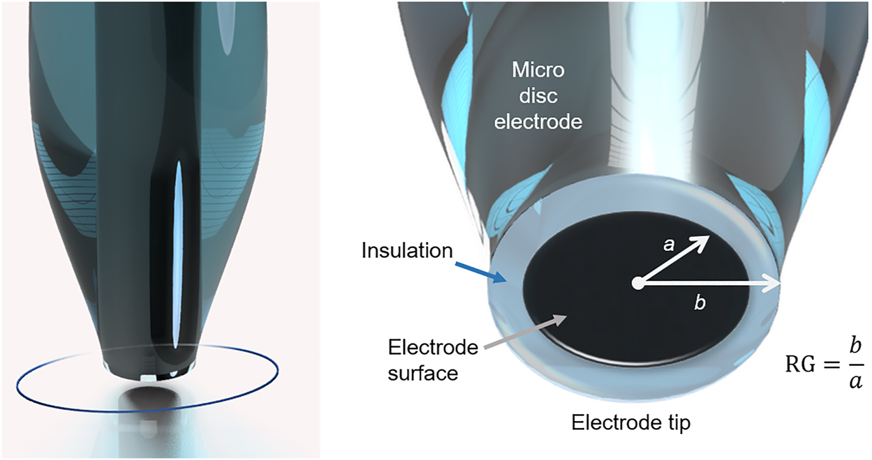
Illustration of an UME tip with markings for used radii for RG value calculations.
To obtain a very close approach of the tip, the glass shielding and thereby the RG value must be very small. RG values <10 are desirable, because the risk of crashing the tip during a tip approach is reduced. Other advantages are that with a small RG nanometre tip-to-substrate distances can be achieved resulting in a high sensitivity and large currents due to a better back diffusion of the mediator (Danis et al. 2015; Forster 1994; Leonhardt et al. 2011; Polcari et al. 2016; Shen et al. 2011).
4.1 Shape of UMEs
UMEs can have a variety of different geometries, with each shape having different characteristics (Mirkin et al. 1992). Possible UME electrode geometries are disc, conical, hemispherical, ring or combinations such as ring-disc (Lee et al. 1991b; Zoski 2002). Detailed information on the different UME geometries can be found in Bard and Faulkner (2000) as well as in Mirkin et al. 1992. It is also possible to fabricate multi barrel electrodes, where two separate electrodes are intertwisted, which allow the simultaneous measurement in amperometric and potentiometric mode (Filotas et al. 2012; Wen and Oakley 1990; Wei et al. 1995). This has the advantage that improved results can be obtained. It is also possible to perform two potentiometric measurements at the same time, for which the adequate ionophores must be injected into the two different electrode capillaries. An example would be the simultaneous detection of zinc and hydrogen ions without changing the UME in between (Filotas et al. 2017a; Filotas et al. 2017b).
4.2 Fabrication of UMEs for corrosion analysis
There are two established techniques for producing UMEs. One technique is based on a micropipette puller system, in which a metal wire is pulled into a glass capillary under vacuum conditions and sealed. In the second method, a metal wire is placed in a glass capillary with one end closed. The capillary is then sealed around the wire by placing it in a heated coil and polishing it to expose the electroactive surface. Detailed information about the fabrication can be found in Ballesteros Katemann and Schuhmann (2002), Danis et al. (2015) and Zoski (2002).
The following subsections describe four categories of electrode types commonly used in corrosion studies, with a clear focus on MD electrodes, as they are most commonly used in the study of corrosion processes. The manufacturing routes and applications are summarised for (transition) metal and carbon MD electrodes, coated and surface modified electrodes, ion selective electrodes and dual function.
4.2.1 Metal and carbon MD electrodes
A variety of materials can be used to make UMEs, from carbon fibres to noble metals with platinum being the most common, but also gold, silver, iridium, and other transition metals such as antimony, cobalt or tungsten (Danis et al. 2015). Danis et al. (2015) fabricated UMEs with different metal wires (Pt, Au, Ag) and also with carbon fibres. A micropipette puller equipped with a CO2 laser was used to seal the respective electrode wire in the drawn pipette tip. The electrodes were characterised and tested for quality of sealing and polishing, RG value and electrochemical behaviour. They were able to produce UMEs with small RG values between 2.5 and 3.6 and high reproducibility of the UME geometries. Moreover, the fabrication time for these UMEs was less than one hour (Ballesteros Katemann and Schuhmann 2002; Zoski 2002).
An alternative to the application of a wire is the use of beads, as done by (Miles et al. 1997) to obtain a gold UME. After pulling the glass capillary to reduce the opening to a certain size, the capillary was filled with a suspension of fine micro gold beads. The filled capillary was then placed in an oven and heated to almost 600 °C to sinter the gold beads. The capillary was then sealed with epoxy resin to create a leak-proof tip. Advantages are that microspheres are often cheaper than wires, are available in certain size ranges down to very small particle diameters and are easier to handle than microwires.
Izquierdo et al. (2011) applied a different method for the fabrication of metal electrodes. In a first step, antimony powder was melted and filled into a glass capillary by suction. In a second step, the antimony-filled capillary was inserted into a second glass capillary so that the small capillary protruded about 15 mm. The electrical contact of the antimony electrode tip is achieved by filling the outer capillary with mercury and providing it with a metal contact wire and then filling it with mercury to make the electrical contact. The antimony UME was used to study the corrosion process of a zinc-iron model system. An outstanding advantage of this tip is the dual function property of the antimony tip, which allows combined amperometric and potentiometric operation.
Another alternative is the use of Hg microdroplets deposited on metal wires. Souto and his group used platinum microelectrodes coated with mercury to detect, for example, the release of zinc ions and the consumption of dissolved oxygen in a galvanic system of iron and zinc and to identify anodic and cathodic sites of galvanic systems (Souto et al. 2012a; Souto et al. 2012b).
Silva et al. (2012) employed boron-doped nanocrystalline diamond microelectrodes to detect zinc ions and dissolved oxygen. Here, tungsten wires were used as electrode bodies and the diamond films were produced by hot filament chemical vapour deposition. Table 3 gives a selected summary of different metal and carbon MD electrodes and their application for various sample systems in corrosion research.
Selected summary of various metal and carbon MD electrodes.
| Electrode material | dtip (µm) | Sample | Medium | Mode | Research application | Source |
|---|---|---|---|---|---|---|
| Au | 12.5 | Stainless steel (AISI 304) and high purity Al | 10 mM Cl− solution at pH 3 | GC | Initiation and localization of pitting corrosion | Wipf (1994) |
| Au | 1 | Cu | 0.5 M NaCl | SG/TC | Study of copper dissolution | Izquierdo et al. (2017) |
| Carbon fibre | 8 | Ti/TiO2 | 10 mM H2SO4 + 50 mM KBr | SG/TC | Characterization of redox-active sites | Basame and White (1998) |
| Carbon fibre | 15 | Ti | 0.1 M NaCl + 2 mM FcMeOH | FB | Investigation of the conductivity of passive films on Ti-based materials | Asserghine et al. (2021) |
| Carbon fibre | 9 | Al/Al2O3 | CH3CN + 0.1 M TBAPF6 + 40 mM NB | SG/TC | Spatial detection of electroactive defect sites in the native oxide film | Serebrennikova and White (2001) |
| Carbon fibre | 8 | SS 303/304 | 10 mM KI + 0.1 M NaCl | TG/SC | Dissolution behaviour of MnS | Paik et al. (2000) |
| Pt | 2–7 | 316 Ti SS | 1 mM KCl | AC-SECM | Corrosion pit initiation and growth | Eckhard et al. (2007a) |
| Pt | 10 | Silyl-covered AA2024-T3 | 0.05 mM NaCl + 5 mM FcMeOH | FB, RC | Self-healing of anticorrosive coatings | (Gonzalez-Garcia et al. 2011) |
| Pt | 10 | Al alloys AA1050 and AA3003 |
10 mM NaCl + 5 mM KI | EC-AFM/SECM | Localized corrosion behaviour | Davoodi et al. (2005) |
| Pt | 10 | Mild steel with polyurethane coating | 0.1 M KCl | FB | Effect of oxygen in localized degradation processes in coated metals | (Gonzalez et al. 2011) |
| Pt | 10 | UNS S46500 martensitic stainless steel | 5 mM NaCl + NaOH pH 10 | RC | Mapping of the relative concentration of hydrogen | Schaller et al. (2015) |
| Pt | 10 | Mild steel with galfan polyester painted | 0.1 M KCl + 0.5 mM FcMeOH | FB | Coating degradation initiation and progress | Souto et al. (2009) |
| Pt | 10 | Mild steel coated with polyurethane | 0.1 M KCl or K2SO4, 0.5 mM FcMeOH | FB | Cl−-induced degradation processes | Santana et al. (2010) |
| Pt | 25 | Cu + BTAH, MBTAH, MBI, EX | 1 mM Na2SO4 | AC-SECM | Inhibitor-specific protection characteristics | Santana et al. (2012a) |
| Pt | 25 | Cu-based quaternary bronze | 1 mM FcMeOH + 0.1 M KNO3 or K2SO4 | FB | Characterization of (corroding) Cu alloy patinas | Guadagnini et al. (2011) |
| Pt | 25 | Cu + benzotriazole | 0.067 M Na2SO4 + 0.33 mM BTAH + 0.37 mM FcMeOH | FB | Formation and characterization of inhibitor films | Izquierdo et al. (2010) |
| Pt | 10, 25 | Mg alloy AZ31 | 5.4 g L−1 NaCl | SG/TC | Corrosion behaviour including H2 evolution | Jamali et al. (2015) |
| Pt | 25 | Mild and stainless steel with Inconel 625 thin film | 1 mM FcMeOH + 0.1 M K2SO4 | FB | Characterization of anti-corrosion superalloys | Johnson et al. (2011) |
| Pt | 10 | AA 2024-T351 Al alloy | 1 mM Na2SO4 | AC-SECM | H mapping and investigation of the effect of H on corrosion | Lafouresse et al. (2017) |
| Pt | 12.5 | Electrogalvanized steel | 10 mM NaCl + FcMeOH | FB, RC | Cut edge corrosion as function of pH | Marques et al. (2015) |
| Pt | 10 | Zn-Fe | 0.1 M NaCl | GC | Galvanic corrosion | Simões et al. (2007) |
| Pt | 10 | Inconel 625 | 1 mM FcMeOH + 0.1 M Na2SO4 | FB | Local electrochemical activity of thermal-sprayed anti-corrosion coatings | Walsh et al. (2008) |
| Pt | 10 | Al | 0.5 M NaCl | FB | Local impact of O2 measurement and the effects on the corrosion process | Abodi et al. (2014) |
| Pt | 10 | Galvanized steel with an epoxy resin coating | 1 mM K4[Fe(CN)6] + 0.1 M NaCl | FB, SG/TC | Delamination of damaged polymeric coatings | Bastos et al. (2004) |
| Pt | 10 | Mg | 0.05 M NaCl (pH = 12) + 0.5 mM K3[Fe(CN)6] | Corrosion resistance of pure Mg | Liu et al. (2009) | |
| Pt | 12.5 | Mg | 1 mM NaCl | FB, SG/TC | Hydrogen evolution on corroding Mg | (Izquierdo et al. 2016a) |
| Pt | 25 | Mg | 0.1 M NaCl + FcMeOH | Mg dissolution | Thomas et al. (2016) | |
| Pt | 10 | AA2024-T3 | 1.25 mM DMAFc + 10 mM Na₂[B₄O₅(OH)₄]·8H₂O pH 8.25 (adjusted with 0.5 M H3BO3) | FB | Heterogeneous redox activity of intermetallic particles | Seegmiller and Buttry (2003) |
| Pt-Ir | – | 316F SS | 0.15/0.03 mol L−1 NaCl + 0.15/0.3 mol L−1 NaClO4 | Pitting corrosion | Williams et al. (1998) | |
| Pt, Sb | 10, 40 | Galvanized carbon steel with polyester coating | 1 mM NaCl | FB | Localized corrosion reactions and pH evolution at cut edges | (Fernandez-Perez et al. 2014) |
| Sb | 15–20 | Zn-Fe | 0.1 M NaCl | GC | pH distribution during (galvanic) corrosion processes | Izquierdo et al. (2011) |
| Sb | 30 | Ti | phosphate buffer saline | monitoring the pH distribution during self-healing of passive titanium dioxide layer | Asserghine et al. (2018) |
4.2.2 pH electrodes
Depending on the target application, the electrode surface of the UME can be modified or coated with inorganic or organic materials. Since the manufacturing processes for coated electrodes are very similar, only one manufacturing process is explained in detail here. Further details on the other manufacturing processes are given in Table 4.
Selected summary of pH electrodes and their application.
| Coating material/coating substrate | Coating solution | Application | Advantages/disadvantages | Source |
|---|---|---|---|---|
| α+β Pd-hydride /Au, Pt< | (NH4)2PdCl4, surfactants, heptane, water | pH-measurements; investigation of pH gradients in the crevice corrosion of Fe | + High electroactive surface areas of Pd films despite small electrode diameter | (Bartlett et al. 2002; Imokawa et al. 2006; Serrapede et al. 2013; Wolfe et al. 2005) |
| + Rapid and stable pH response over pH 2-12 | ||||
| + Rapid potential determining process | ||||
| − 1–3 h lifetime in deaerated solution | ||||
| Ir /Pt | IrCl4·xH2O, H2O2, oxalic acid | pH-measurements, monitoring localized corrosion processes (pitting corrosion) on stainless steel in chloride containing acidic solution in potentiometric mode | + High sensitivity, stability | Zhu et al. (2018a) |
| + Long lifetime | ||||
| + Stable pH response over whole pH range | ||||
| - Potential-pH response strongly dependent from potential range of electrochemical deposition process | ||||
| Ir /carbon fibres | Na3IrCl6, HCl, NaOH | pH-measurements | + Carbon fibres inexpensive | Wipf et al. (2000) |
| + Small size | ||||
| Ir /carbon | IrCl4, H2O2, C2H2O4 | pH-measurements | + Inexpensive method for fabrication of Ir oxide electrodes | Nadappuram et al. (2013) |
| Prussian blue /Pt C modified Au | FeCl3, K3[Fe(CN)6] in KCl and HCl | mapping of H2O2 generation | + Long term stability | Voronin et al. (2012) |
| + Minimized interference of other electroactive species | ||||
| polyaniline /Pt | C6H5NH2·HCl + HCl | pH-measurements, investigation of anodic dissolution in the Fe-H2SO4 system | (Kilmartin et al. 2008; Liu et al. 2017) | |
| Au /11-mercaptoundecanoic acid | AuCN + KCN, HOOC-C11SH + EtOH | pH measurements | + Strong pH dependence | Boldt et al. (2005) |
| - Measurement characteristics dependent on monolayer formation | ||||
| W/WO3 | Growing own oxide layer in H2SO4 | pH-measurements | + Wide pH range | Yamamoto et al. (2003) |
| + Fast response | ||||
| + Simple fabrication | ||||
| Pt/nitrophenyl | 4-nitrophenyldiazonium tetrafluoroborate, tetrabutylammonium tetrafluoroborate, acetonitrile | + High stability | Janin et al. (2009) |
Bartlett et al. (2002) designed a coated palladium hydride pH microsensor for SECM measurements. A polished gold disc electrode was used as substrate for the coating and a plating mixture consisting of (NH4)2PdCl4, surfactants (Brij® or C16EO8), heptane and water. To obtain a uniform composition, the coating mixture was stirred during heating and then cooled again. A potential of 0.341 V versus SHE was then applied to deposit the palladium on the electrode. Throughout the process, the amount of charge was controlled to ensure uniform deposition. The advantages of this method are that it results in uniform, controllable and thin layers of palladium and the deposition time is quite short with a few minutes.
4.2.3 Ion selective electrodes
For a corrosion investigation the selective detection of one specific corrosive species might be necessary to reveal the corrosion mechanism and to quantify the progress of corrosive degradation. Most ion-selective electrodes, described in literature, are based on so-called ionophores. Ionophores have the advantage that only a certain type of ions can be detected, depending on the application. For example, Nazarov et al. (2013) developed sodium- and chloride-selective electrodes for studying the distribution of Cl− and Na+ at corroding cut edges of Zn-Al coated metal samples. In Izquierdo et al. (2012a), the thin glass capillaries of UMEs were silanised to obtain a hydrophobic layer. The UME was then filled with the respective ionophore mixture and an internal filling solution that was in contact with the ionophore. Table 5 summarizes different ionophores that have been successfully applied in corrosion research. In Ortuño et al. (2014) an extended list of ionophores can be found, including all research areas.
Selected summary of successfully used ionophores for ion selective electrodes.
| Ion of interest | Ionophore | pH-range/response timea | Application | Advantages/disadvantages | Source |
|---|---|---|---|---|---|
| Mg2+ | bis-N,N-dicyclohexyl-malonamide based Mg2+ ionophore | N/A | Mg corrosion and galvanic corrosion (Mg/Fe couple) | + Inexpensive | Salleh et al. (2018) |
| Mg2+ | N,N′,N’’-tris[3-(heptylmethylamino)-3-oxopropionyl]-8,8′-iminodioctylamine (ETH 7025), potassium tetrakis(4-chlorophenyl) borate (KTpClPB), o-nitrophenyl-n-octylether (o-NPOE) | N/A /1.7 s (τ95) | Mg2+ release during Mg alloy corrosion | + Large dynamic range | Dauphin-Ducherme et al. (2015) |
| + Good selectivity | |||||
| H+ | benzyldioctadecylamine in the presence of NaCl as background | 2.6–12.3 /0.39 s (τ95) | pH measurements over corroding cut edges of Zn-Al-Mg coated steel samples | + Fast response | Zdrachek et al. (2015) |
| Zn2+ | N-phenyliminodiacetic acid bis-N′,N′-dicyclohexylamide zinc | N/A | Zn2+ spatial distribution during galvanic corrosion, influence of corrosion product formation | + High spatial resolution | (Izquierdo et al. 2012b) |
| Na+, Cl− | Na+-ionophore II | 2-12 /710 ms (τ95) | Distribution of Na+ and Cl− during corrosion of metal coated steel cut edges | + Long-term stability | Nazarov et al. (2013) |
| Na+-ionophore VI | |||||
| Na+-ionophore VIII | |||||
| + Good reproducibility of potential | |||||
| Na+-ionophore X | |||||
| Cl−-ionophore I | |||||
| Cl−-ionophore II | |||||
| Cu2+, Zn2+ | Zn ionophore I or Cu(II) ionophore IV | N/A | Monitoring of Zn2+ and Cu2+ in a Zn-Cu galvanic corrosion process | + Simultaneous monitoring of Zn2+ and Cu2+ (double-barrel electrode) | Filotás et al. (2017b) |
| + THF, oNPOE, PVC, PTCB | |||||
| Cu2+ | Tetraethylthiuram disulphide | 3–5.5 /16 s (τ90) | Measurements of copper substrates | − Partially long response times | Csoka and Mekhalif (2009) |
-
aTime until a certain percentage of the peak signal is reached. N/A, no information available.
5 Conclusions
In this review we have summarized various possibilities for the use of scanning electrochemical microscopy in corrosion research. To give an overview of the variety of applications, we have prepared extensive tables listing the different redox mediators according to their redox properties, as well as a selected overview of metal and carbon MD electrodes, pH electrodes and ion selective electrodes and their implementation.
Regarding the diverse developments in the past years, which were highlighted in the present work, SECM has become one of the essential methods for the clarification of corrosion mechanisms. Among the many advantages of the technique, its mutability, with a great set of modes and electrodes to choose to meet the demands of the respective research task, and its high spatial resolution, which makes it an ideal method to study the electrochemical processes occurring in (localised) corrosive degradation of materials, are especially convincing.
About the authors
Ines Traxler is PhD student at CEST – Competence Centre for Electrochemical Surface Technology in Linz. Her research topic focuses on hydrogen embrittlement of zinc and zinc-alloy coated high strength steels.

Tanja D. Singewald is a PhD student at CEST – Competence Centre for Electrochemical Surface Technology in Linz. Her research topic is the interfacial delamination processes of polymer-coated galvanized steel surfaces.

Gabriela Schimo-Aichhorn studied chemical engineering at JKU Linz and is currently a post-doc researcher at CEST Linz. Her main research fields are corrosion and hydrogen in metals.

Sabine Hild is a university professor and head of the Institute of Polymer Science at JKU Linz. She did her PhD in physical chemistry at TU Clausthal (Germany) and received her habilitation in physics of macromolecular materials in Ulm (Germany). Her research focuses on the high-resolution physicochemical characterisation of polymer surfaces/interfaces under various conditions using scanning probe microscopy methods and confocal-Raman spectroscopy. As a council member of the Upper Austrian Council for Research and Technology, she contributes her experience to applied university research.
Markus Valtiner is a university professor at TU Wien. He did his PhD at the Max Planck Institute in Düsseldorf with Prof. Dr. Martin Stratmann and worked as a post-doc with Jacob Israelachvili at UC Santa Barbara (USA). He is an expert in surface analytics, and solid-liquid interfaces, (electrochemical) non-equilibrium processes and material degradation/corrosion. His expertise includes the development of operando methods to characterize material stability under reaction conditions and the characterization of the properties of complex solid-electrolyte interfaces.
Acknowledgments
This work originates from research in the strategic project CIMCA (FFG 865864 CEST-K1, since 2019). The authors thank Hannes Schedlberger for the support.
-
Author contributions: All the authors have accepted responsibility for the entire content of this submitted manuscript and approved submission.
-
Research funding: The COMET Centre CEST is funded within the framework of COMET — Competence Centers for Excellent Technologies by BMVIT, BMDW as well as the Province of Lower Austria and Upper Austria. The COMET programme is run by FFG.
-
Conflicts of interest: The authors declare no conflicts of interest regarding this article.
References
Abodi, L.C., Gonzalez-Garcia, Y., Dolgikh, O., Dan, C., Deconinck, D., Mol, J.M.C., Terryn, H., and Deconinck, J. (2014). Simulated and measured response of oxygen SECM-measurements in presence of a corrosion process. Electrochim. Acta 146: 556–563, https://doi.org/10.1016/j.electacta.2014.09.010.Suche in Google Scholar
Amemiya, S., Bard, A.J., Fan, F.-R.F., Mirkin, M.V., and Unwin, P.R. (2008). Scanning electrochemical microscopy. Annu. Rev. Anal. Chem. 1: 95–131, https://doi.org/10.1146/annurev.anchem.1.031207.112938.Suche in Google Scholar PubMed
Amemiya, S., Chen, R., Nioradze, N., and Kim, J. (2016). Scanning electrochemical microscopy of carbon nanomaterials and graphite. Accounts Chem. Res. 49: 2007–2014, https://doi.org/10.1021/acs.accounts.6b00323.Suche in Google Scholar PubMed
Amphlett, J.L. and Denuault, G. (1998). Scanning electrochemical microscopy (SECM): an investigation of the effects of tip geometry on amperometric tip response. J. Phys. Chem. B 102: 9946–9951, https://doi.org/10.1021/jp982829u.Suche in Google Scholar
Aouina, N., Balbaud-Célérier, F., Huet, F., Joiret, S., Perrot, H., Rouillard, F., and Vivier, V. (2011). Single pit initiation on 316L austenitic stainless steel using scanning electrochemical microscopy. Electrochim. Acta 56: 8589–8596, https://doi.org/10.1016/j.electacta.2011.07.044.Suche in Google Scholar
Asserghine, A., Filotás, D., Németh, B., Nagy, L., and Nagy, G. (2018). Potentiometric scanning electrochemical microscopy for monitoring the pH distribution during the self-healing of passive titanium dioxide layer on titanium dental root implant exposed to physiological buffer (PBS) medium. Electrochem. Commun. 95: 1–4, https://doi.org/10.1016/j.elecom.2018.08.008.Suche in Google Scholar
Asserghine, A., Medvidović-Kosanović, M., Stanković, A., Nagy, L., Souto, R.M., and Nagy, G. (2020). A scanning electrochemical microscopy characterization of the localized corrosion reactions occurring on nitinol in saline solution after anodic polarization. Sens. Actuators B: Chem. 321: 128610, https://doi.org/10.1016/j.snb.2020.128610.Suche in Google Scholar
Asserghine, A., Ashrafi, A.M., Mukherjee, A., Petrlak, F., Heger, Z., Svec, P., Richtera, L., Nagy, L., Souto, R.M., Nagy, G., et al.. (2021). In situ investigation of the cytotoxic and interfacial characteristics of titanium when galvanically coupled with magnesium using scanning electrochemical microscopy. ACS Appl. Mater. Interfaces 13: 43587–43596, https://doi.org/10.1021/acsami.1c10584.Suche in Google Scholar PubMed
Bae, J.H., Yu, Y., and Mirkin, M.V. (2017). Diffuse layer effect on electron-transfer kinetics measured by scanning electrochemical microscopy (SECM). J. Phys. Chem. Lett. 8: 1338–1342, https://doi.org/10.1021/acs.jpclett.7b00161.Suche in Google Scholar PubMed
Ballesteros Katemann, B. and Schuhmann, W. (2002). Fabrication and characterization of needle-type Pt-disk nanoelectrodes. Electroanalysis 14: 22–28, https://doi.org/10.1002/1521-4109(200201)14:1<22::aid-elan22>3.0.co;2-f.10.1002/1521-4109(200201)14:1<22::AID-ELAN22>3.0.CO;2-FSuche in Google Scholar
Ballesteros Katemann, B., Schulte, A., Calvo, E.J., Koudelka-Hep, M., and Schumann, W. (2002). Localised electrochemical impedance spectroscopy with high lateral resolution by means of alternating current scanning electrochemical microscopy. Electrochem. Commun. 4: 134–138, https://doi.org/10.1016/s1388-2481(01)00294-6.Suche in Google Scholar
Ballesteros Katemann, B., Inchauspe, C.G., Castro, P.A., Schulte, A., Calvo, E.J., and Schuhmann, W. (2003). Precursor sites for localised corrosion on lacquered tinplates visualised by means of alternating current scanning electrochemical microscopy. Electrochim. Acta 48: 1115–1121, https://doi.org/10.1016/s0013-4686(02)00822-8.Suche in Google Scholar
Baltes, N., Thouin, L., Amatore, C., and Heinze, J. (2004). Imaging concentration profiles of redox-active species with nanometric amperometric probes: effect of natural convection on transport at microdisk electrodes. Angew. Chem. Int. Ed. 43: 1431–1435, https://doi.org/10.1002/anie.200352662.Suche in Google Scholar PubMed
Baranski, A. and Diakowski, P. (2004). Application of AC impedance techniques to scanning electrochemical microscopy. J. Solid State Electrochem. 8: 683–692, https://doi.org/10.1007/s10008-004-0533-x.Suche in Google Scholar
Bard, A.J. (2010). Inner-sphere heterogeneous electrode reactions. Electrocatalysis and photocatalysis: the challenge. J. Am. Chem. Soc. 132: 7559–7567, https://doi.org/10.1021/ja101578m.Suche in Google Scholar PubMed
Bard, A.J. and Faulkner, L.R. (2000). Electrochemical methods: fundamentals and applications, 2nd ed. Hoboken: Wiley & Sons Ltd.Suche in Google Scholar
Bard, A.J. and Mirkin, M.V. (Eds.) (2012). Scanning electrochemical microscopy, 2nd ed. Abingdon: CRC Press.10.1201/b11850Suche in Google Scholar
Bard, A.J., Fan, F.R.F., Kwak, J., and Lev, O. (1989). Scanning electrochemical microscopy. Introduction and principles. Anal. Chem. 61: 132–138, https://doi.org/10.1021/ac00177a011.Suche in Google Scholar
Bard, A.J., Denuault, G., Lee, C., Mandler, D., and Wipf, D.O. (1990). Scanning electrochemical microscopy – a new technique for the characterization and modification of surfaces. Accounts Chem. Res. 23: 357–363, https://doi.org/10.1021/ar00179a002.Suche in Google Scholar
Bartlett, P.N., Gollas, B., Guerin, S., and Marwan, J. (2002). The preparation and characterisation of H 1-e palladium films with a regular hexagonal nanostructure formed by electrochemical deposition from lyotropic liquid crystalline phases. Phys. Chem. Chem. Phys. 4: 3835–3842, https://doi.org/10.1039/b201845d.Suche in Google Scholar
Basame, S.B. and White, H.S. (1995). Scanning electrochemical microscopy of native titanium oxide films. Mapping the potential dependence of spatially-localized electrochemical reactions. J. Phys. Chem. 99: 16430–16435, https://doi.org/10.1021/j100044a034.Suche in Google Scholar
Basame, S.B. and White, H.S. (1998). Scanning electrochemical microscopy: measurement of the current density at microscopic redox-active sites on titanium. J. Phys. Chem. B 102: 9812–9819, https://doi.org/10.1021/jp982088x.Suche in Google Scholar
Basame, S.B. and White, H.S. (1999a). Chemically-selective and spatially-localized redox activity at Ta/Ta2O5 Electrodes. Langmuir 15: 819–825, https://doi.org/10.1021/la9808216.Suche in Google Scholar
Basame, S.B. and White, H.S. (1999b). Scanning electrochemical microscopy of metal/metal oxide electrodes. Analysis of spatially localized electron-transfer reactions during oxide growth. Anal. Chem. 71: 3166–3170, https://doi.org/10.1021/ac9902897.Suche in Google Scholar PubMed
Bastos, A.C., Simões, A.M., González, S., González-García, Y., and Souto, R.M. (2004). Imaging concentration profiles of redox-active species in open-circuit corrosion processes with the scanning electrochemical microscope. Electrochem. Commun. 6: 1212–1215, https://doi.org/10.1016/j.elecom.2004.09.022.Suche in Google Scholar
Bastos, A.C., Simões, A.M., González, S., González-García, Y., and Souto, R.M. (2005). Application of the scanning electrochemical microscope to the examination of organic coatings on metallic substrates. Prog. Org. Coating 53: 177–182, https://doi.org/10.1016/j.porgcoat.2005.02.005.Suche in Google Scholar
Battistel, D., Daniele, S., Gerbasi, R., and Baldo, M.A. (2010). Characterization of metal-supported Al2O3 thin films by scanning electrochemical microscopy. Thin Solid Films 518: 3625–3631, https://doi.org/10.1016/j.tsf.2009.09.080.Suche in Google Scholar
Bertoncello, P. (2010). Advances on scanning electrochemical microscopy (SECM) for energy. Energy Environ. Sci. 3: 1620–1633, https://doi.org/10.1039/c0ee00046a.Suche in Google Scholar
Boldt, F.-M., Baltes, N., Borgwarth, K., and Heinze, J. (2005). Investigation of carboxylic-functionalized and n-alkanethiol self-assembled monolayers on gold and their application as pH-sensitive probes using scanning electrochemical microscopy. Surf. Sci. 597: 51–64, https://doi.org/10.1016/j.susc.2004.04.062.Suche in Google Scholar
Cannan, S., Cervera, J., Steliaros, R.J., Bitziou, E., Whitworth, A.L., and Unwin, P.R. (2011). Scanning electrochemical microscopy (SECM) studies of catalytic EC’ processes: theory and experiment for feedback, generation/collection and imaging measurements. Phys. Chem. Chem. Phys. 13: 5403–5412, https://doi.org/10.1039/c0cp02530e.Suche in Google Scholar PubMed
Casillas, N., Charlebois, S.J., Smyrl, W.H., and White, H.S. (1993). Scanning electrochemical microscopy of precursor sites for pitting corrosion on titanium. J. Electrochem. Soc. 140: L142–L145, https://doi.org/10.1149/1.2220897.Suche in Google Scholar
Chen, X., Eckhard, K., Zhou, M., Bron, M., and Schuhmann, W. (2009). Electrocatalytic activity of spots of electrodeposited noble-metal catalysts on carbon nanotubes modified glassy carbon. Anal. Chem. 81: 7597–7603, https://doi.org/10.1021/ac900937k.Suche in Google Scholar PubMed
Ciani, I. and Daniele, S. (2004). Voltammetric determination of the geometrical parameters of inlaid microdisks with shields of thickness comparable to the electrode radius. Anal. Chem. 76: 6575–6581, https://doi.org/10.1021/ac049041u.Suche in Google Scholar PubMed
Ciani, I., Daniele, S., Bragato, C., and Baldo, M.A. (2003). Stability of mercury-coated platinum microelectrodes upon touching a solid surface in scanning electrochemical microscopy (SECM) experiments. Electrochem. Commun. 5: 354–358, https://doi.org/10.1016/s1388-2481(03)00068-7.Suche in Google Scholar
Cornut, R. and Lefrou, C. (2008). New analytical approximation of feedback approach curves with a microdisk SECM tip and irreversible kinetic reaction at the substrate. J. Electroanal. Chem. 621: 178–184, https://doi.org/10.1016/j.jelechem.2007.09.021.Suche in Google Scholar
Cornut, R., Griveau, S., and Lefrou, C. (2010). Accuracy study on fitting procedure of kinetics SECM feedback experiments. J. Electroanal. Chem. 650: 55–61, https://doi.org/10.1016/j.jelechem.2010.09.007.Suche in Google Scholar
Csoka, B. and Mekhalif, Z. (2009). Carbon paste-based ion-selective dual function microelectrodes for SECM measurements. Electrochim. Acta 54: 3225–3232, https://doi.org/10.1016/j.electacta.2008.10.067.Suche in Google Scholar
Danis, L., Polcari, D., Kwan, A., Gateman, S.M., and Mauzeroll, J. (2015). Fabrication of carbon, gold, platinum, silver, and mercury ultramicroelectrodes with controlled geometry. Anal. Chem. 87: 2565–2569, https://doi.org/10.1021/ac503767n.Suche in Google Scholar PubMed
Da Silva, R.M.P., Izquierdo, J., Milagre, M.X., Betancor-Abreu, A.M., de Oliveira, L.A., Antunes, R.A., Souto, R.M., and Costa, I. (2021). On the local corrosion behavior of coupled welded zones of the 2098-T351 Al-Cu-Li alloy produced by Friction Stir Welding (FSW): an amperometric and potentiometric microelectrochemical investigation. Electrochim. Acta 373: 137910, https://doi.org/10.1016/j.electacta.2021.137910.Suche in Google Scholar
Dauphin-Ducherme, P., Asmussen, R.M., Shoesmith, D.W., and Mauzeroll, J. (2015). In-situ Mg2+ release monitored during magnesium alloy corrosion. J. Electroanal. Chem. 736: 61–68, https://doi.org/10.1016/j.jelechem.2014.10.030.Suche in Google Scholar
Davoodi, A., Pan, J., Leygraf, C., and Norgren, S. (2005). In situ investigation of localized corrosion of aluminium alloys in chloride solution using integrated EC-AFM/SECM techniques. Electrochem. Solid State Lett. 8: B21–B24, https://doi.org/10.1149/1.1911900.Suche in Google Scholar
Davoodi, A., Pan, J., Leygraf, C., and Norgren, S. (2007). Integrated AFM and SECM for in situ studies of localized corrosion of Al alloys. Electrochim. Acta 52: 7697–7705, https://doi.org/10.1016/j.electacta.2006.12.073.Suche in Google Scholar
Denuault, G., Frank, M.H.T., and Nugues, S. (1995). A description of the scanning electrochemical microscope (SECM) and of its applications. In: Gewirth, A.A. and Siegenthaler, H. (Eds.), Nanoscale probes of the solid/liquid interface. NATO ASI series (series E: applied sciences), Vol. 288. Springer, Dordrecht, pp. 69–82.10.1007/978-94-015-8435-7_5Suche in Google Scholar
Dimeski, G., Badrick, T., and John, A.S. (2010). Ion selective electrodes (ISEs) and interferences-A review. Clin. Chim. Acta, J. Int. Clin. Chem. 411: 309–317, https://doi.org/10.1016/j.cca.2009.12.005.Suche in Google Scholar PubMed
Dong, C.F., Luo, H., Xiao, K., Li, X.G., and Cheng, Y.F. (2012). In situ characterization of pitting corrosion of stainless steel by a scanning electrochemical microscopy. J. Mater. Eng. Perform. 21: 406–410, https://doi.org/10.1007/s11665-011-9899-y.Suche in Google Scholar
Dufek, E.J. and Buttry, D.A. (2009). Characterization of Zr(IV)–phosphonate thin films which inhibit O2 reduction on AA2024-T3. J. Electrochem. Soc. 156: C322–C330, https://doi.org/10.1149/1.3174384.Suche in Google Scholar
Duarte, R.G., González, S., Castela, A.S., Ferreira, M.G.S., and Souto, R.M. (2012). Sensing polymer inhomogenity in coated metals during the early stages of coating degradation. Prog. Org. Coating 74: 365–370, https://doi.org/10.1016/j.porgcoat.2011.11.009.Suche in Google Scholar
Eckhard, K. and Schuhmann, W. (2007). Localised visualisation of O2 consumption and H2O2 formation by means of SECM for the characterisation of fuel cell catalyst activity. Electrochim. Acta 53: 1164–1169, https://doi.org/10.1016/j.electacta.2007.02.028.Suche in Google Scholar
Eckhard, K. and Schuhmann, W. (2008). Alternating current techniques in scanning electrochemical microscopy (AC-SECM). The Analyst 133: 1486–1497, https://doi.org/10.1039/b806721j.Suche in Google Scholar PubMed
Eckhard, K., Chen, X., Turcu, F., and Schuhmann, W. (2006). Redox competition mode of scanning electrochemical microscopy (RC-SECM) for visualisation of local catalytic activity. Phys. Chem. Chem. Phys. 8: 5359–5365, https://doi.org/10.1039/b609511a.Suche in Google Scholar PubMed
Eckhard, K., Etienne, M., Schulte, A., and Schuhmann, W. (2007a). Constant-distance mode AC-SECM for the visualisation of corrosion pits. Electrochem. Commun. 9: 1793–1797, https://doi.org/10.1016/j.elecom.2007.03.035.Suche in Google Scholar
Eckhard, K., Kranz, C., Shin, H., Mizaikoff, B., and Schuhmann, W. (2007b). Frequency dependence of the electrochemical activity contrast in AC-scanning electrochemical microscopy and atomic force microscopy-AC-scanning electrochemical microscopy imaging. Anal. Chem. 79: 5435–5438, https://doi.org/10.1021/ac070605e.Suche in Google Scholar PubMed
Eckhard, K., Erichsen, T., Stratmann, M., and Schuhmann, W. (2008). Frequency-dependent alternating-current scanning electrochemical microscopy (4D AC-SECM) for local visualisation of corrosion sites. Chem. Eur J. 14: 3968–3976, https://doi.org/10.1002/chem.200701861.Suche in Google Scholar PubMed
Edwards, M.A., Martin, S., Whitworth, A.L., Macpherson, J.V., and Unwin, P.R. (2006). Scanning electrochemical microscopy: principles and applications to biophysical systems. Physiol. Meas. 27: R63–R108, https://doi.org/10.1088/0967-3334/27/12/r01.Suche in Google Scholar PubMed
Engstrom, R.C. and Pharr, C.M. (1989). Scanning electrochemical microscopy. Anal. Chem. 61: 1099A–1104A, https://doi.org/10.1021/ac00194a735.Suche in Google Scholar
Fernández, J.L. and Bard, A.J. (2003). Scanning electrochemical microscopy. 47. Imaging electrocatalytic activity for oxygen reduction in an acidic medium by the tip generation-substrate collection mode. Anal. Chem. 75: 2967–2974, https://doi.org/10.1021/ac0340354.Suche in Google Scholar PubMed
Fernández, J.L. and Bard, A.J. (2004). Scanning electrochemical microscopy 50. Kinetic study of electrode reactions by the tip generation-substrate collection mode. Anal. Chem. 76: 2281–2289, https://doi.org/10.1021/ac035518a.Suche in Google Scholar PubMed
Fernández-Pérez, B.M., Izquierdo, J., González, S., and Souto, R.M. (2014). Scanning electrochemical microscopy studies for the characterization of localized corrosion reactionsat cut edges of coil-coated steel. J. Solid State Electrochem. 18: 2983–2992, https://doi.org/10.1007/s10008-014-2397-z.Suche in Google Scholar
Filotás, D., Fernández-Pérez, B.M., Izquierdo, J., Nagy, L., Nagy, G., and Souto, R.M. (2016). Combined amperometric/potentiometric probes for improved chemical imaging of corroding surfaces using scanning electrochemical microscopy. Electrochim. Acta 221: 48–55, https://doi.org/10.1016/j.electacta.2016.10.142.Suche in Google Scholar
Filotás, D., Fernández-Pérez, B.M., Izquierdo, J., Kiss, A., Nagy, L., Nagy, G., and Souto, R.M. (2017a). Improved potentiometric SECM imaging of galvanic corrosion reactions. Corrosion Sci. 129: 136–145, https://doi.org/10.1016/j.corsci.2017.10.006.Suche in Google Scholar
Filotás, D., Fernández-Pérez, B.M., Izquierdo, J., Nagy, L., Nagy, G., and Souto, R.M. (2017b). Novel dual microelectrode probe for the simultaneous visualization of local Zn2+ and pH distributions in galvanic corrosion processes. Corrosion Sci. 114: 37–44, https://doi.org/10.1016/j.corsci.2016.10.014.Suche in Google Scholar
Filotás, D., Fernández-Pérez, B.M., Kiss, A., Nagy, L., Nagy, G., and Souto, R.M. (2018). Double barrel microelectrode assembly to prevent electrical field effects in potentiometric SECM imaging of galvanic corrosion processes. J. Electrochem. Soc. 165: C270–C277, https://doi.org/10.1149/2.0671805jes.Suche in Google Scholar
Filotás, D., Fernández-Pérez, B.M., Nagy, L., Nagy, G., and Souto, R.M. (2019). Multi-barrel electrodes containing an internal micro-reference for the improved visualization of galvanic corrosion processes in magnesium-based materials using potentiometric scanning electrochemical microscopy. Sensor. Actuator. B Chem. 296: 126625, https://doi.org/10.1016/j.snb.2019.126625.Suche in Google Scholar
Filotás, D., Fernández-Pérez, B.M., Nagy, L., Nagy, G., and Souto, R.M. (2020). A novel scanning electrochemical microscopy strategy for the investigation of anomalous hydrogen evolution from AZ63 magnesium alloy. Sensor. Actuator. B Chem. 308: 127691, https://doi.org/10.1016/j.snb.2020.127691.Suche in Google Scholar
Filotás, D., Nagy, L., Nagy, G., and Souto, R.M. (2021). New insights on the influence of aluminum on the anomalous hydrogen evolution of anodized magnesium using scanning electrochemical microscopy. Electrochim. Acta 391: 138915, https://doi.org/10.1016/j.electacta.2021.138915.Suche in Google Scholar
Forster, R.J. (1994). Microelectrodes: new dimensions in electrochemistry. Chem. Soc. Rev. 23: 289–297, https://doi.org/10.1039/cs9942300289.Suche in Google Scholar
Fushimi, K., Lill, K.A., and Habazaki, H. (2007). Heterogeneous hydrogen evolution on corroding Fe–3 at.% Si surface observed by scanning electrochemical microscopy. Electrochim. Acta 52: 4246–4253, https://doi.org/10.1016/j.electacta.2006.12.006.Suche in Google Scholar
Gabrielli, C., Joiret, S., Keddam, M., Portail, N., Rousseau, P., and Vivier, V. (2008). Single pit on iron generated by SECM: an electrochemical impedance spectroscopy investigation. Electrochim. Acta 53: 7539–7548, https://doi.org/10.1016/j.electacta.2008.01.013.Suche in Google Scholar
González, S., Santana, J.J., González-García, Y., Fernández-Mérida, L., and Souto, R.M. (2011). Scanning electrochemical microscopy for the investigation of localized degradation processes in coated metals: effect of oxygen. Corrosion Sci. 53: 1910–1915.10.1016/j.corsci.2011.02.008Suche in Google Scholar
González-Garcĭa, Y., Burstein, G.T., González, S., and Souto, R.M. (2004). Imaging metastable pits on austenitic stainless steel in situ at the open-circuit corrosion potential. Electrochem. Commun. 6: 637–642, https://doi.org/10.1016/j.elecom.2004.04.018.Suche in Google Scholar
González-García, Y., Santana, J.J., González-Guzmán, J., Izquierdo, J., González, S., and Souto, R.M. (2010). Scanning electrochemical microscopy for the investigation of localized degradation processes in coated metals. Prog. Org. Coating 69: 110–117, https://doi.org/10.1016/j.porgcoat.2010.04.006.Suche in Google Scholar
González-García, Y., González, S.J., Hughes, A.E., and Mol, J.M.C. (2011). A combined redox-competition and negative-feedback SECM study of self-healing anticorrosive coatings. Electrochem. Commun. 13: 1094–1097, https://doi.org/10.1016/j.elecom.2011.07.009.Suche in Google Scholar
Guadagnini, L., Maljusch, A., Chen, X., Neugebauer, S., Tonelli, D., and Schuhmann, W. (2009). Visualization of electrocatalytic activity of microstructured metal hexacyanoferrates by means of redox competition mode of scanning electrochemical microscopy (RC-SECM). Electrochim. Acta 54: 3753–3758, https://doi.org/10.1016/j.electacta.2009.01.076.Suche in Google Scholar
Guadagnini, L., Chiavari, C., Martini, C., Bernardi, E., Morselli, L., and Tonelli, D. (2011). The use of scanning electrochemical microscopy for the characterisation of patinas on copper alloys. Electrochim. Acta 56: 6598–6606, https://doi.org/10.1016/j.electacta.2011.04.080.Suche in Google Scholar
Hampel, M., Schenderlein, M., Schary, C., Dimper, M., and Ozcan, O. (2019). Efficient detection of localized corrosion processes on stainless steel by means of scanning electrochemical microscopy (SECM) using a multi-electrode approach. Electrochem. Commun. 101: 52–55, https://doi.org/10.1016/j.elecom.2019.02.019.Suche in Google Scholar
He, H., Qin, Z., and Shoesmith, D.W. (2010). Characterizing the relationship between hyperstoichiometry, defect structure and local corrosion kinetics of uranium dioxide. Electrochim. Acta 56: 53–60, https://doi.org/10.1016/j.electacta.2010.09.064.Suche in Google Scholar
Heinze, J. (1993). Ultramicroelectrodes in electrochemistry. Angew. Chem. Int. Ed. 32: 1268–1288, https://doi.org/10.1002/anie.199312681.Suche in Google Scholar
Horrocks, B.R., Mirkin, M.V., Pierce, D.T., Bard, A.J., Nagy, G., and Toth, K. (1993a). Scanning electrochemical microscopy. 19. Ion-selective potentiometric microscopy. Anal. Chem. 65: 1213–1224, https://doi.org/10.1021/ac00057a019.Suche in Google Scholar
Horrocks, B.R., Schmidtke, D., Heller, A., and Bard, A.J. (1993b). Scanning electrochemical microscopy. 24. Enzyme ultramicroelectrodes for the measurement of hydrogen peroxide at surfaces. Anal. Chem. 65: 3605–3614, https://doi.org/10.1021/ac00072a013.Suche in Google Scholar PubMed
Huang, L., Li, Z., Lou, Y., Cao, F., Zhang, D., and Li, X. (2018). Recent advances in scanning electrochemical microscopy for biological applications. Materials 11: 1389, https://doi.org/10.3390/ma11081389.Suche in Google Scholar PubMed PubMed Central
Imokawa, T., Williams, K.-J., and Denuault, G. (2006). Fabrication and characterization of nanostructured Pd hydride pH microelectrodes. Anal. Chem. 78: 265–271, https://doi.org/10.1021/ac051328j.Suche in Google Scholar PubMed
Izquierdo, J., Santana, J.J., González, S., and Souto, R.M. (2010). Use of scanning electrochemical microscopy for the characterization of thin inhibitor films on reactive metals: the protection of copper surfaces by benzotriazole. Electrochim. Acta 55: 8791–8800, https://doi.org/10.1016/j.electacta.2010.08.020.Suche in Google Scholar
Izquierdo, J., Nagy, L., Varga, Á., Santana, J.J., Nagy, G., and Souto, R.M. (2011). Spatially resolved measurement of electrochemical activity and pH distributions in corrosion processes by scanning electrochemical microscopy using antimony microelectrode tips. Electrochim. Acta 56: 8846–8850, https://doi.org/10.1016/j.electacta.2011.07.076.Suche in Google Scholar
Izquierdo, J., Nagy, L., Varga, Á., Bitter, I., Nagy, G., and Souto, R.M. (2012a). Scanning electrochemical microscopy for the investigation of corrosion processes: measurement of Zn2+ spatial distribution with ion selective microelectrodes. Electrochim. Acta 59: 398–403, https://doi.org/10.1016/j.electacta.2011.10.076.Suche in Google Scholar
Izquierdo, J., Santana, J.J., González, S., and Souto, R.M. (2012b). Scanning microelectrochemical characterization of the anti-corrosion performance of inhibitor films formed by 2-mercaptobenzimidazole on copper. Prog. Org. Coating 74: 526–533, https://doi.org/10.1016/j.porgcoat.2012.01.019.Suche in Google Scholar
Izquierdo, J., Kiss, A., Santana, J.J., Nagy, L., Bitter, I., Isaacs, H.S., Nagy, G., and Souto, R.M. (2013a). Development of Mg2+ ion-selective microelectrodes for potentiometric scanning electrochemical microscopy monitoring of galvanic corrosion processes. J. Electrochem. Soc. 160: C451–C459, https://doi.org/10.1149/2.001310jes.Suche in Google Scholar
Izquierdo, J., Nagy, L., González, S., Santana, J.J., Nagy, G., and Souto, R.M. (2013b). Resolution of the apparent experimental discrepancies observed between SVET and SECM for the characterization of galvanic corrosion reactions. Electrochem. Commun. 27: 50–53, https://doi.org/10.1016/j.elecom.2012.11.002.Suche in Google Scholar
Izquierdo, J., Martín-Ruíz, L., Fernández-Pérez, B.M., Fernández-Mérida, L., Santana, J.J., and Souto, R.M. (2014a). Imaging local surface reactivity on stainless steels 304 and 316 in acid chloride solution using scanning electrochemical microscopy and the scanning vibrating electrode technique. Electrochim. Acta 134: 167–175, https://doi.org/10.1016/j.electacta.2014.04.161.Suche in Google Scholar
Izquierdo, J., Martín-Ruíz, L., Fernández-Pérez, B.M., Rodríguez-Raposo, R., Santana, J.J., and Souto, R.M. (2014b). Scanning microelectrochemical characterization of the effect of polarization on the localized corrosion of 304 stainless steel in chloride solution. J. Electroanal. Chem. 728: 148–157, https://doi.org/10.1016/j.jelechem.2014.06.009.Suche in Google Scholar
Izquierdo, J., Eifert, A., Kranz, C., and Souto, R.M. (2015). In situ monitoring of pit nucleation and growth at an iron passive oxide layer by using combined atomic force and scanning electrochemical microscopy. Chemelectrochem 2: 1847–1856, https://doi.org/10.1002/celc.201500100.Suche in Google Scholar
Izquierdo, J., Fernández-Pérez, B.M., Eifert, A., Souto, R.M., and Kranz, C. (2016a). Simultaneous atomic force-scanning electrochemical microscopy (AFM-SECM) imaging of copper dissolution. Electrochim. Acta 201: 320–332, https://doi.org/10.1016/j.electacta.2015.12.160.Suche in Google Scholar
Izquierdo, J., Fernández-Pérez, B.M., FilotásŐri, Z., Kiss, A., Martín-Gómez, R.T., Nagy, L., Nagy, G., and Souto, R.M. (2016b). Imaging of concentration distributions and hydrogen evolution on corroding magnesium exposed to aqueous environments using scanning electrochemical microscopy. Electroanalysis 28: 2354–2366, https://doi.org/10.1002/elan.201600265.Suche in Google Scholar
Izquierdo, J., González-Marrero, M., BozorgFernández-Pérez, B.M., Vasconcelos, H.C., Santana, J.J., and Souto, R.M. (2016c). Multiscale electrochemical analysis of the corrosion of titanium and nitinol for implant applications. Electrochim. Acta 203: 366–378, https://doi.org/10.1016/j.electacta.2016.01.146.Suche in Google Scholar
Izquierdo, J., Eifert, A., Kranz, C., and Souto, R.M. (2017). In situ investigation of copper corrosion in acidic chloride solution using atomic force-scanning electrochemical microscopy. Electrochim. Acta 247: 588–599, https://doi.org/10.1016/j.electacta.2017.07.042.Suche in Google Scholar
Jamali, S.S., Moulton, S.E., Tallman, D.E., Forsyth, M., Weber, J., and Wallace, G.G. (2014). Applications of scanning electrochemical microscopy (SECM) for local characterization of AZ31 surface during corrosion in a buffered media. Corrosion Sci. 86: 93–100, https://doi.org/10.1016/j.corsci.2014.04.035.Suche in Google Scholar
Jamali, S.S., Moulton, S.E., Tallman, D.E., Forsyth, M., Weber, J., and Wallace, G.G. (2015). Evaluating the corrosion behaviour of magnesium alloy in simulated biological fluid by using SECM to detect hydrogen evolution. Electrochim. Acta 152: 294–301, https://doi.org/10.1016/j.electacta.2014.11.012.Suche in Google Scholar
Janin, M., Ghilane, J., Randriamahazaka, H., and Lacroix, J.-C. (2009). Microelectrodes modification through the reduction of aryl diazonium and their use in scanning electrochemical microscopy (SECM). Electrochem. Commun. 11: 647–650, https://doi.org/10.1016/j.elecom.2009.01.004.Suche in Google Scholar
Janotta, M., Rudolph, D., Kueng, A., Kranz, C., Voraberger, H.-S., Waldhauser, W., and Mizaikoff, B. (2004). Analysis of corrosion processes at the surface of diamond-like carbon protected zinc selenide waveguides. Langmuir 20: 8634–8640, https://doi.org/10.1021/la049042h.Suche in Google Scholar PubMed
Jensen, M.B. and Tallman, D.E. (2012). Applications of SECM to corrosion studies. In: Bard, A.J. and Zoski, C.G. (Eds.), Electroanalytical chemistry: a series of advances, Vol. 24. CRC Press, Boca Raton, pp. 171–286.10.1201/b11480-4Suche in Google Scholar
Jensen, M.B., Guerard, A., Tallman, D.E., and Bierwagen, G.P. (2008). Studies of electron transfer at aluminum alloy surfaces by scanning electrochemical microscopy. J. Electrochem. Soc. 155: C324–C332, https://doi.org/10.1149/1.2916734.Suche in Google Scholar
Jiang, M.-Y., Wu, L.-K., Hu, J.-M., and Zhang, J.-Q. (2015). Silane-incorporated epoxy coatings on aluminum alloy (AA2024). Part 1: improved corrosion performance. Corrosion Sci. 92: 118–126, https://doi.org/10.1016/j.corsci.2014.11.046.Suche in Google Scholar
Johnson, L. and Walsh, D.A. (2012). Tip generation–substrate collection–tip collection mode scanning electrochemical microscopy of oxygen reduction electrocatalysts. J. Electroanal. Chem. 682: 45–52, https://doi.org/10.1016/j.jelechem.2012.06.024.Suche in Google Scholar
Johnson, L., Niaz, A., Boatwright, A., Voisey, K.T., and Walsh, D.A. (2011). Scanning electrochemical microscopy at thermal sprayed anti-corrosion coatings: effect of thermal spraying on heterogeneous electron transfer kinetics. J. Electroanal. Chem. 657: 46–53, https://doi.org/10.1016/j.jelechem.2011.03.009.Suche in Google Scholar
Kai, T., Zoski, C.G., and Bard, A.J. (2018). Scanning electrochemical microscopy at the nanometer level. Chem. Commun. 54: 1934–1947, https://doi.org/10.1039/c7cc09777h.Suche in Google Scholar PubMed
Karnicka, K., Eckhard, K., Guschin, D.A., Stoica, L., Kulesza, P.J., and Schuhmann, W. (2007). Visualisation of the local bio-electrocatalytic activity in biofuel cell cathodes by means of redox competition scanning electrochemical microscopy (RC-SECM). Electrochem. Commun. 9: 1998–2002, https://doi.org/10.1016/j.elecom.2007.05.015.Suche in Google Scholar
Kaufmann, E.N. (Ed.) (2012). Characterization of materials, 2nd ed. Chichester: Wiley VCH.Suche in Google Scholar
Keddam, M., Sánchez-Sánchez, C.M., and Vivier, V. (2018). Scanning electrochemical microscopy in the AC-mode. In: Wandelt, K. (Ed.), Encyclopedia of interfacial chemistry. Surface science and electrochemistry, Vol. 1. Elsevier, USA, pp. 453–464.10.1016/B978-0-12-409547-2.13625-XSuche in Google Scholar
Kilmartin, P.A., Martinez, A., and Bartlett, P.N. (2008). Polyaniline-based microelectrodes for sensing ascorbic acid in beverages. Curr. Appl. Phys. 8: 320–323, https://doi.org/10.1016/j.cap.2007.10.021.Suche in Google Scholar
Kiss, A., Filotás, D., Souto, R.M., and Nagy, G. (2017). The effect of electric field on potentiometric scanning electrochemical microscopic imaging. Electrochem. Commun. 77: 138–141, https://doi.org/10.1016/j.elecom.2017.03.011.Suche in Google Scholar
Kong, D., Dong, C., Zheng, Z., Mao, F., Xu, A., Ni, X., Man, C., Yao, J., Xiao, K., and Li, X. (2018). Surface monitoring for pitting evolution into uniform corrosion on Cu-Ni-Zn ternary alloy in alkaline chloride solution: ex-situ LCM and in-situ SECM. Appl. Surf. Sci. 440: 245–257, https://doi.org/10.1016/j.apsusc.2018.01.116.Suche in Google Scholar
Kranz, C. (2014). Recent advancements in nanoelectrodes and nanopipettes used in combined scanning electrochemical microscopy techniques. The Analyst 139: 336–352, https://doi.org/10.1039/c3an01651j.Suche in Google Scholar PubMed
Kranz, C., Lötzbeyer, T., Schmidt, H.-L., and Schuhmann, W. (1997). Lateral visualization of direct electron transfer between microperoxidase and electrodes by means of scanning electrochemical microscopy. Biosens. Bioelectron. 12: 257–266, https://doi.org/10.1016/s0956-5663(96)00056-5.Suche in Google Scholar
Kuznetsov, V., Maljusch, A., Souto, R.M., Bandarenka, A.S., and Schuhmann, W. (2014). Characterisation of localised corrosion processes using scanning electrochemical impedance microscopy. Electrochem. Commun. 44: 38–41, https://doi.org/10.1016/j.elecom.2014.04.011.Suche in Google Scholar
Kuznetsov, V., Estrada-Vargas, A., Maljusch, A., Berkes, B.B., Bandarenka, A.S., Souto, R.M., and Schuhmann, W. (2016). Kinetic passivation effect of localized differential aeration on brass. ChemPlusChem 81: 49–57, https://doi.org/10.1002/cplu.201600014.Suche in Google Scholar PubMed
Kwak, J. and Bard, A.J. (1989a). Scanning electrochemical microscopy. Apparatus and two-dimensional scans of conductive and insulating substrates. Anal. Chem. 61: 1794–1799, https://doi.org/10.1021/ac00192a003.Suche in Google Scholar
Kwak, J. and Bard, A.J. (1989b). Scanning electrochemical microscopy. Theory of the feedback mode. Anal. Chem. 61: 1221–1227, https://doi.org/10.1021/ac00186a009.Suche in Google Scholar
Lafouresse, M.C., Bonfils-Lahovary, M.-L.de, Laffont, L., and Blanc, C. (2017). Hydrogen mapping in an aluminum alloy using an alternating current scanning electrochemical microscope (AC-SECM). Electrochem. Commun. 80: 29–32, https://doi.org/10.1016/j.elecom.2017.05.007.Suche in Google Scholar
Lee, C., Kwak, J., and Anson, F.C. (1991a). Application of scanning electrochemical microscopy to generation/collection experiments with high collection efficiency. Anal. Chem. 63: 1501–1504, https://doi.org/10.1021/ac00014a030.Suche in Google Scholar
Lee, C., Miller, C.J., and Bard, A.J. (1991b). Scanning electrochemical microscopy: preparation of submicrometer electrodes. Anal. Chem. 63: 78–83, https://doi.org/10.1021/ac00001a016.Suche in Google Scholar
Lefrou, C. and Cornut, R. (2010). Analytical expressions for quantitative scanning electrochemical microscopy (SECM). ChemPhysChem 11: 547–556, https://doi.org/10.1002/cphc.200900600.Suche in Google Scholar PubMed
Leonhardt, K., Avdic, A., Lugstein, A., Pobelov, I., Wandlowski, T., Wu, M., Gollas, B., and Denuault, G. (2011). Atomic force microscopy-scanning electrochemical microscopy: influence of tip geometry and insulation defects on diffusion controlled currents at conical electrodes. Anal. Chem. 83: 2971–2977 https://doi.org/10.1021/ac103083y.Suche in Google Scholar PubMed
LeSuer, R.J., Fan, F.-R.F., and Bard, A.J. (2004). Scanning electrochemical microscopy, 52. Bipolar conductance technique at ultramicroelectrodes for resistance measurements. Anal. Chem. 76: 6894–6901, https://doi.org/10.1021/ac048867t.Suche in Google Scholar PubMed
Li, Y., Ning, X., Ma, Q., Qin, D., and Lu, X. (2016). Recent advances in electrochemistry by scanning electrochemical microscopy. TrAC, Trends Anal. Chem. 80: 242–254, https://doi.org/10.1016/j.trac.2016.02.002.Suche in Google Scholar
Lin, T.-E., Bondarenko, A., Lesch, A., Pick, H., Cortés-Salazar, F., and Girault, H.H. (2016). Monitoring tyrosinase expression in non-metastatic and metastatic melanoma tissues by scanning electrochemical microscopy. Angew. Chem. Int. Ed. 55: 3813–3816, https://doi.org/10.1002/anie.201509397.Suche in Google Scholar PubMed
Liu, X., Zhang, T., Shao, Y., Meng, G., and Wang, F. (2009). Effect of alternating voltage treatment on the corrosion resistance of pure magnesium. Corrosion Sci. 51: 1772–1779, https://doi.org/10.1016/j.corsci.2009.05.002.Suche in Google Scholar
Liu, W., Cao, F., Xia, Y., Chang, L., and Zhang, J. (2014). Localized corrosion of magnesium alloys in NaCl solutions explored by scanning electrochemical microscopy in feedback mode. Electrochim. Acta 132: 377–388, https://doi.org/10.1016/j.electacta.2014.04.044.Suche in Google Scholar
Liu, R., Zhang, M., Meng, Q., Yuan, B., Zhu, Y., Li, L., and Wang, C. (2017). Oscillations of pH at the Fe│H2SO4 interface during anodic dissolution. Electrochem. Commun. 82: 103–106, https://doi.org/10.1016/j.elecom.2017.08.006.Suche in Google Scholar
Lu, G., Cooper, J.S., and McGinn, P.J. (2007). SECM imaging of electrocatalytic activity for oxygen reduction reaction on thin film materials. Electrochim. Acta 52: 5172–5181, https://doi.org/10.1016/j.electacta.2007.02.022.Suche in Google Scholar
Malel, E., Colleran, J., and Mandler, D. (2011). Studying the localized deposition of Ag nanoparticles on self-assembled monolayers by scanning electrochemical microscopy (SECM). Electrochim. Acta 56: 6954–6961, https://doi.org/10.1016/j.electacta.2011.06.017.Suche in Google Scholar
Malik, M.A. and Kulesza, P.J. (2007). Monitoring of conductivity changes in passive layers by scanning electrochemical microscopy in feedback mode: localization of pitting precursor sites on surfaces of multimetallic phase materials. Anal. Chem. 79: 3996–4005, https://doi.org/10.1021/ac061664f.Suche in Google Scholar PubMed
Malik, M.A., Kulesza, P.J., and Pawlowska, G. (2009). Surface analysis with scanning electrochemical microscopy in the feedback mode: monitoring of reactivity and pitting precursor sites on the Nd–Fe–B-type magnet. Electrochim. Acta 54: 5537–5543, https://doi.org/10.1016/j.electacta.2009.04.054.Suche in Google Scholar
Marques, A.G., Izquierdo, J., Souto, R.M., and Simões, A.M. (2015). SECM imaging of the cut edge corrosion of galvanized steel as a function of pH. Electrochim. Acta 153: 238–245, https://doi.org/10.1016/j.electacta.2014.11.192.Suche in Google Scholar
Martin, R.D. and Unwin, P.R. (1998). Theory and experiment for the substrate generation/tip collection mode of the scanning electrochemical microscope: application as an approach for measuring the diffusion coefficient ratio of a redox couple. Anal. Chem. 70: 276–284, https://doi.org/10.1021/ac970681p.Suche in Google Scholar
Miles, D.T., Knedlik, A., and Wipf, D.O. (1997). Construction of gold microbead ultramicroelectrodes. Anal. Chem. 69: 1240–1243, https://doi.org/10.1021/ac960970y.Suche in Google Scholar
Minguzzi, A., Alpuche-Aviles, M.A., Rodríguez López, J., Rondinini, S., and Bard, A.J. (2008). Screening of oxygen evolution electrocatalysts by scanning electrochemical microscopy using a shielded tip approach. Anal. Chem. 80: 4055–4064, https://doi.org/10.1021/ac8001287.Suche in Google Scholar PubMed
Mirkin, M.V. and Horrocks, B.R. (2000). Electroanalytical measurements using the scanning electrochemical microscope. Anal. Chim. Acta 406: 119–146, https://doi.org/10.1016/s0003-2670(99)00630-3.Suche in Google Scholar
Mirkin, M.V., Fan, F.-R.F., and Bard, A.J. (1992). Scanning electrochemical microscopy part 13. Evaluation of the tip shapes of nanometer size microelectrodes. J. Electroanal. Chem. 328: 47–62, https://doi.org/10.1016/0022-0728(92)80169-5.Suche in Google Scholar
Mirkin, M.V., Nogala, W., Velmurugan, J., and Wang, Y. (2011). Scanning electrochemical microscopy in the 21st century. Update 1: five years after. Phys. Chem. Chem. Phys. 13: 21196–21212, https://doi.org/10.1039/c1cp22376c.Suche in Google Scholar PubMed
Nadappuram, P.M., McKelvey, K., Al Botros, R., Colburn, A.W., and Unwin, P.R. (2013). Fabrication and characterization of dual function nanoscale pH-scanning ion conductance microscopy (SICM) probes for high resolution pH mapping. Anal. Chem. 85: 8070–8074, https://doi.org/10.1021/ac401883n.Suche in Google Scholar PubMed
Nagaiah, T.C., Schäfer, D., Schuhmann, W., and Dimcheva, N. (2013). Electrochemically deposited Pd-Pt and Pd-Au codeposits on graphite electrodes for electrocatalytic H2O2 reduction. Anal. Chem. 85: 7897–7903, https://doi.org/10.1021/ac401317y.Suche in Google Scholar PubMed
Nazarov, V.A., Taryba, M.G., Zdrachek, E.A., Andronchyk, K.A., Egorov, V.V., and Lamaka, S.V. (2013). Sodium- and chloride-selective microelectrodes optimized for corrosion studies. J. Electroanal. Chem. 706: 13–24, https://doi.org/10.1016/j.jelechem.2013.07.034.Suche in Google Scholar
Niu, L., Yin, Y., Guo, W., Lu, M., Qin, R., and Chen, S. (2009). Application of scanning electrochemical microscope in the study of corrosion of metals. J. Mater. Sci. 44: 4511–4521, https://doi.org/10.1007/s10853-009-3654-x.Suche in Google Scholar
Ortuño, J.A., Tomás-Alonso, F., and Rubio, A.M. (2014). Chapter 9 – ion-selective electrodes based on ionic liquids. In: Perez De Los Rios, A. and Hernandez Fernandez, F.J. (Eds.). Ionic liquids in separation technology. Elsevier, Amsterdam, pp. 275–299.10.1016/B978-0-444-63257-9.00009-2Suche in Google Scholar
Paik, C.H. and Alkire, R.C. (2001). Role of sulfide inclusions on localized corrosion of Ni200 in NaCl solutions. J. Electrochem. Soc. 148: B276–B281, https://doi.org/10.1149/1.1379030.Suche in Google Scholar
Paik, C.H., White, H.S., and Alkire, R.C. (2000). Scanning electrochemical microscopy detection of dissolved sulfur species from inclusions in stainless steel. J. Electrochem. Soc. 147: 4120–4124, https://doi.org/10.1149/1.1394028.Suche in Google Scholar
Payne, N.A., Stephens, L.I., and Mauzeroll, J. (2017). The application of scanning electrochemical microscopy to corrosion research. Corrosion 73: 759–780, https://doi.org/10.5006/2354.Suche in Google Scholar
Pilbáth, A., Szabó, T., Telegdi, J., and Nyikos, L. (2012). SECM study of steel corrosion under scratched microencapsulated epoxy resin. Prog. Org. Coating 75: 480–485, https://doi.org/10.1016/j.porgcoat.2012.06.006.Suche in Google Scholar
Polcari, D., Dauphin-Ducharme, P., and Mauzeroll, J. (2016). Scanning electrochemical microscopy: a comprehensive review of experimental parameters from 1989 to 2015. Chem. Rev. 116: 13234–13278, https://doi.org/10.1021/acs.chemrev.6b00067.Suche in Google Scholar PubMed
Pust, S.E., Scharnweber, D., Baunack, S., and Wittstock, G. (2007a). Electron transfer kinetics at oxide films on metallic biomaterials: scanning electrochemical microscopy studies of Ti6Al4V. J. Electrochem. Soc. 154: C508–C514, https://doi.org/10.1149/1.2754080.Suche in Google Scholar
Pust, S.E., Scharnweber, D., Nunes Kirchner, C., and Wittstock, G. (2007b). Heterogeneous distribution of reactivity on metallic biomaterials: scanning probe microscopy studies of the biphasic Ti alloy Ti6Al4V. Adv. Mater. 19: 878–882, https://doi.org/10.1002/adma.200601446.Suche in Google Scholar
Pust, S.E., Maier, W., and Wittstock, G. (2008). Investigation of localized catalytic and electrocatalytic processes and corrosion reactions with scanning electrochemical microscopy (SECM). Z. Phys. Chem. 222: 1463–1517, https://doi.org/10.1524/zpch.2008.5426.Suche in Google Scholar
Radtke, V., Heß, C., Souto, R.M., and Heinze, J. (2006). Electroless, electrolytic and galvanic copper deposition with the scanning electrochemical microscope (SECM). Z. Phys. Chem. 220: 393–406, https://doi.org/10.1524/zpch.2006.220.4.393.Suche in Google Scholar
Ramanavicius, A., Morkvenaite-Vilkonciene, I., Kisieliute, A., Petroniene, J., and Ramanaviciene, A. (2017). Scanning electrochemical microscopy based evaluation of influence of pH on bioelectrochemical activity of yeast cells − Saccharomyces cerevisiae. Colloids Surf. B Biointerfaces 149: 1–6, https://doi.org/10.1016/j.colsurfb.2016.09.039.Suche in Google Scholar PubMed
Salleh, S.H., Thomas, S., Yuwono, J.A., Venkatesan, K., and Birbilis, N. (2015). Enhanced hydrogen evolution on Mg (OH)2 covered Mg surfaces. Electrochim. Acta 161: 144–152, https://doi.org/10.1016/j.electacta.2015.02.079.Suche in Google Scholar
Salleh, S.H., Birbilis, N., Musameh, M., Venkatesan, K., and Thomas, S. (2018). On the development and application of an in-house fabricated Mg2+ ion selective microelectrode (ISME) for assessing Mg corrosion. J. Electrochem. Soc. 165: C771–C776, https://doi.org/10.1149/2.0591811jes.Suche in Google Scholar
Sánchez-Sánchez, C.M. and Bard, A.J. (2009). Hydrogen peroxide production in the oxygen reduction reaction at different electrocatalysts as quantified by scanning electrochemical microscopy. Anal. Chem. 81: 8094–8100, https://doi.org/10.1021/ac901291v.Suche in Google Scholar PubMed
Sánchez-Sánchez, C.M., Rodríguez-López, J., and Bard, A.J. (2008). Scanning electrochemical microscopy. 60. Quantitative calibration of the SECM substrate generation/tip collection mode and its use for the study of the oxygen reduction mechanism. Anal. Chem. 80: 3254–3260, https://doi.org/10.1021/ac702453n.Suche in Google Scholar PubMed
Santana, J.J., González-Guzmán, J., Fernández-Mérida, L., González, S., and Souto, R.M. (2010). Visualization of local degradation processes in coated metals by means of scanning electrochemical microscopy in the redox competition mode. Electrochim. Acta 55: 4488–4494, https://doi.org/10.1016/j.electacta.2010.02.091.Suche in Google Scholar
Santana, J.J., Pähler, M., Schuhmann, W., and Souto, R.M. (2012a). Investigation of copper corrosion inhibition with frequency-dependent alternating-current scanning electrochemical microscopy. ChemPlusChem 77: 707–712, https://doi.org/10.1002/cplu.201200091.Suche in Google Scholar
Santana, J.J., Pähler, M., Souto, R.M., and Schuhmann, W. (2012b). Direct evidence of early blister formation in polymer-coated metals from exposure to chloride-containing electrolytes by alternating-current scanning electrochemical microscopy. Electrochim. Acta 77: 60–64, https://doi.org/10.1016/j.electacta.2012.05.062.Suche in Google Scholar
Santos, C.S., Lima, A.S., Battistel, D., Daniele, S., and Bertotti, M. (2016). Fabrication and use of dual-function iridium oxide coated gold SECM tips. An application to pH monitoring above a copper electrode surface during nitrate reduction. Electroanalysis 28: 1441–1447, https://doi.org/10.1002/elan.201501082.Suche in Google Scholar
Schaller, R.F., Thomas, S., Birbilis, N., and Scully, J.R. (2015). Spatially resolved mapping of the relative concentration of dissolved hydrogen using the scanning electrochemical microscope. Electrochem. Commun. 51: 54–58, https://doi.org/10.1016/j.elecom.2014.12.004.Suche in Google Scholar
Schulte, A., Nebel, M., and Schuhmann, W. (2010). Scanning electrochemical microscopy in neuroscience. Annu. Rev. Anal. Chem. 3: 299–318, https://doi.org/10.1146/annurev.anchem.111808.073651.Suche in Google Scholar PubMed
Seegmiller, J.C. and Buttry, D.A. (2003). A SECM study of heterogeneous redox activity at AA2024 surfaces. J. Electrochem. Soc. 150: B413–B418, https://doi.org/10.1149/1.1593041.Suche in Google Scholar
Seegmiller, J.C., Pereira da Silva, J.E., Buttry, D.A., Córdoba de Torresi, S.I., and Torresi, R.M. (2005). Mechanism of action of corrosion protection coating for AA2024-T3 based on poly (aniline)-poly (methylmethacrylate) blend. J. Electrochem. Soc. 152: B45–B53, https://doi.org/10.1149/1.1839472.Suche in Google Scholar
Serebrennikova, I. and White, H.S. (2001). Scanning electrochemical microscopy of electroactive defect sites in the native oxide film on aluminum. Electrochem. Solid State Lett. 4: B4–B6, https://doi.org/10.1149/1.1344278.Suche in Google Scholar
Serebrennikova, I., Lee, S., and White, H.S. (2002). Visualization and characterization of electroactive defects in the native oxide film on aluminium. Faraday Discuss 121: 199–210, https://doi.org/10.1039/b201539k.Suche in Google Scholar PubMed
Serrapede, M., Denuault, G., Sosna, M., Pesce, G.L., and Ball, R.J. (2013). Scanning electrochemical microscopy: using the potentiometric mode of SECM to study the mixed potential arising from two independent redox processes. Anal. Chem. 85: 8341–8346, https://doi.org/10.1021/ac4017055.Suche in Google Scholar PubMed
Shen, Y., Träuble, M., and Wittstock, G. (2008). Detection of hydrogen peroxide produced during electrochemical oxygen reduction using scanning electrochemical microscopy. Anal. Chem. 80: 750–759, https://doi.org/10.1021/ac0711889.Suche in Google Scholar PubMed
Shen, M., Arroyo-Currás, N., and Bard, A.J. (2011). Achieving nanometer scale tip-to-substrate gaps with micrometer-size ultramicroelectrodes in scanning electrochemical microscopy. Anal. Chem. 83: 9082–9085, https://doi.org/10.1021/ac2021294.Suche in Google Scholar PubMed
Silva, E.L., Bastos, A.C., Neto, M.A., Silva, R.F., Zheludkevich, M.L., Ferreira, M.G.S., and Oliveira, F.J. (2012). Boron doped nanocrystalline diamond microelectrodes for the detection of Zn2+ and dissolved O2. Electrochim. Acta 76: 487–494, https://doi.org/10.1016/j.electacta.2012.05.074.Suche in Google Scholar
Simões, A.M., Bastos, A.C., Ferreira, M.G., González-García, Y., González, S., and Souto, R.M. (2007). Use of SVET and SECM to study the galvanic corrosion of an iron–zinc cell. Corrosion Sci. 49: 726–739, https://doi.org/10.1016/j.corsci.2006.04.021.Suche in Google Scholar
Souto, R.M., González-García, Y., González, S., and Burstein, G.T. (2004). Damage to paint coatings caused by electrolyte immersion as observed in situ by scanning electrochemical microscopy. Corrosion Sci. 46: 2621–2628, https://doi.org/10.1016/j.corsci.2004.06.002.Suche in Google Scholar
Souto, R.M., González-Garcĭa, Y., and González, S. (2005). In situ monitoring of electroactive species by using the scanning electrochemical microscope. Application to the investigation of degradation processes at defective coated metals. Corrosion Sci. 47: 3312–3323, https://doi.org/10.1016/j.corsci.2005.07.005.Suche in Google Scholar
Souto, R.M., Fernández Mérida, L., and González, S. (2009). SECM imaging of interfacial processes in defective organic coatings applied on metallic substrates using oxygen as redox mediator. Electroanalysis 21: 2640–2646, https://doi.org/10.1002/elan.200900232.Suche in Google Scholar
Souto, R.M., Lamaka, S., and González, S. (2010). Uses of scanning electrochemical microscopy in corrosion research. In: Méndez-Vilas, A. and Díaz, J. (Eds.), Microscopy: science, technology, applications and education, Vol. III. Formatex, Badajoz, pp. 1769–1780.Suche in Google Scholar
Souto, R.M., Santana, J.J., Fernández-Mérida, L., and González, S. (2011). Sensing electrochemical activity in polymer coated metals during the early stages of coating degradation-Effect of the polarization of the substrate. Electrochim. Acta 56: 9596–9601, https://doi.org/10.1016/j.electacta.2011.03.077.Suche in Google Scholar
Souto, R.M., González-García, Y., Battistel, D., and Daniele, S. (2012a). In situ scanning electrochemical microscopy (SECM) detection of metal dissolution during zinc corrosion by means of mercury sphere-cap microelectrode tips. Chem. Eur J. 18: 230–236, https://doi.org/10.1002/chem.201102325.Suche in Google Scholar PubMed
Souto, R.M., González-García, Y., Battistel, D., and Daniele, S. (2012b). On the use of mercury-coated tips in scanning electrochemical microscopy to investigate galvanic corrosion processes involving zinc and iron. Corrosion Sci. 55: 401–406, https://doi.org/10.1016/j.corsci.2011.11.003.Suche in Google Scholar
Souto, R.M., Izquierdo, J., Santana, J.J., Kiss, A., Nagy, L., and Nagy, G. (2012c). Idiot box: progress in scanning electrochemical microscopy by coupling potentiometric and amperometric measurement modes. In: Méndez-Vilas, A. (Ed.). Current microscopy contributions to advances in science and technology. Formatex Research Center, Badajoz, pp. 1407–1415.Suche in Google Scholar
Souto, R.M., Kiss, A., Izquierdo, J., Nagy, L., Bitter, I., and Nagy, G. (2013). Spatially-resolved imaging of concentration distributions on corroding magnesium-based materials exposed to aqueous environments by SECM. Electrochem. Commun. 26: 25–28, https://doi.org/10.1016/j.elecom.2012.10.001.Suche in Google Scholar
Still, J.W. and Wipf, D.O. (1997). Breakdown of the iron passive layer by use of the scanning electrochemical microscope. J. Electrochem. Soc. 144: 2657–2665, https://doi.org/10.1149/1.1837879.Suche in Google Scholar
Sun, P., Laforge, F.O., and Mirkin, M.V. (2007). Scanning electrochemical microscopy in the 21st century. Phys. Chem. Chem. Phys. 9: 802–823, https://doi.org/10.1039/b612259k.Suche in Google Scholar PubMed
Tefashe, U.M., Snowden, M.E., Ducharme, P.D., Danaie, M., Botton, G.A., and Mauzeroll, J. (2014). Local flux of hydrogen from magnesium alloy corrosion investigated by scanning electrochemical microscopy. J. Electroanal. Chem. 720–721: 121–127, https://doi.org/10.1016/j.jelechem.2014.03.002.Suche in Google Scholar
Thomas, S., Gharbi, O., Salleh, S.H., Volovitch, P., Ogle, K., and Birbilis, N. (2016). On the effect of Fe concentration on Mg dissolution and activation studied using atomic emission spectroelectrochemistry and scanning electrochemical microscopy. Electrochim. Acta 210: 271–284, https://doi.org/10.1016/j.electacta.2016.05.164.Suche in Google Scholar
Torbati-Sarrof, H. and Poursaee, A. (2018). Corrosion of coupled steels with different microstructures in concrete environment. Construct. Build. Mater. 167: 680–687, https://doi.org/10.1016/j.conbuildmat.2018.02.083.Suche in Google Scholar
Trinh, D., Keddam, M., Novoa, X.R., and Vivier, V. (2011a). Alternating-current measurements in scanning electrochemical microscopy, part 1: principle and theory. ChemPhysChem 12: 2169–2176, https://doi.org/10.1002/cphc.201001084.Suche in Google Scholar PubMed
Trinh, D., Keddam, M., Novoa, X.R., and Vivier, V. (2011b). Alternating current measurements in scanning electrochemical microscopy, Part 2. Detection of adsorbates. ChemPhysChem 12: 2177–2183, https://doi.org/10.1002/cphc.201001085.Suche in Google Scholar PubMed
Unwin, P.R. and Bard, A.J. (1991). Scanning electrochemical microscopy. 9. Theory and application of the feedback mode to the measurement of following chemical reaction rates in electrode processes. J. Phys. Chem. 95: 7814–7824, https://doi.org/10.1021/j100173a049.Suche in Google Scholar
Varvara, S., Caniglia, G., Izquierdo, J., Bostan, R., Găină, L., Bobis, O., and Souto, R.M. (2020). Multiscale electrochemical analysis of the corrosion control of bronze in simulated acid rain by horse-chestnut (Aesculus hippocastanum L.) extract as green inhibitor. Corrosion Sci. 165: 108381, https://doi.org/10.1016/j.corsci.2019.108381.Suche in Google Scholar
Velmurugan, J., Agrawal, A., An, S., Choudhary, E., and Szalai, V.A. (2017). Fabrication of scanning electrochemical microscopy-atomic force microscopy (SECM-AFM) probes to image surface topography and reactivity at the nanoscale. Anal. Chem. 89: 2687–2691, https://doi.org/10.1021/acs.analchem.7b00210.Suche in Google Scholar PubMed PubMed Central
Ventosa, E. and Schuhmann, W. (2015). Scanning electrochemical microscopy of Li-ion batteries. Phys. Chem. Chem. Phys. 17: 28441–28450, https://doi.org/10.1039/c5cp02268a.Suche in Google Scholar PubMed
Völker, E., Inchauspe, C.G., and Calvo, E.J. (2006). Scanning electrochemical microscopy measurement of ferrous ion fluxes during localized corrosion of steel. Electrochem. Commun. 8: 179–183, https://doi.org/10.1016/j.elecom.2005.10.003.Suche in Google Scholar
Voronin, O.G., Hartmann, A., Steinbach, C., Karyakin, A.A., Khokhlov, A.R., and Kranz, C. (2012). Prussian blue-modified ultramicroelectrodes for mapping hydrogen peroxide in scanning electrochemical microscopy (SECM). Electrochem. Commun. 23: 102–105, https://doi.org/10.1016/j.elecom.2012.07.017.Suche in Google Scholar
Walsh, D.A., Li, L.E., Bakare, M.S., and Voisey, K.T. (2008). Visualisation of the local electrochemical activity of thermal sprayed anti-corrosion coatings using scanning electrochemical microscopy. Electrochim. Acta 54: 4647–4654, https://doi.org/10.1016/j.electacta.2009.03.057.Suche in Google Scholar
Wei, C., Bard, A.J., Nagy, G., and Toth, K. (1995). Scanning electrochemical microscopy. 28. Ion-selective neutral carrier-based microelectrode potentiometry. Anal. Chem. 67: 1346–1356, https://doi.org/10.1021/ac00104a008.Suche in Google Scholar
Wen, R. and Oakley, B. (1990). Ion-selective microelectrodes suitable for recording rapid changes in extracellular ion concentration. J. Neurosci. Methods 31: 207–213, https://doi.org/10.1016/0165-0270(90)90112-s.Suche in Google Scholar PubMed
Williams, D.E., Mohiuddin, T.F., and Zhu, Y.Y. (1998). Elucidation of a trigger mechanism for pitting corrosion of stainless steels using submicron resolution scanning electrochemical and photoelectrochemical microscopy. J. Electrochem. Soc. 145: 2664–2672, https://doi.org/10.1149/1.1838697.Suche in Google Scholar
Wipf, D.O. (1994). Initiation and study of localized corrosion by scanning electrochemical microscopy. Colloids Surf. A Physicochem. Eng. Asp. 93: 151–261, https://doi.org/10.1016/0927-7757(94)02872-9.Suche in Google Scholar
Wipf, D.O., Ge, F., Spaine, T.W., and Baur, J.E. (2000). Microscopic measurement of pH with iridium oxide microelectrodes. Anal. Chem. 72: 4921–4927, https://doi.org/10.1021/ac000383j.Suche in Google Scholar PubMed
Wolfe, R.C., Weil, K.G., Shaw, B.A., and Pickering, H.W. (2005). Measurement of pH gradients in the crevice corrosion of iron using a palladium hydride microelectrode. J. Electrochem. Soc. 152: B82–B88, https://doi.org/10.1149/1.1851053.Suche in Google Scholar
Xu, F., Beak, B., and Jung, C. (2012). In situ electrochemical studies for Li+ ions dissociation from the LiCoO2 electrode by the substrate-generation/tip-collection mode in SECM. J. Solid State Electrochem. 16: 305–311, https://doi.org/10.1007/s10008-011-1325-8.Suche in Google Scholar
Yamamoto, K., Shi, G., Zhou, T., Xu, F., Zhu, M., Liu, M., Kato, T., Jin, J.-Y., and Jin, L. (2003). Solid-state pH ultramicrosensor based on a tungstic oxide film fabricated on a tungsten nanoelectrode and its application to the study of endothelial cells. Anal. Chim. Acta 480: 109–117, https://doi.org/10.1016/s0003-2670(02)01602-1.Suche in Google Scholar
Ye, Z., Zhu, Z., Zhang, Q., Liu, X., Zhang, J., and Cao, F. (2018). In situ SECM mapping of pitting corrosion in stainless steel using submicron Pt ultramicroelectrode and quantitative spatial resolution analysis. Corrosion Sci. 143: 221–228, https://doi.org/10.1016/j.corsci.2018.08.014.Suche in Google Scholar
Yin, Y., Niu, L., Lu, M., Guo, W., and Chen, S. (2009). In situ characterization of localized corrosion of stainless steel by scanning electrochemical microscope. Appl. Surf. Sci. 255: 9193–9199, https://doi.org/10.1016/j.apsusc.2009.07.003.Suche in Google Scholar
Zeradjanin, A.R., Schilling, T., Seisel, S., Bron, M., and Schuhmann, W. (2011). Visualization of chlorine evolution at dimensionally stable anodes by means of scanning electrochemical microscopy. Anal. Chem. 83: 7645–7650, https://doi.org/10.1021/ac200677g.Suche in Google Scholar PubMed
Zeradjanin, A.R., Menzel, N., Schuhmann, W., and Strasser, P. (2014). On the faradaic selectivity and the role of surface inhomogeneity during the chlorine evolution reaction on ternary Ti-Ru-Ir mixed metal oxide electrocatalysts. Phys. Chem. Chem. Phys. 16: 13741–13747, https://doi.org/10.1039/c4cp00896k.Suche in Google Scholar PubMed
Zhang, Q., Ye, Z., Zhu, Z., Liu, X., Zhang, J., and Cao, F. (2018). Separation and kinetic study of iron corrosion in acidic solution via a modified tip generation/substrate collection mode by SECM. Corrosion Sci. 139: 403–409, https://doi.org/10.1016/j.corsci.2018.05.021.Suche in Google Scholar
Zhang, Q., Liu, P., Zhu, Z., Zhang, J., and Cao, F. (2020). The study of the H2O2 during oxygen reduction process on typically corroding metal surface using tip generation-substrate collection mode of SECM. Corrosion Sci. 164: 108312, https://doi.org/10.1016/j.corsci.2019.108312.Suche in Google Scholar
Zhou, F., Unwin, P.R., and Bard, A.J. (1992). Scanning electrochemical microscopy. 16. Study of second-order homogeneous chemical reactions via the feedback and generation/collection modes. J. Phys. Chem. 96: 4917–4924, https://doi.org/10.1021/j100191a036.Suche in Google Scholar
Zhu, Z., Liu, X., Ye, Z., Zhang, J., Cao, F., and Zhang, J. (2018a). A fabrication of iridium oxide film pH micro-sensor on Pt ultramicroelectrode and its application on in-situ pH distribution of 316L stainless steel corrosion at open circuit potential. Sensor. Actuator. B Chem. 255: 1974–1982, https://doi.org/10.1016/j.snb.2017.08.219.Suche in Google Scholar
Zdrachek, E.A., Karotkaya, A.G., Nazarov, V.A., Andronchyk, K.A., Stanishevskii, L.S., Egorov, V.V., Taryba, M.G., Snihirova, D., Sviatlana, L.S., and Lamaka, V. (2015). H+-selective microelectrodes with optimized measuring range for corrosion studies. Sensor. Actuator. B Chem.B 207: 967–975, https://doi.org/10.1016/j.snb.2014.07.106.Suche in Google Scholar
Zoski, C.G. (2002). Ultramicroelectrodes: design, fabrication, and characterization. Electroanalysis 14: 1041–1051, https://doi.org/10.1002/1521-4109(200208)14:15/16<1041::aid-elan1041>3.0.co;2-8.10.1002/1521-4109(200208)14:15/16<1041::AID-ELAN1041>3.0.CO;2-8Suche in Google Scholar
Zoski, C.G. (2016). Review — advances in scanning electrochemical microscopy (SECM). J. Electrochem. Soc. 163: H3088–H3100, https://doi.org/10.1149/2.0141604jes.Suche in Google Scholar
© 2022 Ines Traxler et al., published by De Gruyter, Berlin/Boston
This work is licensed under the Creative Commons Attribution 4.0 International License.
Artikel in diesem Heft
- Frontmatter
- Editor’s Note
- A demonstration of resilience
- Reviews
- Scanning electrochemical microscopy methods (SECM) and ion-selective microelectrodes for corrosion studies
- Corrosion inhibitors for AA6061 and AA6061-SiC composite in aggressive media: a review
- Original Articles
- Oxidation–reduction reactions and hydrogenation of steels of different structures in chloride-acetate solutions in the presence of iron sulfides
- The golden alloy Cu5Zn5Al1Sn: effect of immersion time and anti-corrosion activity of cysteine in 3.5 wt% NaCl solution
- Corrosion inhibition of hydrazine on carbon steel in water solutions with different pH adjusted by ammonia
- Comparison of protective coatings prepared from various trialkoxysilanes and possibilities of spectroelectrochemical approaches for their investigation
- Corrosion behaviour OF HVOF deposited Zn–Ni–Cu and Zn–Ni–Cu–TiB2 coatings on mild steel