Esophageal xanthoma: presence of M2 macrophages suggests association with late inflammatory and reparative processes
-
Karina Uehara


Abstract
Esophageal xanthoma is a rare lesion which is an asymptomatic small yellowish polyp, and most of the reported cases were solitary lesion. Histologically, aggregations of foam cells are found under the papillary hypertrophic squamous epithelium and the foam cells express CD68. The etiology of esophageal xanthoma is unknown. The focal irritation of the esophageal mucosa and infiltrated inflammatory cells are presumed to contribute to its pathogenesis. Although the pathogenesis may be associated with inflammation, the type and nature of the macrophages remain unclear. Here we report a 46-year-old male with esophageal xanthoma, which was incidentally found by endoscopy. Histologically, acute inflammation was not noted, and immunohistochemistry revealed that the foam cells seen in this case of esophageal xanthoma expressed increased levels of M2 macrophage markers. These findings suggest that esophageal xanthoma is associated with late inflammatory and reparative processes long after the initial inflammation of esophageal squamous epithelium.
1 Introduction
Esophageal xanthoma, first reported in 1984, is a very rare tumor of the esophagus [1]. Because the published literature on esophageal xanthoma is very limited, its precise incidence is unknown. Esophageal xanthoma is an asymptomatic small yellowish polyp, mostly reported as a solitary lesion [1-5]. Histologically, islands of foam cells are found under the papillary hypertrophic squamous epithelium [2-5].
The etiology of esophageal xanthoma is unknown. Skin xanthoma is frequently caused by chronic hyperlipidemia; however, only one of the reported cases on esophageal xanthoma was associated with hyperlipidemia [6], whereas the others were not correlated [1-5]. Because two reported cases on esophageal xanthoma were associated with mediastinal radiation therapy, the focal irritation and infiltrated inflammatory cells were presumed to contribute to the pathogenesis [3, 5]. Immunohistochemical studies on the foam cells in esophageal xanthoma demonstrated that they were positive for CD68 but expressed neither S-100 nor CD1a, suggesting that they were from the monocyte/macrophage lineage rather than being Langerhans cells [2-5]. Although the pathogenesis may be associated with inflammation, the type and nature of macrophages in esophageal xanthoma remain unclear.
In inflammatory reactions, macrophages have some important functions. In acute inflammation, microbial stimuli or inflammatory cytokines (e.g., IFN-γ, TNF-α, and GM-CSF) induce M1 macrophages. M1 macrophages recruit lymphocytes and neutrophils by secreting immunostimulatory cytokines (e.g., IL-12, IL-1β, TNF-α, IL-6, and IL-23) and augment the inflammatory response. Furthermore, M1 macrophages produce other inflammatory effector molecules such as inducible nitrogen oxidase (iNOS) for generating reactive oxygen species and nitrogen intermediates to associate directly with acute inflammation [7-10]. In the late inflammation phase, IL-4, IL-13, IL-10, and TGF-β induce M2 macrophages to suppress inflammation. M2 macrophages express CD163 and CD206, of which expression in macrophages is a characteristic of the tissues responding to late phase inflammation, and elicit reparative processes, such as scavenging cell debris, producing angiogenic factors, secreting fibrogenic cytokines, and remodeling collagenesis. M1 macrophages can differentiate into M2 macrophages by M2-inducing signals or vice versa. These phenomena represent functionally extreme phenotypes of macrophages, which show a broad range of differentiation states [7, 8].
Regarding their origins, the foam cells in oral verruciform xanthoma were reported to show characteristics of late-inflammatory macrophages. The results implied that the foam cells in oral verruciform xanthoma are related not to acute inflammation but rather to the anti-inflammatory and re-constitutive phases in late inflammation [11].
Here we report a 46-year-old male with esophageal xanthoma, which was incidentally found by endoscopy. To reveal the subtype and nature of the foam cells in the lesion, we examined the esophageal xanthoma histologically and immunohistochemically. The finding of acute inflammation was not noted in the lesion and the foam cells seen in the esophageal xanthoma expressed increased levels of M2 macrophage markers, such as CD163; however, the M1 marker (iNOS) was only slightly positive in the foam cells. These findings suggest that esophageal xanthoma is associated with late inflammatory and reparative process rather than acute inflammation.
2 Case report
A 46-year-old man visited a medical clinic to undergo an annual health examination. He had no past medical history and no clinical symptoms. The physical examination did not reveal any significant finding. Laboratory blood tests showed normal findings including LDL, HDL, and triglyceride. Routine upper gastro-intestinal endoscopy revealed no abnormality in the stomach. However, a small yellowish granular lesion approximately 10 mm in diameter was noted near the esophageal orifice (Figure 1), and a biopsy was carried out.
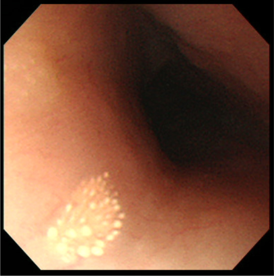
Endoscopic findings of esophageal xanthoma. A small yellowish granular lesion is noted in the esophageal mucosa
Histologically, the lesion comprised a mildly hyperplastic squamous epithelium with papillomatosis, but atypism was not noted. In the papilla just beneath the squamous epithelium, various sized aggregations of foam cells were found (Figure 2A). The foam cells had small round shaped nuclei located at the periphery of the cytoplasm, and had abundant granular cytoplasm (Figure 2B). In the deep submucosal layer, some lymphocytes and histiocytes were noted around small vessels. A small number of tiny foam cells were also found; however, acute inflammatory findings, such as neutrophilic infiltrate and edema were not noted (Figure 2C). Neither glycogen deposition nor organisms, such as fungus, were demonstrated on Periodic Acid Schiff (PAS) and Diastase-pretreated Periodic Acid Schiff (d-PAS) stained sections (data not shown).
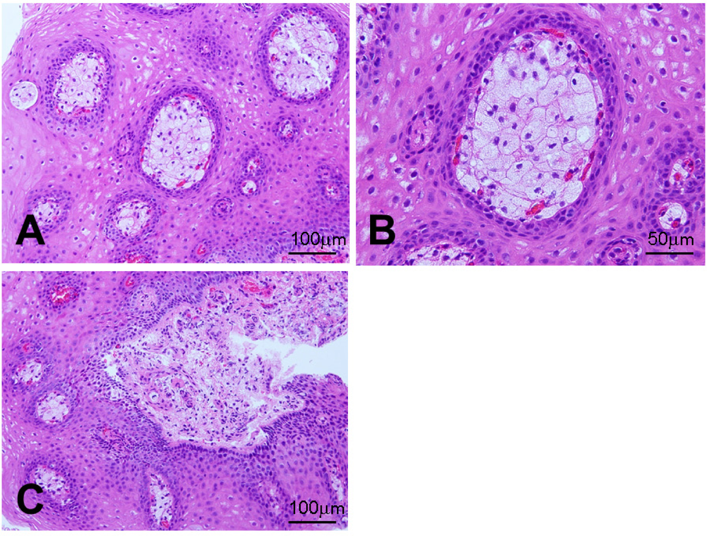
Histological findings of esophageal xanthoma. (A) Various sized aggregations of foam cells are found in the papillae (×200, HE). (B) The foam cells have small round-shaped nuclei located at the periphery of the cytoplasm, and have abundant granular cytoplasm (×400, HE). (C) In the deep submucosal layer, some lymphocytes and histiocytes are noted, however, acute inflammatory findings, such as neutrophilic infiltrate, edema, and small vessel formation, are not noted (×200, HE)
The immunohistochemistry revealed that the foam cells were positive for CD68, CD163, and TGF-β while only weakly positive for iNOS (Figure 3A-D) and negative for CD30 (data not shown).

Expression of macrophage lineage markers in foam cells. Foam cells in xanthoma are strongly positive for CD68 (A, x400), moderately positive for CD163 (B, x400) and TGF-β (C, x400), and weakly positive for iNOS (D, x400)
Ethical approval
The research related to human use has been complied with all the relevant national regulations, institutional policies and in accordance the tenets of the Helsinki Declaration.
Informed consent
Informed consent has been obtained from all individuals included in this study.
3 Discussion
Although esophageal xanthoma is thought to be rare, its precise incidence is not known, because only a limited number of cases have been reported. Esophageal xanthoma was first reported in 1984 [1], and since then, 23 cases have been published in the English literature. According to the reported cases, esophageal xanthoma is generally a solitary, small (usually 2-5 mm, maximum 10 mm), yellowish elevated lesion [2, 4, 5]. Most patients have mild or no gastrointestinal symptoms and the malignant transformation of esophageal xanthoma has never been reported.
The etiology of esophageal xanthoma remains unknown. Although one case had hyperlipidemia [6], the others had no association with abnormal lipid metabolism. These findings suggest that the pathogenesis of esophageal xanthoma is different from that of skin xanthoma, which is frequently seen in patients with chronic hyperlipidemia. Herrera-Goepfert et al. reported esophageal verruciform xanthoma after mediastinal irradiation and suggested that the lesion may be associated with physical stimuli causing irritation or trauma to the esophagus [3]. Becheanu et al. suggested that a response to focal mucosal damage may have contributed to the pathogenesis of esophageal xanthoma. They also suggested that the reason why gastric xanthoma seen more frequently than esophageal xanthoma was that trauma and inflammation are better tolerated by the esophageal squamous epithelium than by the gastric columnar epithelium [2]. However, only two cases of radiotherapy-related esophageal xanthomas have been reported, and no definitive etiology has been established in the remainder including the present case.
Rawal et al. reported on the macrophage subpopulations in oral verruciform xanthoma, and demonstrated that RM3/1 (reparative) and 25F9 (resident) positive macrophages were predominant over 27E10 (inflammatory) positive macrophages. They suggest that the findings of oral verruciform xanthoma are consistent either with a chronic reactive process or a pathogenetic mechanism where the role of acute inflammation-associated macrophages is limited [11].
Recently, it has been revealed that macrophages are involved in various inflammatory processes. As described earlier, the distinction between M1 and M2 macrophages reflects functional extreme phenotypes among a broad range of differentiation states. In general, M1 macrophages are associated with acute inflammatory reactions, such as phagocytosis of pathogens, antigen presentation, and cytokine secretion inducing inflammatory cell recruitment. In contrast to M1 macrophages, M2 macrophages are associated with late phase of inflammation, involving phagocytosis of cell debris and cytokine secretion, which induce fibroblast proliferation and connective tissue synthesis and deposition, thereby contributing to tissue regeneration [7, 8]. As oral verruciform xanthoma is regarded as one of the healing processes of local inflammation, Hirokawa et al. suggested that esophageal xanthoma is caused by chronic inflammatory processes [4].
The present case showed that the macrophages seen in the lesion of esophageal xanthoma expressed not only CD68 but also CD163; however, they expressed low levels of iNOS, and were negative for CD30, which is one of TNF-α receptor [10], demonstrating characteristics of M2 macrophages. We also found that the CD163 expressing macrophages produced TGF-β, which is related with late remodeling phase of inflammation [8]. Furthermore, neutrophilic infiltrate and edema were not found; instead, a small number of lymphocytes and histiocytes were noted in the subepithelial layer. These findings suggest that esophageal xanthoma is associated with the late inflammatory and reparative phase, rather than the acute inflammatory phase. According to the two cases of esophageal xanthoma after mediastinal radiation therapy, the lesions were found 3 years and 33 months after the therapy, respectively [3, 5]. The duration between radiation therapy and endoscopy may support the idea that esophageal xanthoma is associated with late inflammatory and reparative processes after initial injury of the esophageal squamous epithelium, however, more evidence is needed to support this claim. Endoscopists have a possibility to encounter a case of esophageal xanthoma among patients with a history of thoracic lesions. Differential diagnosis includes ectopic sebaceous glands, squamous papilloma, granular cell tumor, verrucous carcinoma and papillary squamous cell carcinoma. To make definitive diagnosis, histological examination should be performed.
4 Conclusion
In conclusion, we reported a case of esophageal xanthoma with no apparent thoracic injury or trauma. Histologically, no acute inflammatory phase was noted. Immunohistochemistry revealed that the foam cells seen in the lesion consisted of M2 macrophages. These findings suggest that esophageal xanthoma is associated with late inflammatory and reparative processes long after the initial injury of the esophageal squamous epithelium. Endoscopists have a possibility to encounter a case of esophageal xanthoma among patients with a history of thoracic lesions. Because differential diagnosis includes malignancies such as verrucous carcinoma and papillary squamous cell carcinoma, histological examination should be performed for definitive diagnosis.
Conflicts of interest: The authors declare that they have no conflicts of interest.
Acknowledgements
We thank Mr. Zensei Toyoda (University of the Ryukyus) for his technical assistance.
Authors’ contributions: KU wrote the draft and reviewed the literature. HI performed endoscopic examination. YT, KK and SK analyzed immunohistochemical data and reviewed the manuscript. MO, AO and AI made the pathological diagnosis. TK conceived and designed the report. All authors approved the final version of the manuscript.
References
[1] Remmele W, Engelsing B. Lipid island of the esophagus. Case report. Endoscopy. 1984;16:240-24110.1055/s-2007-1018590Search in Google Scholar
[2] Becheanu G, Dumbrava M, Arbanas T, Diculescu M, Hoyeau-Idrissi N, Flejou JF. Esophageal xanthoma--report of two new cases and review of the literature. J Gastrointestin Liver Dis. 2011;20:431-433Search in Google Scholar
[3] Herrera-Goepfert R, Lizano-Soberon M, Garcia-Perales M. Verruciform xanthoma of the esophagus. Hum Pathol. 2003;34:814-81510.1016/S0046-8177(03)00236-3Search in Google Scholar
[4] Hirokawa M, Takenaka R, Takahashi A, Sugihara K, Wada H, Tashiro T, et al. Esophageal xanthoma: report of two cases and a review of the literature. J Gastroenterol Hepatol. 2003;18:1105-110810.1046/j.1440-1746.2003.02844.xSearch in Google Scholar PubMed
[5] Salamanca J, Alemany I, Sosa G, Pinedo F, Hernando S, Martin-Acosta P. Esophageal verruciform xanthoma following radiotherapy. Gastroenterol Hepatol. 2012, 35:317-32010.1016/j.gastrohep.2011.12.009Search in Google Scholar PubMed
[6] Stolte M, Seifert E. Lipid islands in the esophagus. Leber Magen Darm. 1985;15:137-139Search in Google Scholar
[7] Dey A, Allen J, Hankey-Giblin PA. Ontogeny and polarization of macrophages in inflammation: blood monocytes versus tissue macrophages. Front Immunol. 2014;5:68310.3389/fimmu.2014.00683Search in Google Scholar PubMed PubMed Central
[8] Sica A, Erreni M, Allavena P, Porta C. Macrophage polarization in pathology. Cell Mol Life Sci. 2015;72:4111-412610.1007/s00018-015-1995-ySearch in Google Scholar PubMed
[9] Jurisic V, Terzic T, Colic S, Jurisic M. The concentration of TNF-α correlate with number of inflammatory cells and degree of vascularization in radicular cysts. Oral Dis. 2008;14:600-60510.1111/j.1601-0825.2007.01426.xSearch in Google Scholar PubMed
[10] Jurisic V, Srdic-Rajic T, Konjevic G, Bogdanovic G, Colic M. TNF-α induced apoptosis is accompanied with rapid CD30 and slower CD45 shedding from K-562 cells. J Membrane Biol. 2011;239:115-12210.1007/s00232-010-9309-7Search in Google Scholar PubMed
[11] Rawal SY, Kalmar JR, Tatakis DN. Verruciform xanthoma: immunohistochemical characterization of xanthoma cell phenotypes. J Periodontol. 2007;78:504-50910.1902/jop.2007.060196Search in Google Scholar PubMed
© 2017 Karina Uehara et al.
This work is licensed under the Creative Commons Attribution-NonCommercial-NoDerivatives 4.0 License.
Articles in the same Issue
- Regular Articles
- Intravascular treatment of left subclavian artery aneurysm coexisting with aortic coarctation in an adult patient
- Regular Articles
- Effect of electrical stimulation on blood flow velocity and vessel size
- Regular Articles
- Live birth pregnancy outcome after first in vitro fertilization treatment in a patient with Systemic Lupus Erythematosus and isolated high positive IgA anti-β2glycoprotein I antibodies: a case report
- Regular Articles
- Periodontal ligament stem cells regulate apoptosis of neutrophils
- Regular Articles
- Platelet-rich fibrin (PRF) in implants dentistry in combination with new bone regenerative flapless technique: evolution of the technique and final results
- Regular Articles
- The significance of strong ion gap for predicting return of spontaneous circulation in patients with cardiopulmonary arrest
- Regular Articles
- Clinicopathology of EpCAM and EGFR in human epithelial ovarian carcinoma
- Regular Articles
- Intraosseous lipoma of the mandibula: A case report and review of the literature
- Regular Articles
- Transurethral resection of the prostate, bladder explosion and hyponatremic encephalopathy: a rare case report of malpractice
- Regular Articles
- Brain strokes related to aortic aneurysma – the analysis of three cases
- Regular Articles
- Effect of Bicyclol tablets on drug induced liver injuries after kidney transplantation
- Regular Articles
- Plasma free fatty acids in hyperemesis gravidarum pregnancy
- Regular Articles
- Impact of chromosomal rearrangement upon DNA methylation patterns in leukemia
- Regular Articles
- Gefitinib versus docetaxel in treated non-small-cell lung cancer: a meta-analysis
- Regular Articles
- The clinical characteristics of patients with chronic idiopathic anal pain
- Regular Articles
- Bone tunnel impaction reduced the tibial tunnel enlargement
- Regular Articles
- Effects of S-1 combined with radiotherapy in the treatment of nasopharyngeal cancer: a meta-analysis based on randomized controlled trials
- Regular Articles
- Predictions and outcomes of atrial fibrillation in the patients with acute myocardial infarction
- Regular Articles
- An accuracy study of the Intracavitary Electrocardiogram (IC-ECG) guided peripherally inserted central catheter tip placement among neonates
- Regular Articles
- Serum CA125, CA199 and CEA combined detection for epithelial ovarian cancer diagnosis: A meta-analysis
- Regular Articles
- Surface coil intensity correction in magnetic resonance imaging in spinal metastases
- Regular Articles
- Muscle stem cell and physical activity: what point is the debate at?
- Regular Articles
- MicroRNA let-7g directly targets forkhead box C2 (FOXC2) to modulate bone metastasis in breast cancer
- Regular Articles
- Monitoring health inequalities at the municipal level: Lithuanian experience
- Regular Articles
- Role of Epstein-Barr virus in the development of nasopharyngeal carcinoma
- Regular Articles
- Thrombectomy combined with indwelling-catheter thrombolysis is more effective than pure thrombectomy for the treatment of lower extremity deep venous thrombosis
- Regular Articles
- Expression of Hepcidin and Neogenin in colorectal cancer
- Regular Articles
- Carnitine and adiponectin levels in breast cancer after radiotherapy
- Regular Articles
- Pathophysiology of meningioma growth in pregnancy
- Regular Articles
- Causal neuro-immune relationships at patients with chronic pyelonephritis and cholecystitis. Correlations between parameters EEG, HRV and white blood cell count
- Regular Articles
- Measuring efficiency of secondary healthcare providers in Slovenia
- Regular Articles
- Galectin-3 expression in colorectal cancer and its correlation with clinical pathological characteristics and prognosis
- Regular Articles
- Model for studying anti- allergic drugs for allergic conjunctivitis in animals
- Regular Articles
- Barriers perceived by nurses in the optimal treatment of postoperative pain
- Regular Articles
- Tumor microenvironment in treatment of glioma
- Regular Articles
- Delirium risk of dexmedetomidine and midazolam in patients treated with postoperative mechanical ventilation: A meta-analysis
- Regular Articles
- Hemangioma of the rib: a rare case report and literature review
- Regular Articles
- The diagnostic accuracy of conventional forceps biopsy compared to ESD
- Regular Articles
- Increased miR-25 expression in serum of gastric cancer patients is correlated with CA19-9 and acts as a potential diagnostic biomarker
- Regular Articles
- Therapeutic nanomedicine surmounts the limitations of pharmacotherapy
- Regular Articles
- Relationship between PD-L1 expression and clinical characteristics in patients with breast invasive ductal carcinoma
- Regular Articles
- Trypsinogen activation peptide induces HMGB1 release from rat pancreatic acinar cells
- Regular Articles
- The effective regulation of pro- and anti-inflammatory cytokines induced by combination of PA-MSHA and BPIFB1 in initiation of innate immune responses
- Regular Articles
- Cell based therapeutic approach in vascular surgery: application and review
- Regular Articles
- Clinical efficacy of alprostadil combined with α-lipoic acid in the treatment of elderly patients with diabetic nephropathy
- Regular Articles
- Professional burnout and concurrent health complaints in neonatal nursing
- Regular Articles
- Esophageal xanthoma: presence of M2 macrophages suggests association with late inflammatory and reparative processes
- Regular Articles
- Cone beam computed tomography analysis in 3D position of maxillary denture
- Regular Articles
- CK20 mRNA expression in serum as a biomarker for colorectal cancer diagnosis: A meta-analysis
- Regular Articles
- Serum AFU, 5’-NT and AFP as biomarkers for primary hepatocellular carcinoma diagnosis
- Regular Articles
- Absolute reliability and concurrent validity of hand held dynamometry and isokinetic dynamometry in the hip, knee and ankle joint: systematic review and meta-analysis
- Regular Articles
- The Fountain of Youth: A tale of parabiosis, stem cells, and rejuvenation
- Regular Articles
- Foam sclerotherapy during shunt surgery for portal hypertension and varices
- Regular Articles
- Insomnia and depression: Japanese hospital workers questionnaire survey
- Regular Articles
- Serum NF-κBp65, TLR4 as biomarker for diagnosis of preeclampsia
- Regular Articles
- Docetaxel/cisplatin therapy in myasthenia gravis with hypertension/diabetes
- Regular Articles
- Fluid resuscitation and markers of glycocalyx degradation in severe sepsis
- Regular Articles
- Modified Sauve-Kapandji procedure for patients with old fractures of the distal radius
- Regular Articles
- Bile leakage after liver transplantation
- Regular Articles
- VEGF overexpression predicts poor survival in hepatocellular carcinoma
- Regular Articles
- Galen vein aneurysm– challenge for treatment
- Regular Articles
- Retrieval of a broken sewing needle from the sacrum aided by a permanent magnet: a case report and literature review
- Regular Articles
- HIV/STI prevention interventions: A systematic review and meta-analysis
- Regular Articles
- Aortic aneurysm as a complication of myeloperoxidase-antineutrophil cytoplasmic antibody-associated vasculitis
- Regular Articles
- Real-time monitoring of contrast-enhanced ultrasound for radio frequency ablation
- Regular Articles
- Successful drug-eluting stent implantation in a male patient with dextrocardia: a case report
- Regular Articles
- Primary pleomorphic liposarcoma of fallopian tube with recurrence: a case report and review of the literature
- Regular Articles
- Color Doppler Ultrasound in Uterine Arterial Embolization
- Regular Articles
- Pattern of alcohol consumption by young people from North Eastern Portugal
- Regular Articles
- Effects of out-of-hospital continuing nursing on schizophrenia patients' rehabilitation and quality of life