Association between RS3763040 polymorphism of the AQP4 and idiopathic intracranial hypertension in a Spanish Caucasian population
-
Juan José Tellería-Orriols


Abstract
Background
Idiopathic intracranial hypertension (IIH) is a condition of increased intracranial pressure of unknown aetiology. Principal symptoms are headache, visual disturbances, and obesity, together with elevated intracranial pressure. Unspecified MRI, despite normal ventricle size, suggests alterations in the water flux cellular mediated by the brain water channel aquaporin-4 (AQP4). The association among IIH, cerebral spinal fluid malfunction, reabsorption, and functional or regulatory modifications of AQP4 is a hypothesis not confirmed.
Methods
Blood samples were collected from 72 Spanish Caucasian patients with IIH. A genetic association study was performed with bi-allelic SNPs rs1049305 and rs10244884 in AQ1 and rs2075575, rs3763043, and rs3763040 in AQ4. Genetic data were compared with 94 healthy Caucasian control. Statistics studies were assessed by Pearson’s χ 2 tests for 2 × 2 (alleles) or 3 × 2 (genotypes) contingency tables. A P value < 0.05 was considered to be statistically significant.
Results
Statistically significant differences were found when comparing the results of the rs3763040 polymorphism of the AQ4 locus of IIH patients with controls, in genotypic frequencies (P = 0.0442) and allele frequencies (P = 0.0171). Furthermore, a statistically significant difference (P = 0.0207) was found in individuals carrying and not carrying the minor allele (GG + GA individuals vs GG homozygotes). No statistically significant differences were found when comparing allele and genotypic frequencies for SNPs rs1049305 and rs10244884 of AQ1 and rs2075575 and rs3763043 of AQ4.
Conclusions
The association of AQP4 and specifically of its polymorphic variant rs3763040 with IIH should be validated in other ethnic groups in order to assess more precisely the role of AQP4 in the etiopathogenesis of IIH.
1 Introduction
Idiopathic intracranial hypertension (IIH), also known as pseudotumour cerebri, is a rare neurological disorder characterized by increased intracranial pressure (ICP) over 25 cm/H2O or 18 mm/Hg, in the absence of hydrocephalus or intracranial pathology, and otherwise normal cerebrospinal fluid (CSF) contents [1,2].
The etiology of IIH remains unknown. Although this disorder was once called benign intracranial hypertension to differentiate it from secondary intracranial hypertension caused by malignancy, IIH should not be considered a benign disorder as symptoms include disabling headaches, obesity, papilledema, and blurred vision, which evolve slowly over time and can cause severe and permanent vision loss in 10% of affected individuals due to pressure on the optic nerve [3,4].
The incidence of IIH in the general population is thought to be about 1–2 per 100,000. Although it does occur in men, the disorder is most common in obese women of childbearing age with an incidence of 10 per 100,000 [1].
Imaging of the brain with MRI is essential in patients with suspected IIH to exclude elevated CSF pressure due to other causes. MRI may show slit-like ventricles or a flattened pituitary suggesting a diffuse accumulation of water in the skull causing optic nerve tortuosity, optic nerve sheath distension [5], and increased CSF clearance activity seen as hypertrophic Paccioni’s granulations which can produce stenosis when located close to the transverse sinus, causing blood retention and a further increase in ICP.
Hyperintensity and increased optic nerve diameter, suggestive of optic nerve oedema, observed in T2-weighted and angio-venous MRI showing uni- or bilateral transverse sinus stenosis due to hypertrophic Pacchioni’s granulations support the diagnosis of IIH [6].
Genetic predisposition to IIH has been previously proposed [7] and indeed several cases of multiple incidences in families have been described [8,9] showing that IIH occurrence within a family is more common than previously believed, and its incidence in families is more common than in the general population. Nevertheless, there does not appear to be a Mendelian pattern of inheritance. IIH would be more likely caused by the concurrence of environmental factors such as overweight in individuals with increased genetic susceptibility [10].
Aquaporins (AQPs) are a family of water-channel proteins, which in many cells provide the main route for bidirectional water movement across the membrane by increasing plasma membrane osmotic permeability [11]. AQPs have been found in different cerebral structures. Among these, the most common are AQP1 and AQP4. The roles of AQPs have been demonstrated in several diseases such as cerebral edema, various central nervous system (CNS) tumours, Alzheimer’s disease, and epilepsy [12], but there are few previous studies on the role of AQPs in IIH and the results are inconclusive.
Here, we study the association between different polymorphisms in AQP1 and AQP4 in order to better understand the role of AQPs in the pathogenesis of and predisposition to IIH.
2 Materials and methods
2.1 Patients’ samples
A total of 72 Caucasian patients, 55 female (73.6%) and 17 male (23.6%), with a mean age of 39.01 years and an age range of 11–72 years, diagnosed of having IIH according to Friedman criteria [2], were included in the study. All patients had undergone brain MRI, computed tomography scan of the brain, or both. Brain angio-MRI was performed in patients with neuroimaging signs suggestive of transverse sinus venous stenosis in order to ensure that they met the entry criteria of the study. Clinical data included the most representative symptoms of IIH such as headache, papilledema, obesity, sinus stenosis, and intracranial pressure of above 20 mm Hg.
Intracranial pressure was determined by lumbar puncture, in the L4–L5 interspace, with the patient in the supine decubitus position.
The presence of disorders such as metabolic diseases, liver dysfunction, or infectious diseases, as well as other neurological diseases such as tumours or hydrocephalus seen on neuroimaging were considered exclusion criteria. No pregnant women were included.
The results were compared with those of a control group of 94 healthy Caucasian individuals of Spanish origin.
About 5–10 mL of venous blood was obtained from all participants. From these samples, genomic DNA was isolated using an automated nucleic acid purification system (Roche® Nucleic Acid Purification System MagNA Pure Compact).
2.2 Genotyping
Five bi-allelic SNPs rs1049305 and rs10244884 in AQ1 and rs2075575, rs3763043, and rs3763040 in AQ4 were selected and genotyped to determine their allele and genotypic frequencies in the study population and controls.
Samples were genotyped using KASP (Kompetitive allele-specific PCR) genotyping technology (Biosearch Technologies®) that allows biallelic discrimination of known SNPs and indels, following the manufacturer’s instructions.
2.3 Statistical analysis
The results are presented as genotype and allele frequencies for both IIH patients and control groups. The differences in genotype and allele frequency between the two groups were assessed by Pearson’s χ 2 tests for 2 × 2 (alleles) or 3 × 2 (genotypes) contingency tables. A P value < 0.05 was considered statistically significant. We applied Hardy–Weinberg’s law and its equation which states that in an equilibrium population, given allele frequencies p and q, the genotypic distribution will be p2 + 2pq + q2. In our study, the distribution of each genotype in our population was considered to be in equilibrium when the comparison between the observed and expected genotypic frequencies applying the Hardy–Weinberg law was <0.05.
-
Ethical approval: This research related to human use complied with all the relevant national regulations, and institutional policies, and is in accordance with the tenets of the Helsinki Declaration, and has been approved by the authors’ institutional review board or equivalent committee. All experiments conformed to the Declaration of Helsinki and were approved by the Ethics Committee of Hospital Clínico Universitario de Valladolid.
-
Informed consent: Informed consent has been obtained from all individuals included in this study.
3 Results
3.1 Clinical findings
The clinical data of the patients are included in Table 1.
Relation between intracranial pressure and clinic dates
| Clinical datesPatients n 72 (%) | ||||||||
|---|---|---|---|---|---|---|---|---|
| Intracranial pressure | Headache | Papilledema | Obesity | Sinus venous stenosis | ||||
| 60 (83%) | 42 (58%) | 37 (51%) | 25 (34%) | |||||
| Yes | No | Yes | No | Yes | No | Yes | No | |
| <30 mm Hg | 34 | 12 | 23 | 23 | 14 | 17 | 17 | 29 |
| >30 mm Hg | 26 | 0 | 19 | 7 | 23 | 18 | 8 | 18 |
Headache was the most frequent symptom, affecting 60 patients (83%). Papilledema was found in 42 patients (58%). Obesity was present in 27 patients (51%). Radiologically, venous sinus stenosis was observed in 25 patients (34%).
Lumbar CSF pressure was elevated in all patients with a range of 20–57 mm Hg and a median of 28.64 mm Hg.
No statistically significant difference was observed between clinical data and intracranial pressure. However, when we compare the data with intracranial pressure equal to or higher than 30 mm Hg, there is a statistically significant difference with headache (P = 0.0116). The increase in headaches with increased pressure is a clear cause-and-effect situation. The other clinical data, although related to intracranial pressure, are due to another type of cause.
3.2 Analysis of polymorphism
Genotypic and allele frequencies are summarized in Tables 2 and 3, respectively.
Genotypic frequencies in IIH patients and controls
| Polymorphism | Genotypes | P | ||
|---|---|---|---|---|
| rs3763040 | GG | GA | AA | |
| IIH patients | 56 (77.8%) | 14 (19.4%) | 2 (2.8%) | 0.0442 |
| Controls | 56 (59.6%) | 32 (34.0%) | 6 (6.4%) | |
| rs2075575 | GG | GA | AA | |
| IIH patients | 23 (31.94%) | 35 (48.61%) | 14 (19.44%) | 0.9371 |
| Controls | 28 (29.78%) | 46 (48.93%) | 20 (21.27%) | |
| rs3763043 | CC | CT | CC | |
| IIH patients | 31 (43.05%) | 34 (47.22%) | 7 (9.72%) | 0.1703 |
| Controls | 50 (53.19%) | 31 (32.97%) | 13 (13.82%) | |
| rs1049305 | GC | GC | CC | |
| IIH patients | 21 (29.16%) | 38 (52.77%) | 13 (18%) | 0.3296 |
| Controls | 34 (36.17%) | 50 (53.19%) | 10 (13.51%) | |
| rs10244884 | TT | TC | CC | |
| IIH patients | 14 (19.44%) | 36 (50.00%) | 22 (30.55%) | 0.8437 |
| Controls | 21 (22.34%) | 43 (45.74%) | 30 (31%) | |
A = Adenine; C = Cytosine; G = Guanine; T = Thymine.
Allele frequencies in IIH patients and controls
| Polymorphism | Allele frequencies | P | |
|---|---|---|---|
| rs3763040 | G | A | |
| IIH patients | 126 (87.5%) | 18 (12.5%) | 0.0171 |
| Controls | 144 (76.6%) | 44 (23.4%) | |
| rs2075575 | G | A | |
| IIH patients | 81 (56.25%) | 63 (43.75%) | 0.8065 |
| Controls | 102 (54.25%) | 86 (45.74%) | |
| rs3763043 | C | T | |
| IIH patients | 96 (66.66%) | 48 (33.33%) | 0.639 |
| Controls | 131 (69.68%) | 57 (30.31%) | |
| rs1049305 | G | C | |
| IIH patients | 80 (55.55%) | 64 (44.44%) | 0.2253 |
| Controls | 118 (62.76%) | 70 (37.23%) | |
| rs10244884 | T | C | |
| IIH patients | 64 (44.44%) | 80 (55.55%) | 1 |
| Controls | 85 (45.21%) | 103 (54.78%) | |
A = Adenine; C = Cytosine; G = Guanine; T = Thymine.
No statistically significant differences were found when comparing allele and genotypic frequencies for SNPs rs2075575 and rs3763043 of AQ4 and rs1049305 and rs10244884 of AQ1. In contrast, differences were observed when comparing the results of the rs3763040 polymorphism of the AQ4 locus. Specifically, differences in genotypic frequencies (P = 0.0442) and allele frequencies (P = 0.0171) were observed between patients and controls.
Furthermore, when comparing individuals carrying and not carrying the minor allele (AA + GA) individuals vs. GG homozygotes, a statistically significant difference (P = 0.0207) was also observed between the groups studied (Figure 1).
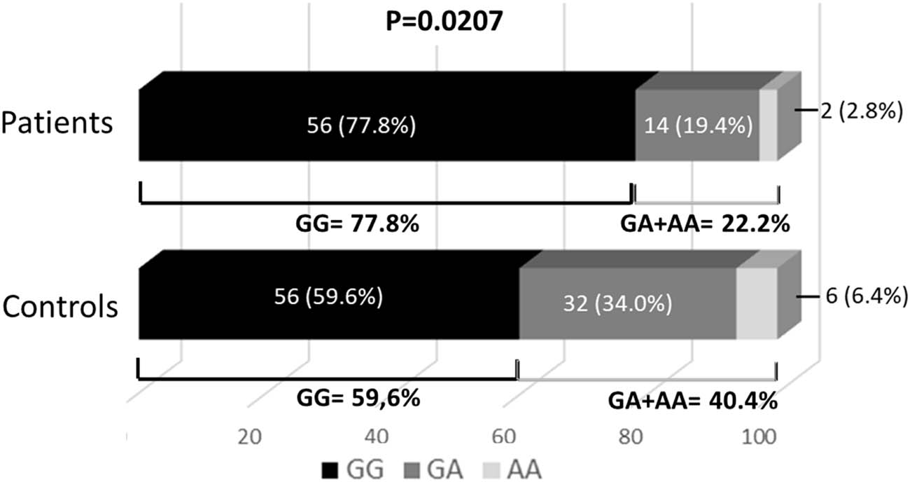
Comparison between individuals carrying the minor allele (A): genotype GA + AA vs homozygotes for the major allele: genotype GG.
The odds ratio of A allele carriers (AA + GA) to GG homozygotes was 2.3750, with a confidence interval of 1.289–4.743 at a 95% confidence level.
No statistically significant differences were found when comparing polymorphism and clinical dates.
4 Discussion
The higher incidence of IIH in individuals with family history than in the general population, the reported parent–child transmission, and concordance in siblings [7,13] suggest the existence of genetic susceptibility factors that are inherited in a non-Mendelian multifactorial pattern. IIH would occur when non-genetic triggers act in individuals with increased susceptibility.
AQPs are a family of tetrameric membrane proteins whose primary function is to transport water across cell membranes in response to osmotic gradients created by active solute transport.
Given the known role of AQPs in the regulation of water flow, in the present study, we have analysed whether different genetic polymorphisms of AQPs that are expressed in the brain (namely, AQP1 and AQP4) are related to individual susceptibility to IIH.
We found no association between four of the SNPs studied (the two in AQP1 and two of the three in AQP4) and IIH. However, the study of the rs3763040 SNP in the intron region showed that both allele and genotypic frequencies were different when comparing the patient and control groups. The major allele (G) was more frequent in patients with IIH than in controls, with P = 0.0442, so the G allele is associated with an increased risk of developing IIH, especially when found in homozygosis, with patients carrying the minor allele (A) in both homozygosis and heterozygosis showing a lower risk of IIH (P = 0.0207) (Figure 1).
AQP4 is the principal AQP in the mammalian brain and is found in supporting cells in the CNS facilitating water movement into and out of the brain [11].
The hypothesis of AQP4 involvement in the pathogenesis and progression of IIH is supported by the observation that AQP4-deficient mice develop greater ICP than wild-type mice when blood–brain barrier disruption is induced [14]. Moreover, acetazolamide is known to inhibit water conduction by AQP-4 [15,16] and its use in IIH patients has a direct effect on papilledema and intracranial pressure, and significantly improves visual field function in patients with IIH [17].
The AQP4 intronic rs3763040 polymorphism has also been related to neuromyelitis optica [18] and to the progression of Alzheimer’s disease [19], meaning that it is an SNP with functional consequences or in linkage disequilibrium with another polymorphism that has functional consequences.
Our case–control genetic study is at odds with another study in 28 Norwegian patients with IIH in which no correlation was found between genetic variants in AQP4 and IIH [20]. In this study, the authors themselves stress that an etiopathogenic link between AQP4 and IIH remains attractive and that further association studies should be performed in larger sample sizes despite the difficulty given the rarity of the condition.
Based on a precise definition of the IIH phenotype and a plausible etiopathogenic hypothesis and its candidate genes, we have conducted a case–control study which, to our knowledge, is the first work to demonstrate an association between genetic variants in AQP4 and IIH defined phenotype, which had an apparent a priori hypothesis and a strong biological plausibility.
The association of AQP4 and specifically of its polymorphic variant rs3763040 with IIH should be validated in other ethnic groups to assess more precisely the role of AQP4 in the etiopathogenesis of IIH.
This study is limited by the number of patients (72). Considering that IIH is a rare disease, it is difficult to obtain a larger sample, which would be very useful to validate the results. On the other hand, the findings of statistical significance for the rs3763040 polymorphism of AQP4, without being able to say that it is the cause, open the discussion of its role. Further studies to test the gene, as well as its pathway relationship with other genes, may provide interesting data to determine what appears to be a multifactorial disease.
-
Funding information: This work was partly supported by ADEFHIC (Hispanic Association of Intracranial Idiopathic Hypertension of sick people and their families).
-
Conflict of interest: The authors state no conflict of interest.
-
Data availability statement: The datasets generated during and/or analysed during the current study are available from the corresponding author on reasonable request.
References
[1] Dhungana S, Sharrack B, Woodroofe N. Idiopathic intracranial hypertension. Acta Neurol Scand. 2010;121:71–82.10.1111/j.1600-0404.2009.01172.xSearch in Google Scholar PubMed
[2] Friedman DI, Jacobson DM. Diagnostic criteria for idiopathic intracranial hypertension. Neurology. 2002;59:1492–5.10.1212/01.WNL.0000029570.69134.1BSearch in Google Scholar PubMed
[3] Kanagalingam S, Subramanian PS. Update on idiopathic intracranial hypertension. Curr Treat Options Neurol. 2018;20(7):24.10.1007/s11940-018-0512-7Search in Google Scholar PubMed
[4] Madriz G, Cestari DM. An update of idiopathic intracranial hypertension. Neuro-Ophthalmology. 2018;29:495–502.10.1097/ICU.0000000000000518Search in Google Scholar PubMed
[5] Barkatullah AF, Leishangthem L, Moss HE. MRI findings as markers of idiopathic intracranial hypertension. Curr Opin Neurol. 2021;34(1):75–83.10.1097/WCO.0000000000000885Search in Google Scholar PubMed PubMed Central
[6] Holbrook J, Saindane AM. Imaging of intracranial pressure disorders. Neurosurgery. 2017;80(3):341–54.10.1227/NEU.0000000000001362Search in Google Scholar PubMed
[7] Corbett JJ. The first Jacobson Lecture. Familial idiopathic intracranial hypertension. J Neuroophthalmol. 2008;28:337–47.10.1097/WNO.0b013e31818f12a2Search in Google Scholar PubMed
[8] Qiao L, Wei Y. Familial idiopathic intracranial hypertension in two non-obese Chinese sisters. Front Neurol. 2020;11:569432.10.3389/fneur.2020.569432Search in Google Scholar PubMed PubMed Central
[9] Behbehani R, Ali A, Al-Mousa AJ, Albuloushi SN. Familial non-obese idiopathic intracranial hypertension. Am J Ophthalmol Case Rep. 2022;27:101619.10.1016/j.ajoc.2022.101619Search in Google Scholar PubMed PubMed Central
[10] Klein A, Dotan G, Kesler A. Familial Occurrence of Idiopathic Intracranial Hypertension. Isr Med Assoc J. 2018;Sep;20(9):557–60.Search in Google Scholar
[11] Tait MJ, Saadoun S, Bell BA, Papadopoulos MC. Water movements in the brain: role of aquaporins. Trends Neurosci. 2008;31(1):37–43.10.1016/j.tins.2007.11.003Search in Google Scholar PubMed
[12] Dasdelen D, Mogulkoc R, Baltaci AK. Aquaporins and roles in brain health and brain injury. Mini Rev Med Chem. 2020;20(6):498–512.10.2174/1389557519666191018142007Search in Google Scholar PubMed
[13] Beri S, Chandratre S, Chow G. Familial idiopathic intracranial hypertension with variable phenotype. Eur J Paediatr Neurol. 2011;15(1):81–3.10.1016/j.ejpn.2010.02.005Search in Google Scholar PubMed
[14] Papadopoulos MC, Manley GT, Krishna S, Verkman AS. Aquaporin-4 facilitates reabsorption of excess fluid in vasogenic brain edema. FASEB J. 2004;18:1291–3.10.1096/fj.04-1723fjeSearch in Google Scholar PubMed
[15] Tanimura Y, Hiroaki Y, Fujiyoshi Y. Acetazolamide reversibly inhibits water conduction by aquaporin-4. J Struct Biol. 2009;166(1):16–21.10.1016/j.jsb.2008.11.010Search in Google Scholar PubMed
[16] Millichap JG, Millichap JJ. Mechanism of action of acetazolamide and idiopathic intracranial hypertension. Front Neurol. 2015 Feb 3;6:13.10.3389/fneur.2015.00013Search in Google Scholar PubMed PubMed Central
[17] Wall M, McDermott M, Kupersmith M. Idiopathic intracranial hypertension – reply. JAMA. 2014;312:1060. 10.1001/jama.2014.8903.Search in Google Scholar PubMed
[18] Qiu W, Chang Y, Li R, Long Y, Huang J, Mai W, et al. [Correlation of AQP4 gene polymorphism with NMO clinical phenotypes and its underlying mechanism]. Zhonghua Yi Xue Za Zhi. 2015;1795(7):501–6.Search in Google Scholar
[19] Burfeind KG, Murchison CF, Westaway SK, Simon MJ, Erten-Lyons D, Kaye JA, et al. The effects of noncoding aquaporin-4 single-nucleotide polymorphisms on cognition and functional progression of Alzheimer’s disease. Alzheimers Dement (N Y). 2017 26;3(3):348–59.10.1016/j.trci.2017.05.001Search in Google Scholar PubMed PubMed Central
[20] Kerty E, Heuser K, Indahl UG, Berg PR, Nakken S, Lien S, et al. Is the brain water channel aquaporin-4 a pathogenetic factor in idiopathic intracranial hypertension? Results from a combined clinical and genetic study in a Norwegian cohort. Acta Ophthalmol. 2013;91(1):88–91.10.1111/j.1755-3768.2011.02231.xSearch in Google Scholar PubMed
© 2023 the author(s), published by De Gruyter
This work is licensed under the Creative Commons Attribution 4.0 International License.
Articles in the same Issue
- Research Articles
- HIF-1α participates in secondary brain injury through regulating neuroinflammation
- Omega-3 polyunsaturated fatty acids alleviate early brain injury after traumatic brain injury by inhibiting neuroinflammation and necroptosis
- The correlation between non-arteritic anterior ischemic optic neuropathy and cerebral infarction
- Enriched environment can reverse chronic sleep deprivation-induced damage to cellular plasticity in the dentate gyrus of the hippocampus
- Middle cerebral artery dynamic cerebral autoregulation is impaired by infarctions in the anterior but not the posterior cerebral artery territory in patients with mild strokes
- Leptin ameliorates Aβ1-42-induced Alzheimer’s disease by suppressing inflammation via activating p-Akt signaling pathway
- TIPE2 knockdown exacerbates isoflurane-induced postoperative cognitive impairment in mice by inducing activation of STAT3 and NF-κB signaling pathways
- Does the patellar tendon reflex affect the postural stability in stroke patients with blocked vision?
- Inactivation of CACNA1H induces cell apoptosis by initiating endoplasmic reticulum stress in glioma
- miR-101-3p improves neuronal morphology and attenuates neuronal apoptosis in ischemic stroke in young mice by downregulating HDAC9
- A custom-made weight-drop impactor to produce consistent spinal cord injury outcomes in a rat model
- Arterial spin labeling for moyamoya angiopathy: A preoperative and postoperative evaluation method
- Thyroid hormone levels paradox in acute ischemic stroke
- Geniposide protected against cerebral ischemic injury through the anti-inflammatory effect via the NF-κB signaling pathway
- The clinical characteristics of acute cerebral infarction patients with thalassemia in a tropic area in China
- Comprehensive behavioral study of C57BL/6.KOR-ApoEshl mice
- Incomplete circle of Willis as a risk factor for intraoperative ischemic events during carotid endarterectomies performed under regional anesthesia – A prospective case-series
- HOTAIRM1 knockdown reduces MPP+-induced oxidative stress injury of SH-SY5Y cells by activating the Nrf2/HO-1 pathway
- Esmolol inhibits cognitive impairment and neuronal inflammation in mice with sepsis-induced brain injury
- EHMT2 affects microglia polarization and aggravates neuronal damage and inflammatory response via regulating HMOX1
- Hematoma evacuation based on active strategies versus conservative treatment in the management of moderate basal ganglia hemorrhage: A retrospective study
- Knockdown of circEXOC6 inhibits cell progression and glycolysis by sponging miR-433-3p and mediating FZD6 in glioma
- CircYIPF6 regulates glioma cell proliferation, apoptosis, and glycolysis through targeting miR-760 to modulate PTBP1 expression
- Relationship between serum HIF-1α and VEGF levels and prognosis in patients with acute cerebral infarction combined with cerebral-cardiac syndrome
- The promoting effect of modified Dioscorea pills on vascular remodeling in chronic cerebral hypoperfusion via the Ang/Tie signaling pathway
- Effects of enriched environment on the expression of β-amyloid and transport-related proteins LRP1 and RAGE in chronic sleep-deprived mice
- An interventional study of baicalin on neuronal pentraxin-1, neuronal pentraxin-2, and C-reactive protein in Alzheimer’s disease rat model
- PD98059 protects SH-SY5Y cells against oxidative stress in oxygen–glucose deprivation/reperfusion
- TPVB and general anesthesia affects postoperative functional recovery in elderly patients with thoracoscopic pulmonary resections based on ERAS pathway
- Brain functional connectivity and network characteristics changes after vagus nerve stimulation in patients with refractory epilepsy
- Association between RS3763040 polymorphism of the AQP4 and idiopathic intracranial hypertension in a Spanish Caucasian population
- Effects of γ-oryzanol on motor function in a spinal cord injury model
- Electroacupuncture inhibits the expression of HMGB1/RAGE and alleviates injury to the primary motor cortex in rats with cerebral ischemia
- Effects of edaravone dexborneol on neurological function and serum inflammatory factor levels in patients with acute anterior circulation large vessel occlusion stroke
- CST3 alleviates bilirubin-induced neurocytes’ damage by promoting autophagy
- Excessive MALAT1 promotes the immunologic process of neuromyelitis optica spectrum disorder by upregulating BAFF expression
- Evaluation of cholinergic enzymes and selected biochemical parameters in the serum of patients with a diagnosis of acute subarachnoid hemorrhage
- 7-Day National Institutes of Health Stroke Scale as a surrogate marker predicting ischemic stroke patients’ outcome following endovascular therapy
- Cdk5 activation promotes Cos-7 cells transition towards neuronal-like cells
- 10.1515/tnsci-2022-0313
- PPARα agonist fenofibrate prevents postoperative cognitive dysfunction by enhancing fatty acid oxidation in mice
- Predicting functional outcome in acute ischemic stroke patients after endovascular treatment by machine learning
- EGCG promotes the sensory function recovery in rats after dorsal root crush injury by upregulating KAT6A and inhibiting pyroptosis
- Preoperatively administered single dose of dexketoprofen decreases pain intensity on the first 5 days after craniotomy: A single-centre placebo-controlled, randomized trial
- Myeloarchitectonic maps of the human cerebral cortex registered to surface and sections of a standard atlas brain
- The BET inhibitor apabetalone decreases neuroendothelial proinflammatory activation in vitro and in a mouse model of systemic inflammation
- Carthamin yellow attenuates brain injury in a neonatal rat model of ischemic–hypoxic encephalopathy by inhibiting neuronal ferroptosis in the hippocampus
- Functional connectivity in ADHD children doing Go/No-Go tasks: An fMRI systematic review and meta-analysis
- Review Articles
- Human prion diseases and the prion protein – what is the current state of knowledge?
- Nanopharmacology as a new approach to treat neuroinflammatory disorders
- Case Report
- Deletion as novel variants in VPS13B gene in Cohen syndrome: Case series
- Commentary
- Translation of surface electromyography to clinical and motor rehabilitation applications: The need for new clinical figures
- Revealing key role of T cells in neurodegenerative diseases, with potential to develop new targeted therapies
- Retraction
- Retraction of “Eriodictyol corrects functional recovery and myelin loss in SCI rats”
- Special Issue “Advances in multimedia-based emerging technologies...”
- Evaluation of the improvement of walking ability in patients with spinal cord injury using lower limb rehabilitation robots based on data science