Abstract
Background
Ca2+ channels are abnormally expressed in various tumor cells and are involved in the progression of human glioma. Here, we explored the role of a calcium channel, voltage-dependent, T-type, alpha 1H subunit (CACNA1H), which encodes T-type Ca2+ channel Cav3.2 in glioma cells.
Methods
Cell viability and apoptosis were detected using cell-counting kit-8 and flow cytometry, respectively. The expression of target protein was determined using western blot analysis.
Results
Cell viability of U251 cells was inhibited significantly after the knockdown of CACNA1H. The apoptosis of U251 cells was enhanced significantly after the knockdown of CACNA1H. Importantly, knockdown of CACNA1H decreased the levels of p-PERK, GRP78, CHOP, and ATF6, indicating that CACNA1H knockdown activated endoplasmic reticulum stress (ERS) in U251 cells. In addition, T-type Ca2+ channel inhibitor NNC55-0396 also induced apoptosis through the activation of ERS in U251 cells. ERS inhibitor UR906 could block CACNA1H inhibitor ABT-639-induced apoptosis.
Conclusion
Suppression of CACNA1H activated the ERS and thus induced apoptosis in glioma cells. T-type Ca2+ channel inhibitors ABT-639 and NNC55-0396 also induced apoptosis through ERS in glioma cells. Our data highlighted the effect of CACNA1H as an oncogenic gene in human glioma.
1 Introduction
Gliomas are the most common central nervous system malignancies, which are characterized by low surgical resection rate, high recurrence, and poor sensitivity to chemoradiotherapy [1]. Intracellular Ca2+ is an important signal regulating cell proliferation, differentiation, and apoptosis [2,3]. Ca2+ channels are abnormally expressed in various tumor cells and are involved in the progression of human glioma [4,5]. T-type Ca2+ channels are low-voltage-gated channels that are widely expressed in normal tissues, including brain, kidney, heart, and smooth muscle [6,7]. Studies have shown that T-type Ca2+ channels are highly expressed in various types of gliomas, and their increased expression can promote tumor cell proliferation [6]. Valerie et al. found that treatment with the T-type selective antagonist mibefradil or knockdown of T-type Ca2+ channel reduced cell viability and induced apoptosis in U251, T98G, and U87 cells [8]. At present, the specific mechanism by which T-type Ca2+ channel regulates glioma progression remains unclear.
T-type Ca2+ channels include three subtypes, Cav3.1, Cav3.2, and Cav3.3, encoded by CACNA1G, CACNA1H, and CACNA1I, respectively [9]. Studies have found that CACNA1H mutations exist in childhood absence epilepsy [10], generalized epilepsy with febrile seizures plus [11], and autism spectrum disorders [12], suggesting that CACNA1H plays an important role in neurological lesions. NNC-55 (T-channel inhibitor) or knockdown of CACNA1H can activate endoplasmic reticulum stress (ERS), thus increasing autophagy inhibition and apoptotic signals in CACNA1H−/− mice [13]. Tumor cells are maintained in a hypoxic, glucose-deficient, low pH microenvironment due to their rapid growth, which induces ERS and unfolded protein responses (UPRs) [14]. ERS participates in the process of tumorigenesis, development, and drug resistance by inducing autophagy, invasion, and apoptosis of tumor cells [15]. However, there is no report on whether CACNA1H participates in tumor progression through ERS. Here, we investigated this hypothesis in a glioma cell model in vitro.
2 Materials and methods
2.1 Cell culture and treatment
The U251 human glioma cells were cultured in Dulbecco’s Modified Eagle Medium supplemented with 10% fetal bovine serum (Thermo Fisher Scientific, Shanghai, China). CACNA1H-specific small interfering RNA (siRNA) was synthesized in GeneChem Co., Ltd (Shanghai, China) and transfected into the U251 cells using lipo2000 (Invitrogen, Carlsbad, CA, USA) according to the manufacturer’s instruction. NNC 55-0396 hydrate (NNC-55), ABT-639, and UR906 were purchased from MedChemExpress (Monmouth Junction, NJ, USA). Cells were seeded into the six-well plate and treated with NNC-55 (10 μM), ABT-639 (10 μM), or UR906 (30 μM) for 12 h.
2.2 Detection of cell viability
Cell-counting kit-8 (CCK8) (MedChemExpress, Monmouth Junction, NJ, USA) assay was performed to determine cell viability. The U251 cells (1,000 cells/well) were transferred into a 96-well plate. For viability detection, cells were incubated with 10 μl of CCK8 for 1.5 h at 37°C. Optical density value at 450 nm was measured using a microplate reader.
2.3 Western blot analysis
Total protein was extracted from U251 cells using RIPA buffer and separated using sodium dodecyl sulfate-polyacrylamide gel electrophoresis. Then, protein samples were transferred to a polyvinylidene difluoride membrane. After blocking with 5% non-fat milk, the membrane was incubated with primary antibodies at 4℃ overnight followed by secondary antibodies at room temperature for 1 h. The primary antibodies Bcl-2 (26593-1-AP), GRP78 (11587-1-AP), CHOP (15204-1-AP), and ATF6 (24169-1-AP) were purchased from PTG (Chicago, IL, USA). The primary antibodies pro-Caspase12 (35965), Cleaved-Caspase12 (35965), p-PERK (3179), and PERK (3192) were purchased from Cell Signaling Technology (Beverly, MA, USA). The specific bands were visualized using an enhanced chemiluminescence kit (Solarbio Science & Technology Co. Ltd., Beijing, China). ImageJ software was used for the quantification of target protein.
2.4 Apoptosis detection
Flow cytometry was performed to detect cell apoptosis using the AnnexinV-FITC/PI kit (Beijing 4A Biotech Co., Ltd, Beijing, China) according to the operating instruction. Cell signal was collected using flow cytometry (BD, Franklin Lakes, NJ, USA). Apoptotic cells were analyzed using FlowJo software (BD Biosciences).
2.5 Data analysis
Data were analyzed using GraphPad Prism 9 and presented as mean ± SD. Student’s t test and one-way analysis of variance with post-hoc Bonferroni were used for data analysis. P < 0.05 was considered statistically significant.
3 Results
3.1 Knockdown of CACNA1H inhibits proliferation and induces apoptosis in glioma cells
To explore the specific role of CACNA1H in glioma cells, we inactivated CACNA1H using siRNA technology (Figure 1a) or CACNA1H inhibitor (ABT-639). ABT-639 is a Cav3.2-specific inhibitor which is less active at other Ca2+ channels (Cav3.1 and Cav3.3). The viability decreased significantly 48 h after the knockdown of CACNA1H. The effect of ABT-639 on cell proliferation was consistent with that of CACNA1H siRNA (Figure 1b). On the contrary, the percentage of apoptosis increased significantly after CACNA1H knockdown or ABT-639 treatment (Figure 1c and d). Then we detected apoptosis-related proteins using western blot (Figure 1e). Our results proved that the expression of Bcl-2 (Figure 1f) and pro-Caspase12 (Figure 1g) decreased significantly, while the expression of Cleaved-Caspase12 (Figure 1h) increased significantly in CACNA1H-inactivated U251 cells. Decreased proliferation and increased apoptosis confirmed the inhibitory effect of CACNA1H inactivation on glioma cell growth.
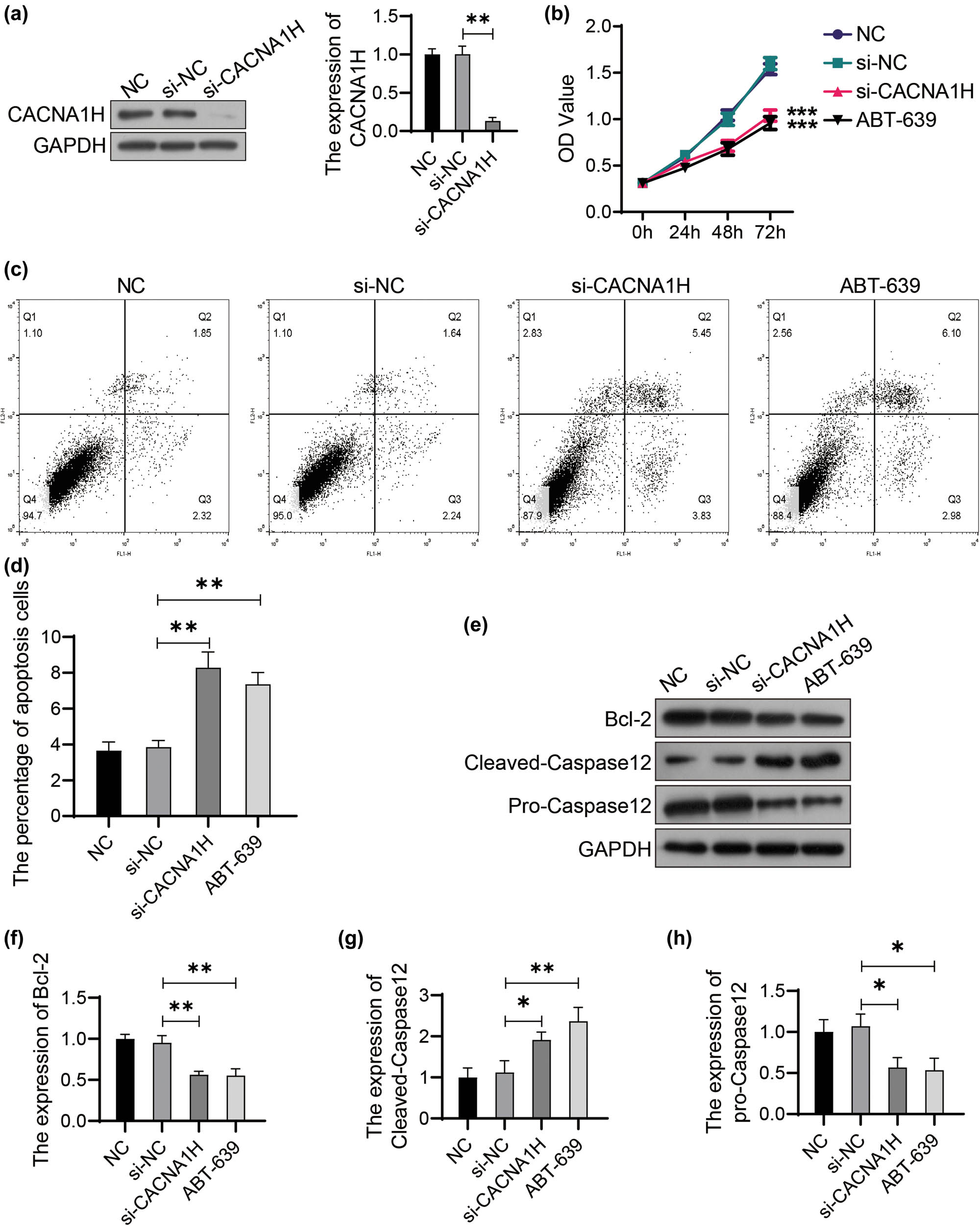
Knockdown of CACNA1H inhibits proliferation and induces apoptosis in glioma cells. (a) The efficiency of siRNA knockdown was detected using western blot. The U251 cells transfected with nonspecific siRNA were used as the negative control (NC). (b) CACNA1H was knocked down by specific siRNA or ABT-639 (10 μM) in U251 cells. Cell viability was determined using CCK8 assay. (c) Flow cytometry was performed to detect cell apoptosis. (d) Apoptotic cells were analyzed using FlowJo software. (e) The expression of target protein was detected using western bolt. The relative expression of Bcl-2 (f), pro-Caspase12 (g), and Cleaved-Caspase12 (h) was analyzed using ImageJ software. *P < 0.05; **P < 0.01; ***P < 0.001.
3.2 Knockdown of CACNA1H activates ERS in U251 cells
Since knockdown of CACNA1H induced tumor cell apoptosis, we further investigated whether CACNA1H was involved in apoptosis regulation through ERS pathway. As shown in Figure 2, the expression levels of ERS-related proteins were detected using western bolt. The phosphorylation level of PERK increased significantly in CACNA1H knockdown cells compared with that of control cells. The GRP78, CHOP, and ATF6 proteins were upregulated in the CACNA1H knockdown cells. The expression levels of ERS markers p-PERK, GRP78, CHOP, and ATF6 were also inhibited in ABT-639-treated U251 cells (Figure 2).
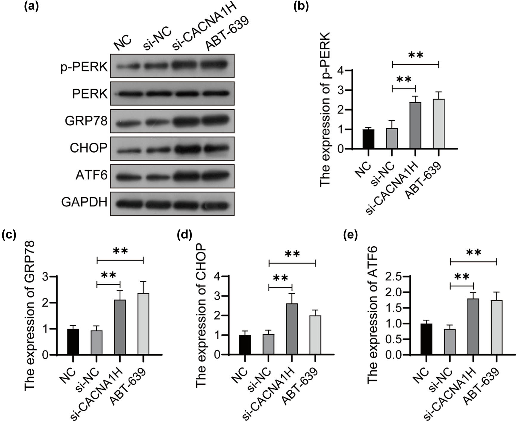
Knockdown of CACNA1H activates ERS in U251 cells. (a) The expression of ERS-related proteins was detected using western bolt. The relative expression of p-PERK (b), GRP78 (c), CHOP (d), and ATF6 (e) was analyzed using ImageJ software. **P < 0.01.
3.3 NNC55-0396 induces apoptosis through the activation of ERS in U251 cells
NNC55-0396 is a potent T-type Ca2+ channel inhibitor, which has been found to induce apoptosis of various tumor cells in recent years. Here, we investigated its effect on U251 cells. As shown in Figure 3a, the proliferation of cells treated with NNC55-0396 decreased significantly compared with the control group.
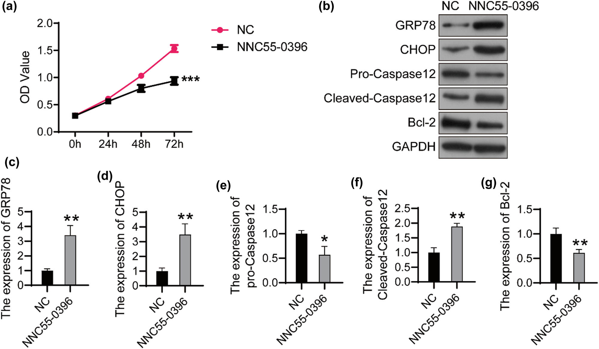
NNC55-0396 activates ERS and induces apoptosis in U251 cells (a) The U251 cells were treated with 10 μM of NNC-55, and cell viability was detected using CCK8 every 24 h. (b) The expression of target proteins was detected using western bolt. The relative expression of GRP78 (c), CHOP (d), pro-Caspase12 (e), Cleaved-Caspase12 (f) and Bcl-2 (g) was analyzed using ImageJ software. *P < 0.05; **P < 0.01; ***P < 0.001.
Importantly, the expression of ERS makers GRP78 and CHOP reduced significantly after the treatment with NNC55-0396 (Figure 3b–d). Bcl-2 and pro-Caspase12 were downregulated and Cleaved-Caspase12 was upregulated in NNC55-0396-treated U251 cells (Figure 3e–g). These results indicated that NNC55-0396 induces apoptosis through the activation of ERS in glioma cells.
3.4 UR906 blocked the apoptosis induced by ABT-639
Next, we investigated the effect of ERS inhibitor UR906 in U251 cells treated with ABT-639. As shown in Figure 4a, cell proliferation increased significantly after the treatment of UR906 in ABT-639-pretreated U251 cells. As predicted, the percentage of apoptosis decreased significantly after the treatment of UR906 in ABT-639-pretreated U251 cells (Figure 4b and c). UR906 also decreased the expression of Bcl-2 and pro-Caspase12, and reduced the expression of Bcl-2 in ABT-639-pretreated U251 cells (Figure 4d–g).
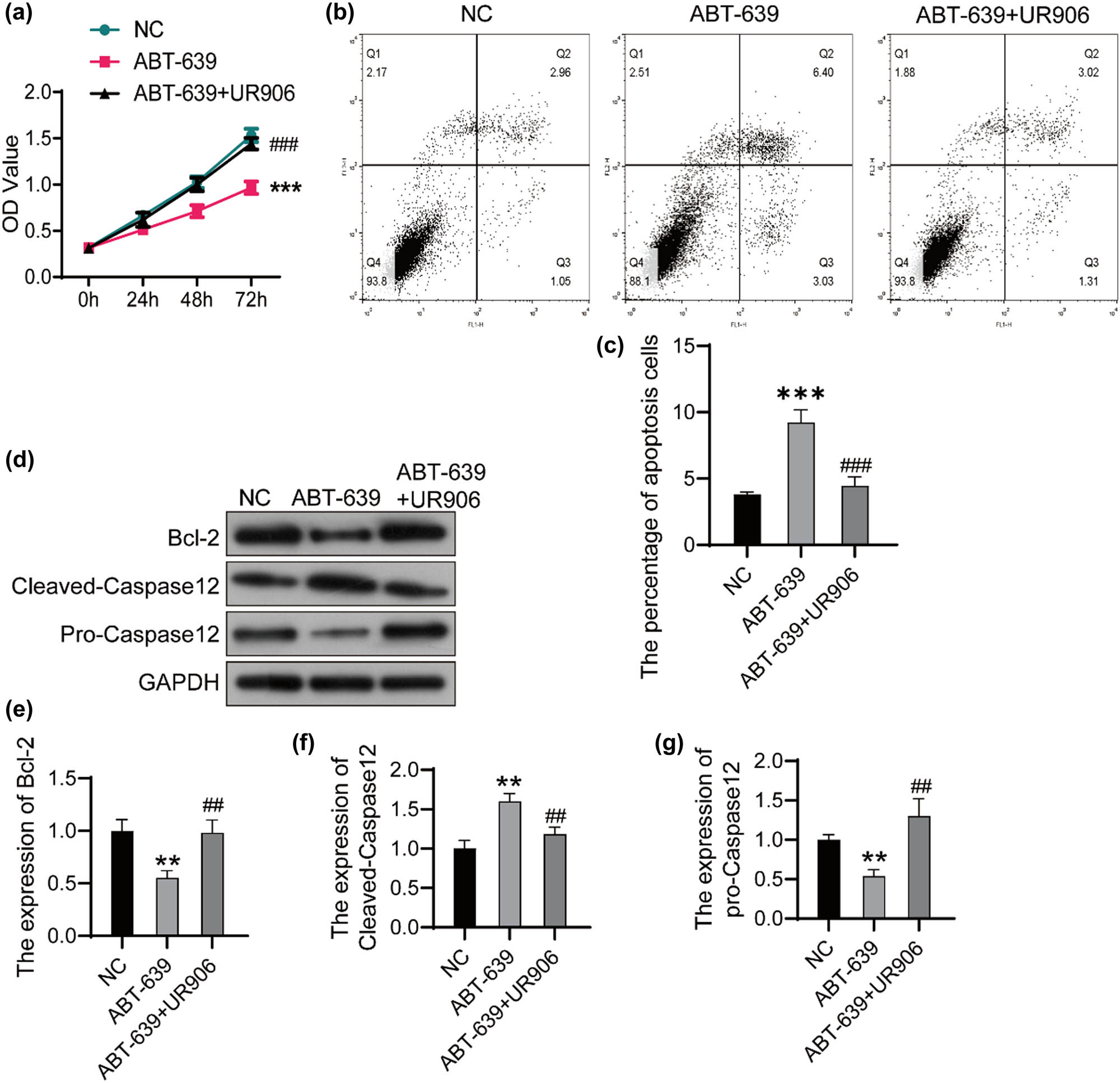
ERS inhibitor UR906 blocked the apoptosis induced by ABT-639. (a) The U251 cells were treated with both ABT-639 (10 μM) and UR906 (30 μM) or only ABT-639 (10 μM) with PBS as the control (NC). Cell viability was determined by CCK8 assay. (b) Flow cytometry was performed to detect cell apoptosis. (c) Apoptotic cells were analyzed using FlowJo software. (d) The expression of target protein was detected using western bolt. The relative expression of Bcl-2 (e), Cleaved-Caspase12 (f), and pro-Caspase12 (g) was analyzed using ImageJ software. **P < 0.01 vs NC group; ***P < 0.001 vs NC group; ## P < 0.01 vs ABT-639 group; ### P < 0.001 vs ABT-639 group.
3.5 UR906 blocked the activation of ERS induced by ABT-639
Finally, we detected the expression of ERS markers in U251 cells. The phosphorylation level of PERK decreased significantly after the treatment of UR906 in ABT-639-pretreated U251 cells. The GRP78, CHOP, and ATF6 proteins were downregulated after the treatment of UR906 in ABT-639-pretreated U251 cells (Figure 5). These data proved that blocking ERS attenuated CACNA1H knockdown-induced apoptosis in glioma cells.
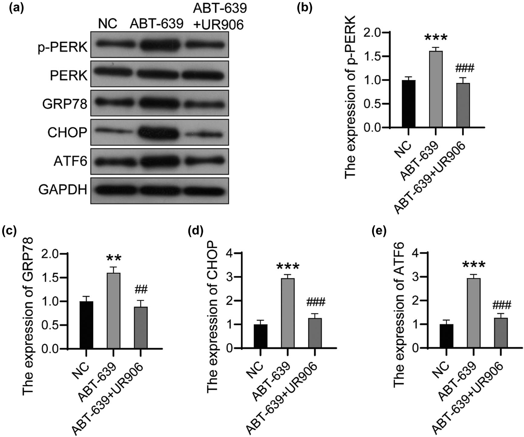
UR906 blocked the activation of ERS induced by ABT-639. (a) The expression of ERS-related proteins was detected using western bolt. The relative expression of p-PERK (b), GRP78 (c), CHOP (d), and ATF6 (e) was analyzed using ImageJ software. **P < 0.01 vs NC group; ***P < 0.001 vs NC group; ## P < 0.01 vs ABT-639 group; ### P < 0.001 vs ABT-639 group.
4 Discussion
CACNA1H encodes a low-voltage-activated calcium channel, which is expressed in thalamocortical circuits and is involved in the formation of neuronal burst firing and rhythm generation, as well as sleep and the development of epilepsy [16,17]. In recent years, the role of CACNA1H in tumors has been gradually discovered [18–21]. The genetic defect and mutation of CACNA1H involves in the progression of benign aldosterone-producing tumors [18,22]. As the cellular antenna for breast cancer-specific frequencies, CACNA1H mediates the targeted inhibition of breast cancer brain metastasis [23]. In human glioma, endostatin inhibited proliferation and migration by inhibiting the channel currents of Ca(V) 3.1 and Ca(V) 3.2 [24]. Here, we proved that the knockdown of CACNA1H inhibited proliferation and induced apoptosis in U251 cells. CACNA1H inhibitor ABT639 also suppressed proliferation and increased the percentage of apoptotic cells. Subsequently, we detected the expression of Bcl-2 and Caspase12 and found that the knockdown of CACNA1H could activate Caspase12 and inhibit Bcl-2 expression.
Bcl-2 inhibits cell death caused by various cytotoxic factors and plays a critical role in tumor cell apoptosis [25]. Interestingly, Bcl-2 also inhibits the transmembrane flow of Ca2+. Lam et al. have reported that the apoptosis induced by the calcium pump-specific inhibitor thapsigargin can be inhibited by Bcl-2. The reason is that Bcl-2 inhibits the transmembrane flow of Ca2+, suggesting that Bcl-2 can regulate apoptosis by regulating the intracellular Ca2+ [26]. Recent studies have shown that Bcl-2 family proteins interact with inositol 1,4,5-triphosphate receptor in endoplasmic reticulum (ER) to regulate its channel switch [27,28]. Anti-apoptotic proteins inhibit the excessive release of Ca2+ from ER and support cell survival, while pro-apoptotic proteins enhance the release of Ca2+ from ER and upregulate the concentration of Ca2+ in mitochondria and trigger apoptosis [27,29]. However, whether Bcl-2 affects CACNA1H activity and forms feedback regulation needs further experimental study.
Studies have shown that the inhibition of CACNA1H activates ERS, thereby inducing skeletal muscle atrophy [13]. ERS can promote the processing of unfolded or misfolded proteins in the ER, which facilitates the restoration of homeostasis and maintenance of cell survival, but persistent ERS leads to UPR and cell apoptosis [30]. The main ER transmembrane proteins involved in UPR signaling are PERK, IRE-1, and ATF6 [31]. Under normal conditions, they usually bind to GRP78 in an inactive state [32]. In the presence of ERS, GRP78 dissociates from these proteins and then binds to higher affinity unfolded and misfolded proteins. At the same time, PERK, IRE-1, and ATF6 dissociated from GRP78 were activated, eventually inducing UPR and apoptosis. Our results proved that the knockdown of CACNA1H and T-type Ca2+ channel inhibitor increased the expression of PERK, GRP78, CHOP, and ATF6, indicating the suppression of CACNA1H-activated ERS in U251 cells. Then, we found that ERS inhibitor UR906 blocked the apoptosis induced by CACNA1H inhibitor, suggesting that CACNA1H downregulation induced the apoptosis of tumor cells by activating ERS.
In addition, we also analyzed the effects of another T-type Ca2+ channel inhibitor, NNC55-0396, on the proliferation and ERS of glioma cells. The results confirmed that NNC55-0396 not only decreased cell activity, but also activated ERS in U251 cells. NNC55-0396 is a highly selective T-type calcium channel blocker for the Cav3.1 T-type channels, and its role in tumor cells is rarely reported. This result suggested that different subtypes of T-type Ca2+ channels might have a universal regulatory effect on ERS and apoptosis in glioma cells, which is worthy of our in-depth study. This result also indicated that NNC55-0396 might be considered as a new small molecule with anti-cancer effect.
5 Conclusion
Our data highlighted the effect of CACNA1H as an oncogenic gene in human glioma. Suppression of CACNA1H activated the ERS and thus induced apoptosis in glioma cells. T-type Ca2+ channel inhibitors ABT-639 and NNC55-0396 also induced apoptosis through ERS in glioma cells.
-
Author contributions: SL was responsible for doing experiments and writing the manuscript. YB assisted in the experiment. CL-L assisted in the analysis of experimental data. GM-X drafted and revised the manuscript. All the authors have read and agreed to the final manuscript.
-
Conflict of interest: The authors state no conflict of interest.
-
Data availability statement: The datasets generated during and/or analyzed during the current study are available from the corresponding author on reasonable request.
References
[1] Xu S, Tang L, Li X, Fan F, Liu Z. Immunotherapy for glioma: Current management and future application. Cancer Lett. 2020;476:1–12.10.1016/j.canlet.2020.02.002Search in Google Scholar PubMed
[2] Wang M, Tan J, Miao Y, Li M, Zhang Q. Role of Ca²⁺ and ion channels in the regulation of apoptosis under hypoxia. Histol Histopathol. 2018;33(3):237–46.Search in Google Scholar
[3] Marchi S, Patergnani S, Missiroli S, Morciano G, Rimessi A, Wieckowski MR, et al. Mitochondrial and endoplasmic reticulum calcium homeostasis and cell death. Cell Calcium. 2018;69:62–72.10.1016/j.ceca.2017.05.003Search in Google Scholar PubMed
[4] Catacuzzeno L, Sforna L, Esposito V, Limatola C, Franciolini F. Ion channels in glioma malignancy. Rev Physiol Biochem Pharmacol. 2021;181:223–67.10.1007/112_2020_44Search in Google Scholar PubMed
[5] Çiğ B, Derouiche S, Jiang LH. Editorial: Emerging roles of TRP channels in brain pathology. Front Cell Dev Biol. 2021;9:705196.10.3389/fcell.2021.705196Search in Google Scholar PubMed PubMed Central
[6] Antal L, Martin-Caraballo M. T-type calcium channels in cancer. Cancers (Basel). 2019;11(2):134–52.10.3390/cancers11020134Search in Google Scholar PubMed PubMed Central
[7] Sallán MC, Visa A, Shaikh S, Nàger M, Herreros J, Cantí C. T-type Ca2+ channels: T for targetable. Cancer Res. 2018;78(3):603–9.10.1158/0008-5472.CAN-17-3061Search in Google Scholar PubMed
[8] Valerie NC, Dziegielewska B, Hosing AS, Augustin E, Gray LS, Brautigan DL, et al. Inhibition of T-type calcium channels disrupts Akt signaling and promotes apoptosis in glioblastoma cells. Biochem Pharmacol. 2013;85(7):888–97.10.1016/j.bcp.2012.12.017Search in Google Scholar PubMed
[9] Tjaden J, Eickhoff A, Stahlke S, Gehmeyr J, Vorgerd M, Theis V, et al. Expression pattern of T-type Ca2+ Channels in cerebellar purkinje cells after VEGF treatment. Cells. 2021;10(9):2277–94.10.3390/cells10092277Search in Google Scholar PubMed PubMed Central
[10] Glauser TA, Holland K, O’Brien VP, Keddache M, Martin LJ, Clark PO, et al. Pharmacogenetics of antiepileptic drug efficacy in childhood absence epilepsy. Ann Neurol. 2017;81(3):444–53.10.1002/ana.24886Search in Google Scholar PubMed PubMed Central
[11] Gardiner M. Genetics of idiopathic generalized epilepsies. Epilepsia. 2005;46(Suppl 9):15–20.10.1111/j.1528-1167.2005.00310.xSearch in Google Scholar PubMed
[12] Han JY, Jang JH, Park J, Lee IG. Targeted next-generation sequencing of Korean patients with developmental delay and/or intellectual disability. Front Pediatr. 2018;6:391.10.3389/fped.2018.00391Search in Google Scholar PubMed PubMed Central
[13] Li S, Hao M, Li B, Chen M, Chen J, Tang J, et al. CACNA1H downregulation induces skeletal muscle atrophy involving endoplasmic reticulum stress activation and autophagy flux blockade. Cell Death Dis. 2020;11(4):279.10.1038/s41419-020-2484-2Search in Google Scholar PubMed PubMed Central
[14] Zanetti M, Xian S, Dosset M, Carter H. The unfolded protein response at the tumor-immune interface. Front Immunol. 2022;13:823157.10.3389/fimmu.2022.823157Search in Google Scholar PubMed PubMed Central
[15] Chen X, Cubillos-Ruiz JR. Endoplasmic reticulum stress signals in the tumour and its microenvironment. Nat Rev Cancer. 2021;21(2):71–88.10.1038/s41568-020-00312-2Search in Google Scholar PubMed PubMed Central
[16] Chourasia N, Ossó-Rivera H, Ghosh A, Von Allmen G, Koenig MK. Expanding the phenotypic spectrum of CACNA1H mutations. Pediatr Neurol. 2019;93:50–5.10.1016/j.pediatrneurol.2018.11.017Search in Google Scholar PubMed
[17] Tatsuki F, Sunagawa GA, Shi S, Susaki EA, Yukinaga H, Perrin D, et al. Involvement of Ca2+-dependent hyperpolarization in sleep duration in mammals. Neuron. 2016;90(1):70–85.10.1016/j.neuron.2016.02.032Search in Google Scholar PubMed
[18] Kamilaris CDC, Hannah-Shmouni F, Stratakis CA. Adrenocortical tumorigenesis: Lessons from genetics. Best Pract Res Clin Endocrinol Metab. 2020;34(3):101428.10.1016/j.beem.2020.101428Search in Google Scholar PubMed PubMed Central
[19] Reimer EN, Walenda G, Seidel E, Scholl UI. CACNA1H(M1549V) mutant calcium channel causes autonomous aldosterone production in HAC15 cells and is inhibited by mibefradil. Endocrinology. 2016;157(8):3016–22.10.1210/en.2016-1170Search in Google Scholar PubMed
[20] Phan NN, Wang CY, Chen CF, Sun Z, Lai MD, Lin YC. Voltage-gated calcium channels: Novel targets for cancer therapy. Oncol Lett. 2017;14(2):2059–74.10.3892/ol.2017.6457Search in Google Scholar PubMed PubMed Central
[21] Sekiguchi F, Kawabata A. Role of Ca(v)3.2 T-type Ca2+ channels in prostate cancer cells. Nihon Yakurigaku Zasshi. 2019;154(3):97–102.10.1254/fpj.154.97Search in Google Scholar PubMed
[22] Nanba K, Blinder AR, Rege J, Hattangady NG, Else T, Liu CJ, et al. Somatic CACNA1H mutation as a cause of aldosterone-producing adenoma. Hypertension. 2020;75(3):645–9.10.1161/HYPERTENSIONAHA.119.14349Search in Google Scholar PubMed PubMed Central
[23] Sharma S, Wu SY, Jimenez H, Xing F, Zhu D, Liu Y, et al. Ca2+ and CACNA1H mediate targeted suppression of breast cancer brain metastasis by AM RF EMF. EBioMedicine. 2019;44:194–208.10.1016/j.ebiom.2019.05.038Search in Google Scholar PubMed PubMed Central
[24] Zhang Y, Zhang J, Jiang D, Zhang D, Qian Z, Liu C, et al. Inhibition of T-type Ca²⁺ channels by endostatin attenuates human glioblastoma cell proliferation and migration. Br J Pharmacol. 2012;166(4):1247–60.10.1111/j.1476-5381.2012.01852.xSearch in Google Scholar PubMed PubMed Central
[25] Zhu R, Li L, Nguyen B, Seo J, Wu M, Seale T, et al. FLT3 tyrosine kinase inhibitors synergize with BCL-2 inhibition to eliminate FLT3/ITD acute leukemia cells through BIM activation. Signal Transduct Target Ther. 2021;6(1):186.10.1038/s41392-021-00578-4Search in Google Scholar PubMed PubMed Central
[26] Distelhorst CW, Lam M, McCormick TS. Bcl-2 inhibits hydrogen peroxide-induced ER Ca2+ pool depletion. Oncogene. 1996;12(10):2051–5.Search in Google Scholar
[27] Ivanova H, Vervliet T, Monaco G, Terry LE, Rosa N, Baker MR, et al. Bcl-2-protein family as modulators of IP(3) receptors and other organellar Ca2+ channels. Cold Spring Harb Perspect Biol. 2020;12:4.10.1101/cshperspect.a035089Search in Google Scholar PubMed PubMed Central
[28] Callens M, Kraskovskaya N, Derevtsova K, Annaert W, Bultynck G, Bezprozvanny I, et al. The role of Bcl-2 proteins in modulating neuronal Ca2+ signaling in health and in Alzheimer’s disease. Biochim Biophys Acta Mol Cell Res. 2021;1868(6):118997.10.1016/j.bbamcr.2021.118997Search in Google Scholar PubMed PubMed Central
[29] Pinton P, Rizzuto R. Bcl-2 and Ca2+ homeostasis in the endoplasmic reticulum. Cell Death Differ. 2006;13(8):1409–18.10.1038/sj.cdd.4401960Search in Google Scholar PubMed
[30] Senft D, Ronai ZA. UPR, autophagy, and mitochondria crosstalk underlies the ER stress response. Trends Biochem Sci. 2015;40(3):141–8.10.1016/j.tibs.2015.01.002Search in Google Scholar PubMed PubMed Central
[31] Kania E, Pająk B, Orzechowski A. Calcium homeostasis and ER stress in control of autophagy in cancer cells. Biomed Res Int. 2015;2015:352794.10.1155/2015/352794Search in Google Scholar PubMed PubMed Central
[32] Hernandez I, Cohen M. Linking cell-surface GRP78 to cancer: From basic research to clinical value of GRP78 antibodies. Cancer Lett. 2022;524:1–14.10.1016/j.canlet.2021.10.004Search in Google Scholar PubMed
© 2023 the author(s), published by De Gruyter
This work is licensed under the Creative Commons Attribution 4.0 International License.
Articles in the same Issue
- Research Articles
- HIF-1α participates in secondary brain injury through regulating neuroinflammation
- Omega-3 polyunsaturated fatty acids alleviate early brain injury after traumatic brain injury by inhibiting neuroinflammation and necroptosis
- The correlation between non-arteritic anterior ischemic optic neuropathy and cerebral infarction
- Enriched environment can reverse chronic sleep deprivation-induced damage to cellular plasticity in the dentate gyrus of the hippocampus
- Middle cerebral artery dynamic cerebral autoregulation is impaired by infarctions in the anterior but not the posterior cerebral artery territory in patients with mild strokes
- Leptin ameliorates Aβ1-42-induced Alzheimer’s disease by suppressing inflammation via activating p-Akt signaling pathway
- TIPE2 knockdown exacerbates isoflurane-induced postoperative cognitive impairment in mice by inducing activation of STAT3 and NF-κB signaling pathways
- Does the patellar tendon reflex affect the postural stability in stroke patients with blocked vision?
- Inactivation of CACNA1H induces cell apoptosis by initiating endoplasmic reticulum stress in glioma
- miR-101-3p improves neuronal morphology and attenuates neuronal apoptosis in ischemic stroke in young mice by downregulating HDAC9
- A custom-made weight-drop impactor to produce consistent spinal cord injury outcomes in a rat model
- Arterial spin labeling for moyamoya angiopathy: A preoperative and postoperative evaluation method
- Thyroid hormone levels paradox in acute ischemic stroke
- Geniposide protected against cerebral ischemic injury through the anti-inflammatory effect via the NF-κB signaling pathway
- The clinical characteristics of acute cerebral infarction patients with thalassemia in a tropic area in China
- Comprehensive behavioral study of C57BL/6.KOR-ApoEshl mice
- Incomplete circle of Willis as a risk factor for intraoperative ischemic events during carotid endarterectomies performed under regional anesthesia – A prospective case-series
- HOTAIRM1 knockdown reduces MPP+-induced oxidative stress injury of SH-SY5Y cells by activating the Nrf2/HO-1 pathway
- Esmolol inhibits cognitive impairment and neuronal inflammation in mice with sepsis-induced brain injury
- EHMT2 affects microglia polarization and aggravates neuronal damage and inflammatory response via regulating HMOX1
- Hematoma evacuation based on active strategies versus conservative treatment in the management of moderate basal ganglia hemorrhage: A retrospective study
- Knockdown of circEXOC6 inhibits cell progression and glycolysis by sponging miR-433-3p and mediating FZD6 in glioma
- CircYIPF6 regulates glioma cell proliferation, apoptosis, and glycolysis through targeting miR-760 to modulate PTBP1 expression
- Relationship between serum HIF-1α and VEGF levels and prognosis in patients with acute cerebral infarction combined with cerebral-cardiac syndrome
- The promoting effect of modified Dioscorea pills on vascular remodeling in chronic cerebral hypoperfusion via the Ang/Tie signaling pathway
- Effects of enriched environment on the expression of β-amyloid and transport-related proteins LRP1 and RAGE in chronic sleep-deprived mice
- An interventional study of baicalin on neuronal pentraxin-1, neuronal pentraxin-2, and C-reactive protein in Alzheimer’s disease rat model
- PD98059 protects SH-SY5Y cells against oxidative stress in oxygen–glucose deprivation/reperfusion
- TPVB and general anesthesia affects postoperative functional recovery in elderly patients with thoracoscopic pulmonary resections based on ERAS pathway
- Brain functional connectivity and network characteristics changes after vagus nerve stimulation in patients with refractory epilepsy
- Association between RS3763040 polymorphism of the AQP4 and idiopathic intracranial hypertension in a Spanish Caucasian population
- Effects of γ-oryzanol on motor function in a spinal cord injury model
- Electroacupuncture inhibits the expression of HMGB1/RAGE and alleviates injury to the primary motor cortex in rats with cerebral ischemia
- Effects of edaravone dexborneol on neurological function and serum inflammatory factor levels in patients with acute anterior circulation large vessel occlusion stroke
- CST3 alleviates bilirubin-induced neurocytes’ damage by promoting autophagy
- Excessive MALAT1 promotes the immunologic process of neuromyelitis optica spectrum disorder by upregulating BAFF expression
- Evaluation of cholinergic enzymes and selected biochemical parameters in the serum of patients with a diagnosis of acute subarachnoid hemorrhage
- 7-Day National Institutes of Health Stroke Scale as a surrogate marker predicting ischemic stroke patients’ outcome following endovascular therapy
- Cdk5 activation promotes Cos-7 cells transition towards neuronal-like cells
- 10.1515/tnsci-2022-0313
- PPARα agonist fenofibrate prevents postoperative cognitive dysfunction by enhancing fatty acid oxidation in mice
- Predicting functional outcome in acute ischemic stroke patients after endovascular treatment by machine learning
- EGCG promotes the sensory function recovery in rats after dorsal root crush injury by upregulating KAT6A and inhibiting pyroptosis
- Preoperatively administered single dose of dexketoprofen decreases pain intensity on the first 5 days after craniotomy: A single-centre placebo-controlled, randomized trial
- Myeloarchitectonic maps of the human cerebral cortex registered to surface and sections of a standard atlas brain
- The BET inhibitor apabetalone decreases neuroendothelial proinflammatory activation in vitro and in a mouse model of systemic inflammation
- Carthamin yellow attenuates brain injury in a neonatal rat model of ischemic–hypoxic encephalopathy by inhibiting neuronal ferroptosis in the hippocampus
- Functional connectivity in ADHD children doing Go/No-Go tasks: An fMRI systematic review and meta-analysis
- Review Articles
- Human prion diseases and the prion protein – what is the current state of knowledge?
- Nanopharmacology as a new approach to treat neuroinflammatory disorders
- Case Report
- Deletion as novel variants in VPS13B gene in Cohen syndrome: Case series
- Commentary
- Translation of surface electromyography to clinical and motor rehabilitation applications: The need for new clinical figures
- Revealing key role of T cells in neurodegenerative diseases, with potential to develop new targeted therapies
- Retraction
- Retraction of “Eriodictyol corrects functional recovery and myelin loss in SCI rats”
- Special Issue “Advances in multimedia-based emerging technologies...”
- Evaluation of the improvement of walking ability in patients with spinal cord injury using lower limb rehabilitation robots based on data science

