MRI and bone scintigraphy for breast cancer bone metastase: a meta-analysis
-
Yue Rong


Abstract
Objective
The aim of this study was to compare the diagnostic value of magnetic resonance image (MRI) and bone scintigraphy (BS) in the diagnosis of breast cancer bone metastases.
Methods
Searching in the databases including PubMed, Embase about the comparative study of MRI and bone scintigraphy in the diagnosis of breast cancer bone metastases during 2000~2018. After we screened further, the extracted effective data were calculated by Meta-Disc 1.4 software.
Results
We obtained 4 articles. The pooled estimates for sensitivity of MRI, BS were 0.99 (95% CI, [0.95, 1.00]) and 0.93 (95% CI, [0.88, 0.97]) respectively; For specificity were 0.99 (95% CI, [0.95, 1.00]) and 0.86 (95% CI, [0.79, 0.92]) respectively. The AUC of SROC curve for MRI and BS were 0.9948 and 0.9675 respectively.
Conclusion
MRI remains to be a satisfactory method for the diagnosis of breast cancer bone metastases and should first be considered for patients.
1 Introduction
Breast cancer is the most common malignant tumor in women, accounting for 23% of all malignant tumors. In recent years, the incidence of breast cancer has increased rapidly, more than 1 million new cases each year worldwide [1]. Bone metastasis occurs in 60% to 70% of recurrence breast cancer patients [2]. Bone metastases cause a series of symptom such as pain, pathological fractures and spinal cord compression, which shortens the survival time from 20 years to 2 years [3]. In addition, bone metastases affect the quality of life of patients. Therefore, accurate and early diagnosis of bone metastasis in breast cancer is of great significance for the treatment and prognosis of breast cancer patients.
At present, the widely used method of bone metastasis screening and examination is bone scan (BS), which has high sensitivity and can obtain images of whole body bone [4], but its specificity is low, and it is easy to cause false positives. Magnetic resonance imaging has the advantages of multi-parameter, multi-azimuth imaging, good soft tissue contrast and high spatial resolution. MRI can clearly distinguish between fat and liquid tissue [5, 6]. With the development of new MRI scanners and coil technology, whole-body diffusion-weighted imaging (WB-DWI) with background body signal suppression, has been recognized as a new imaging modality for the assessment of metastases of various malignancies. MRI has high value in the identification of benign and malignant lesions of the bone [7, 8]. Due to the strong magnetic field of MRI, the examination of patients with metal implants is limited [9], except for WB-DWI, the other MRI techniques can not complete one-time whole body bone imaging compared with bone scan. Therefore, this study collected all the domestic and international MRI and bone scan diagnosis of breast cancer bone metastasis, using meta-analysis to analyze the data, evaluate the diagnostic value of MRI and bone scan for breast cancer bone metastasis, to provide scientific evidence for clinical application.
2 Methods
2.1 Searching method
We conducted a search of PubMed, Embase databases that were published between 2000 and 2018. We limited the search to study published in English. The medical subject heading terms and keywords used included “breast cancer or breast carcinoma or breast neoplasm”, “Osseous metastases or bone metastases”, “magnetic resonance image or MRI”, “Bone scan or Bone scintigraphy”, “sensitivity and specificity”, “diagnoses or diagnosis”. According to the specific database, all retrieval strategies are determined by multiple pre-searches. In order to minimize the missing literature, the author also combines manual search and conducts a second search for the references included in the literature.
2.2 Inclusion criteria
Studies were selected carefully on the basis of the following criteria: MRI is compared with gold standards, or bone scans are compared with gold standards for the diagnosis of breast cancer bone metastases. The gold standard is pathological examination and follow-up; Pathologically diagnosed breast cancer, and patients with bone metastases such as bone pain or patients with high degree of suspicion of bone metastases, without limiting age, gender, ethnicity;
2.3 Excluding standard
Non-clinical controlled trials; Non-breast cancer research; Incomplete data; Case reports; Review literature; Data published repeatedly.
2.4 Data extraction
Two authors independently assessed each literature, and then download and extracted all the data by using standardized data-abstraction forms. The data extracted included year of publication, true positive, false positive, false negative, true negative, sensitivity, specificity. For each study, 2×2 contingency tables were constructed. We calculated the sensitivity, specificity, likelihood ratio (LR).
2.5 Statistical analysis
Data analysis was performed using Meta-Disc 1.4 software. Heterogeneity test was performed, and the receiver operating characteristic curve (SROC curve) was drawn and the sensitivity, specificity, positive likelihood ratio, negative likelihood ratio, and diagnostic test odds ratio of each diagnostic method were calculated.
3 Results
3.1 Literature searches and characteristics of eligible study
According to search strategy, 4 full articles were finally considered eligible for the review after evaluation. Figure 1 shows the flow diagram of study selection process. The detailed characteristics for the 4 eligible studies are summarized in Table 1.
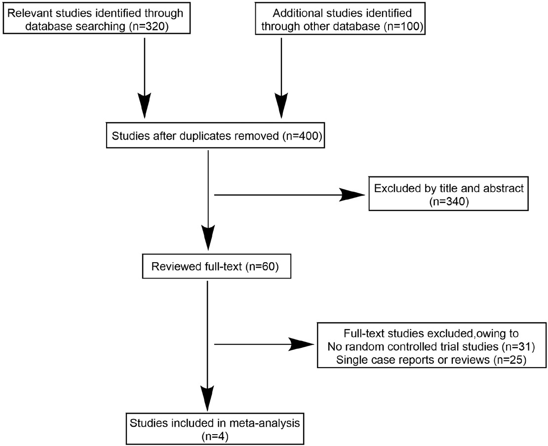
The study selection process
3.2 Quality assessment
We assessed the quality of the included studies according to QUADAS (10). Each study was evaluated respectively by two independent investigators. On average, the investigators disagreed on 3 of 11 items (range, 0–6). All disagreements were resolved by consensus. The quality evaluation of the included studies is shown in Table 2.
Quality assessment of included studies
| Included studies | QUADAS | |||||||||||||
|---|---|---|---|---|---|---|---|---|---|---|---|---|---|---|
| 1 | 2 | 3 | 4 | 5 | 6 | 7 | 8 | 9 | 10 | 11 | 12 | 13 | 14 | |
| Kim 2009 | Yes | Yes | Yes | Yes | Yes | Yes | Yes | Yes | Yes | No | No | Yes | Yes | No |
| Engelhard 2004 | Yes | Yes | Yes | Yes | Yes | Yes | Yes | Yes | Yes | No | No | Yes | Yes | No |
| Altehoefer 2001 | Yes | Yes | Yes | Yes | Yes | Yes | Yes | Yes | Yes | No | No | Yes | Yes | No |
| Layer 1999 | Yes | Yes | Yes | No | Yes | Yes | Yes | Yes | Yes | No | No | Yes | No | No |
3.3 The diagnostic sensitivity and specificity of MRI
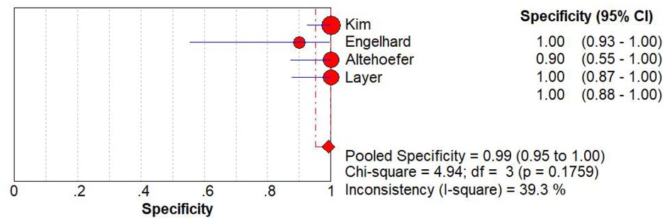
The pooled diagnostic sensitivity and specificity of MRI are 0.99 (95% CI, [0.95, 1.00]), 0.99 (95% CI, [0.95, 1.00]) respectively. Significant heterogeneity was found among these studies (I2=34.5% and 39.3%). Due to significant heterogeneity of the data, we used a random effects model (Figure 2 and 3).
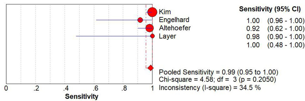
The plot for the sensitivity of MRI
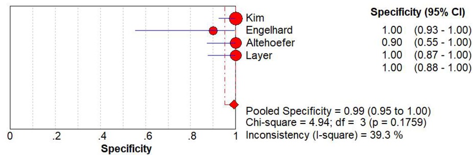
The plot for the specificity of MRI
3.4 The diagnostic sensitivity and specificity of BS
The pooled diagnostic sensitivity and specificity of BS are 0.93 (95% CI, [0.88, 0.97]), 0.86 (95% CI, [0.79, 0.92]) respectively. Significant heterogeneity was found among these studies (I2=64.8% and 87.2%). Due to significant heterogeneity of the data, we used a random effects model (Figure 4 and 5).
The plot for the sensitivity of BS
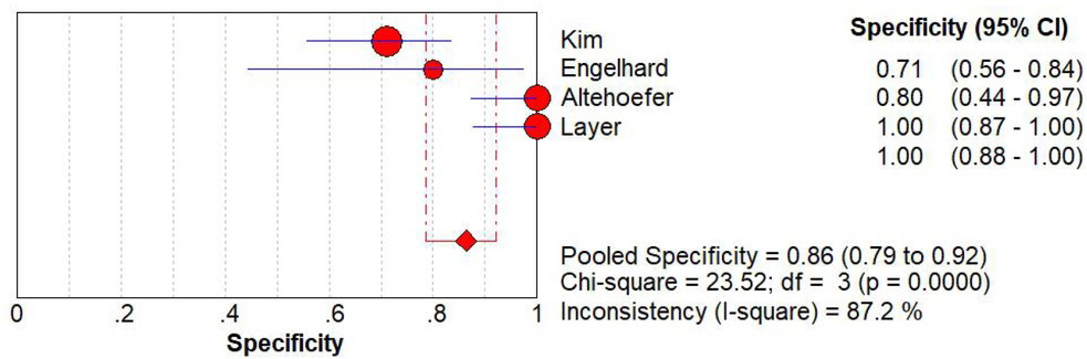
The plot for the specificity of BS
3.5 The negative LR, positive LR and SROC curve of MRI
The pooled negative LR and positive LR of MRI are 0.04 (95% CI, [0.01, 0.12]), 29.36 (95% CI, [8.41, 102.57]) respectively. (Figure 6 and 7). We successfully plotted the SROC
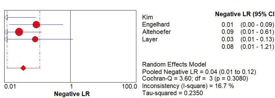
The plot for the negative LR of MRI
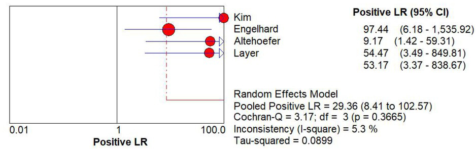
The plot for the positive LR of MRI
curve. The area under the SROC curve (AUC) is 0.9948 and the Q* is 0.9720 (Figure 8).
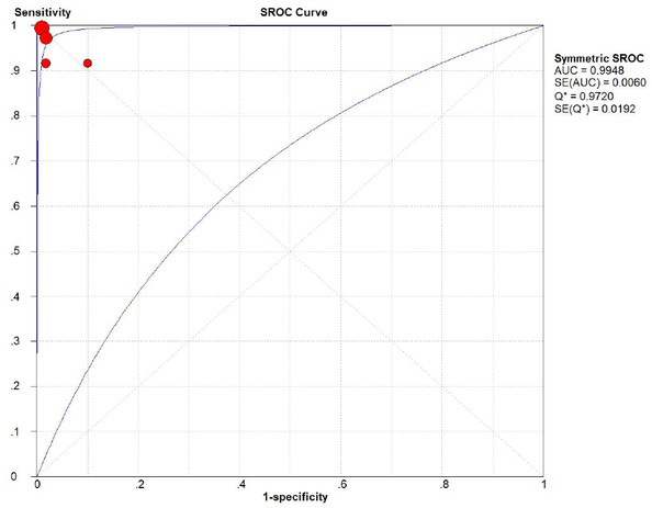
The SROC curve of MRI
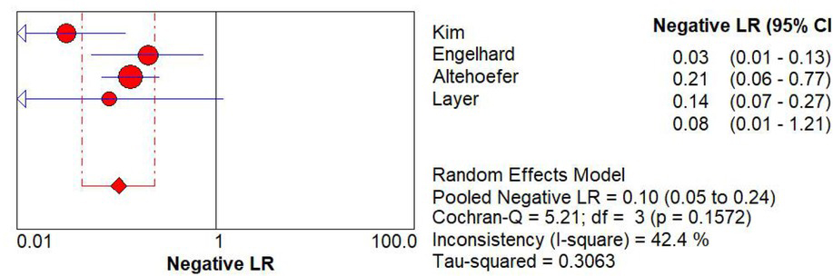
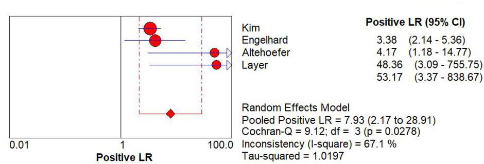
3.6 The negative LR, positive LR and SROC curve of BS
The pooled negative LR and positive LR of BS are 0.10 (95% CI, [0.05, 0.24]), 7.93 (95% CI, [2.17, 28.91]) respectively. (Figure 9 and 10). We successfully plotted the SROC curve. The area under the SROC curve (AUC) is 0.9675 and the Q* is 0.9159 (Figure 11).
The plot for the specificity of MRI
The plot for the sensitivity of BS
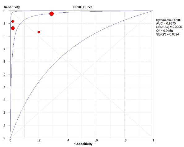
TThe SROC curve of BS
4 Discussion
A total of 4 studies were included in this systematic review. Meta-analysis results showed that the combined sensitivity and specificity of MRI for the diagnosis of breast cancer with bone metastases were 99%, indicating that the rate of missed diagnosis and misdiagnosis was very low (1%); the area under the curve of MRI and bone scan was 0.9948 and 0.9675, their diagnostic performance are high. The combined sensitivity of bone scan for the diagnosis of breast cancer with bone metastases was 93%, indicating a missed diagnosis rate of 7%; combined specificity was 86%, indicating a misdiagnosis rate of 14%. Therefore,
compared with bone scan, the missed diagnosis rate and misdiagnosis rate of MRI in the diagnosis of breast cancer bone metastasis are very low, and the SROC curve of MRI is close to the upper left corner, that is, the area under the curve is larger, indicating the accuracy of MRI for bone metastasis of breast cancer. Higher, combined with MRI’s image quality is high. Therefore, MRI is a more accurate method for diagnosing bone metastasis in breast cancer.
The 4 studies included in this systematic review were from different countries, and the combination of computer and manual search was used in the search process, and the data collection was relatively comprehensive. In the diagnosis of diseases, the diagnosis results of MRI and bone scan are related to the severity of the disease and the diagnostic level of doctor. However, all included studies do not describe such problems, which may influence the interpretation of the results. Due to included studies were lack of negative trials, there may be potential publish bias, although it was reported that the low detection rate and high cost of MRI is little value for diagnosis of breast cancer bone metastases [11].
In this study, as long as there is a metastasis, there is a positive case. If the same patient has multiple metastases, the number of lesions detected by MRI and bone scan is different from the difference between the good and the malignant. Bone scans sometimes show multiple abnormal uptake areas, but both benign and malignant lesions coexist, and it is not possible to clearly distinguish between benign and malignant specific lesions [12]. The advantages of MRI are multi-azimuth imaging and multi-sequence multi-parameter imaging, with high specificity and high quality images. In recent years, the PET-like technique, magnetic resonance systemic diffusion-weighted imaging (WB-DWI), has a very high sensitivity to malignant tumor examination, and can obtain system-specific tumor imaging similar to PET. Inspection costs are much lower than PET and are widely used in clinical practice [8].
In summary, MRI can be an effective and feasible method for diagnosing bone metastasis in breast cancer. However, the limitations of methodology, high-quality studies are needed to further confirm the clinical value of MRI in the diagnosis of breast cancer bone metastasis.
Acknowledgements
This research was supported by Zhejiang Provincial Natural Science Foundation of China under Grant No. LGF19H180016.
-
Conflict of interest: Authors state no conflict of interest.
Reference
[1] Siegel RL, Miller KD, Jemal A. Cancer statistics, 2018. Ca A Cancer J Clin. 2018; 68:7–3010.3322/caac.21442Search in Google Scholar PubMed
[2] Chen HH, Su WC, Guo HR, Lee BF, Su WR, Wu PS, et al. Clinical significance and outcome of one or two rib lesions on bone scans in breast cancer patients without known metastases. Nucl Med Commun. 2003; 24:1167–117410.1097/00006231-200311000-00007Search in Google Scholar PubMed
[3] Hibberd CS, Quan GMY. Risk factors for pathological fracture and metastatic epidural spinal cord compression in patients with spinal metastases. Orthopedics. 2018; 41:1–810.3928/01477447-20171106-06Search in Google Scholar PubMed
[4] Takesh M, Odat Allh K, Adams S, Zechmann C. Diagnostic role of (18) F-FECH-PET/CT compared with bone scan in evaluating the prostate cancer patients referring with biochemical recurrence. ISRN Oncol. 2012:81523410.5402/2012/815234Search in Google Scholar PubMed PubMed Central
[5] Catalá V, Salas D, Esquena S, Mateu S, Algaba F, Palou J, et al. Questions and answers on prostate multiparameter magnetic resonance imaging: Everything a urologist should know. Actas Urol Esp. 2016; 40:339–35210.1016/j.acuroe.2016.05.001Search in Google Scholar
[6] Tarachkova EV, Strel’Tsova ON, Panov VO, Bazaeva IY, Tyurin IE. Multiparameter magnetic resonance imaging in the diagnosis of cancer of the cervix uteri. Vestn Rentgenol Radiol. 2015:43–55Search in Google Scholar
[7] Kim SH, Yoo HJ, Kang Y, Choi JY, Hong SH. MRI findings of new uptake in the femoral head detected on follow-up bone scans. AJR Am J Roentgenol. 2015; 204:608–61410.2214/AJR.14.12968Search in Google Scholar PubMed
[8] Pasoglou V, Michoux N, Tombal B, Jamar F, Lecouvet FE. wbMRI to detect bone metastases: critical review on diagnostic accuracy and comparison to other imaging modalities. Clin Transl Imaging. 2015; 3:141–15710.1007/s40336-015-0120-4Search in Google Scholar
[9] Parsons TM, Satchithananda K, Berbe R, Siddiqui IA, Robinson E, Hart AJ, et al. MRI investigations in patients with problems due to metal-on-metal implants. Orthopde. 2013; 42:629–636Search in Google Scholar
[10] Whiting P, Rutjes AW, Reitsma JB, Bossuyt PM, Kleijnen J. The development of QUADAS: a tool for the quality assessment of studies of diagnostic accuracy included in systematic reviews. BMC Med Res Methodol. 2003; 3:2510.1186/1471-2288-3-25Search in Google Scholar PubMed PubMed Central
[11] Jones AL, Williams MP, Powles TJ, Oliff JFC, Hardy JR, Cherryman G, et al. Magnetic resonance imaging in the detection of skeletal metastases in patients with breast cancer. Br J Cancer. 1990; 62:296–29810.1038/bjc.1990.281Search in Google Scholar PubMed PubMed Central
[12] Kim JY, Choi YY, Kim CW, Sung YK, Yoo DH. Bone Scintigraphy in the Diagnosis of Rheumatoid Arthritis: Is There Additional Value of Bone Scintigraphy with Blood Pool Phase over Conventional Bone Scintigraphy? J Korean Med Sci. 2016; 31:502–50910.3346/jkms.2016.31.4.502Search in Google Scholar PubMed PubMed Central
[13] Kim DS, Hong SJ. Magnetic resonance imaging diagnoses of bone scan abnormalities in breast cancer patients. Nucl Med Commun. 2009; 30:736–74110.1097/MNM.0b013e32832ed375Search in Google Scholar
[14] Engelhard K, Hollenbach HP, Wohlfart K, Imhoff EV, Fellner FA. Comparison of whole-body MRI with automatic moving table technique and bone scintigraphy for screening for bone metastases in patients with breast cancer. Eur Radiol. 2004; 14:99–10510.1007/s00330-003-1968-7Search in Google Scholar
[15] Altehoefera C, Högerle S, Moser E, Langer M. Comparative detectability of bone metastases and impact on therapy of magnetic resonance imaging and bone scintigraphy in patients with breast cancer. Eur J Radiol. 2001; 40:16–2310.1016/S0720-048X(01)00313-8Search in Google Scholar
[16] Layer G, Steudel A, Schüller H, van Kaick G, Grünwald F, Reiser M, et al. Magnetic resonance imaging to detect bone marrow metastases in the initial staging of small cell lung carcinoma and breast carcinoma. Cancer. 2015; 85:1004–100910.1002/(SICI)1097-0142(19990215)85:4<1004::AID-CNCR31>3.0.CO;2-2Search in Google Scholar
© 2019 Yue Rong et al. published by De Gruyter
This work is licensed under the Creative Commons Attribution-NonCommercial-NoDerivatives 4.0 International License.
Articles in the same Issue
- Research Article
- Prostate Cancer-Specific of DD3-driven oncolytic virus-harboring mK5 gene
- Case Report
- Pediatric acute paradoxical cerebral embolism with pulmonary embolism caused by extremely small patent foramen ovale
- Research Article
- Associations between ambient temperature and acute myocardial infarction
- Case Report
- Discontinuation of imatinib mesylate could improve renal impairment in chronic myeloid leukemia
- Research Article
- METTL3 promotes the proliferation and mobility of gastric cancer cells
- The C677T polymorphism of the methylenetetrahydrofolate reductase gene and susceptibility to late-onset Alzheimer’s disease
- microRNA-1236-3p regulates DDP resistance in lung cancer cells
- Review Article
- The link between thyroid autoimmunity, depression and bipolar disorder
- Research Article
- Effects of miR-107 on the Chemo-drug sensitivity of breast cancer cells
- Analysis of pH dose-dependent growth of sulfate-reducing bacteria
- Review Article
- Musculoskeletal clinical and imaging manifestations in inflammatory bowel diseases
- Research Article
- Regional hyperthermia combined with chemotherapy in advanced gastric cancer
- Analysis of hormone receptor status in primary and recurrent breast cancer via data mining pathology reports
- Morphological and isokinetic strength differences: bilateral and ipsilateral variation by different sport activity
- The reliability of adjusting stepped care based on FeNO monitoring for patients with chronic persistent asthma
- Comparison of the clinical outcomes of two physiological ischemic training methods in patients with coronary heart disease
- Analysis of ticagrelor’s cardio-protective effects on patients with ST-segment elevation acute coronary syndrome accompanied with diabetes
- Computed tomography findings in patients with Samter’s Triad: an observational study
- Case Report
- A spinal subdural hematoma induced by guidewire-based lumbar drainage in a patient with ruptured intracranial aneurysms
- Research Article
- High expression B3GAT3 is related with poor prognosis of liver cancer
- Effects of light touch on balance in patients with stroke
- Oncoprotein LAMTOR5 activates GLUT1 via upregulating NF-κB in liver cancer
- Effects of budesonide combined with noninvasive ventilation on PCT, sTREM-1, chest lung compliance, humoral immune function and quality of life in patients with AECOPD complicated with type II respiratory failure
- Prognostic significance of lymph node ratio in ovarian cancer
- Case Report
- Brainstem anaesthesia after retrobulbar block
- Review Article
- Treating infertility: current affairs of cross-border reproductive care
- Research Article
- Serum inflammatory cytokines comparison in gastric cancer therapy
- Behavioural and psychological symptoms in neurocognitive disorders: Specific patterns in dementia subtypes
- MRI and bone scintigraphy for breast cancer bone metastase: a meta-analysis
- Comparative study of back propagation artificial neural networks and logistic regression model in predicting poor prognosis after acute ischemic stroke
- Analysis of the factors affecting the prognosis of glioma patients
- Compare fuhrman nuclear and chromophobe tumor grade on chromophobe RCC
- Case Report
- Signet ring B cell lymphoma: A potential diagnostic pitfall
- Research Article
- Subparaneural injection in popliteal sciatic nerve blocks evaluated by MRI
- Loneliness in the context of quality of life of nursing home residents
- Biological characteristics of cervical precancerous cell proliferation
- Effects of Rehabilitation in Bankart Lesion in Non-athletes: A report of three cases
- Management of complications of first instance of hepatic trauma in a liver surgery unit: Portal vein ligation as a conservative therapeutic strategy
- Matrix metalloproteinase 2 knockdown suppresses the proliferation of HepG2 and Huh7 cells and enhances the cisplatin effect
- Comparison of laparoscopy and open radical nephrectomy of renal cell cancer
- Case Report
- A severe complication of myocardial dysfunction post radiofrequency ablation treatment of huge hepatic hemangioma: a case report and literature review
- Solar urticaria, a disease with many dark sides: is omalizumab the right therapeutic response? Reflections from a clinical case report
- Research Article
- Binge eating disorder and related features in bariatric surgery candidates
- Propofol versus 4-hydroxybutyric acid in pediatric cardiac catheterizations
- Nasointestinal tube in mechanical ventilation patients is more advantageous
- The change of endotracheal tube cuff pressure during laparoscopic surgery
- Correlation between iPTH levels on the first postoperative day after total thyroidectomy and permanent hypoparathyroidism: our experience
- Case Report
- Primary angiosarcoma of the kidney: case report and comprehensive literature review
- Research Article
- miR-107 enhances the sensitivity of breast cancer cells to paclitaxel
- Incidental findings in dental radiology are concerning for family doctors
- Suffering from cerebral small vessel disease with and without metabolic syndrome
- A meta-analysis of robot assisted laparoscopic radical prostatectomy versus laparoscopic radical prostatectomy
- Indications and outcomes of splenectomy for hematological disorders
- Expression of CENPE and its prognostic role in non-small cell lung cancer
- Barbed suture and gastrointestinal surgery. A retrospective analysis
- Using post transplant 1 week Tc-99m DTPA renal scan as another method for predicting renal graft failure
- The pseudogene PTTG3P promotes cell migration and invasion in esophageal squamous cell carcinoma
- Lymph node ratio versus TNM system as prognostic factor in colorectal cancer staging. A single Center experience
- Review Article
- Minimally invasive pilonidal sinus treatment: A narrative review
- Research Article
- Anatomical workspace study of Endonasal Endoscopic Transsphenoidal Approach
- Hounsfield Units on Lumbar Computed Tomography for Predicting Regional Bone Mineral Density
- Communication
- Aspirin, a potential GLUT1 inhibitor in a vascular endothelial cell line
- Research Article
- Osteopontin and fatty acid binding protein in ifosfamide-treated rats
- Familial polyposis coli: the management of desmoid tumor bleeding
- microRNA-27a-3p down-regulation inhibits malignant biological behaviors of ovarian cancer by targeting BTG1
- PYCR1 is associated with papillary renal cell carcinoma progression
- Prediction of recurrence-associated death from localized prostate cancer with a charlson comorbidity index–reinforced machine learning model
- Colorectal cancer in the elderly patient: the role of neo-adjuvant therapy
- Association between MTHFR genetic polymorphism and Parkinson’s disease susceptibility: a meta-analysis
- Metformin can alleviate the symptom of patient with diabetic nephropathy through reducing the serum level of Hcy and IL-33
- Case Report
- Severe craniofacial trauma after multiple pistol shots
- Research Article
- Echocardiography evaluation of left ventricular diastolic function in elderly women with metabolic syndrome
- Tailored surgery in inguinal hernia repair. The role of subarachnoid anesthesia: a retrospective study
- The factors affecting early death in newly diagnosed APL patients
- Review Article
- Oncological outcomes and quality of life after rectal cancer surgery
- Research Article
- MiR-638 repressed vascular smooth muscle cell glycolysis by targeting LDHA
- microRNA-16 via Twist1 inhibits EMT induced by PM2.5 exposure in human hepatocellular carcinoma
- Analyzing the semantic space of the Hippocratic Oath
- Fournier’s gangrene and intravenous drug abuse: an unusual case report and review of the literature
- Evaluation of surgical site infection in mini-invasive urological surgery
- Dihydromyricetin attenuates inflammation through TLR4/NF-kappaB pathway
- Clinico-pathological features of colon cancer patients undergoing emergency surgery: a comparison between elderly and non-elderly patients
- Case Report
- Appendix bleeding with painless bloody diarrhea: A case report and literature review
- Research Article
- Protective effects of specneuzhenide on renal injury in rats with diabetic nephropathy
- PBF, a proto-oncogene in esophageal carcinoma
- Use of rituximab in NHL malt type pregnant in I° trimester for two times
- Cancer- and non-cancer related chronic pain: from the physiopathological basics to management
- Case report
- Non-surgical removal of dens invaginatus in maxillary lateral incisor using CBCT: Two-year follow-up case report
- Research Article
- Risk factors and drug resistance of the MDR Acinetobacter baumannii in pneumonia patients in ICU
- Accuracy of tumor perfusion assessment in Rat C6 gliomas model with USPIO
- Lemann Index for Assessment of Crohn’s Disease: Correlation with the Quality of Life, Endoscopic Disease activity, Magnetic Resonance Index of Activity and C- Reactive Protein
- Case report
- Münchausen syndrome as an unusual cause of pseudo-resistant hypertension: a case report
- Research Article
- Renal artery embolization before radical nephrectomy for complex renal tumour: which are the true advantages?
- Prognostic significance of CD276 in non-small cell lung cancer
- Potential drug-drug interactions in acute ischemic stroke patients at the Neurological Intensive Care Unit
- Effect of vitamin D3 on lung damage induced by cigarette smoke in mice
- CircRNA-UCK2 increased TET1 inhibits proliferation and invasion of prostate cancer cells via sponge miRNA-767-5p
- Case report
- Partial hydatidiform mole and coexistent live fetus: a case report and review of the literature
- Research Article
- Effect of NGR1 on the atopic dermatitis model and its mechanisms
- Clinical features of infertile men carrying a chromosome 9 translocation
- Review Article
- Expression and role of microRNA-663b in childhood acute lymphocytic leukemia and its mechanism
- Case Report
- Mature cystic teratoma of the pancreas: A rare cystic neoplasm
- Research Article
- Application of exercised-based pre-rehabilitation in perioperative period of patients with gastric cancer
- Case Report
- Predictive factors of intestinal necrosis in acute mesenteric ischemia
- Research Article
- Application of exercised-based pre-rehabilitation in perioperative period of patients with gastric cancer
- Effects of dexmedetomidine on the RhoA /ROCK/ Nox4 signaling pathway in renal fibrosis of diabetic rats
- MicroRNA-181a-5p regulates inflammatory response of macrophages in sepsis
- Intraventricular pressure in non-communicating hydrocephalus patients before endoscopic third ventriculostomy
- CyclinD1 is a new target gene of tumor suppressor miR-520e in breast cancer
- CHL1 and NrCAM are primarily expressed in low grade pediatric neuroblastoma
- Epidemiological characteristics of postoperative sepsis
- Association between unstable angina and CXCL17: a new potential biomarker
- Cardiac strains as a tool for optimization of cardiac resynchronization therapy in non-responders: a pilot study
- Case Report
- Resuscitation following a bupivacaine injection for a cervical paravertebral block
- Research Article
- CGF treatment of leg ulcers: A randomized controlled trial
- Surgical versus sequential hybrid treatment of carotid body tumors