Some microelectrochemical methods for the investigation of passivity and corrosion
-
Koji Fushimi
Koji Fushimi graduated from the Graduate School of Engineering, Hokkaido University (Japan), in 1993. He then worked at Toyo Seikan Co., Ltd. (Japan). He joined Hokkaido University as a research assistant of Prof. Masahiro Seo in 1996 and obtained his PhD in applied chemistry at Hokkaido University in 2001, receiving a Young Researcher Award from the Japan Society of Corrosion Engineering (JSCE) in 2002. From 2002 to 2003, he held a postdoctoral research position at the Max Planck Institute for Iron Research (Germany). Since 2008, when he became an Associate Professor at Hokkaido University, he has studied the passivity and corrosion of materials with microelectrochemical methods, receiving The Best Year’s Paper Award from JSCE in 2017.
 and
Ryogo Nakagawa
and
Ryogo Nakagawa
Ryogo Nakagawa graduated from the Faculty of Engineering, Hokkaido University (Japan) in 2017. Since then, he has studied at the Graduate School of Chemical Sciences and Engineering, Hokkaido University.

Abstract
Microelectrochemical approaches using a microcapillary cell (MCC), scanning electrochemical microscopy (SECM), and in situ ellipsomicroscopy for studying heterogeneous passive or corroding surfaces of materials are reviewed. An MCC can be used to investigate the localized behavior of a site of interest on a material by various electrochemical methods, including electrochemical impedance spectroscopy. SECM has often been used for imaging corroding surfaces or passive films on materials. Moreover, the use of a liquid-phase ion gun, a mode of SECM that forms a local aggressive environment at the solution/material interphase, can reveal the depassivation mechanism and kinetics of the surface of a material. The heterogeneous growth or degradation of a thin passive film on a material has been monitored using in situ ellipsomicroscopy, and a depassivation site has been successfully found before a film breakdown is initiated. These microelectrochemical methods are useful for monitoring the heterogeneous distribution of a passive film and for investigating the heterogeneity of the passivity of materials.
1 Introduction
A passive film formed on a metallic material plays an important role in limiting the rate of corrosion of the material. The properties of a passive film as well as the mechanism and kinetics of passivity have attracted the attention of corrosion scientists (Sato, 1978; Sato & Okamoto, 1981; Schultze & Hassel, 2003). Although a passive film is generally a thin oxide layer (10−9–10−8 m), it can sustain a significantly high electric field (108–109 V m−1) at the solution/material interface. This barrier results in one of the noteworthy passive behaviors for the corrosion of materials. Conversely, the existence of defects and/or parts showing inferior properties in the passive film allows a charge transfer reaction through the oxide film, as the electric field can no longer be sustained. Sato pointed out that the resonance of the two-dimensional perturbation of mass transport on the surface results in localized corrosion (Sato, 1987). Thus, it is important to understand the heterogeneity of a passive film as well as the degradation of a passive film and to determine the fundamentals of not only pitting corrosion protection but also an anodic protection technique.
Because the formation of a passive film on a material is mainly determined by an electrochemical reaction, electrochemical methods have been used to investigate the mechanism and kinetics of passivity and to analyze the passive film itself. For example, dc methods, such as potentiometry, amperometry, and dynamic polarization, and ac methods, including electrochemical impedance spectroscopy (EIS), have been frequently used to measure the specific potential, current (density), and resistance, which correspond to the phenomena of passivity and corrosion of materials. However, practical metals and alloys such as steel, titanium alloy, and copper alloy are polycrystalline materials in which crystallites have different crystallographic orientations and in which there is a considerable amount of impurities as well as a large amount of grain boundaries. Therefore, passive oxide films formed on materials are greatly influenced by the surface inhomogeneity and typically do not have homogeneous properties. It is difficult to obtain not only electrochemical information but also geometric information using a simple electrochemical method, as a general electrochemical apparatus can measure the whole electrode potential and current flowing through the whole electrode but cannot measure the partial potential or current flowing through a part of the electrode. This also suggests that classical methods do not enable the monitoring of the heterogeneous passivity or imaging of a heterogeneous passive film on a sample surface. Microelectrochemical methods have been developed to obtain geometrical information as well as electrochemical information, and physicochemical tools have been combined with an electrochemical method. Here, several methods, including a microcapillary cell (MCC) technique, scanning electrochemical microscopy (SECM), and in situ ellipsomicroscopy, which are useful for studying the heterogeneous surfaces of passive materials, are reviewed and the heterogeneous passivity/corrosion and properties of a heterogeneous passive film are discussed.
2 An MCC for studying a local part of a passive surface
An MCC is a small three-electrode electrochemical system in which an electrolyte solution is supplied from a capillary tip such as a glass tube or plastic pipette to a sample surface. A counter electrode and a reference electrode are located inside the capillary, and a small surface area of the sample in contact with the electrolyte solution is used as a working electrode, namely, a specimen. To keep an area of the specimen in contact with the electrolyte solution, a silicone rubber ring (Suter & Böhni, 1997, 1998) or a meniscus, which is a triple-phase interface balancing surface tension of the solution between the sample and capillary tip (Hassel & Lohrengel, 1997; Lohrengel, 1997), is used as a gasket. When a silicone rubber ring is used, it is easy to keep a rigid specimen area, but it is difficult to scan the electrode in a horizontal direction on the sample surface, whereas it is easy to scan the electrode but difficult to maintain the specimen area when a meniscus is used. Figure 1 shows a block diagram of the MCC set-up in which a silicone rubber gasket is used for the tip of the glass capillary (Takabatake et al., 2014). The use of a bended microcapillary enables the MCC to be combined with an optical microscope and to be positioned on the surface area of interest. Most of the electrochemical techniques, including EIS, can be used with MCCs, although the impedance between the specimen and counter electrodes becomes higher than that in cells used generally. The volume of the solution used in an MCC is generally so small that electrolysis, which consumes a relatively large electric charge and induces significant contamination with products or dilution of reactants, becomes difficult. Because there is little risk of solution contamination and dilution, an MCC has been used to study passive films on stainless steels (Suter & Böhni, 1997, 1998), aluminum (Hassel & Lohrengel, 1997), titanium (Schneider et al., 2009), iron (Fushimi et al., 2013), and other metals.
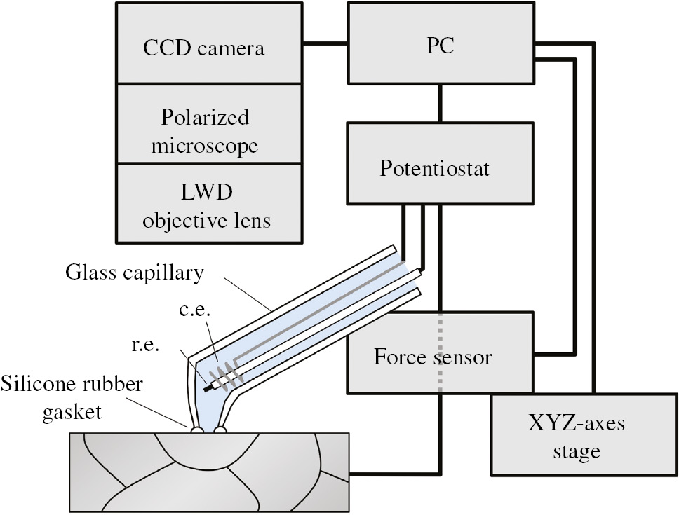
Schematic set-up of the MCC (Takabatake et al., 2014). Reproduced with permission from The Electrochemical Society.
Figure 2 shows the cross-section corrosion potential of aluminum-alloy brazing sheets consisting of an Al-Zn alloy sacrificial anode layer, an Al-Mn-Cu core layer, and an Al-Si brazing filler for a heat exchanger of an automobile in NaCl solution measured using an MCC with a diameter of 200 μm (Fushimi et al., 2008). The lateral resolution of the potential measurement of <20 μm was obtained by scanning the MCC on a tilted sample surface with an angle of 3.7° at steps of 200 μm. It is clear that heat treatment of the brazing sheet at 868 K results in a decrease in the corrosion potential of the outermost layer of the sacrificial anode layer. In a separate experiment, electron probe microanalysis (EPMA) revealed that the shift of the potential is due to the diffusion of Zn to the core layer.
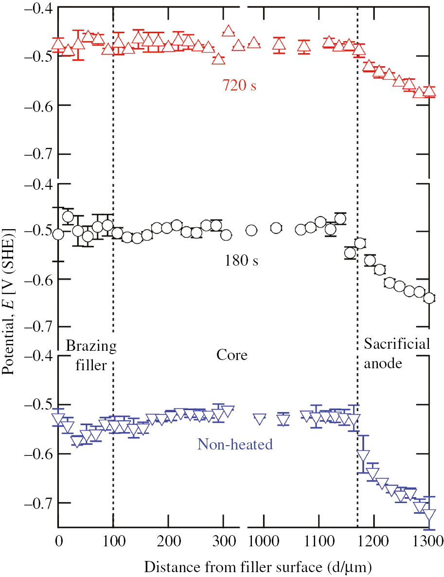
Corrosion potential profile at a cross-section of an aluminum-alloy brazing sheet when an MCC in 0.53 m NaCl solution with a diameter of 200 μm was scanned in steps of 200 μm with intervals of 30 s. The specimen was heated for 0, 180, or 720 s at 868 K (Fushimi et al., 2008).
An MCC has also been used to evaluate the electrochemical behavior of welded materials. Postweld heat treatment of low C-13% Cr martensitic stainless steel weld joints resulted in nobler and more stable potentials for the heat-affected zone in comparison to those in the as-welded condition (Hashizume et al., 2009). In a heat-tinted zone of stainless steel weldment, the depletion of Cr at the substrate surface and the formation of an Fe-rich oxide layer on the surface were mainly observed, although their distances from the welding bead were not the same and different corrosion behaviors were observed. The Cr depletion led to the lowering of the pitting potential due to the activation of the substrate steel, whereas the formation of an Fe-rich oxide layer led to preferential rusting in a cyclic corrosion test due to the deterioration of the surface protective layer (Kawano et al., 2015).
Figure 3 shows a double-logarithmic plot for current-time transients of iron single grains, {001}, {101} and {111} grains, measured using an MCC with a diameter of 70 μm when the iron electrode was polarized at 1.2 V (RHE) in 0.05 m H2SO4 (Takabatake et al., 2014). Initially, large currents flow due to the charging of the electric double layer and subsequent oxidation of the iron surface regardless of the crystallographic orientation. However, the current decreases to the magnitude of 10−6 A cm−2 during 3.6 ks polarization. Linear negative slopes of transients in the double-logarithmic plot after 10 s are about 0.65 due to the formation of an insulating oxide film and the dissolution of iron through the film. However, the slope after 500 s is dependent on the orientation, 0.56, 0.37, and 0.06 for {101}, {111} and {001} grains, respectively, and the current density at 3.6 ks becomes greater in the order of {101} < {111} < {001} grains. This order was also found for the charge transfer resistance measured in EIS with the MCC, although the donor density of the film on each grain showed a different order. It is thought that the electron transfer reaction (ETR) and/or ion transport reaction (ITR) through the passive film indirectly depend on the crystallographic orientation of the substrate iron single grains.
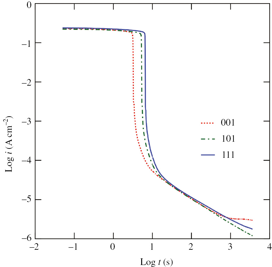
Double-logarithmic plot of current density-time transients of iron {001}, {101} and {111} single grains under the condition of potentiostatic polarization at 1.2 V (SHE) in 0.05 m H2SO4 (Takabatake et al., 2014). Reproduced with permission from The Electrochemical Society.
3 SECM imaging
SECM is a kind of scanning probe microscopy that was developed to image a specimen electrode surface immersed in a solution using a microelectrode as a probe (Engstrom et al., 1986; Bard et al., 1989, 1991). Figure 4 shows a schematic diagram of the SECM set-up. The principle of SECM and its applications to the electrochemistry of solution/material interphases have been described in detail elsewhere (Bard & Mirkin, 2001, 2012). In SECM, two independent working electrodes, a probe microelectrode and a specimen electrode, form parts of an electrochemical cell and are connected to a bipotentiostat together with counter and reference electrodes. The probe microelectrode, the diameter of which is less than 5×10⁻⁵ m, is capable of sensing potential in a potentiometric condition or detecting Faradaic current in an amperometric condition and of imaging the surface to show the distribution of electrochemically active species by repeated scanning across the specimen surface in a horizontal direction at a constant height.
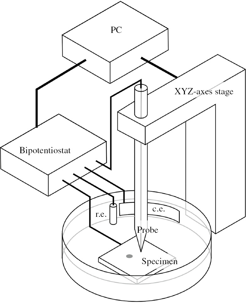
Schematic set-up of the SECM.
When the probe is an ion-selective microelectrode such as an oxide-covered tungsten electrode or a micropipette electrode, the tip of which is covered with a membrane containing an ionophore of a special ion (e.g. Cl−), SECM can potentiometrically monitor the distribution of the acidity of a solution or the concentration of Cl−. For example, the pH value in a pit of stainless steel was monitored with a tungsten microelectrode probe (Tanabe et al., 1998). A Cl−-sensing microelectrode was also used to monitor the accumulation of Cl− on stainless steel (Lin et al., 2000). The resolution of an image obtained using a potentiometric probe is not good as not only the probe size but also the diffusion of ions affect the resolution.
On the contrary, the amperometric application of a microelectrode probe enables the characterization of a specimen surface with a higher resolution. In this case, a probe electrode is generally polarized in a mass-transport control condition to detect electroactive species against the specimen surface. Because the current detected by the amperometric probe electrode is in proportion to the concentration of electroactive species in the vicinity of the electrode, the probe is capable of monitoring the electrochemical activity of the specimen surface if the probe is located close to the specimen surface.
Two types of electroactive species are detected in amperometric SECM: one is a product of electrolysis of the specimen surface or the solution itself and the other is an additive redox mediator in the solution. A corroding specimen can simply generate an electroactive species for the probe microelectrode, and the SECM system for detecting the species generated from the specimen is more straightforward than that for detecting additives in the solution. However, the SECM system for detecting corrosion products shows blurred images compared to those of SECM for detecting an additive mediator, as corrosion products can be generated from the entire specimen surface. The measurement for a relatively long time and/or the measurement of a specimen that generates a relatively large amount of corrosion products result in the accumulation of a large amount of electroactive species over the specimen surface. On the contrary, the redox mediator for the additives is generally chosen so as not to affect passivity or corrosion of the specimen and so as to be detected near the probe microelectrode. Thus, SECM images with a relatively high resolution are only obtained in the SECM system for detecting the corrosion products when there are reactive sites of small sizes and with a small density on a mostly inactive specimen surface.
Ferrous and ferric species generated from a polycrystalline iron electrode in pH 2.3 sulfate solution were monitored by a microelectrode of graphite-reinforced carbon for mechanical pencil lead as follows (Fushimi & Seo, 2001a):
The generation of ferrous and ferric species was strongly dependent on the potential of the iron electrode: ferrous and ferric species were generated in active and transpassive regions, respectively. It was also revealed that the active-passive transition led to the formation and removal of a salt film on the iron electrode surface. The detection of ferrous ions by an SECM probe was also used to monitor the behavior of the active dissolution of ferritic Fe-7.5Al-7Cr steel in H2SO4 (Lill, 2008; Lill et al., 2008). It is clear from Figure 5 that dissolution activity is strongly dependent on the crystallographic structure of the steel; ferrous ions are actively dissolved in the order of {101} grain < {111} grain < {001} grain < boundary of two grains < triple point of the boundary. A manner similar to that for detecting ferrous species was used to monitor the corrosion of polymer-coated steels (Souto et al., 2004). It was reported that the localized dissolution of a substrate metal occurred at defects of the coating.
Probe current image of an Fe7.5Al7Cr alloy electrode polarized at 0.42 V (SHE) in deaerated 0.5 m H2SO4 solution. For SECM imaging, the probe electrode, which was a carbon microdisk electrode with a diameter of 7 μm, was polarized at 1.14 V (SHE). The crystallographic orientation was identified using electron backscatter diffraction patterning (Lill, 2008).
Bromide ions in solution work not only as aggressive ions of a passive film on titanium but also as a redox mediator of SECM when film depassivation occurs at potentials higher than the bromine formation potential. After the breakdown of the passive film, pitting sites propagating on passive titanium were successfully monitored in H2SO4-containing bromide ions as follows (Casillas et al., 1993; Zhu & Williams, 1997):
Moreover, oxygen generated from polycrystalline titanium during anodic polarization in H2SO4 was used to monitor the heterogeneity of a passive film (Fushimi et al., 2000a):
It was revealed that needle-like titanium grains, which seemed to be twins such as 1012 grains, form relatively active oxide films and change from amorphous to crystalline oxide such as anatase. The compressive stress that accumulated in the oxide is thought to be one of the origins of the crystallization and the increasing electron transfer paths.
The corrosion of metals always involves a cathodic reaction of oxidants in the solution. The reduction of oxygen dissolved in a neutral solution or protons in an acidic solution leads to a decrease in the acidity of the solution.
There are two main ways for detecting solution acidity. One is a direct sensing of OH− or H2 generated from the specimen surface with the inverse reaction of the specimen. The detection of evolved hydrogen was used for the quantitative evaluation of galvanic corrosion of carbon steel coupled with magnetite in sulfate solution (Fushimi et al., 2002). Hydrogen evolution was also monitored from corroding Fe-3Si in diluted hydrochloric acid (Fushimi et al., 2007). The other way is the so-called “competition” mode in which the alkalinity of the solution rather than the acidity is sensed using a similar reaction on the probe electrode (Eckhard et al., 2006). The redox competition mode was also applied to monitor the localized reactive sites arising from a circular holiday on painted metal (Santana et al., 2010).
Another SECM mode with a mediator excels in the monitoring of a surface on which large reactive sites are located. In this mode, a redox mediator of electroactive species such as Fe(CN)64−, Ru(NH3)63+, or ferrocenemethanol (FcMeOH) is intentionally added to the solution to generate electroactive species for the local specimen surface. Because the reaction occurs only at a site of the specimen surface near the probe microelectrode, the microelectrode is capable of flowing a current depending on the electroactivity of a local site of interest. The lateral resolution of the SECM image measured with this mode is of the same magnitude as the diameter of the probe microelectrode at interelectrode distances less than half of the probe diameter.
Although most passive films have semiconductive properties, the ETR of a redox mediator on the film is due to the elastic exchange of electrons between occupied and unoccupied states in the oxide and electrolyte. The probability W of electron tunneling through the film strongly depends on the tunneling distance dt in the film as well as the energy barrier height Δε as follows (Schultze, 1978):
where m is an effective electron mass and ħ is the quantum Planck constant. In the case of an n-type semiconductive film, the indirect tunneling of electrons via donor levels in the film enables electrons to be conducted through the film, and the thickness of the electron tunneling is given as follows (Memming, 1983):
where e is the elementary charge, k is the Boltzmann constant, and LD,eff is an effective Debye length that is inversely proportional to the square root of the donor density ND as follows (Memming, 1983):
where ε0 and ε are the vacuum permittivity and relative permittivity of the film, respectively. Using the Marcus model for the distribution functions in the electrolyte and the band model for the electron states in the oxide, the redox current is finally given as follows (Schultze & Hassel, 2003):
where Dvac,ox and Dred,el are the densities of empty sites in the oxide and occupied states in the electrolyte, respectively. Thus, the donor density in the film strongly affects the redox current through the film.
The heterogeneities of passive films formed on iron (Fushimi et al., 1999), titanium (Fushimi et al., 2000a), and their base alloys (Zhu et al., 2007; Lill et al., 2008) were observed by the detection of the ETR of the redox mediator through the film. The heterogeneous distribution of a passive film was found to be dependent on the metallographic structure of the substrate on which the film was formed. A passive film formed on iron allowed the ETR frequently in the order of {001} < {101} ≤ {111} grains (Fushimi et al., 1999). The hexagonal close-packed structure of a titanium substrate showed a clear anisotropic oxide formation, in which 0001 grains form an oxide allowing the ETR more than that by other grains of XXX0. The ETR is accelerated with an increase in the donor density of a semiconductive film (Kudelka et al., 1996). Because the basal plane is composed of close-packed atoms, a relatively large donor density in the oxide formed on the basal plane seems to be due to the accumulation of a larger compressive stress than that of other planes (Fushimi et al., 2000a) and the presence of energetically favorable sites for structural transformation and impurity species (Matykina et al., 2008). These lead to the modification of the local electronic properties of the oxide, an increase in donor density, and then the enhancement of the ETR through the film.
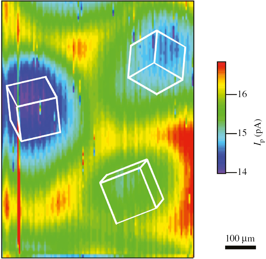
The anisotropic oxide formation on polycrystalline materials was also observed on some alloys. In the case of ferritic FeAlCr alloy (Figure 6; Lill et al., 2008), the conductivity of the oxide formed was in the order of {101} grain < {111} grain < {001} grain. Furthermore, the film formed on grain boundaries had higher ETR reactivity than that of the film formed on single grains, and the film formed on the triple point of a boundary showed higher reactivity than that of the film formed on the boundary of two grains. Oxide films formed on grain boundaries of ASTM Grade 2 titanium also showed a distribution of reactivity that was higher than that of films formed on single grains due to the accumulation of defects in the film and the presence of Fe impurities from the substrate (Zhu et al., 2007). On the contrary, a passive film formed on carbon steel in pH 8.4 borate buffer showed a distribution of ETR reactivity that depended on the metallographic structure of the substrate as shown in Figure 7. It is clear that the film formed on eutectoid perlite is more active than that formed on primary ferrite. Imaging Raman spectroscopy revealed the enrichment of amorphous carbon and the depletion of magnetite on the perlite. The differences in the chemical structure and defect density of the film seem to give rise to the heterogeneous distribution of reactivity.
![Figure 6:
Probe current image obtained in deaerated pH 8.4 borate buffer containing 0.1 m K4[Fe(CN)6] after the Fe7.5Al7Cr sample had been passivated in 0.5 m H2SO4 solution for 3.6 ks at 0.7 V. The probe of the platinum microelectrode with a diameter of 10 μm was polarized at 1.2 V, whereas the sample was polarized at 0 V (Lill et al., 2008).](/document/doi/10.1515/corrrev-2017-0050/asset/graphic/j_corrrev-2017-0050_fig_032.jpg)
Probe current image obtained in deaerated pH 8.4 borate buffer containing 0.1 m K4[Fe(CN)6] after the Fe7.5Al7Cr sample had been passivated in 0.5 m H2SO4 solution for 3.6 ks at 0.7 V. The probe of the platinum microelectrode with a diameter of 10 μm was polarized at 1.2 V, whereas the sample was polarized at 0 V (Lill et al., 2008).
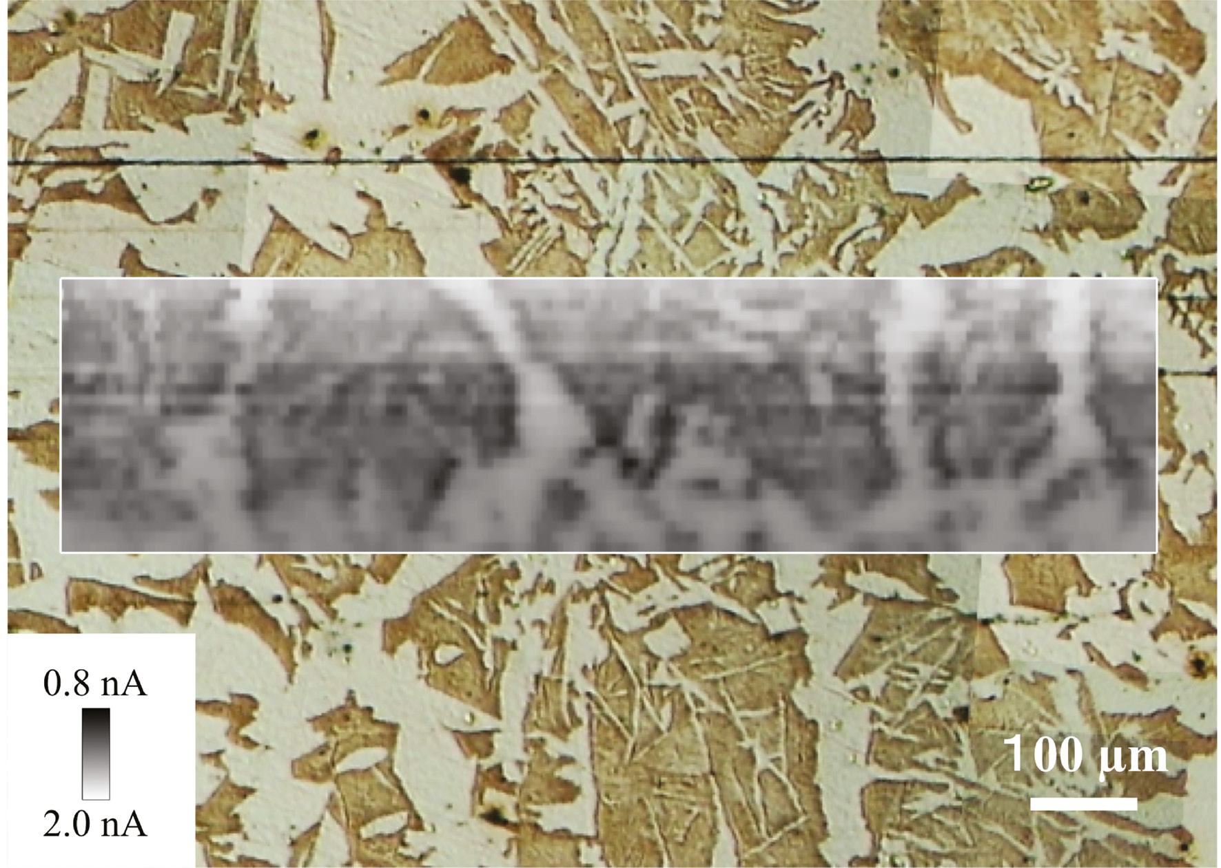
Optical microscopic image of S45C carbon steel. (Inset) Probe current image of S45C carbon steel obtained in pH 8.4 buffer containing 1 mm FcMeOH after passivating in pH 8.4 borate buffer at 1 V (SHE) for 3.6 ks. The carbon steel specimen electrode and the platinum microelectrode with a diameter of 10 μm were polarized at 0.1 and 0.8 V (SHE), respectively.
4 Liquid-phase ion gun (LPIG)
SECM is a useful tool not only for monitoring a passive or corroding surface but also for studying corrosion events such as passive film breakdown. The use of an LPIG is an SECM application in which the microelectrode of the SECM probe is used as a source for generating aggressive species toward the specimen surface to induce localized electrochemical or chemical reactions on the surface. It is well known that halide ions play a role in the initiation of localized corrosion such as pitting. It is expected that the application of an LPIG to passive materials will be useful for investigating the mechanism and kinetics of localized corrosion.
The reduction of trichloroacetic acid in an aqueous solution using an inert platinum microelectrode leads to the generation of chloride ions as follows:
It has been used to initiate the formation of pits at a site of interest on iron (Still & Wipf, 1997) or stainless steel (Wipf, 1994) and to investigate the susceptibility of a passive film to the localized breakdown and initiation of localized corrosion. The breakdown was found to depend strongly on the passivation potential and the site of chloride ion generation. Because the reduction of trichloroacetic acid also involves a change of solution acidity as well as the generation of chloride ions, it is difficult to analyze the mechanism and kinetics of passive film breakdown.
A silver chloride-coated silver microelectrode can be used as a source of chloride ions in a solution without changing the solution acidity as follows:
It was used to induce localized corrosion on passivated iron (Fushimi & Seo, 2001b; Fushimi et al., 2000b) and stainless steel (Aouina et al., 2011). Figure 8 shows the transients of currents flowing through an iron electrode and a silver chloride-coated silver microelectrode in pH 6.5 borate solution when the iron electrode was passivated at 0.7 V [standard hydrogen electrode (SHE)] for 3.6 ks and polarized at the same potential and the microelectrode potential was changed to −0.1 V (SHE; Fushimi et al., 2000b). A cathodic current flowing through the microelectrode basically corresponds to the generation of chloride ions from the microelectrode, whereas an anodic current is dependent on the passive property of the iron electrode surface. A negligibly small anodic current of the iron electrode means that the surface is in a fine passive state, whereas a significantly large anodic current flowing through the iron electrode is due to the depassivation and corrosion of the iron electrode. If ferric ions are generated as corrosion products of the iron electrode and are present in the vicinity of the microelectrode, the polarization of the microelectrode at −0.1 V (SHE) enables the microelectrode to detect ferric ions by Eq. (2). On the contrary, although an iron electrode allows the flowing anodic current to dissolve iron as ferrous ions, the ferrous ions generated do not lead to a change in the current flowing through the microelectrode as the microelectrode in this polarization condition is inert for ferrous species. The generation of chloride ions from the microelectrode finally resulted in the formation of a concavity on the passivated iron surface due to localized corrosion. The experimental behavior for the initiation of localized corrosion could be classified from the transients into the following three domains: (I) induction and accumulation of chloride ions on local sites of the passive film, (II) breakdown of the passive film at a site with highly concentrated chloride ions, and (III) propagation of localized corrosion or, at lower potentials, repassivation. It is notable that, in the second domain, the passive film of iron dissolved as ferrous ions in the local environment with highly concentrated chloride ions, as the cathodic polarization of the microelectrode for the generation of chloride ions also enabled the detection of ferric species in the vicinity of the microelectrode and positive feedback of the oxidation of ferrous ions on the iron electrode, and the reduction of ferric ions on the microelectrode was observed. The monitoring of intermediate species of ferric species during corrosion initiation is important. The LPIG technique has been used for the evaluation of localized corrosion resistance of steels (Fushimi & Seo, 2001b). The induction period for the initiation of film breakdown was dependent on the ETR activity of the passive film.
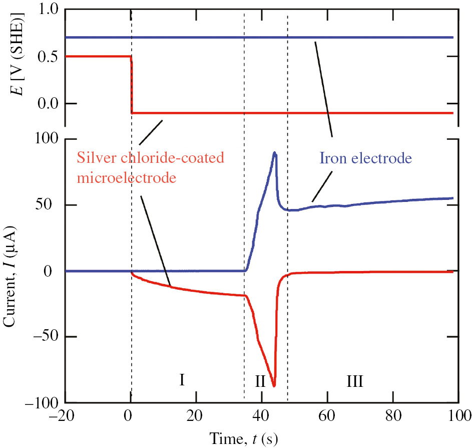
Transients of currents flowing through an iron electrode and a silver chloride-coated microelectrode in pH 6.5 borate solution when the iron electrode was polarized at 0.7 V and the microelectrode potential was changed to −0.1 V (Fushimi et al., 2000b). Reproduced with permission from The Electrochemical Society.
Similarly, a silver sulfide-coated silver microelectrode was used to generate HS− in an aqueous solution:
Using the LPIG generating HS−, the localized sulfidation of silver (Lee et al., 2015a) and that of stainless steel (Lee et al., 2015b, 2016) were investigated and the quantitative mechanism and kinetics for the sulfidation were discussed.
In the case of small interelectrode distances, electrolysis by the microelectrode leads to a change in the acidity of the solution in the vicinity of not only the microelectrode but also the specimen as follows:
The anodic polarization of the inert platinum microelectrode can initiate the localized breakdown of the passive film due to the acidification of the local solution near the passive materials. It is also expected to be effective for investigating the localized corrosion of the materials.
5 In situ ellipsomicroscopy for studying a passive film
Ellipsometry detects the change in the elliptical shape of polarized light occurring by reflection at a solid surface and enables the observation of a surface film with a thickness of subnanometer. Because visible polarized light can penetrate an aqueous solution, ellipsometry using visible polarized light is effective for the in situ detection of passive oxide films on various materials. Ellipsomicroscopy is an application of ellipsometry for surveying the microscopic heterogeneity of passive oxide films by combining with an optical microscope. Figure 9 shows a schematic view of an in situ ellipsomicroscope (Fushimi et al., 2014), in which the measurement is performed based on manual null-method ellipsometry (Azzum & Bashara, 1987) with an optical arrangement of polarizer P – phase compensator C – reflection sample surface S – analyzer A. The light from individual positions on the reflection surface is focused and the surface is imaged by the CCD or CMOS camera. In the null method, C is fixed at the azimuth of −45° and the extinction state of the reflected light intensity on the CMOS camera is sought by manually controlling the azimuths of P and A. If heterogeneity of the surface oxide in thickness and optical property emerges, the extinction state is not kept on the whole surface, but places having different thicknesses and optical properties deviate from the extinction and brighten slightly. A place with larger deviation exhibits greater brightness.
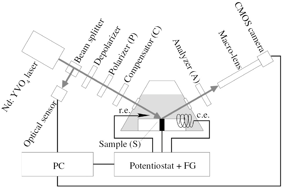
Schematic set-up of the in situ ellipsomicroscope (Fushimi et al., 2014).
The heterogeneous growth of titanium passive oxide in 0.05 m H2SO4 is shown in Figure 10, with the potential being increased from the open circuit potential (OCP) to 8 V (SSE) at a sweep rate 5 mV s−1 (Fushimi et al., 2014). During the potential sweep in the positive direction, the PCSA optical configuration was kept constant, which was initially controlled in the extinction state at the OCP. When oxide growth occurs with an increase in potential, the brightness gradually increases. The reflected light intensities were averaged at all pixels on the CMOS camera. The average reflection intensity increases with the increase in potential, indicating a gradual growth of the oxide film. However, the brightness is not homogeneous, and the heterogeneity of the brightness is larger at potentials higher than 2 V. It can be seen that the differences in thickness and optical property among the sites are increased at higher potentials. The sizes of individual sites can be seen to be several 10 μm in diameter, which may be an average diameter of the surface grain. The quantitative distribution of thickness and optical property is strongly dependent on the crystallographic orientation of the substrate as well as the potential applied as shown in Figure 11 (Fushimi et al., 2014), which is an inverse pole figure of the intensity measured in the ellipsomicroscopic image of the oxide film formed on polycrystalline titanium at 4 V in H2SO4. The crystallographic orientation of each place on the substrate titanium was measured by electron backscatter diffraction patterning, and the intensity was normalized by the highest intensity in the ellipsomicroscopic image. Assuming that the intensity corresponds to the film thickness, the 0001 grain is covered with a thinner film than the other grains such as XXX0 grains. This is in good agreement with the results obtained using other microelectrochemical techniques as described above.
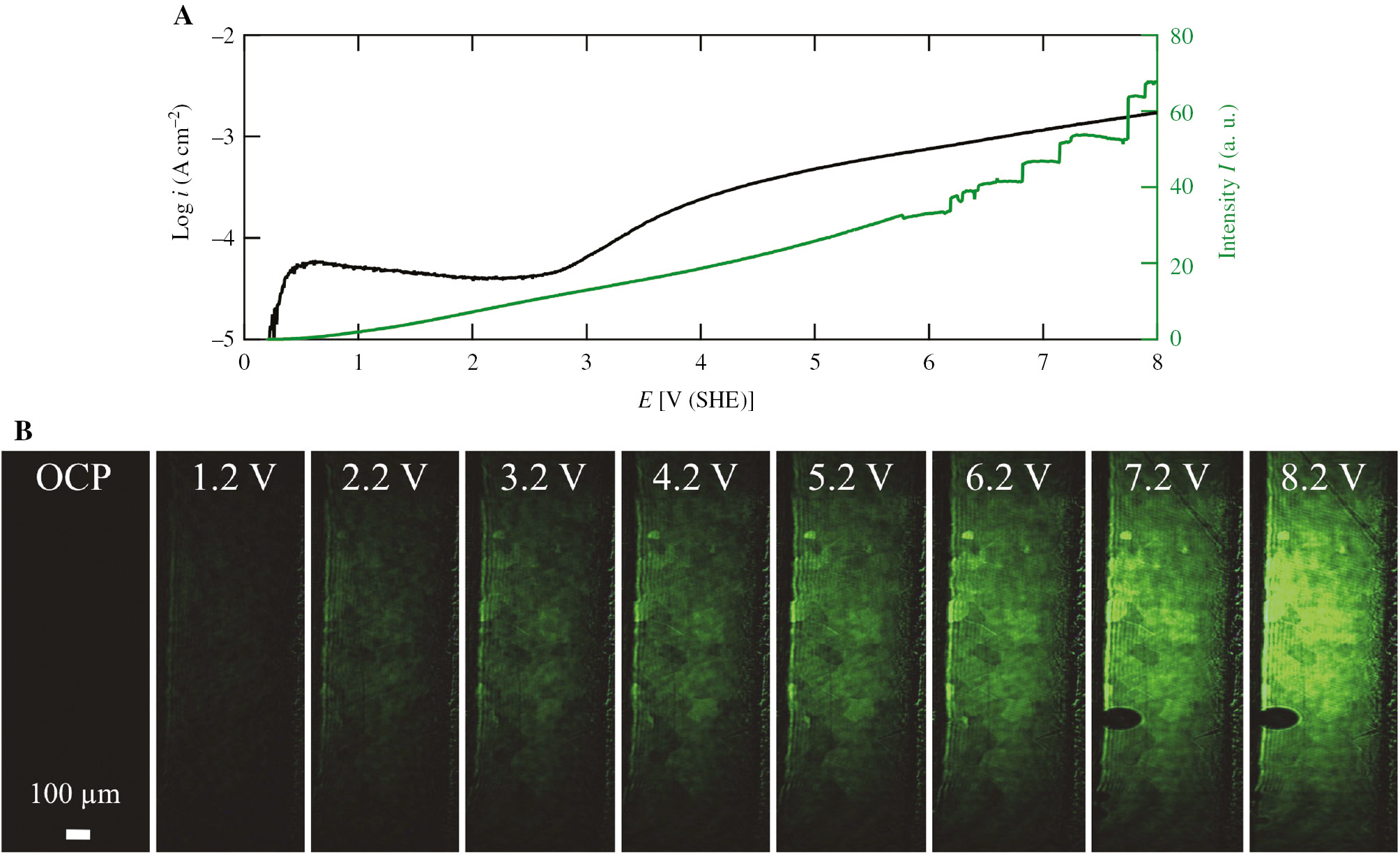
(A) Anodic current and total intensity of an ellipsomicroscopic image as a function of potential during the dynamic polarization of titanium in 0.05 m H2SO4 solution and (B) ellipsomicroscopic images at the initial null condition at OCP and at potentials of 1.2, 2.2, 3.2, 4.2, 5.2, 6.2, 7.2 and 8.2 V (SHE). The potential was swept at a rate of 5 mV s−1 (Fushimi et al., 2014).
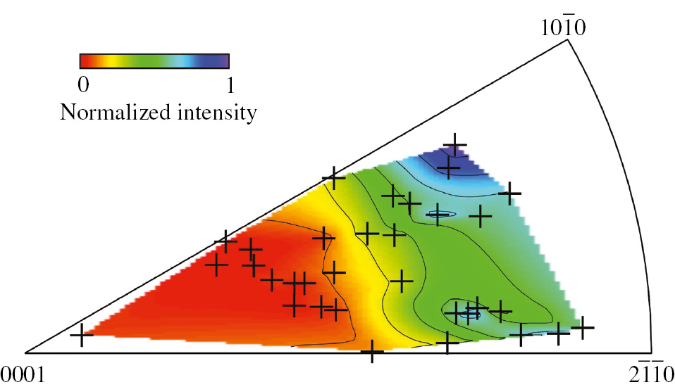
Contour map of light intensity in the ellipsomicroscopic image for an oxide film formed at 4 V (Figure 6E) drawn on an inversed pole figure. The light intensity at each spot in the image was normalized by the largest intensity and marked with a cross in the map. The crystallographic orientations of individual crystallites were obtained by electron backscatter diffraction patterning (Fushimi et al., 2014).
Ellipsomicroscopy can be used for imaging an oxide film not only during formation but also during degradation. The photoelectrochemical degradation of an oxide film on titanium (Fushimi et al., 2015) and oxide breakdown occurring in a solution containing bromide ions (Fushimi et al., 2014) were also observed using in situ ellipsomicroscopy. Figure 12 shows a current transient during the dynamic polarization of titanium in 0.05 m H2SO4 containing 1.5 m KBr. At 1081.40 s (5.37 V), the titanium oxide allows a catastrophically large current flow due to the breakdown of the passivity on titanium by the attack of bromide ions. Immediately before the large current flow indicating the initiation of passivity breakdown, an increase in current was detected from 1081.07 s. Simultaneously, a dark spot in the ellipsomicroscopic image appeared (1081.07 s), and the area of the dark spot increased with time. The dark spot initially appearing is due to the thinning or change of the oxide. Ellipsomicroscopy with a sufficiently rapid response enables the identification of the site at which the localized breakdown of the film is initiated. The Auger electron spectroscopy measurement of the specimen surface before the large current flow revealed a small amount of Br on the surface of the dark spot. It was thought that the locally absorbed bromide ions caused the thinning and breakdown of the oxide film and the ETR through the locally thin oxide film, and finally depassivation was induced.
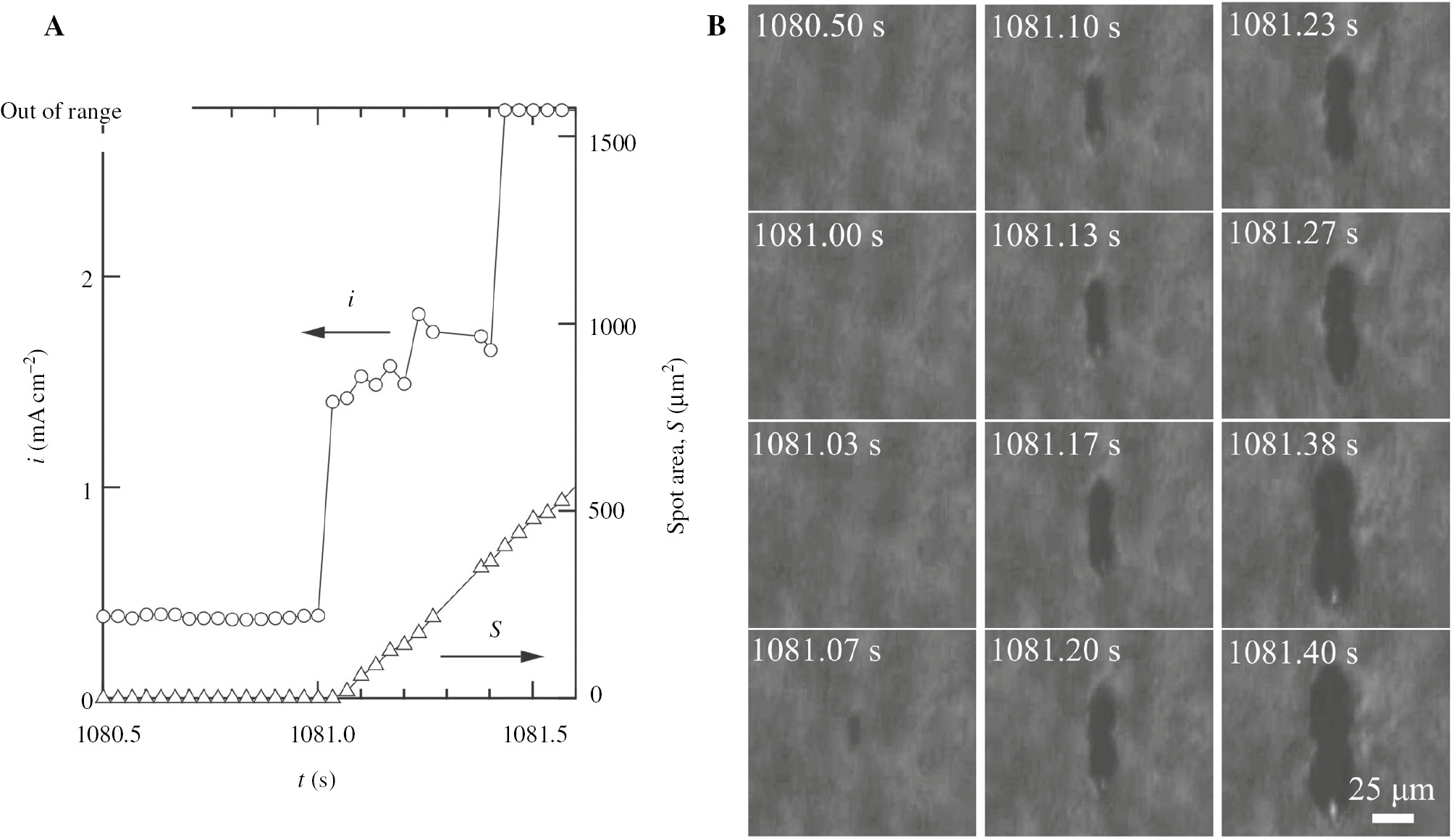
(A) Time variations of current and area of the spot that appeared in the image during the dynamic polarization of titanium with a sweep rate of 5 mV s−1 in 0.05 m H2SO4 solution containing 1.5 m KBr and (B) ellipsomicroscopic images of the titanium surface. The current was measured with a fixed detection range. The spot area was estimated after binominal image filtering (Fushimi et al., 2014).
6 Summary
Because corrosion reactions are mainly electrochemical reactions, the behaviors of passivity and corrosion of materials have recently been investigated using electrochemical methods. Microelectrochemical methods have been used to study the passivity and corrosion in detail. An MCC can be used to investigate a site of interest on a material with various electrochemical methods. SECM not only provides images of a corroding surface or passive film on a material but also shows the reactivity of the surface against localized aggressive environments. In situ ellipsomicroscopy enables the monitoring of the heterogeneous growth or degradation of a thin passive film. It is also useful for finding a depassivation site before a film breakdown is initiated. These microelectrochemical methods are useful for monitoring the heterogeneous distribution of a passive film and for investigating the heterogeneity of the passivity of materials.
About the authors

Koji Fushimi graduated from the Graduate School of Engineering, Hokkaido University (Japan), in 1993. He then worked at Toyo Seikan Co., Ltd. (Japan). He joined Hokkaido University as a research assistant of Prof. Masahiro Seo in 1996 and obtained his PhD in applied chemistry at Hokkaido University in 2001, receiving a Young Researcher Award from the Japan Society of Corrosion Engineering (JSCE) in 2002. From 2002 to 2003, he held a postdoctoral research position at the Max Planck Institute for Iron Research (Germany). Since 2008, when he became an Associate Professor at Hokkaido University, he has studied the passivity and corrosion of materials with microelectrochemical methods, receiving The Best Year’s Paper Award from JSCE in 2017.

Ryogo Nakagawa graduated from the Faculty of Engineering, Hokkaido University (Japan) in 2017. Since then, he has studied at the Graduate School of Chemical Sciences and Engineering, Hokkaido University.
References
Aouina N, Balbaud-Célérier F, Huet F, Joiret S, Rouillard F, Vivier V. Single pit initiation on 316L austenitic stainless steel using scanning electrochemical microscopy. Electrochim Acta 2011; 56: 8589–8596.10.1016/j.electacta.2011.07.044Search in Google Scholar
Azzum RMA, Bashara NM. Ellipsometry and polarized light. Amsterdam, The Netherlands: North-Holland Personal Library, 1987.10.1016/S0003-2670(00)82849-4Search in Google Scholar
Bard AJ, Mirkin MV. Scanning electrochemical microscopy. New York, NY: Marcel Dekker, 2001.10.1201/9780203910771Search in Google Scholar
Bard AJ, Mirkin MV. Scanning electrochemical microscopy, 2nd ed., New York, NY: CRC Press, 2012.10.1201/b11850Search in Google Scholar
Bard AJ, Fan FRF, Kwak J, Lev O. Scanning electrochemical microscopy. Introduction and principles. Anal Chem 1989; 61: 132–138.10.1021/ac00177a011Search in Google Scholar
Bard AJ, Fan FRF, Pierce DT, Unwin PR, Wipf DO, Zhou F. Chemical imaging of surfaces with the scanning electrochemical microscope. Science 1991; 254: 68–74.10.1126/science.254.5028.68Search in Google Scholar PubMed
Casillas N, Charlebois SJ, Smyrl WH, White HS. Scanning electrochemical microscopy of precursor sites for pitting corrosion on titanium. J Electrochem Soc 1993; 140: L142–L145.10.1149/1.2220897Search in Google Scholar
Eckhard K, Chen X, Turcu F, Schuhmann W. Redox competition mode of scanning electrochemical microscopy (RC-SECM) for visualisation of local catalytic activity. Phys Chem Chem Phys 2006; 8: 5359–5365.10.1039/b609511aSearch in Google Scholar PubMed
Engstrom RC, Weber M, Wunder DJ, Burgess R, Winquist S. Measurements within the diffusion layer using a microelectrode probe. Anal Chem 1986; 58: 844–848.10.1021/ac00295a044Search in Google Scholar
Fushimi K, Seo M. An SECM observation of dissolution distribution of ferrous or ferric ion from a polycrystalline iron electrode. Electrochim Acta 2001a; 46: 121–127.10.1016/S0013-4686(01)00557-6Search in Google Scholar
Fushimi K, Seo M. Initiation of a local breakdown of passive film on iron due to chloride ions generated by a liquid-phase ion gun. J Electrochem Soc 2001b; 148: B450–B456.10.1149/1.1407832Search in Google Scholar
Fushimi K, Azumi K, Seo M. Evaluation of heterogeneity in thickness of passive films on pure iron by scanning electrochemical microscopy. ISIJ Int 1999; 39: 346–351.10.2355/isijinternational.39.346Search in Google Scholar
Fushimi K, Okawa T, Seo M. A scanning electrochemical microscopic observation of heterogeneous oxygen evolution on a polycrystalline titanium during anodic oxidation. Electrochemistry 2000a; 68: 950–954.10.5796/electrochemistry.68.950Search in Google Scholar
Fushimi K, Azumi K, Seo M. Liquid-phase ion gun for local breakdown of the passive film on iron. J Electrochem Soc 2000b; 147: 552–557.10.1149/1.1393231Search in Google Scholar
Fushimi K, Yamamuro T, Seo M. Hydrogen generation from a single crystal magnetite coupled galvanically with a carbon steel in sulfate solution. Corros Sci 2002; 44: 611–623.10.1016/S0010-938X(01)00093-2Search in Google Scholar
Fushimi K, Lill KA, Habazaki H. Heterogeneous hydrogen evolution on corroding Fe-3 at.% Si surface observed by scanning electrochemical microscopy. Electrochim Acta 2007; 52: 4246–4253.10.1016/j.electacta.2006.12.006Search in Google Scholar
Fushimi K, Yamamoto S, Ozaki R, Habazaki H. Cross-section corrosion-potential profiles of aluminum-alloy brazing sheets observed by the flowing electrolyte scanning-droplet-cell technique. Electrochim Acta 2008; 53: 2529–2537.10.1016/j.electacta.2007.10.044Search in Google Scholar
Fushimi K, Takabatake Y, Nakanishi T, Hasegawa Y. Microelectrode techniques for corrosion research of iron. Electrochim Acta 2013; 113: 741–747.10.1016/j.electacta.2013.03.021Search in Google Scholar
Fushimi K, Kurauchi K, Tamamoto Y, Nakanishi T, Hasegawa Y, Ohtsuka T. Growth and degradation of an anodic oxide film on titanium in sulphuric acid observed by ellipso-microscopy. Electrochim Acta 2014; 144: 56–63.10.1016/j.electacta.2014.08.082Search in Google Scholar
Fushimi K, Kurauchi K, Ikeyama I, Kitagawa Y, Nakanishi T, Hasegawa Y, Ueda M, Ohtsuka T. Titanium surface anodized under UV light irradiation observed by ellipso-microscopy. J Sol Electrochem 2015; 19: 3579–3588.10.1007/s10008-015-2753-7Search in Google Scholar
Hashizume S, Nakayama T, Sakairi M, Fushimi S. Effect of PWHT on electrochemical behavior of low C-13%Cr welded joints with the use of a solution flow type micro-droplet cell. In: Corrosion 2009, Atlanta, GA: NACE International, 2009; 09089: 1–13.10.5006/C2009-09089Search in Google Scholar
Hassel AW, Lohrengel MM. The scanning droplet cell and its application to structured nanometer oxide films on aluminium. Electrochim Acta 1997; 42: 3327–3333.10.1016/S0013-4686(97)00184-9Search in Google Scholar
Kawano T, Ishii T, Kajiyama H, Kimura M, Fushimi K. Microelectrochemistry at heat-tinted zone of stainless steel weldment. Zairyo-to-Kankyo 2015; 64: 552–557.10.3323/jcorr.64.552Search in Google Scholar
Kudelka S, Michaelis A, Schultze JW. Effect of texture and formation rate on ionic and electronic properties of passive layers on Ti single grains. Electrochim Acta 1996; 41: 863–870.10.1016/0013-4686(95)00375-4Search in Google Scholar
Lee JS, Fushimi K, Kitagawa Y, Nakanishi T, Hasegawa Y. Development of a liquid-phase ion gun and its application for sulfidation of silver surface. J. Electrochem Soc 2015a; 162: C115–C120.10.1149/2.0071504jesSearch in Google Scholar
Lee JS, Kitagawa Y, Nakanishi T, Hasegawa Y, Fushimi K. Effect of hydrogen sulfide ions on the passive behavior of type 316L stainless steel. J Electrochem Soc 2015b; 162: C685–C692.10.1149/2.0861512jesSearch in Google Scholar
Lee JS, Kitagawa Y, Nakanishi T, Hasegawa Y, Fushimi K. Passivation behavior of type-316L stainless steel in presence of hydrogen sulfide ions generated from a local anion generating system. Electrochim Acta 2016; 220: 304–311.10.1016/j.electacta.2016.10.124Search in Google Scholar
Lill KA. Investigations with scanning electrochemical microscopy. Electrochemical investigations on the corrosion properties of new classes of light weight steels. Dissertation. Düsseldorf, Germany, 2008: 83–112.Search in Google Scholar
Lill KA, Fushimi K, Seo M, Hassel AW. Reactivity imaging of a passive ferritic FeAlCr steel. J Appl Electrochem 2008; 38: 1339–1345.10.1007/s10800-008-9564-9Search in Google Scholar
Lin CJ, Du RG, Nguyen T. In-situ imaging of chloride ions at the metal/solution interface by scanning combination microelectrodes. Corrosion 2000; 56: 41–47.10.5006/1.3280521Search in Google Scholar
Lohrengel MM. Interface and volume effects in biological cells and electrochemical microcells. Electrochim Acta 1997; 42: 3265–3271.10.1016/S0013-4686(97)00177-1Search in Google Scholar
Matykina E, Arrabal R, Skeldon P, Thompson GE, Habazaki H. Influence of grain orientation on oxygen generation in anodic titania. Thin Solid Films 2008; 516: 2296–2305.10.1016/j.tsf.2007.08.104Search in Google Scholar
Memming M. Processes at semiconductor electrodes. In: Bockris JOM, Conway BE, Yeager E, Khan SUM, White RE, editors. Comprehensive treatise of electrochemistry. Vol. 7. New York, NY: Plenum Press, 1983: 529–592.10.1007/978-1-4613-3584-9_9Search in Google Scholar
Santana JJ, González-Guzmán J, Fernández-Mérida L, González S, Souto RM. Visualization of local degradation processes in coated metals by means of scanning electrochemical microscopy in the redox competition mode. Electrochim Acta 2010; 55: 4488–4494.10.1016/j.electacta.2010.02.091Search in Google Scholar
Sato N. The passivity of metals and passivating films. In: Frankenthal RP, Kruger J, editors. Passivity of metals. Princeton, NJ: The Electrochemical Society, 1978: 29–58.Search in Google Scholar
Sato N. Some concepts of corrosion fundamentals. Corros Sci 1987; 27: 421–433.10.1016/0010-938X(87)90086-2Search in Google Scholar
Sato N, Okamoto G. Electrochemical passivation of metals. In: Bockris JOM, Conway BE, Yeager E, White RE, editors. Comprehensive treatise of electrochemistry, Vol. 4. New York, NY: Plenum Press, 1981: 193–241.Search in Google Scholar
Schneider M, Schroth S, Schilm J, Michaelis A. Micro-EIS of anodic thin oxide films on titanium for capacitor applications. Electrochim Acta 2009; 54: 2663–2671.10.1016/j.electacta.2008.11.003Search in Google Scholar
Schultze JW. Electron transfer reactions on passive films. In: Frankenthal RP, Kruger J, editors. Passivity of metals. Princeton, NJ: The Electrochemical Society, 1978; 82–101.Search in Google Scholar
Schultze JW, Hassel AW. 3.2 Passivity of metals, alloys, and semiconductors. In: Bard AJ, Frankel GS, editors. Encyclopedia of electrochemistry, Vol. 4. Weinheim, Germany: Wiley-VCH, 2003; 216–270.10.1002/9783527610426.bard040302Search in Google Scholar
Souto RM, González-Garcia Y, González S, Burstein GT. Damage to paint coatings caused by electrolyte immersion as observed in situ by scanning electrochemical microscopy. Corros Sci 2004; 46: 2621–2628.10.1016/j.corsci.2004.06.002Search in Google Scholar
Still JW, Wipf DO. Breakdown of the iron passive layer by use of the scanning electrochemical microscope. J Electrochem Soc 1997; 144: 2657–2665.10.1149/1.1837879Search in Google Scholar
Suter T, Böhni H. A new microelectrochemical methods to study pit initiation on stainless steels. Electrochim Acta 1997; 42: 3275–3280.10.1016/S0013-4686(70)01783-8Search in Google Scholar
Suter T, Böhni H. Microelectrodes for studies of localised corrosion processes. Electrochim Acta 1998; 43: 2843–2849.10.1016/S0013-4686(98)00025-5Search in Google Scholar
Takabatake Y, Fushimi K, Nakanishi T, Hasegawa Y. Grain-dependent passivation of iron in sulfuric acid solution. J Electrochem Soc 2014; 161: C594–C600.10.1149/2.0901414jesSearch in Google Scholar
Tanabe H, Togashi K, Misawa T, Mudali UK. In situ pH measurements during localised corrosion of type 316N stainless steel using scanning electrochemical microscopy. J Mater Sci Lett 1998; 17: 551–553.10.1023/A:1006517503049Search in Google Scholar
Wipf DO. Initiation and study of localized corrosion by scanning electrochemical microscopy. Colloid Surf A Physicochem Eng Asp 1994; 93: 251–261.10.1016/0927-7757(94)02872-9Search in Google Scholar
Zhu Y, Williams DE. Scanning electrochemical microscopic observation of a precursor state to pitting corrosion of stainless steel. J Electrochem Soc 1997; 144: L43–L45.10.1149/1.1837487Search in Google Scholar
Zhu R, Nowierski C, Ding Z, Noël JJ, Shoesmith DW. Insights into grain structures and their reactivity on grade-2 Ti alloy surfaces by scanning electrochemical microscopy. Chem Mater 2007: 19: 2533–2543.10.1021/cm070023dSearch in Google Scholar
©2018 Walter de Gruyter GmbH, Berlin/Boston
Articles in the same Issue
- Frontmatter
- In this issue
- Editorial
- Special issue on Recent advances in corrosion science: celebrating the 90th birthday of Professor Norio Sato
- Reviews
- Some microelectrochemical methods for the investigation of passivity and corrosion
- Inhibition effect of underpotential deposition of metallic cations on aqueous corrosion of metals
- Role of anodic oxide films in the corrosion of aluminum and its alloys
- Development of novel surface treatments for corrosion protection of aluminum: self-repairing coatings
- Mini review
- High-temperature corrosion resistance of SiO2-forming materials
- Original articles
- Cyclic carburization-oxidation behavior of Hastelloy-X at 1000°C
- Growth of passive oxide films on iron and titanium under non-stationary state
- Influence of metal cations on inhibitor performance of gluconates in the corrosion of mild steel in fresh water