
Community news
-
Andreas F. Thoß


Introducing Microscopy Australia
World-class research infrastructure drives world-class research, which in turn drives discovery and innovation. Microscopy Australia, enabled by the National Collaborative Research Infrastructure Strategy, is ensuring that researchers are able to access high-end platforms with dedicated platform experts, placed in strategic locations, throughout the nationwide network.
Microscopy Australia, established in 2007, provides open-access, university-based microscopy and microanalysis facilities around Australia. This stable and successful research infrastructure is united by values of collaboration, accessibility, excellence and innovation. This means that researchers from any institution or industry can use any of the instruments across our network. Access is subsidised for publicly funded researchers at rate that is affordable on most Australian research grants, and reduced rates are offered to international researchers. Microscopy Australia’s facilities also provide training and fee-for-service analysis (for more information: https://micro.org.au/academic/access-guide/).
Microscopy Australia has invested in four state-of-the-art technologies to underpin Australia’s future research and industry advances:
atomic scale microscopy
cryo-electron microscopy
correlative microscopy techniques
high-sensitivity microanalytical tools.
Located all around Australia, our facilities are at:
The University of Queensland, the Centre for Microscopy and Microanalysis
The University of Sydney, Sydney Microscopy and Microanalysis
UNSW Sydney, Electron Microscope Unit
Australian National University, Centre for Advanced Microscopy and the CTlab
Monash University, the Monash Centre for EM and the Ramaciotti Centre for Cryo-EM
The University of Western Australia, the Centre for Microscopy, Characterisation and Analysis
The University of Adelaide, Adelaide Microscopy
The University of South Australia, Future Industries Institute
Flinders University, Flinders Microscopy and Microanalysis.
Microscopy Australia is connected to an additional nine affiliated linked laboratories, increasing the national skilled microscopy workforce by providing professional development opportunities, including Staff Shadowing visits and training programs.
With the help of our experts, Microscopy Australia provides free online microscopy training through MyScope. Technique Finder helps users who aren’t sure which microscopy technique is right for their project to select the one that can help them best. Our YouTube channel includes a wide range of videos, from theory through to hands on demonstrations. Keep an eye on our Twitter for events and career opportunities. Check out our public engagement initiatives, including two exhibitions and ‘MyScope Explore’ here (https://micro.org.au/micro-wow/curious-minds/).
Microscopy Australia is enabled by the Australian federal government’s National Collaborative Research Infrastructure Strategy, in partnership with state government and institutional support.
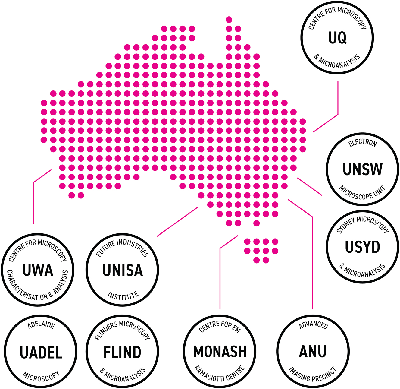
Microscopy Australia hosts nine facilities at different locations across the continent.
Array Tomography Workshop Munich 2024
Munich, Germany | November 4 – 8, 2024
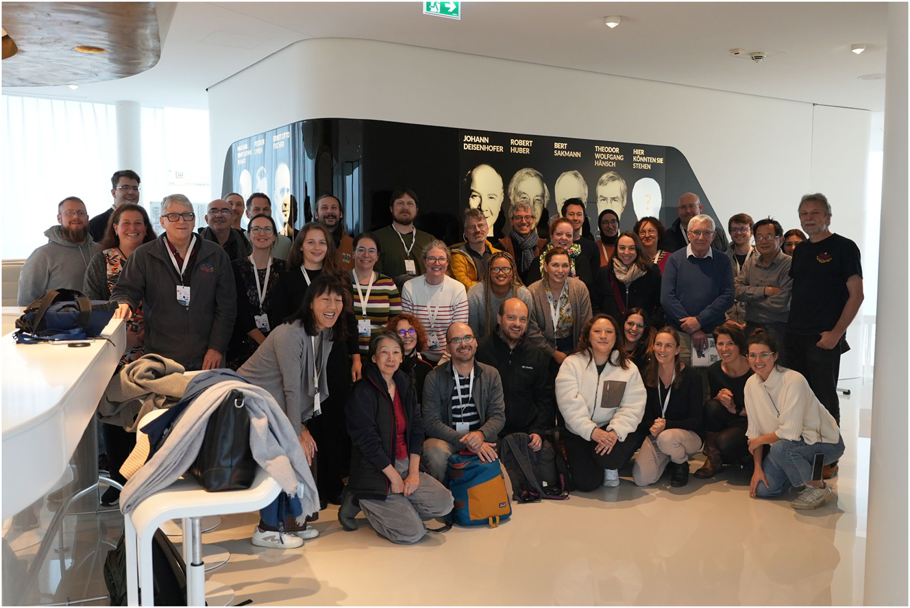
Our Chan Zuckerberg Initiative funded “Array Tomography (AT) Training and Network” organized a first workshop at the German Center of Neurodegenerative Diseases (DZNE) in Munich beginning of November 2024.
For a real hands-on experience, only 20 participants were selected and, besides our team of nine CZI-funded AT enthusiasts, another ten speakers and instructors as well as industry representants were added. The welcome evening at the Campus Home Lounge provided the opportunity to closely interact by scientific speed dating, creative name tags and interactive posters.
On the first day, Helmut Gnägi paved the way with a technically insightful opening talk on the art and science of ultramicrotomy while Bruno Humbel laid a solid foundation by talking about sample preparation. The morning sessions were packed with scientific talks on the application and development of diverse AT methods, ranging from traditional automated tape collecting ultramicrotomy (ATUM) and rigid support AT to magnetic collection, correlated (CLEM) and multimodal approaches.
Richard Schalek and Kevin Briggman reported about technical challenges and achievements of AT for large scale projects in connectomics while Mark Terasaki, Jemima Burden and others revealed the diverse spectrum of applications and scales of AT. The other mornings were dedicated to CLEM, with Kristina Micheva, Mariano Soiza-Reilly and Thomas Misgeld and an overview of image analysis for AT by Josh Morgan. Naomi Kamasawa and Jemima Burden provided overviews of opportunities and limitations of the diverse AT approaches.
In the afternoons we had four different hands-on courses on rigid support AT, ATUM, imaging and image analysis as well as immunofluorescence AT. This provided a lot of time for intense interaction and direct consulting of the participants.
On the last day, we focused on novel developments and an intense feedback round where the participants reported on lessons learned and their next steps. We all agreed that we learned so much from this interaction and thought about ways to further develop and apply AT in the future and lower entry barriers. The participants appreciated the tight interaction with academic and industry partners, a key element for successful technology application and development of the diverse AT approaches.
The evening events provided the opportunity to explore Munich in small groups during a city rally and learn about the history of microscopy at the Deutsches Museum. Finally, the golden radish spiralizer was awarded at the Bavarian radish serial sectioning contest, although we agreed that this technique won’t make it to the novel development section at next year’s workshop in Buenos Aires, Argentina.
The report and the photo were kindly provided by Martina Schifferer.
https://www.arraytomography.org/workshop-2024.html
A special highlight of the Array Tomography Workshop Munich 2024 was the visit of the Deutsches Museum (German Museum), a historical collection of science and technology.
Spotlight on the 13th Asia Pacific Microscopy Congress (APMC13)
Brisbane, Australia | February 2–7, 2025
The countdown is on for APMC13, set to take place in Brisbane, Australia, from February 2–7, 2025. Hosted by the Australian Microscopy and Microanalysis Society (AMMS), the event will bring together leading experts, researchers, and industry professionals to explore advancements in light, ion, and electron microscopy. With its unique integration of indigenous culture and cutting-edge science, APMC13 promises to be a transformative gathering, highlighting the role of analytical technologies in advancing a sustainable, cyclic green economy.
Exciting Announcement: Methods in Microscopy Partners with APMC13
We are thrilled to announce that Methods in Microscopy (MiM) has partnered with APMC13 to amplify the exchange of scientific innovations within the microscopy community. All three editors of MiM – Roger Wepf, Martin Friedrich, and Andreas Thoss – will attend the conference and actively engage with attendees. MiM will also host an exclusive editorial dinner, fostering deeper collaboration and dialogue, and sponsor a Best Method Award to recognize outstanding contributions in the field.
Furthermore, we will test a new way of publishing the abstracts of all APMC-contributors. We have found and contracted a platform where all abstracts receive an unique DOI (link) and where they are registered with google scholar and other search engines.
Invited Speakers: An Impressive Lineup
The APMC13 organizing committee has unveiled an inspiring list of invited speakers, featuring renowned scientists from across the globe.
PLENARY SPEAKERS
Rafal Dunin-Borkowski Germany; Li-An Chu Taiwan; Joanne Etheridge Australia; Dylan Owen UK; Asim Tewari India; Xiuzhen Yu Japan; Ernst Stelzer Germany; Michelle Digman US; Frances M. Ross US
INVITED SESSION SPEAKERS
Shirin Ansari AUS; Sonu Bhaskar AUS; Maté Biro AUS; Chittanon Buranachai Thailand; Tim A. Butcher Germany; Julie Cairney AUS; Shery Chang AUS; Chien-Chun Chen Taiwan; Kok Hao Chen Singapore; Zhen Chen China; Cheng-Hao Chuang Taiwan; Paul Dastoor Australia; Elizabeth Duke Germany; Itia Favre-Bulle AUS; Scott Findlay AUS; Sonja Frolich AUS; Chuanbo Gao China; Debnath Ghosal AUS; Swagatha Ghosh Japan; Enrique Gomez US; Laurent Grocm France; Sarah Harmer AUS; Anja Henss Germany; Haydee Hernandez-Aviña Mexico; Zhehao Huang China; Ryo Ishikawa Japan; Izzy Jayasinghe AUS; Errin Johnson AUS; Se-Ho Kim South Korea; Yumi Konagaya Japan; Shu Ying Lee Singapore; Xiaopeng Li China; Yung-Chang Lin Japan; Shee-Mei Lok Singapore; Michael Matthews UK; Michael Moody AUS; Hironori Nakao Japan; Kayla Nguyen US; Hiroshi Okamoto Japan; Kousuke Ooe AUS; Laura Otter AUS; Robert Parton AUS; Darrin Pochan US; Hikaru Saito Japan; Deniz Saltukoglu Germany: Vanessa Schendel AUS; Jian-Ren Shen Japan; Jean-Baptiste Sibarita France; Kevin Spring AUS; Rhonda Stroud US; Etsuo Susaki Japan; Dai-Ming Tang Japan; Satoko Toyama Japan; Johan Verbeeck Belgium; Wu Yajun Singapore; Kazuo Yamamoto Japan; Jiawei Yan China; Yongsoo Yang South Korea; Koji Yonekura Japan; Nestor Zaluzec US; Nadia Zatsepin AUS; Dongping Zhan China
We look forward to an unforgettable event that celebrates the intersection of culture, innovation, and community. Be sure to join us and contribute to the future of microscopy!
Back to the beginnings: ELMI 2025 in Heidelberg
Heidelberg, Germany | June 3–6, 2025
After this year’s [2024] highly successful meeting in Liverpool, the 25th international meeting of the European Light Microscopy Initiative (ELMI) will take place from the 3rd to the 6th of June 2025 in Heidelberg, Germany at the European Molecular Biology Laboratory (EMBL) and it will be a return to where it all started.
The roots
25 years ago, a group of imaging specialists from all over Europe met in a seminar room at EMBL in Heidelberg to get acquainted with each other’s work, needs and challenges and to discuss how one could efficiently network on such issues inside Europe. The meeting was hosted by the recently created Advanced Light Microscopy Facility at EMBL and many of the participants actively provided imaging support for projects of other scientists in a form that subsequently crystallized into the current model of core facility operation. The European Light Microscopy Initiative was born and one of its core activities since then is the annual ELMI meeting that brings together imaging technology developers, service providers, users and companies in the microscopy field.
Outlook on the 2025 meeting
The annual meeting provides information on the latest developments in light microscopy through plenary sessions on specific topics in the mornings that are followed by workshops on new instrumentation and methods in the afternoon. With its unique format the ELMI meeting is a perfect place for knowledge exchange and networking both with academic partners and with companies. The main meeting is preceded by a core facility satellite meeting which is an essential platform for service providers in the different institutes in Europe and worldwide before the ELMI meeting starts in the evening with an opening lecture and a get-together.
As a major European science hub Heidelberg’s research landscape hosts many research centres with strong imaging groups and units and the ELMI meeting organization brings together representatives of several such centers: Elisa d’ Este (Max Planck Institute for Medical Research), Ulrike Engel (Heidelberg University), Elisa May (German Cancer Research Center) and Rainer Pepperkok, Stefan Terjung and Timo Zimmermann (EMBL). The 2025 meeting will take place at EMBL’s Advanced Training Centre on the EMBL Heidelberg campus in the forest hills overlooking Heidelberg and the Rhine valley.
This year’s topics will range from nanoscopy over probes and biosensors, imaging in health applications and tissue imaging to data analysis. The overarching impact of artificial intelligence technology will be reflected inside all sessions. The scientific program will be complemented by ELMI’s signature afternoon workshops and will be rounded off by a social event highlighting Heidelberg’s beautiful city and surroundings and by the traditional “friendly” ELMI football match between two teams fielded by the academic and the company sides. We look forward to welcoming you next year to the place where the question “So, are we going to do it then?” started it all. Registration will open very soon and more information can be found at https://s.embl.org/elmi2025.
The report and the photo were kindly provided by Timo Zimmermann.
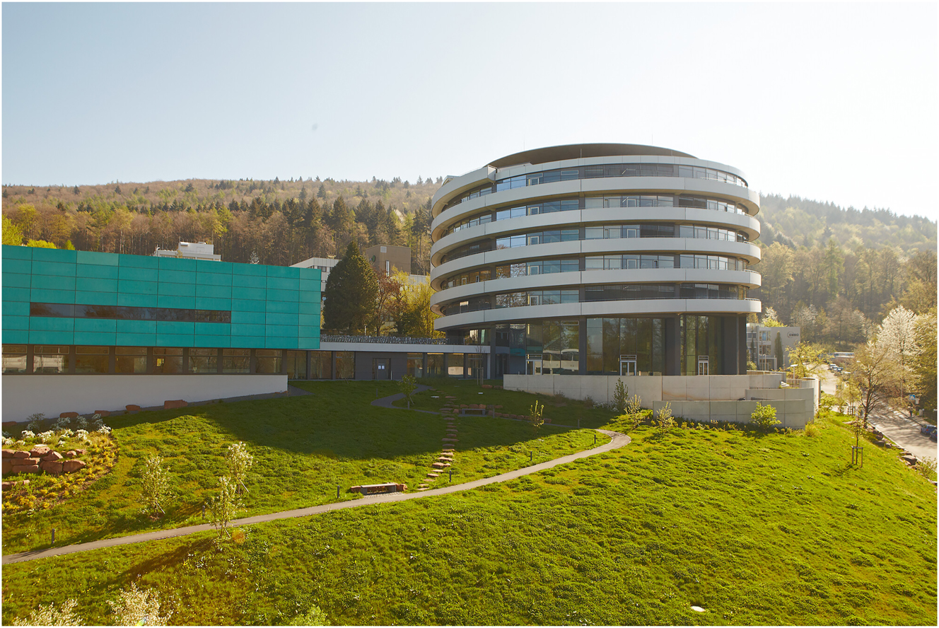
EMBL’s Advanced Training Centre (ATC) is shaped in the form of a double helix. It will host ELMI 2025. (Photo: Marietta Schupp/EMBL Copyright © EMBL, License CC-BY-NC-ND 4.0).
© 2024 the author(s), published by De Gruyter on behalf of Thoss Media
This work is licensed under the Creative Commons Attribution 4.0 International License.
Artikel in diesem Heft
- Frontmatter
- Editorial
- What is open access and what is it good for?
- News
- Community news
- View
- Optical sectioning in fluorescence microscopies is essential for volumetric measurements
- Review Article
- Wrapped up: advancements in volume electron microscopy and application in myelin research
- Research Articles
- Reducing artifact generation when using perceptual loss for image deblurring of microscopy data for microstructure analysis
- Utilizing collagen-coated hydrogels with defined stiffness as calibration standards for AFM experiments on soft biological materials: the case of lung cells and tissue
- Phase characterisation in minerals and metals using an SEM-EDS based automated mineralogy system
- Corrigendum
- Corrigendum to: FAST-EM array tomography: a workflow for multibeam volume electron microscopy