Community News

APMC13 – What a blast!
A report from Brisbane, Australia by Eric Hanssen, President – Committee of Asia Pacific Societies for Microscopy (CAPSM)
Five years ago, almost to the day, Roger Wepf, Caterina Caderas and I travelled to Hyderabad, India, to attend the 12th Asia Pacific Microscopy Congress and present a bid to host APMC13 in Brisbane as a joint conference with the 28th Australian Microscopy and Microanalysis conference. Whilst this was a successful trip, it was one of our last one as COVID19 hit the world. The uncertainty in international travel over the next two years were too great to take any final decision on conference date early on. After a few changes in the international conference calendar Roger and the new co-chair, A/Prof Louise Cole, were able to finally settle for a date and planning started to unveil for AMC13/ACMM28.
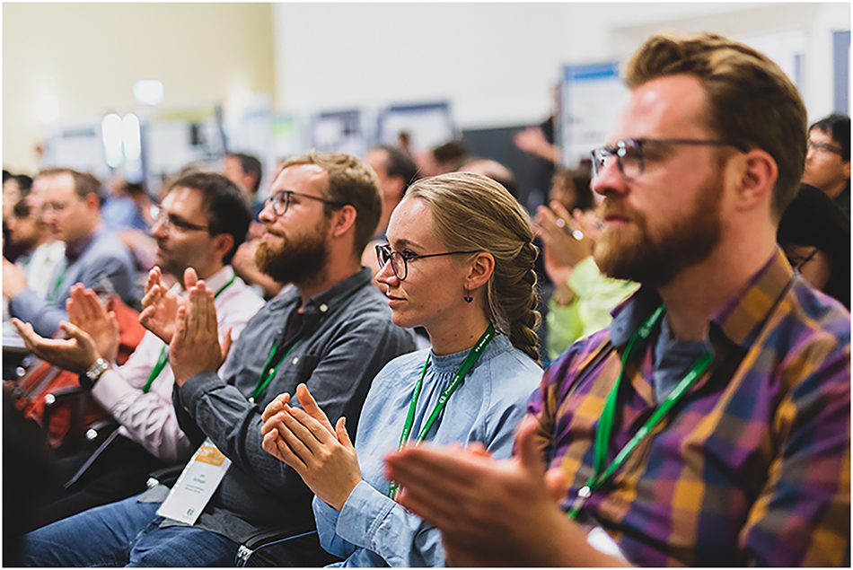
Fast forward to 2025 and here we are, APMC13 has now concluded. As an attendee, the conference ran flawlessly thanks to Roger and Louise’s team tireless efforts. Ten days for all to enjoy hands-on and theoretical workshops, talks from leaders in various field of research but also from students presenting at their first international conference. All the sessions I attended were alive with interactions between the audience and the speakers, regardless of the subject. This not only underline a great choice of speakers but also of the quality of the audience, writer of this page included.
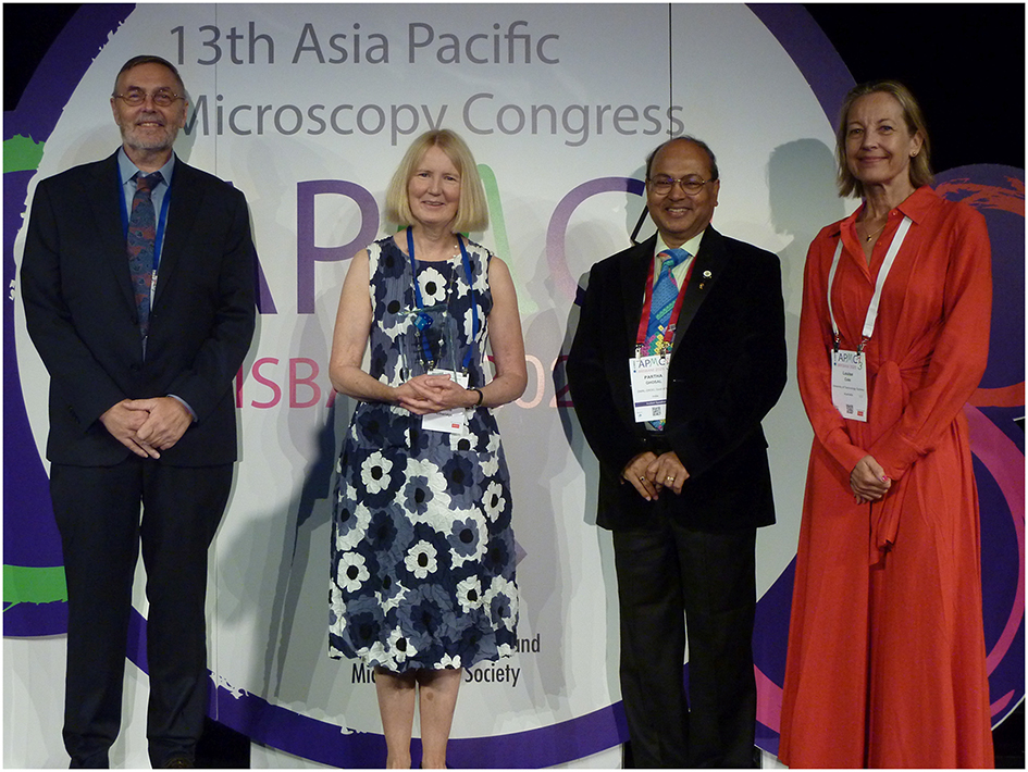
APMC13 began each morning with a keynote. The first on “101 things to do with an energetic electron” was presented by Joanne Etheridge (2nd from left) and introduced by Partha Ghosal (3rd from left, APMC12 chair). They were joined on stage by APMC13 chairs Roger Wepf (left) and Louise Cole (right) (Photo: Martin Friedrich).
Breaking from tradition, the welcome function was held on the Monday night after a few sessions instead of just before the conference on the Sunday. While this saves a considerable amount of funds, hence lowering the already high registration fees it also gives everyone some breathing space especially for the exhibitors that are always pushed to their limits during the bump-in and bump-out of conference, it did lack a little bit of the atmosphere encountered when everyone meets for the first time after a hiatus of 3–4 years but was none-the-less a great experience.
I was lucky enough to attend other functions and events of the course of the week. The editorial team of Methods in Microscopy (MiM), the new method specific journal, organised the second editorial board diner at Popolo, a very nice Italian restaurant. It was a great night to catch up with old and new colleagues and remake the world to our microscopic image. I am sure there will be a fully dedicated page to that evening.
The Following night was organised by the Korean Microscopy Society to support their bid to host the next APMC conference. Labelled the “Jeju night”, it featured the unavoidable presentation for the potential conference venue but also a very fun little raffle with a range of Korean specific gift from fancy teas to Jeju themed folding fans. One quick recovery evening with some friends from around the planet and it was time for the conference diner, which was also a celebration of JEOL Australasia 60th birthday. It was a blast, from sake barrel breaking (and drinking) to Japanese drums (they were awesome), through the award ceremony of the Australian Microscopy and Microanalysis Society. MiM also had two CAPSM labelled awards for the best method talks at the conference, which were a nice surprise for the audience, especially for the recipients.
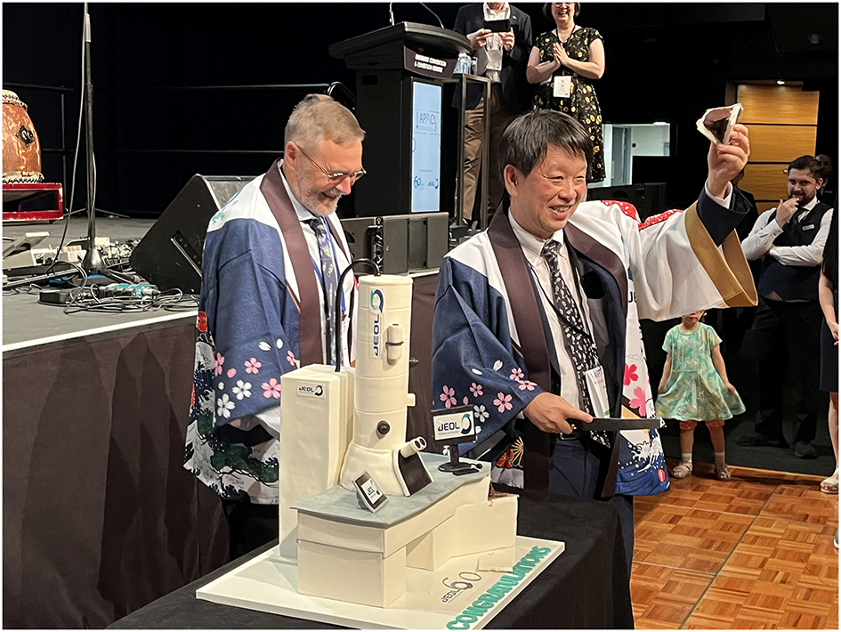
JEOL celebrated its 60th anniversary at CAPSM13. Conference chair Roger Wepf (left) presented to JEOL’s President & CEO Izumi Oi (Photo: Andreas Thoss).
As new president of CAPSM I sincerely thank the Australian Microscopy and Microanalysis Society that hosted APMC13, whish all the best to the Korean Microscopy Society for the next APMC in Jeju Island, sometime towards the end of 2028. One advice for KSM, keep the coffee good and flowing and everyone will remember the conference fondly. Keep an eye out of the confirmation of dates.
Thank you to all the manufacturers present at the exhibition for their time, support and running in booth workshop throughout the week. Finally thank you to the editorial team of Methods in Microscopy supporting the conference and our community. Please reciprocate by submitting manuscript of your newly developed techniques.
Professor Eric Hanssen,
President – Committee of Asia Pacific Societies for Microscopy (CAPSM)
Past-President - Australian Microscopy and Microanalysis Society (AMMS)
Methods in Microscopy at APMC13
A conference review from the editorial team
Meeting in person is a rare opportunity when a team is split between Germany and Australia. So it was a true celebration when the editorial team of Methods in Microscopy reunited in Brisbane for APMC13, a conference that was co-chaired by MiM Editor-in-Chief Roger Wepf. For the Editors Martin Friedrich and Andreas Thoss from Germany, it was a warm introduction to the Australian microscopy community.
Their enthusiasm was shared by several members of the editorial board and guests at a memorable dinner in one of Brisbane's most beautiful Italian restaurants, the Popolo. Situated in South Bank Parklands, it offers stunning views of the Brisbane River and a vibrant skyline, paired withexceptional cuisine. It became a night to remember, where old and new friends gathered from around the world.
Held on the conference's opening Monday, the dinner set stage for days filled with academic sessions and the energy of the exhibition floor. MiM had its own table, welcoming authors, board members and other interested attendees for discussion.
The conference program was packed with highlights, from engaging keynotes in the morning to lively social events in the evening. Yet, one of the most remarkable moments was the conference dinner – not just for the exceptional food or the dynamic Taiko drumming performance by Byron Taiko, but also for the 60th anniversary celebration of the Japanese company JEOL. The event featured sake, a birthday cake shaped like an electron microscope and a seriesof award ceremonies, including the MiM Best Method Awards. The night ended with scientists filling the dance floor, spreading good vibes.
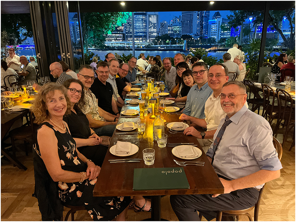
It was a great pleasure for the MiM Editors to meet with members of the Editorial Board and other valued members of the microscopy network at Brisbane’s waterfront (Photo: Martin Friedrich).
Best Methods Awards presented at APMC13 Dinner
The journal Methods in Microscopy was founded to advance technical know-how across all areas of microscopy applications through rigorous publishing. However, direct engagement with the community has always been part of our vision. APMC13 provided an ideal platform for the MiM editorial team, authors and the microscopy community to exchange ideas. We gathered valuable input for future journal issues and ways to improve out publishing process.
Conferences also offer a chance to give back to the community. Sponsored by our publishing partner De Gruyter and MiM Editor-in-Chief Roger Wepf, we were proud to recognize two outstanding scientists with the Best Method Award for their presentation at APMC13.
Felipe Kremer (Centre for Advanced Microscopy, Australian National University, Canberra, ACT, Australia) was given the MIM Award for Best Method in Microscopy in Bio/Life Sciences for his presentation “Using Gatan scripting for improved MicroED data quality”.
Héctor Hugo Pérez Garza, CEO of Dutch startup DENSsolutions B.V., won the Best Method in Materials/Physical Sciences for his presentation “Silicon-Based CryoEM Grids: Revolutionizing Sample Stability and Data Quality with Microfabrication and Graphene”.
The awards include a certificate and a cash prize of AUD 1,500. We extend our gratitude to the session chairs who nominated candidates. The final selections were made by CAPSM President Eric Hannsen and MiM Editor-in-Chief Roger Wepf.
The award-winning abstracts along with most other APMC13 presentations have been published as a special collection on the scienceopen platform. Each abstract has been assigned a Digital Object Identifier (DOI) and can be found via Google Scholar or directly at www.scienceopen.com. This publication was made possible through a unique collaboration between the conference organizers and the Methods in Microscopy editorial team.
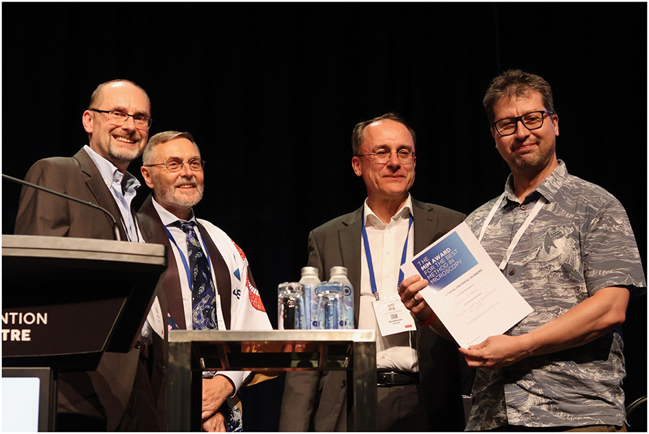
Felipe Kremer (right) was presented the MIM AWARD for the best method in Microscopy in Bio/Life Sciences at the APMC13 conference dinner by Martin Friedrich, Roger Wepf, and Andreas Thoss (from left to right) (Photo with courtesy of Peter Hines).
4th Annual Meeting of the Bridging Nordic Microscopy Infrastructure
Gothenburg, Sweden, August 19–22, 2025
The BNMI aims to strengthen the international competitiveness of Nordic advanced microscopy environments by fostering collaboration, knowledge exchange, and training. Through initiatives such as scientific symposia, workshops, shadowing programs for facility staff, and short-term scientific mobility, BNMI aspires to establish a sustainable Nordic platform that promotes efficient collaboration and long-term partnerships across the region. The BNMI is also an integral part of the Euro-Bioimaging-ERIC, which unites bioimaging infrastructures across Europe to enhance research, diagnostics, and patient care. One of BNMI’s key activities is the organization of scientific symposia. This year, the Centre for Cellular Imaging at the University of Gothenburg, Sweden, will proudly host the BNMI 2025 Symposium, a premier event that will convene imaging facility staff, leading scientists, early-career scientists, and industry representatives for 3 days of impactful discussions and collaboration. The symposium will offer an engaging and dynamic program tailored to imaging researchers across various disciplines. Attendees will have the opportunity to explore the latest advancements in imaging technologies and forge valuable connections within the global imaging community. Topics will span key areas of imaging research, including: Correlative Multimodal Imaging; Nanoscale Imaging; Mesoscopy; Smart Microscopy; New Frontiers in Artificial Intelligence for Microscopy.
With ample opportunities for both formal discussions and informal networking, the symposium is designed to enrich scientific dialogue and foster meaningful collaborations. On behalf of the organizing committee, we look forward to welcoming you to this exciting meeting and wish you a memorable stay in Gothenburg!
These are selected key sessions and speakers covering the most exciting areas of imaging research:
Keynote: Self-organisation of microtubules into a bipolar spindle
Invited Speaker: Eric Karsenti (EMBL, Heidelberg).
Imaging sub-cellular events at high resolution using advanced light
Invited Speakers: Jonas Ries (University of Vienna) and Marine Laporte (University Lyon).
Enabling imaging across scales
Invited Speaker: Evgenia Zagoriy (EMBL, Heidelberg), Nalan Liv (UMC Utrecht) Shigeaki Kanatani (Karolinska Institute, Stockholm), and Jemima Burden (University College London).
Smart Microscopy: from image analysis, artifical inteligence to intelligent acquisition
Invited Speakers: Ilaria Testa (SciLifeLab, Solna) and Benedict Diederich (Leibniz Institute of Photonic Technology, Jena).
Exploring Emerging Frontiers in Artificial Intelligence for Microscopy: An Overview of Trends and Innovations
Invited Speakers: Ivo Sbalzarini (MPI-CBG & CSBD Dresden) and Artur Yakimovich (CASUS HZ Dresden-Rossendorf).
On behalf of the organizing committee, I eagerly look forward to welcoming you to this exciting event and wish you a rewarding and memorable experience in Gothenburg!
Julia Fernandez-Rodriguez, Chair of BNMI 2025
30th PicoQuant Single Molecule Workshop welcomes two Nobel Laureates
Berlin, Germany, September 23 to 26, 2025
PicoQuant proudly announces the 30th anniversary of its Single Molecule Workshop, taking place in Berlin from September 23 to 26, 2025. Over the past three decades, this workshop has established itself as a leading platform for researchers in single molecule spectroscopy and super-resolution microscopy, bringing together experts from physics, chemistry, biology, and life sciences.
This year’s workshop will be a landmark event with two renowned Nobel Laureates as keynote speakers:
Prof. Stefan Hell (Max Planck Institute for Multidisciplinary Sciences, Germany)
Prof. W. E. Moerner (Stanford University, USA)
In addition to keynote lectures, invited speakers will present their latest work. Attendees will experience a diverse program, including informal networking opportunities, such as a welcome reception and a special anniversary celebration, fostering collaboration and idea exchange among participants. As Rainer Erdmann, Managing Director of PicoQuant explains: “The workshop is an outstanding opportunity to meet not only Nobel Laureates but also to discuss latest results with many famous scientists and bright students. With a dedicated ‘student award’ we encourage students to submit their work not only in posters but also as contributed talks.”
The 30th Picoquant Single Molecule Workshop will be held in Berlin Adlershof at the Bunsen Hall (Copyright: Picoquant).
Students are invited to submit abstracts for oral and poster presentations by April 16, 2025. Early-bird registration is available until April 16, 2025, while regular registration closes on August 15, 2025.
Since its inception in 1995, the Single Molecule Workshop has been at the forefront of scientific discovery, providing a forum for exchanging knowledge and fostering innovation in ultra-sensitive optical detection. With its long-standing commitment to advancing single molecule science, PicoQuant continues to provide a platform for collaboration and discovery.
© 2025 the author(s), published by De Gruyter on behalf of Thoss Media
This work is licensed under the Creative Commons Attribution 4.0 International License.
Artikel in diesem Heft
- Frontmatter
- Editorial
- Embracing the power of fluorescence lifetime imaging
- News
- Community News
- Views
- Perspective: fluorescence lifetime imaging and single-molecule spectroscopy for studying biological condensates
- Advanced fluorescence lifetime-enhanced multiplexed nanoscopy of cells
- Tutorials
- From principles to practice: a comprehensive guide to FRET-FLIM in plants
- Fluorochrome separation by fluorescence lifetime phasor analysis in confocal and STED microscopy
- Calibration approaches for fluorescence lifetime applications using time-domain measurements
- FRET-analysis in living cells by fluorescence lifetime imaging microscopy: experimental workflow and methodology
- Protocol for in vivo fluorescence lifetime microendoscopic imaging of the murine femoral marrow
- Research Articles
- Spectro-FLIM for heritage: scanning and analysis of the time resolved luminescence spectra of a fossil shrimp
- Quantifying nucleation in flow by video-FLIM
- Benchmarking of fluorescence lifetime measurements using time-frequency correlated photons