Why do certain cancer cells alter functionality and fuse?
-
Thomas Dittmar
Thomas Dittmar studied Chemistry at the Ruhr-University Bochum and received his diploma in 1995. The he moved to the Institute of Immunology at Witten/Herdecke for his PhD thesis, which he finished in 1999. After a short PostDoc time he was a Junior Professor at the Institute of Immunology at Witten/Herdecke University (2003–2009). Since 2010 he is a full professor at Witten/Herdecke University and since 2018 he is the acting head of the Institute of Immunology at Witten/Herdecke University. His particular focus of research is the role of cell-cell fusion in a cancer context.

 ,
Mareike Sieler
,
Mareike Sieler
Mareike Sieler obtained her Bachelors’ and Masters’ degree in Chemical Biology at the Technical University of Dortmund. The work was carried out at the Chemical Genomics Centre of the Max Planck Institute of Molecular Physiology (B.Sc., 2016) and at the Leibniz Institute for Analytical Sciences - ISAS e.V. (M.Sc., 2018) in Dortmund. Currently, she is working at the Institute for Immunology of the University of Witten/Herdecke in the group of Prof. Dr. Thomas Dittmar to obtain the scientific doctorate degree in biochemistry. The topic of the PhD thesis is the molecular characterization of the mechanisms of cell fusion of human breast cancer cells and breast epithelial cells.
Ralf Hass studied Biochemistry at University of Hannover, Max-Planck Institute for experimental Medicine, Göttingen, and University of California, San Diego, USA to finish his diploma thesis in 1986. Following subsequent studies at New York Medical College, Valhalla, USA, and at University of Hannover he received his PhD in Biochemistry with a PhD award in 1988. As a PostDoc Ralf worked several years at the Dana-Farber Cancer Institute, Harvard Medical School, Boston, USA, where he was appointed ‘Instructor in Medicine’. From 1997 to 2000 he pursued his science as assistant professor at University Clinic Charité, Berlin. Thereafter, Ralf moved to Hannover Medical School as extraordinary professor since 2001. His present projects predominantly focus on interactions of mesenchymal stem cells within damaged and/or neoplastic tissues and potential clinical benefits of these stem cells.

Abstract
Cancer cell fusion represents a rare event. However, the surviving cancer hybrid cells after a post-hybrid selection process (PHSP) can overgrow other cancer cells by exhibiting a proliferation advantage and/or expression of cancer stem-like properties. Addition of new tumor properties during hetero-fusion of cancer cells e.g. with mesenchymal stroma-/stem-like cells (MSC) contribute to enhanced tumor plasticity via acquisition of new/altered functionalities. This provides new avenues for tumor development and metastatic behavior. Consequently, the present review article will also address the question as to whether cancer cell fusion represents a general and possibly evolutionary-conserved program or rather a random process?
1 Introduction
Continuously invasive proliferation of cancer cells is paralleled by interaction with surrounding tissues to establish a dynamic tumor microenvironment (TME) in solid tumors. Such TME represents an orchestration of the extracellular matrix (ECM) together with various different cell types eventually forming an organ-like entity (Egeblad et al. 2010; Ungefroren et al. 2011). Cell populations contributing to the TME include fibroblasts, adipose cells and stromal cells for the tumor architecture and endothelial cells for tumor angiogenesis to support oxygen and nutrient supply. Moreover, immune cells such as T cells, NK cells, dendritic cells, and macrophages accumulate in this pro-inflammatory milieu of invasive tissue lesions (Coussens and Werb 2002; Yang and Zhang 2017). In addition, heterogeneous mesenchymal stroma-/stem-like cells (MSC) are predominantly recruited to tumor sites to assist in repair activities, support neovascularization, modulate the immune response, and directly interact with the cancer cells (Mandel et al. 2013; Melzer et al. 2018a). Accordingly, the TME is underlying continuous changes based on the various TME-associated cell populations to communicate within the TME and mutually acquire new functions (Baghban et al. 2020; Yang et al. 2015). This tumor-directed focus of altered cellular functionalities enables the continuous formation of a complex organ-like tumor consisting of cancer cells in distinct states of development (differentiated, progenitor or cancer stem-like cells) in combination with a variety of differentially organized cell types. Thus, TME-associated fibroblasts can be altered to cancer-associated fibroblasts (CAFs) by reorganizing the tumor architecture and macrophages acquire new properties of tumor-associated macrophages also supporting enhanced cancer cell migration and metastases (Fu et al. 2020). Likewise, endothelial colony-forming progenitors and vascular endothelial cells within the TME form distinct tumor-associated structures. In contrast to a normal healthy vasculature, tumor vessels display an aberrant physiology due to abnormal and leaky endothelial cell layers and an altered density (Munn 2003). This discontinuous endothelium in tumor vessels supplies insufficient oxygen contributing to increased hypoxia within the TME (Muz et al. 2015). A compartmentalization of tumor tissue like in organs with separated local functionalities is supported by MSC. According to their tropism to tumors these cells stimulate regions with both, tumor growth-inhibiting and tumor growth-promoting activities depending on the activation status and external stimuli (Hass 2020). Moreover, MSC can contribute to the formation of flexible cancer stem cell niches for the maintenance of cancer progenitor or cancer stem-like cells (Melzer et al. 2017). Beside the exchange of trophic factors and extracellular vesicles as external stimuli MSC also directly interact with cancer cells at different levels of intensity. Following cell-cell attachment via GAP junctions and notch signaling, MSC can form nanotubes with cancer cells and exchange membrane patches via trogocytosis (Melzer et al. 2016). As an ultimate step of interaction MSC can also fuse with cancer cells to form new cancer hybrid cells expressing new tumorigenic and metastatic properties (Melzer et al. 2018b; Melzer et al. 2021). The possibility to form cancer hybrid cells also applies to other populations within the TME such as CAFs or macrophages (Clawson et al. 2015; Gast et al. 2018; LaBerge et al. 2021; Manjunath et al. 2020b; Shabo and Svanvik 2011; Shabo et al. 2015). In general, cell fusion represents a physiological process required for distinct tissue development and maintenance. However, the necessity of cancer cell-cell fusion and contributing mechanisms are less obvious (Dittmar and Hass 2022). On the one hand, cancer cell-cell fusion within the TME followed by a successful post-hybrid selection process (PHSP) increases tumor plasticity (Hass et al. 2021a). Conversely, the fusogenecity of cancer cells and the overall survival rate of cancer hybrids are very low (Duelli and Lazebnik 2003; Miroshnychenko et al. 2021). In terms of the total number of cells in the tumor, below 0.1 to 1 % of tumor cells fuse (Fortuna et al. 1989; Gast et al. 2018; Lu and Kang 2009; Melzer et al. 2019; Miroshnychenko et al. 2021; Powell et al. 2011; Ramakrishnan et al. 2013; Rizvi et al. 2006; Wakeling et al. 1994; Yan et al. 2015). Similar amounts apply to the PHSP survival rate of cancer hybrids, which is also far less than 1 % (Duelli and Lazebnik 2003).
According to this negligibly low fusion rate why do cancer cells fuse at all? Further distinct questions arise which will be addressed in the present review article: Does cancer cell-cell fusion represent a general or even evolutionary-conserved mechanism for a facilitated adaptation to the microenvironment to increase tumor plasticity and metastatic options? Is it a random process that occurs spontaneously in the tumor tissue despite of tightly regulated extracellular events (Dittmar and Hass 2022) paired with intrinsic molecular signaling factors (Dittmar and Hass 2023)?
2 Cancer cell-cell fusion: a general/evolutionary-conserved mechanism or rather a random spontaneous process?
Cancer cell-cell fusion is commonly associated with tumor progression since various studies have demonstrated that cancer hybrids exhibit an increased malignancy. This is associated with enhanced metastatic capacity, drug resistance and cancer stem/initiating cell (CS/IC) properties (for review see: (Demin et al. 2021; Dittmar 2022; Dittmar and Hass 2022, 2023; Hass et al. 2021a,b; Manjunath et al. 2020b; Shabo et al. 2020; Wang et al. 2021)). Data of Gast et al., Dietz et al., and Manjunath et al. further revealed that disseminated circulating cancer hybrids, harboring macrophages and neoplastic cells, outnumbered conventionally defined circulating tumor cells in cancer patients (Dietz et al. 2021; Gast et al. 2018; Manjunath et al. 2020a). These high numbers of circulating cancer hybrids were correlated with advanced disease progression and overall poor prognosis of pancreatic ductal adenocarcinoma patients (Gast et al. 2018).
Various studies supported the hypothesis of cancer cell fusion as a general and possibly evolutionary-conserved program (Clawson et al. 2012; Dietz et al. 2021; Gast et al. 2018; LaBerge et al. 2017; LaBerge et al. 2021; Lazova et al. 2013; Manjunath et al. 2020a), however, evidences are not clear yet and require further substantiation (Figure 1). In fact, cancer cell-cell fusion appears to be a very inefficient process. As indicated above, the overall fusion frequency of cancer cells and the overall survival rate of cancer hybrids are both far less than 0.1 %. Consequently, only a rare fraction of cancer cells will be able to merge with other cells at all whereby the majority of the emerged cancer hybrids will die due to a failed reorganization of aneuploid chromosomal nuclei (Dornen et al. 2020b; Duelli and Lazebnik 2003; Hass et al. 2021a; Sieler et al. 2021). Only a very rare fraction of cancer hybrids, who have successfully passed through the PHSP, will survive and proliferate (Hass et al. 2021a).
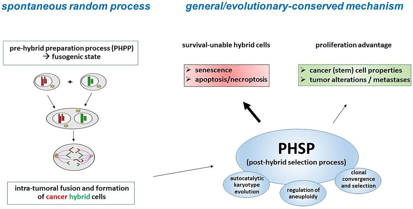
Hypothetic model of spontaneous randomized cancer cell fusion associated with the subsequent general/evolutionary-conserved PHSP whereby a PHSP may also apply to entosis, cannibalism, or even chromothripsis-like chromosomal aberrations/reorganization (adapted from (Hass et al. 2021a)).
This inefficiency actually does not support a general and evolutionary-conserved mechanism. Moreover, such mechanism would require certain conditions to initiate the process of cell-cell merger. The chronically inflamed TME represents a wide mixture of different cell types (e.g. cancer cells, CAFs, immune competent cells, MSC, stromal cells), connective tissue and intercellular communication structures (gap junctions, nanotubes), extracellular vesicles/exosomes, and soluble factors (cytokines, chemokines, growth factors, proteases and more), all of which are forming an interaction network (Coussens and Werb 2002; Hass et al. 2020; Ungefroren et al. 2011; Yang and Zhang 2017). So far, inflammation/inflammatory cytokines have been identified as well-known triggers of cell-cell fusion (Davies et al. 2009; Johansson et al. 2008). But with regard to cancer cell-cell fusion, the in vitro fusion frequency of cancer cells was only moderately increased from approximately 1 % to 2 % (Mohr et al. 2015; Yan et al. 2017). Appropriate in vivo data are still missing. Similarly, the impact of all other cellular and humoral components of the chronically-inflamed TME on cancer cell-cell fusion remains unclear. Thus, if fusion of cancer cells with other cells would be a general/evolutionary-conserved mechanism, the overall fusion frequency of cancer cells would be expected much higher, as numerous cancer cells would be affected by these environmental conditions by changing their functionality.
Moreover, cancer cell-cell fusion appears as a random mixing and segregation of parental chromosomes to daughter cancer hybrid cells, which is further associated with massive DNA damage and induction of aneuploidy (Dornen et al. 2020b; Hass et al. 2021a; Sieler et al. 2021). However, it is well-known that sexual and parasexual recombination of DNA as well as aneuploidy represent general and evolutionary conserved mechanisms in species development and adaptation processes (Beaupere et al. 2018; Bennett 2015; Chunduri and Storchova 2019; Duncan et al. 2012; Yang et al. 2019). This suggests that not the process of cancer cell-cell fusion itself, but rather the selection events during a subsequent PHSP are general/evolutionary-conserved (Figure 1). Nevertheless, cell-cell fusion is a highly complex, tightly regulated, energy dependent, and still not fully understood process (for review see: (Brukman et al. 2019; Dittmar and Hass 2023; Dittmar et al. 2021; Hass et al. 2021a; Hass et al. 2021b; Hernandez and Podbilewicz 2017; Petrany and Millay 2019; Whitlock and Chernomordik 2021)). Therefore, it cannot be ruled out that certain evolutionary-conserved mechanisms may also play a role in cancer cell-cell fusion after all.
Although the mixing and segregation of parental chromosomes, DNA damages and induction of aneuploidy appear randomized during cancer cell fusion it remains unclear why cancer hybrids preferentially exhibit a more malignant phenotype. At least, cancer hybrids displaying active cell survival and/or stemness signaling pathways may express a selection advantage for surviving the PHSP (Figure 1).
3 The contrasting phenomenon of high syncytin-1 expression but low fusogenecity of cancer cells
A variety of different proteins, cytokines, chemokines, adhesion molecules, cytoskeletal proteins, proteases, have been associated with (cancer) cell-cell fusion. So-called fusogens are essential for cell-cell merger as they catalyze the fusion of two negatively charged and, thus, usually repelling phospholipid bilayer membranes (Brukman et al. 2019; Hernandez and Podbilewicz 2017; Whitlock and Chernomordik 2021). Considering that cancer cells can fuse with other cells, they must express fusogens. The human endogenous retroviral (HERV) element syncytin-1 remains the best characterized human fusogen to date and mediates the syncytialization of villous trophoblasts to syncytiotrophoblasts during placentation (Durnaoglu et al. 2021; Malassine et al. 2010). Besides, syncytin-1 is also involved in osteoclastogenesis (Soe et al. 2011) and myogenesis (Frese et al. 2015). Moreover, syncytin-1 has also been associated with cancer cell-cell fusion (Benesova et al. 2017; Bjerregaard et al. 2006; Chignola et al. 2019; Dittmar and Hass 2022; Fei et al. 2019; Fu et al. 2021; Larsen et al. 2009; Larsson et al. 2007; Liu et al. 2019; Strick et al. 2007; Uygur et al. 2019; Yan et al. 2017; Yu et al. 2014). Whether cancer cell-cell fusion is also mediated by fusogens other than syncytin-1 is unknown, since this has not yet been investigated.
Even though physiological cell-cell fusion events and the merger of cancer cells appear to be different in terms of induction, regulation and outcome of evolving hybrids, they represent mechanistically similar conserved processes (Brukman et al. 2019; Dittmar and Hass 2022; Dittmar and Hass 2023; Hernandez and Podbilewicz 2017; Whitlock and Chernomordik 2021). Cell merger is a multifactorial process and depends on the concerted interplay of the phospholipid phosphatidylserine and various proteins, including fusogens, cell adhesion molecules, cytokines, chemokines, cytoskeletal proteins and proteases (Brukman et al. 2019; Dittmar and Hass 2022; Dittmar and Hass 2023; Hernandez and Podbilewicz 2017; Whitlock and Chernomordik 2021). Thus, the knowledge how physiological cell-cell fusion processes are directed and running might be helpful for the understanding how the merger of cancer cells is facilitated.
As physiological examples for cell fusion placentation and myogenesis represent tightly regulated and highly efficient fusion process during embryonic development (Cheng et al. 2014; Isobe et al. 2022; Johnson et al. 2021; Quinn et al. 2017). Placentation is characterized by formation of high numbers of multinucleated cells which is attributed to a continuous expression of syncytin-1 in villous cytotrophoblasts until about 37 weeks of gestation (Chen et al. 2006). Similarly, a peak expression of fusion relevant proteins and generation of multinucleated muscle fibres was observed during muscle development in animal studies (Chen et al. 2020). This indicates that cells in a physiological context could become fusogenic at a certain point of time due to an inherent up-regulation of fusion relevant protein expression, such as fusogens. Similarly, they retain to a non-fusogenic state later, which is related to down-regulation of the cellular fusion machinery. Whether this turn on/turn off characteristic of physiological cell merger also applies to cancer cells is not clear. Previous work suggested that cancer cell-cell fusion is facilitated by syncytin-1 (Benesova et al. 2017; Bjerregaard et al. 2006; Chignola et al. 2019; Dittmar and Hass 2022; Fei et al. 2019; Fu et al. 2021; Larsen et al. 2009; Larsson et al. 2007; Liu et al. 2019; Strick et al. 2007; Uygur et al. 2019; Yan et al. 2017; Yu et al. 2014) indicating that syncytin-1 expression was induced in cancer cells. However, the underlying mechanism is still unclear.
Thus, the knowledge how syncytin-1 expression in placental cells is induced and terminated might be helpful for the development of anti syncytin-1 expression strategies in cancer cells. Briefly, this applies as well to other fusion relevant proteins.
The fact that increased syncytin-1 expression levels were found in different cancer cells/tissues (Bjerregaard et al. 2006; Chignola et al. 2019; Fei et al. 2019; Fu et al. 2021; Liu et al. 2019; Strick et al. 2007; Yan et al. 2017) suggests that this fusogen might also be significantly involved in cancer cell-cell fusion. However, the degree of syncytin-1 expression in cancer cells/tissues does not correlate with the overall very weak fusogenecity of the cells, which is below 0.1–1 % (Fortuna et al. 1989; Gast et al. 2018; Lu and Kang 2009; Melzer et al. 2019; Miroshnychenko et al. 2021; Powell et al. 2011; Ramakrishnan et al. 2013; Rizvi et al. 2006; Wakeling et al. 1994; Yan et al. 2015). Likewise, little if any multinuclear cancer cells were found in cancer biopsies despite increased syncytin-1 expression levels (Benesova et al. 2017; Bjerregaard et al. 2006; Fei et al. 2019; Fu et al. 2021; Liu et al. 2019; Uygur et al. 2019; Yu et al. 2014; Zhou et al. 2021).
This raises the question why the overall fusogenecity of cancer cells remains rather low despite enhanced syncytin-1 expression levels? Cell-cell fusion is a multifactorial process and depends on the interaction of different proteins/factors in a timely coordinated manner. If fusion-relevant components are missing, mutated or masked, the merger of two cells cannot take place. Interestingly, syncytin-1 or syncytin homologous proteins, respectively, were mainly detectable in the cytoplasm and only slightly in the plasma membrane of cancer cells (Bjerregaard et al. 2006; Chignola et al. 2019; Fei et al. 2019; Fu et al. 2021; Liu et al. 2019; Strick et al. 2007; Yan et al. 2017). Since syncytin-1 only exhibits fusogenic properties when it is localized in the cell membrane, this could be one possible explanation for the low fusion frequency of cancer cells.
The processes by which syncytin-1 translocates to the plasma membrane are unclear. Cellular stresses, such as irradiation, may promote this translocation, but even then only a few cancer cells have fused (Chignola et al. 2019). Hypoxia and TME-related (pro-inflammatory) conditions can both trigger cancer cell-cell fusion (Huang et al. 2018; Mohr et al. 2015; Yan et al. 2017), but it remains unknown whether these conditions support syncytin-1 translocation to the plasma membrane of cancer cells. Interestingly, both syncytin-1 expression and syncytialization of trophoblasts were markedly diminished under hypoxic conditions (Alsat et al. 1996; Kudo et al. 2003). Thus, it cannot be ruled out that cancer cells in neoplastic tissues respond differently to hypoxia and syncytin-1 expression when compared to normal trophoblasts.
Studies in astrocytes and muscle cells revealed that sycncytin-1 impaired the expression of its own receptor alanine, serine, cysteine transporter 2 (ASCT2), which was associated with a decreased fusion frequency (Antony et al. 2007; Frese et al. 2015). It remains to be clarified whether a similar mechanism might exist in syncytin-1 expressing cancer cells. Indeed, markedly lower ASCT2 expression levels were observed in the syncytin-1 overexpressing cell line M13_Syn1 as compared to wildtype human M13SV1 breast epithelial cells (Figure 2). In contrast, high ASCT2 and moderate syncytin-1 expression levels were observed in MDA-MB-231, MDA-MB-435S and HS578T human breast cancer cells (Figure 2). Nonetheless, the overall fusion frequency of these cells was only about 1 % (Mohr et al. 2015), which was most likely attributed to cytosolic localization of syncytin-1.
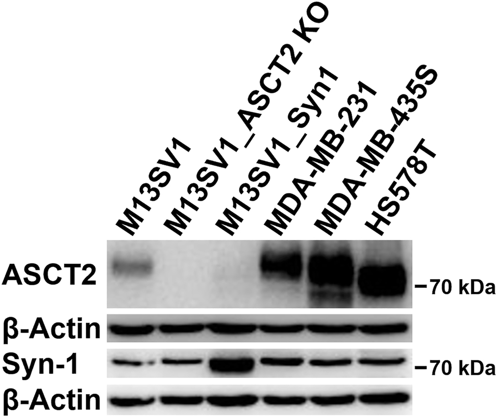
Syncytin-1 and ASCT2 expression in human breast epithelial cells and different cancer cell lines. All wildtype cells possess comparable syncytin-1 expression levels, whereas ASCT2 was predominantly expressed in cancer cells. M13_ASCT2KO cells lack ASCT2 expression due to previous CRISPR/Cas knockout. Interestingly, overexpression of syncytin-1 in M13SV1 breast epithelial cells (M13_Syn1) was associated with a markedly reduced ASCT2 expression.
4 Cell-cell fusion independent effects of syncytin-1 on cancer progression
Syncytin-1 in cancer cells is commonly associated with cell-cell merger due to its fusogenic properties. However, recent findings indicate that proliferation, invasion, metastasis and immune escape of cancer cells are also positively triggered by syncytin-1, which is in line with other HERV elements, such as HERV-K (HML-2) (Gao et al. 2021; Liu et al. 2019; Strick et al. 2007; Zhou et al. 2021) (Dervan et al. 2021; Meyer et al. 2017). This may attribute possible cell-cell fusion independent effects of syncytin-1 on cancer progression. Thus, alternative to its involvement in the fusion of cancer cells and other cells, syncytin-1 also contributes to cellular processes that have a direct impact on tumor growth and metastasis. This assumption is in further agreement with the positive correlation between syncytin-1 expression in tumor tissues, disease progression and overall poor prognosis (Benesova et al. 2017; Fu et al. 2021; Liu et al. 2019; Uygur et al. 2019; Yu et al. 2014; Zhou et al. 2021). While a cytosolic localization of tumor-promoting syncytin-1 effects needs to be verified, these data could indicate alternative syncytin-1 functions according to the intracellular localization in cancer cells.
5 Wound healing-driven cancer cell-cell fusion
With regard to cell-cell fusion in tumors it is commonly assumed that cancer cells “actively” fuse with normal cells. However, as summarized above with high syncytin-1 expression levels but low fusion frequencies of cancer cells there might be further alternative pathways that can explain the fusion of cancer cells with other cells.
The TME resembles chronically inflamed tissue (Balkwill and Mantovani 2001; Dvorak 1986; Mantovani et al. 2008) and various pro-fusogenic cell types, such as fibroblasts, macrophages, bone marrow-derived cells (BMDCs) and MSC (Clawson et al. 2015; Dietz et al. 2021; Dornen et al. 2020a; Gast et al. 2018; Hass et al. 2019; Kemeny et al. 2016; Lizier et al. 2016; Melzer et al. 2018c; Shabo et al. 2015; Sottile et al. 2016) accumulate within this pro-inflammatory milieu of invasive tissue lesions (Coussens and Werb 2002; Mandel et al. 2013; Melzer et al. 2018a; Yang and Zhang 2017). In this regard, it is well-known that MSC, BMDCs and macrophages can regenerate damaged tissues by cell-cell fusion and that this process is triggered by inflammation (Camargo et al. 2004; Davies et al. 2009; Ferrand et al. 2011; Johansson et al. 2008; Silk et al. 2013; Vassilopoulos et al. 2003; Wang et al. 2003; Willenbring et al. 2004). Accordingly, MSC, BMDCs and macrophages recruited to the TME may recognize damaged cancer cells and try to regenerate them by cell-cell fusion. Tissue regeneration by cell merger represents an universal process. MSC, BMDCs and macrophages are not able to distinguish between damaged normal cells/tissue and damaged cancer cells/tumor tissue and exhibit their regenerative capacity according to the requirements of the damaged tissues (Dittmar and Zanker 2015; Dittmar et al. 2006; Hass and Otte 2012; Melzer et al. 2016). Moreover, MSC, BMDCs and macrophages have all been identified as fusion partners for cancer cells (Clawson et al. 2015; Dietz et al. 2021; Dornen et al. 2020a; Gast et al. 2018; Hass et al. 2019; Kemeny et al. 2016, Lizier et al. 2016; Melzer et al. 2018c; Shabo et al. 2015; Sottile et al. 2016). Whether this possible mechanism would be independent of syncytin-1 expression in cancer cells remains to be explored. So far, it is only known that MSC, BMDCs and macrophages could regenerate damaged tissues by cell merger and this process is triggered by inflammation. However, which fusion-relevant proteins/factors are expressed by MSC, BMDCs and macrophages, how the cell fusion machinery is induced in these cells and how damaged cancer cells are recognized by them remains completely unknown.
6 Conclusions
In this review, we tried to address the questions whether fusion of cancer cells might be a general/evolutionary-conserved mechanism and if so, why only a certain fraction of cancer cells alter their functionality and fuse. Although some studies revealed that (circulating) tumor hybrids were found in cancer patients and correlated with disease stage and tumor progression (Dietz et al. 2021; Gast et al. 2018; Manjunath et al. 2020a), these cases are too rare to conclude that cell-cell merger could be general/evolutionary conserved mechanism in cancer. In addition, the process of fusion of cancer cells is very inefficient, as both the fusion rate and the survival rate are very low. Undoubtedly, this contributes to intratumoral heterogeneity and increased tumor plasticity. However, with a general/evolutionary conserved process, it would be expected that more cancer cells should fuse and more tumor hybrids should survive. On the other hand, the process of cell-cell fusion remains scarcely understood, and many processes and regulatory mechanisms have not yet been discovered. Nonetheless, it might be speculated whether evolutionary-conserved processes that enable cells to survive under adverse conditions might help tumor hybrids to successfully survive the PHSP (Figure 1).
Due to its fusogenic properties expression of syncytin-1 in cancer cells is commonly associated with cell-cell merger, but the overall high syncytin-1 expression levels are in contrast to the overall weak fusogenecity of the cancer cells (Dittmar and Hass 2023). This may be due to the fact that syncytin-1 is predominantly localized in the cytoplasm and hardly in the plasma membrane of cancer cells. Why cancer cells express syncytin-1 and why the majority is present in the cytoplasm is poorly understood. The same is true for those processes that control translocation of syncytin-1 to the plasma membrane, which again occurs only in a certain subpopulation of cancer cells. Recent studies suggest cell-cell fusion independent processes controlled by syncytin-1, such as proliferation, metastasis and immune escape. But again, these cases are too rare to conclude a generalized mechanism.
MSC, BMDC and macrophages represent an alternative source of fusogenic cells that could merge with cancer cells. Their fusogenecity and tissue restoration capacity has been demonstrated in several studies and also in the chronically-inflamed TME.
Nevertheless, the fusion of cancer cells with other cell types is still scarcely understood and demands further research activities.
Funding source: Niedersächsische Krebsgesellschaft
Award Identifier / Grant number: Ralf Hass
Funding source: interne Forschungsförderung Witten/Herdecke University
Award Identifier / Grant number: Mareike Sieler
About the authors

Thomas Dittmar studied Chemistry at the Ruhr-University Bochum and received his diploma in 1995. The he moved to the Institute of Immunology at Witten/Herdecke for his PhD thesis, which he finished in 1999. After a short PostDoc time he was a Junior Professor at the Institute of Immunology at Witten/Herdecke University (2003–2009). Since 2010 he is a full professor at Witten/Herdecke University and since 2018 he is the acting head of the Institute of Immunology at Witten/Herdecke University. His particular focus of research is the role of cell-cell fusion in a cancer context.
Mareike Sieler obtained her Bachelors’ and Masters’ degree in Chemical Biology at the Technical University of Dortmund. The work was carried out at the Chemical Genomics Centre of the Max Planck Institute of Molecular Physiology (B.Sc., 2016) and at the Leibniz Institute for Analytical Sciences - ISAS e.V. (M.Sc., 2018) in Dortmund. Currently, she is working at the Institute for Immunology of the University of Witten/Herdecke in the group of Prof. Dr. Thomas Dittmar to obtain the scientific doctorate degree in biochemistry. The topic of the PhD thesis is the molecular characterization of the mechanisms of cell fusion of human breast cancer cells and breast epithelial cells.

Ralf Hass studied Biochemistry at University of Hannover, Max-Planck Institute for experimental Medicine, Göttingen, and University of California, San Diego, USA to finish his diploma thesis in 1986. Following subsequent studies at New York Medical College, Valhalla, USA, and at University of Hannover he received his PhD in Biochemistry with a PhD award in 1988. As a PostDoc Ralf worked several years at the Dana-Farber Cancer Institute, Harvard Medical School, Boston, USA, where he was appointed ‘Instructor in Medicine’. From 1997 to 2000 he pursued his science as assistant professor at University Clinic Charité, Berlin. Thereafter, Ralf moved to Hannover Medical School as extraordinary professor since 2001. His present projects predominantly focus on interactions of mesenchymal stem cells within damaged and/or neoplastic tissues and potential clinical benefits of these stem cells.
-
Author contributions: T.D. and R.H. designed the study, analyzed and interpreted the data and wrote the manuscript. M.S. performed the Western Blot analysis and read and revised the manuscript. All authors have read and agreed to the published version of the manuscript.
-
Research funding: This work was supported by a grant from the “Niedersächsische Krebsgesellschaft e.V.” with respect to the NDR charity campaign ‘Hand in Hand für Norddeutschland 2019’ to Ralf Hass and an internal grant from the “interne Forschungsförderung” of Witten/Herdecke University to Mareike Sieler.
-
Conflict of interest statement: The authors declare no conflict of interest.
References
Alsat, E., Wyplosz, P., Malassine, A., Guibourdenche, J., Porquet, D., Nessmann, C., and Evain-brion, D. (1996). Hypoxia impairs cell fusion and differentiation process in human cytotrophoblast, in vitro. J. Cell. Physiol. 168: 346–353, https://doi.org/10.1002/(sici)1097-4652(199608)168:2<346::aid-jcp13>3.0.co;2-1.10.1002/(SICI)1097-4652(199608)168:2<346::AID-JCP13>3.0.CO;2-1Search in Google Scholar
Antony, J.M., Ellestad, K.K., Hammond, R., Imaizumi, K., Mallet, F., Warren, K.G., and Power, C. (2007). The human endogenous retrovirus envelope glycoprotein, syncytin-1, regulates neuroinflammation and its receptor expression in multiple sclerosis: a role for endoplasmic reticulum chaperones in astrocytes. J. Immunol. 179: 1210–1224, https://doi.org/10.4049/jimmunol.179.2.1210.Search in Google Scholar
Baghban, R., Roshangar, L., Jahanban-Esfahlan, R., Seidi, K., Ebrahimi-kalan, A., Jaymand, M., Kolahian, S., Javaheri, T., and Zare, P. (2020). Tumor microenvironment complexity and therapeutic implications at a glance. Cell Commun. Signal. 18: 59, https://doi.org/10.1186/s12964-020-0530-4.Search in Google Scholar
Balkwill, F. and Mantovani, A. (2001). Inflammation and cancer: back to Virchow? Lancet 357: 539–545, https://doi.org/10.1016/s0140-6736(00)04046-0.Search in Google Scholar
Beaupere, C., Dinatto, L., Wasko, B.M., Chen, R.B., Vanvalkenburg, L., Kiflezghi, M.G., Lee, M.B., Promislow, D.E.L., Dang, W., Kaeberlein, M., et al.. (2018). Genetic screen identifies adaptive aneuploidy as a key mediator of ER stress resistance in yeast. Proc. Natl. Acad. Sci. U.S.A. 115: 9586–9591, https://doi.org/10.1073/pnas.1804264115.Search in Google Scholar
Benesova, M., Trejbalova, K., Kovarova, D., Vernerova, Z., Hron, T., Kucerova, D., and Hejnar, J. (2017). DNA hypomethylation and aberrant expression of the human endogenous retrovirus ERVWE1/syncytin-1 in seminomas. Retrovirology 14: 20, https://doi.org/10.1186/s12977-017-0342-9.Search in Google Scholar
Bennett, R.J. (2015). The parasexual lifestyle of Candida albicans. Curr. Opin. Microbiol. 28: 10–17, https://doi.org/10.1016/j.mib.2015.06.017.Search in Google Scholar
Bjerregaard, B., Holck, S., Christensen, I.J., and Larsson, L.I. (2006). Syncytin is involved in breast cancer-endothelial cell fusions. Cell. Mol. Life Sci. 63: 1906–1911, https://doi.org/10.1007/s00018-006-6201-9.Search in Google Scholar
Brukman, N.G., Uygur, B., Podbilewicz, B., and Chernomordik, L.V. (2019). How cells fuse. J. Cell Biol. 218: 1436–1451, https://doi.org/10.1083/jcb.201901017.Search in Google Scholar
Camargo, F.D., Finegold, M., and Goodell, M.A. (2004). Hematopoietic myelomonocytic cells are the major source of hepatocyte fusion partners. J. Clin. Invest. 113: 1266–1270, https://doi.org/10.1172/jci21301.Search in Google Scholar
Chen, B., You, W., Wang, Y., and Shan, T. (2020). The regulatory role of Myomaker and Myomixer-Myomerger-Minion in muscle development and regeneration. Cell. Mol. Life Sci. 77: 1551–1569, https://doi.org/10.1007/s00018-019-03341-9.Search in Google Scholar PubMed PubMed Central
Chen, C.P., Wang, K.G., Chen, C.Y., Yu, C., Chuang, H.C., and Chen, H. (2006). Altered placental syncytin and its receptor ASCT2 expression in placental development and pre-eclampsia. BJOG 113: 152–158, https://doi.org/10.1111/j.1471-0528.2005.00843.x.Search in Google Scholar PubMed
Cheng, C.S., El-Abd, Y., Bui, K., Hyun, Y.E., Hughes, R.H., Kraus, W.E., and Truskey, G.A. (2014). Conditions that promote primary human skeletal myoblast culture and muscle differentiation in vitro. Am. J. Physiol. Cell Physiol. 306: C385–C395, https://doi.org/10.1152/ajpcell.00179.2013.Search in Google Scholar PubMed PubMed Central
Chignola, R., Sega, M., Molesini, B., Baruzzi, A., Stella, S., and Milotti, E. (2019). Collective radioresistance of T47D breast carcinoma cells is mediated by a Syncytin-1 homologous protein. PLoS One 14: e0206713, https://doi.org/10.1371/journal.pone.0206713.Search in Google Scholar PubMed PubMed Central
Chunduri, N.K. and Storchova, Z. (2019). The diverse consequences of aneuploidy. Nat. Cell Biol. 21: 54–62, https://doi.org/10.1038/s41556-018-0243-8.Search in Google Scholar PubMed
Clawson, G.A., KimchI, E., Patrick, S.D., Xin, P., Harouaka, R., Zheng, S., Berg, A., Schell, T., Staveley-O’carroll, K.F., NeveS, R.I., et al.. (2012). Circulating tumor cells in melanoma patients. PLoS One 7: e41052, https://doi.org/10.1371/journal.pone.0041052.Search in Google Scholar PubMed PubMed Central
Clawson, G.A., Matters, G.L., Xin, P., Imamura-Kawasawa, Y., Du, Z., Thiboutot, D.M., Helm, K.F., Neves, R.I., and Abraham, T. (2015). Macrophage-tumor cell fusions from peripheral blood of melanoma patients. PLoS One 10: e0134320, https://doi.org/10.1371/journal.pone.0134320.Search in Google Scholar PubMed PubMed Central
Coussens, L.M. and Werb, Z. (2002). Inflammation and cancer. Nature 420: 860–867, https://doi.org/10.1038/nature01322.Search in Google Scholar PubMed PubMed Central
Davies, P.S., Powell, A.E., Swain, J.R., and Wong, M.H. (2009). Inflammation and proliferation act together to mediate intestinal cell fusion. PLoS One 4: e6530, https://doi.org/10.1371/journal.pone.0006530.Search in Google Scholar PubMed PubMed Central
Demin, S., Berdieva, M., and Goodkov, A. (2021). Cell-cell fusions and cell-in-cell phenomena in healthy cells and cancer: lessons from protists and invertebrates. Semin. Cancer Biol. 81: 96–105, https://doi.org/10.1016/j.semcancer.2021.03.005.Search in Google Scholar PubMed
Dervan, E., Bhattacharyya, D.D., Mcauliffe, J.D., Khan, F.H., and Glynn, S.A. (2021). Ancient adversary - HERV-K (HML-2) in cancer. Front. Oncol. 11: 658489, https://doi.org/10.3389/fonc.2021.658489.Search in Google Scholar PubMed PubMed Central
Dietz, M.S., Sutton, T.L., Walker, B.S., Gast, C.E., Zarour, L., Sengupta, S.K., Swain, J.R., Eng, J., Parappilly, M., Limbach, K., et al.. (2021). Relevance of circulating hybrid cells as a non-invasive biomarker for myriad solid tumors. Sci. Rep. 11: 13630, https://doi.org/10.1038/s41598-021-93053-7.Search in Google Scholar PubMed PubMed Central
Dittmar, T. (2022). Generation of cancer stem/initiating cells by cell-cell fusion. Int. J. Mol. Sci. 23, https://doi.org/10.3390/ijms23094514.Search in Google Scholar PubMed PubMed Central
Dittmar, T. and Hass, R. (2022). Extracellular events involved in cancer cell-cell fusion. Int. J. Mol. Sci. 23, https://doi.org/10.3390/ijms232416071.Search in Google Scholar PubMed PubMed Central
Dittmar, T. and Hass, R. (2023). Intrinsic signalling factors associated with cancer cell-cell fusion. Cell Commun. Signal. 21: 68, https://doi.org/10.1186/s12964-023-01085-5.Search in Google Scholar PubMed PubMed Central
Dittmar, T., Seidel, J., Zänker, K.S., and Niggemann, B. (2006). Carcinogenesis driven by bone marrow-derived stem cells. Contrib. Microbiol. 13: 156–169, https://doi.org/10.1159/000092971.Search in Google Scholar PubMed
Dittmar, T., Weiler, J., Luo, T., and Hass, R. (2021). Cell-cell fusion mediated by viruses and HERV-derived fusogens in cancer initiation and progression. Cancers 13: 5363, https://doi.org/10.3390/cancers13215363.Search in Google Scholar PubMed PubMed Central
Dittmar, T. and Zanker, K.S. (2015). Tissue regeneration in the chronically inflamed tumor environment: implications for cell fusion driven tumor progression and therapy resistant tumor hybrid cells. Int. J. Mol. Sci. 16: 30362–30381, https://doi.org/10.3390/ijms161226240.Search in Google Scholar PubMed PubMed Central
Dornen, J., Myklebost, O., and Dittmar, T. (2020a). Cell Fusion of mesenchymal stem/stromal cells and breast cancer cells leads to the formation of hybrid cells exhibiting diverse and individual (stem cell) characteristics. Int. J. Mol. Sci. 21: 9636, https://doi.org/10.3390/ijms21249636.Search in Google Scholar PubMed PubMed Central
Dornen, J., Sieler, M., Weiler, J., Keil, S., and Dittmar, T. (2020b). Cell fusion-mediated tissue regeneration as an inducer of polyploidy and aneuploidy. Int. J. Mol. Sci. 21: 1811, https://doi.org/10.3390/ijms21051811.Search in Google Scholar PubMed PubMed Central
Duelli, D. and Lazebnik, Y. (2003). Cell fusion: a hidden enemy? Cancer Cell 3: 445–448, https://doi.org/10.1016/s1535-6108(03)00114-4.Search in Google Scholar PubMed
Duncan, A.W., Hanlon Newell, A.E., Bi, W., Finegold, M.J., Olson, S.B., Beaudet, A.L., and Grompe, M. (2012). Aneuploidy as a mechanism for stress-induced liver adaptation. J. Clin. Invest. 122: 3307–3315, https://doi.org/10.1172/jci64026.Search in Google Scholar
Durnaoglu, S., Lee, S.K., and Ahnn, J. (2021). Syncytin, envelope protein of human endogenous retrovirus (HERV): no longer ’fossil’ in human genome. Anim. Cell Syst. 25: 358–368, https://doi.org/10.1080/19768354.2021.2019109.Search in Google Scholar PubMed PubMed Central
Dvorak, H.F. and Underhill, L.H. (1986). Tumors: wounds that do not heal. Similarities between tumor stroma generation and wound healing. N. Engl. J. Med. 315: 1650–1659, https://doi.org/10.1056/nejm198612253152606.Search in Google Scholar
Egeblad, M., Nakasone, E.S., and Werb, Z. (2010). Tumors as organs: complex tissues that interface with the entire organism. Dev. Cell 18: 884–901, https://doi.org/10.1016/j.devcel.2010.05.012.Search in Google Scholar PubMed PubMed Central
Fei, F., Li, C., Wang, X., Du, J., Liu, K., Li, B., Yao, P., Li, Y., and Zhang, S. (2019). Syncytin 1, CD9, and CD47 regulating cell fusion to form PGCCs associated with cAMP/PKA and JNK signaling pathway. Cancer Med. 8: 3047–3058, https://doi.org/10.1002/cam4.2173.Search in Google Scholar PubMed PubMed Central
Ferrand, J., Noel, D., Lehours, P., Prochazkova-Carlotti, M., Chambonnier, L., Menard, A., Megraud, F., and Varon, C. (2011). Human bone marrow-derived stem cells acquire epithelial characteristics through fusion with gastrointestinal epithelial cells. PLoS One 6: e19569, https://doi.org/10.1371/journal.pone.0019569.Search in Google Scholar PubMed PubMed Central
Fortuna, M.B., Dewey, M.J., and Furmanski, P. (1989). Cell fusion in tumor development and progression: occurrence of cell fusion in primary methylcholanthrene-induced tumorigenesis. Int. J. Cancer 44: 731–737, https://doi.org/10.1002/ijc.2910440430.Search in Google Scholar PubMed
Frese, S., Ruebner, M., Suhr, F., Konou, T.M., Tappe, K.A., Toigo, M., Jung, H.H., Henke, C., Steigleder, R., Strissel, P.L., et al.. (2015). Long-term endurance exercise in humans stimulates cell fusion of myoblasts along with fusogenic endogenous retroviral genes in vivo. PLoS One 10: e0132099, https://doi.org/10.1371/journal.pone.0132099.Search in Google Scholar PubMed PubMed Central
Fu, L.Q., Du, W.L., Cai, M.H., Yao, J.Y., Zhao, Y.Y., and Mou, X.Z. (2020). The roles of tumor-associated macrophages in tumor angiogenesis and metastasis. Cell. Immunol. 353: 104119, https://doi.org/10.1016/j.cellimm.2020.104119.Search in Google Scholar PubMed
Fu, Y., Zhuang, X., Xia, X., Li, X., Xiao, K., and Liu, X. (2021). Correlation between promoter hypomethylation and increased expression of syncytin-1 in non-small cell lung cancer. Int. J. Gen. Med. 14: 957–965, https://doi.org/10.2147/ijgm.s294392.Search in Google Scholar PubMed PubMed Central
Gao, Y., Yu, X.F., and Chen, T. (2021). Human endogenous retroviruses in cancer: expression, regulation and function. Oncol. Lett. 21: 121, https://doi.org/10.3892/ol.2020.12382.Search in Google Scholar PubMed PubMed Central
Gast, C.E., Silk, A.D., Zarour, L., Riegler, L., Burkhart, J.G., Gustafson, K.T., Parappilly, M.S., Roh-Johnson, M., Goodman, J.R., Olson, B., et al.. (2018). Cell fusion potentiates tumor heterogeneity and reveals circulating hybrid cells that correlate with stage and survival. Sci. Adv. 4: eaat7828, https://doi.org/10.1126/sciadv.aat7828.Search in Google Scholar PubMed PubMed Central
Hass, R. (2020). Role of MSC in the tumor microenvironment. Cancers 12: 2107, https://doi.org/10.3390/cancers12082107.Search in Google Scholar PubMed PubMed Central
Hass, R. and Otte, A. (2012). Mesenchymal stem cells as all-round supporters in a normal and neoplastic microenvironment. Cell Commun. Signal. 10: 26, https://doi.org/10.1186/1478-811x-10-26.Search in Google Scholar PubMed PubMed Central
Hass, R., Von Der Ohe, J., and Dittmar, T. (2021a). Cancer cell fusion and post-hybrid selection process (PHSP). Cancers 13: 4636, https://doi.org/10.3390/cancers13184636.Search in Google Scholar PubMed PubMed Central
Hass, R., Von Der Ohe, J., and Dittmar, T. (2021b). Hybrid formation and fusion of cancer cells in vitro and in vivo. Cancers 13: 4496, https://doi.org/10.3390/cancers13174496.Search in Google Scholar PubMed PubMed Central
Hass, R., Von Der Ohe, J., and Ungefroren, H. (2019). Potential role of MSC/cancer cell fusion and EMT for breast cancer stem cell formation. Cancers 11: 1432, https://doi.org/10.3390/cancers11101432.Search in Google Scholar PubMed PubMed Central
Hass, R., Von Der Ohe, J., and Ungefroren, H. (2020). Impact of the tumor microenvironment on tumor heterogeneity and consequences for cancer cell plasticity and stemness. Cancers 12: 3716, https://doi.org/10.3390/cancers12123716.Search in Google Scholar PubMed PubMed Central
Hernandez, J.M. and Podbilewicz, B. (2017). The hallmarks of cell-cell fusion. Development 144: 4481–4495, https://doi.org/10.1242/dev.155523.Search in Google Scholar PubMed
Huang, C.M., Yan, T.L., Xu, Z., Wang, M., Zhou, X.C., Jiang, E.H., Liu, K., Shao, Z. & Shang, Z.J. (2018). Hypoxia enhances fusion of oral squamous carcinoma cells and epithelial cells partly via the epithelial-mesenchymal transition of epithelial cells. BioMed Res. Int., 2018, 5015203, https://doi.org/10.1155/2018/5015203.Search in Google Scholar PubMed PubMed Central
Isobe, M., Suzuki, Y., Sugiura, H., Shibata, M., Ohsaki, Y., and Kametaka, S. (2022). Novel cell-based system to assay cell-cell fusion during myotube formation. Biomed. Res. 43: 107–114, https://doi.org/10.2220/biomedres.43.107.Search in Google Scholar PubMed
Johansson, C.B., Youssef, S., Koleckar, K., Holbrook, C., Doyonnas, R., Corbel, S.Y., Steinman, L., Rossi, F.M., and Blau, H.M. (2008). Extensive fusion of haematopoietic cells with Purkinje neurons in response to chronic inflammation. Nat. Cell Biol. 10: 575–583, https://doi.org/10.1038/ncb1720.Search in Google Scholar PubMed PubMed Central
Johnson, L.J., Azari, S., Webb, A., Zhang, X., Gavrilin, M.A., Marshall, J.M., Rood, K., and Seveau, S. (2021). Human placental trophoblasts infected by Listeria monocytogenes undergo a pro-inflammatory switch associated with poor pregnancy outcomes. Front. Immunol. 12: 709466, https://doi.org/10.3389/fimmu.2021.709466.Search in Google Scholar PubMed PubMed Central
Kemeny, L.V., Kurgyis, Z., Buknicz, T., Groma, G., Jakab, A., Zanker, K., Dittmar, T., Kemeny, L., and Nemeth, I.B. (2016). Melanoma cells can adopt the phenotype of stromal fibroblasts and macrophages by spontaneous cell fusion in vitro. Int. J. Mol. Sci. 17: 826, https://doi.org/10.3390/ijms17060826.Search in Google Scholar PubMed PubMed Central
Kudo, Y., Boyd, C.A., Sargent, I.L., and Redman, C.W. (2003). Hypoxia alters expression and function of syncytin and its receptor during trophoblast cell fusion of human placental BeWo cells: implications for impaired trophoblast syncytialisation in pre-eclampsia. Biochim. Biophys. Acta 1638: 63–71, https://doi.org/10.1016/s0925-4439(03)00043-7.Search in Google Scholar PubMed
Laberge, G., Duvall, E., Grasmick, Z., Haedicke, K., Galan, A., and Pawelek, J. (2021). A melanoma patient with macrophage-cancer cell hybrids in the primary tumor, a lymph node metastasis and a brain metastasis. Cancer Genet. 256–257: 162–164, https://doi.org/10.1016/j.cancergen.2021.05.009.Search in Google Scholar PubMed
Laberge, G.S., DuvalL, E., Grasmick, Z., Haedicke, K., and Pawelek, J. (2017). A melanoma lymph node metastasis with a donor-patient hybrid genome following bone marrow transplantation: a second case of leucocyte-tumor cell hybridization in cancer metastasis. PLoS One 12: e0168581, https://doi.org/10.1371/journal.pone.0168581.Search in Google Scholar PubMed PubMed Central
Larsen, J.M., Christensen, I.J., Nielsen, H.J., Hansen, U., Bjerregaard, B., Talts, J.F., and Larsson, L.I. (2009). Syncytin immunoreactivity in colorectal cancer: potential prognostic impact. Cancer Lett. 280: 44–49, https://doi.org/10.1016/j.canlet.2009.02.008.Search in Google Scholar PubMed
Larsson, L.I., Holck, S., and Christensen, I.J. (2007). Prognostic role of syncytin expression in breast cancer. Hum. Pathol. 38: 726–731, https://doi.org/10.1016/j.humpath.2006.10.018.Search in Google Scholar PubMed
Lazova, R., Laberge, G.S., Duvall, E., Spoelstra, N., Klump, V., Sznol, M., Cooper, D., Spritz, R.A., Chang, J.T., and Pawelek, J.M. (2013). A melanoma brain metastasis with a donor-patient hybrid genome following bone marrow transplantation: first evidence for fusion in human cancer. PLoS One 8: e66731, https://doi.org/10.1371/journal.pone.0066731.Search in Google Scholar PubMed PubMed Central
Liu, C., Xu, J., Wen, F., Yang, F., Li, X., Geng, D., Li, L., Chen, J., and Zheng, J. (2019). Upregulation of syncytin-1 promotes invasion and metastasis by activating epithelial-mesenchymal transition-related pathway in endometrial carcinoma. Onco. Targets Ther. 12: 31–40, https://doi.org/10.2147/ott.s191041.Search in Google Scholar
Lizier, M., Anselmo, A., Mantero, S., Ficara, F., Paulis, M., Vezzoni, P., Lucchini, F., and Pacchiana, G. (2016). Fusion between cancer cells and macrophages occurs in a murine model of spontaneous neu+ breast cancer without increasing its metastatic potential. Oncotarget 20: 60793–60806, https://doi.org/10.18632/oncotarget.11508.Search in Google Scholar PubMed PubMed Central
Lu, X. and Kang, Y. (2009). Efficient acquisition of dual metastasis organotropism to bone and lung through stable spontaneous fusion between MDA-MB-231 variants. Proc. Natl. Acad. Sci. U.S.A. 106: 9385–9390, https://doi.org/10.1073/pnas.0900108106.Search in Google Scholar PubMed PubMed Central
Malassine, A., Lavialle, C., Frendo, J.L., Dupressoir, A., and Evain-Brion, D. (2010). Syncytins in normal and pathological placentas. In: Lever, A.M. (Ed.), Recent advances in retrovirology. Cambridge University Press, Cambridge, UK.10.1142/9789814295314_0008Search in Google Scholar
Mandel, K., Yang, Y., Schambach, A., Glage, S., Otte, A., and Hass, R. (2013). Mesenchymal stem cells directly interact with breast cancer cells and promote tumor cell growth in vitro and in vivo. Stem Cell. Dev. 22: 3114–3127, https://doi.org/10.1089/scd.2013.0249.Search in Google Scholar PubMed
Manjunath, Y., Mitchem, J.B., Suvilesh, K.N., Avella, D.M., Kimchi, E.T., Staveley-O’carroll, K.F., Deroche, C.B., Pantel, K., Li, G., and Kaifi, J.T. (2020a). Circulating giant tumor-macrophage fusion cells are independent prognosticators in non-small cell lung cancer patients. J. Thorac. Oncol. 15: 1460–1471, https://doi.org/10.1016/j.jtho.2020.04.034.Search in Google Scholar PubMed
Manjunath, Y., Porciani, D., Mitchem, J.B., Suvilesh, K.N., Avella, D.M., Kimchi, E.T., Staveley-O’carroll, K.F., Burke, D.H., Li, G., and Kaifi, J.T. (2020b). Tumor-cell-macrophage fusion cells as liquid biomarkers and tumor enhancers in cancer. Int. J. Mol. Sci. 21: 1872, https://doi.org/10.3390/ijms21051872.Search in Google Scholar PubMed PubMed Central
Mantovani, A., Allavena, P., Sica, A., and Balkwill, F. (2008). Cancer-related inflammation. Nature 454: 436–444, https://doi.org/10.1038/nature07205.Search in Google Scholar PubMed
Melzer, C., Von der Ohe, J., Luo, T., and Hass, R. (2021). Spontaneous fusion of MSC with breast cancer cells can generate tumor dormancy. Int. J. Mol. Sci. 22: 5930, https://doi.org/10.3390/ijms22115930.Search in Google Scholar PubMed PubMed Central
Melzer, C., Von der Ohe, J., and Hass, R. (2018a). Concise review: crosstalk of mesenchymal stroma/stem-like cells with cancer cells provides therapeutic potential. Stem Cell. 36: 951–968, https://doi.org/10.1002/stem.2829.Search in Google Scholar PubMed
Melzer, C., Von der Ohe, J., and Hass, R. (2018b). Enhanced metastatic capacity of breast cancer cells after interaction and hybrid formation with mesenchymal stroma/stem cells (MSC). Cell Commun. Signal. 16: 2, https://doi.org/10.1186/s12964-018-0215-4.Search in Google Scholar PubMed PubMed Central
Melzer, C., Von der Ohe, J., and Hass, R. (2018c). In vitro fusion of normal and neoplastic breast epithelial cells with human mesenchymal stroma/stem cells (MSC) partially involves TNF receptor signaling. Stem Cell. 36: 977–989, https://doi.org/10.1002/stem.2819.Search in Google Scholar PubMed
Melzer, C., Von der Ohe, J., and Hass, R. (2019). In vivo cell fusion between mesenchymal stroma/stem-like cells and breast cancer cells. Cancers 11: 185, https://doi.org/10.3390/cancers11020185.Search in Google Scholar PubMed PubMed Central
Melzer, C., Von der Ohe, J., Lehnert, H., Ungefroren, H., and Hass, R. (2017). Cancer stem cell niche models and contribution by mesenchymal stroma/stem cells. Mol. Cancer 16: 28, https://doi.org/10.1186/s12943-017-0595-x.Search in Google Scholar PubMed PubMed Central
Melzer, C., Yang, Y., and Hass, R. (2016). Interaction of MSC with tumor cells. Cell Commun. Signal. 14: 20, https://doi.org/10.1186/s12964-016-0143-0.Search in Google Scholar PubMed PubMed Central
Meyer, T.J., Rosenkrantz, J.L., Carbone, L., and Chavez, S.L. (2017). Endogenous retroviruses: with us and against us. Front. Chem. 5: 23, https://doi.org/10.3389/fchem.2017.00023.Search in Google Scholar PubMed PubMed Central
Miroshnychenko, D., Baratchart, E., FerralL-Fairbanks, M.C., Velde, R.V., Laurie, M.A., Bui, M.M., Tan, A.C., Altrock, P.M., Basanta, D., and Marusyk, A. (2021). Spontaneous cell fusions as a mechanism of parasexual recombination in tumour cell populations. Nat. Ecol. Evol. 5: 379–391, https://doi.org/10.1038/s41559-020-01367-y.Search in Google Scholar PubMed
Mohr, M., Tosun, S., Arnold, W.H., Edenhofer, F., Zanker, K.S., and Dittmar, T. (2015). Quantification of cell fusion events human breast cancer cells and breast epithelial cells using a Cre-LoxP-based double fluorescence reporter system. Cell. Mol. Life Sci. 72: 3769–3782, https://doi.org/10.1007/s00018-015-1910-6.Search in Google Scholar PubMed PubMed Central
Munn, L.L. (2003). Aberrant vascular architecture in tumors and its importance in drug-based therapies. Drug Discov. Today 8: 396–403, https://doi.org/10.1016/s1359-6446(03)02686-2.Search in Google Scholar PubMed
Muz, B., De La Puente, P., Azab, F., and Azab, A.K. (2015). The role of hypoxia in cancer progression, angiogenesis, metastasis, and resistance to therapy. Hypoxia 3: 83–92, https://doi.org/10.2147/hp.s93413.Search in Google Scholar
Petrany, M.J. and Millay, D.P. (2019). Cell fusion: merging membranes and making muscle. Trends Cell Biol. 29: 964–973, https://doi.org/10.1016/j.tcb.2019.09.002.Search in Google Scholar PubMed PubMed Central
Powell, A.E., Anderson, E.C., Davies, P.S., Silk, A.D., Pelz, C., Impey, S., and Wong, M.H. (2011). Fusion between Intestinal epithelial cells and macrophages in a cancer context results in nuclear reprogramming. Cancer Res. 71: 1497–1505, https://doi.org/10.1158/0008-5472.can-10-3223.Search in Google Scholar
Quinn, M.E., Goh, Q., Kurosaka, M., Gamage, D.G., Petrany, M.J., Prasad, V., and Millay, D.P. (2017). Myomerger induces fusion of non-fusogenic cells and is required for skeletal muscle development. Nat. Commun. 8: 15665, https://doi.org/10.1038/ncomms15665.Search in Google Scholar PubMed PubMed Central
Ramakrishnan, M., Mathur, S.R., and Mukhopadhyay, A. (2013). Fusion derived epithelial cancer cells express hematopoietic markers and contribute to stem cell and migratory phenotype in ovarian carcinoma. Cancer Res. 73: 5360–5370, https://doi.org/10.1158/0008-5472.can-13-0896.Search in Google Scholar
Rizvi, A.Z., Swain, J.R., Davies, P.S., Bailey, A.S., Decker, A.D., Willenbring, H., Grompe, M., Fleming, W.H., and Wong, M.H. (2006). Bone marrow-derived cells fuse with normal and transformed intestinal stem cells. Proc. Natl. Acad. Sci. U.S.A. 103: 6321–6325, https://doi.org/10.1073/pnas.0508593103.Search in Google Scholar PubMed PubMed Central
Shabo, I., Midtbo, K., Andersson, H., Akerlund, E., Olsson, H., Wegman, P., Gunnarsson, C., and Lindstrom, A. (2015). Macrophage traits in cancer cells are induced by macrophage-cancer cell fusion and cannot be explained by cellular interaction. BMC Cancer 15: 922, https://doi.org/10.1186/s12885-015-1935-0.Search in Google Scholar PubMed PubMed Central
Shabo, I. and Svanvik, J. (2011). Expression of macrophage antigens by tumor cells. Adv. Exp. Med. Biol. 714: 141–150, https://doi.org/10.1007/978-94-007-0782-5_7.Search in Google Scholar PubMed
Shabo, I., Svanvik, J., Lindstrom, A., Lechertier, T., Trabulo, S., Hulit, J., Sparey, T., and Pawelek, J. (2020). Roles of cell fusion, hybridization and polyploid cell formation in cancer metastasis. World J. Clin. Oncol. 11: 121–135, https://doi.org/10.5306/wjco.v11.i3.121.Search in Google Scholar PubMed PubMed Central
Sieler, M., Weiler, J., and Dittmar, T. (2021). Cell-cell fusion and the roads to novel properties of tumor hybrid cells. Cells 10: 1465, https://doi.org/10.3390/cells10061465.Search in Google Scholar PubMed PubMed Central
Silk, A.D., Gast, C.E., Davies, P.S., Fakhari, F.D., Vanderbeek, G.E., Mori, M., and Wong, M.H. (2013). Fusion between hematopoietic and epithelial cells in adult human intestine. PLoS One 8: e55572, https://doi.org/10.1371/journal.pone.0055572.Search in Google Scholar PubMed PubMed Central
Soe, K., Andersen, T.L., Hobolt-Pedersen, A.S., Bjerregaard, B., Larsson, L.I., and Delaisse, J.M. (2011). Involvement of human endogenous retroviral syncytin-1 in human osteoclast fusion. Bone 48: 837–846, https://doi.org/10.1016/j.bone.2010.11.011.Search in Google Scholar PubMed
Sottile, F., Aulicino, F., Theka, I., and Cosma, M.P. (2016). Mesenchymal stem cells generate distinct functional hybrids in vitro via cell fusion or entosis. Sci. Rep. 6: 36863, https://doi.org/10.1038/srep36863.Search in Google Scholar PubMed PubMed Central
Strick, R., Ackermann, S., Langbein, M., Swiatek, J., Schubert, S.W., Hashemolhosseini, S., Koscheck, T., Fasching, P.A., Schild, R.L., Beckmann, M.W., et al.. (2007). Proliferation and cell-cell fusion of endometrial carcinoma are induced by the human endogenous retroviral Syncytin-1 and regulated by TGF-β. J. Mol. Med. 85: 23–38, https://doi.org/10.1007/s00109-006-0104-y.Search in Google Scholar PubMed
Ungefroren, H., Sebens, S., Seidl, D., Lehnert, H., and Hass, R. (2011). Interaction of tumor cells with the microenvironment. Cell Commun. Signal. 9: 18, https://doi.org/10.1186/1478-811x-9-18.Search in Google Scholar
Uygur, B., Leikina, E., Melikov, K., Villasmil, R., Verma, S.K., Vary, C.P.H., and Chernomordik, L.V. (2019). Interactions with muscle cells boost fusion, stemness, and drug resistance of prostate cancer cells. Mol. Cancer Res. 17: 806–820, https://doi.org/10.1158/1541-7786.mcr-18-0500.Search in Google Scholar
Vassilopoulos, G., Wang, P.R., and Russell, D.W. (2003). Transplanted bone marrow regenerates liver by cell fusion. Nature 422: 901–904, https://doi.org/10.1038/nature01539.Search in Google Scholar PubMed
Wakeling, W.F., Greetham, J., and Bennett, D.C. (1994). Efficient spontaneous fusion between some co-cultured cells, especially murine melanoma cells. Cell Biol. Int. 18: 207–210, https://doi.org/10.1006/cbir.1994.1063.Search in Google Scholar PubMed
Wang, H.F., Xiang, W., Xue, B.Z., Wang, Y.H., Yi, D.Y., Jiang, X.B., Zhao, H.Y., and Fu, P. (2021). Cell fusion in cancer hallmarks: current research status and future indications. Oncol. Lett. 22: 530, https://doi.org/10.3892/ol.2021.12791.Search in Google Scholar PubMed PubMed Central
Wang, X., Willenbring, H., Akkari, Y., Torimaru, Y., Foster, M., Al-Dhalimy, M., Lagasse, E., Finegold, M., Olson, S., and Grompe, M. (2003). Cell fusion is the principal source of bone-marrow-derived hepatocytes. Nature 422: 897–901, https://doi.org/10.1038/nature01531.Search in Google Scholar PubMed
Whitlock, J.M. and Chernomordik, L.V. (2021). Flagging fusion: phosphatidylserine signaling in cell-cell fusion. J. Biol. Chem. 296: 100411, https://doi.org/10.1016/j.jbc.2021.100411.Search in Google Scholar PubMed PubMed Central
Willenbring, H., Bailey, A.S., Foster, M., Akkari, Y., Dorrell, C., Olson, S., Finegold, M., Fleming, W.H., and Grompe, M. (2004). Myelomonocytic cells are sufficient for therapeutic cell fusion in liver. Nat. Med. 10: 744–748, https://doi.org/10.1038/nm1062.Search in Google Scholar PubMed
Yan, B., Wang, J., and Liu, L. (2015). Chemotherapy promotes tumour cell hybridization in vivo. Tumour Biol. 37: 5025–5030, https://doi.org/10.1007/s13277-015-4337-7.Search in Google Scholar PubMed PubMed Central
Yan, T.L., Wang, M., Xu, Z., Huang, C.M., Zhou, X.C., Jiang, E.H., Zhao, X.P., Song, Y., Song, K., Shao, Z., et al.. (2017). Up-regulation of syncytin-1 contributes to TNF-α-enhanced fusion between OSCC and HUVECs partly via Wnt/β-catenin-dependent pathway. Sci. Rep. 7: 40983, https://doi.org/10.1038/srep40983.Search in Google Scholar PubMed PubMed Central
Yang, F., Teoh, F., Tan, A.S.M., Cao, Y., Pavelka, N., and Berman, J. (2019). Aneuploidy enables cross-adaptation to unrelated drugs. Mol. Biol. Evol. 36: 1768–1782, https://doi.org/10.1093/molbev/msz104.Search in Google Scholar PubMed PubMed Central
Yang, L. and Zhang, Y. (2017). Tumor-associated macrophages: from basic research to clinical application. J. Hematol. Oncol. 10: 58, https://doi.org/10.1186/s13045-017-0430-2.Search in Google Scholar PubMed PubMed Central
Yang, Y., Otte, A., and Hass, R. (2015). Human mesenchymal stroma/stem cells exchange membrane proteins and alter functionality during interaction with different tumor cell lines. Stem Cell. Dev. 24: 1205–1222, https://doi.org/10.1089/scd.2014.0413.Search in Google Scholar PubMed PubMed Central
Yu, H., Liu, T., Zhao, Z., Chen, Y., Zeng, J., Liu, S., and Zhu, F. (2014). Mutations in 3’-long terminal repeat of HERV-W family in chromosome 7 upregulate syncytin-1 expression in urothelial cell carcinoma of the bladder through interacting with c-Myb. Oncogene 33: 3947–3958, https://doi.org/10.1038/onc.2013.366.Search in Google Scholar PubMed
Zhou, Y., Liu, L., Liu, Y., Zhou, P., Yan, Q., Yu, H., Chen, X., and Zhu, F. (2021). Implication of human endogenous retrovirus W family envelope in hepatocellular carcinoma promotes MEK/ERK-mediated metastatic invasiveness and doxorubicin resistance. Cell Death Discov. 7: 177, https://doi.org/10.1038/s41420-021-00562-5.Search in Google Scholar PubMed PubMed Central
© 2023 the author(s), published by De Gruyter, Berlin/Boston
This work is licensed under the Creative Commons Attribution 4.0 International License.
Articles in the same Issue
- Frontmatter
- Highlights in biochemistry Bochum 2022
- Highlights in biochemistry Bochum 2022
- Two are not enough: synthetic strategies and applications of unnatural base pairs
- The emerging role of ATP as a cosolute for biomolecular processes
- Intracellular spatially-targeted chemical chaperones increase native state stability of mutant SOD1 barrel
- Nanoscale organization of CaV2.1 splice isoforms at presynaptic terminals: implications for synaptic vesicle release and synaptic facilitation
- Rodent models for mood disorders – understanding molecular changes by investigating social behavior
- Why do certain cancer cells alter functionality and fuse?
- Research Articles/Short Communications
- Cell Biology and Signaling
- MicroRNA-101-3p inhibits nasopharyngeal carcinoma cell proliferation and cisplatin resistance through ZIC5 down-regulation by targeting SOX2