Momordica charantia fruit mediated green synthesis of silver nanoparticles
-
Mst Kamrun Nahar
Mst Kamrun Nahar is currently pursuing her PhD degree at the Institute of Nano Electronic Engineering (INEE), Universiti Malaysia Perlis (UniMAP). Her research interests include slaughter and non-slaughter meat, the green biosynthesis of nanoparticles, the functional and physicochemical properties of proteins.
 ,
Zarina Zakaria
,
Zarina Zakaria
Zarina Zakaria earned her Bachelor’s degree in Botany (1995) from University Malaya and her Master’s degree (1998) from Universiti Putra Malaysia. She obtained her PhD in Biotechnology in 2004 from Universiti Sains Malaysia. Currently, she is a senior lecturer and program chairman of the Faculty of Engineering Technology (FETech), Universiti Malaysia Perlis (UniMAP). Her research interests include biotechnology and tissue culture.
Uda Hashim earned his Bachelor’s degree in Physical and Applied Physics (1987) from Universiti Kebangsaan Malaysia (UKM). He obtained his PhD in Microelectronic from Universiti Kebangsaan Malaysia in 2001. Currently, he is a director of the Institute of Nano Electronic Engineering (INEE), Universiti Malaysia Perlis (UniMAP). His research interests include semiconductor devices, CMOS based sensor, nanoelectronic and nano biochips.
Md Fazlul Bari earned his Bachelor’s (1990) and Master’s degree (1993) in Applied Chemistry from the university of Rajshahi, Bangladesh. He obtained his PhD in Analytical Chemistry from Universiti Sains Malaysia in 2004. Currently, he is an Associate Professor at the School of Materials Engineering, Universiti Malaysia Perlis (UniMAP). His research interests include hydrometallurgy and food chemistry.

Abstract
The synthesis of nanoparticles (NP) is in the spotlight of modern nanotechnology. In recent years, the development of competent green chemistry methods for the synthesis of metal NPs has become the main focus of research. The biological synthesis of NPs using plant extract is currently under exploitation. For the first time, in this paper, we report the green synthesis of silver nanoparticles (AgNPs) by reduction of silver nitrate, using fruit extracts of Momordica charantia Linn (bitter melon), a commonly found plant in southeast Asia. The reaction process for the synthesis of AgNPs is simple, cost-effective, novel, rapid and an eco-friendly route using the fruit extracts of M. charantia plant, which acts simultaneously as a reducing and stabilizing agent at room temperature. The formation of the AgNPs was confirmed by surface Plasmon spectra using UV-Vis spectrophotometer and an absorbance peak at 440 nm. To optimize the biosynthesis of AgNPs, the effect of the process variables such as contact time, silver ion concentration and fruit extract quantity were also investigated. The prepared NPs properties were characterized by UV-Vis spectrophotometer, Fourier transformed infrared (FTIR) spectroscopy, and TEM analysis.
1 Introduction
Nanotechnology is a promising and rapidly growing field with applications in science and technology [1]. Noble metal nanoparticles such as silver, gold and platinum are broadly used in medicinal applications. Silver nanoparticles (AgNPs) are important materials that have been studied widely. There is a growing need to develop an eco-friendly method for the synthesis of nanoparticles (NPs) that does not utilize toxic chemicals. In general, NPs are prepared by a variety of physical and chemical methods [2, 3] which are not eco-friendly. Nowadays, green chemistry procedures are using various biological systems such as bacteria, fungi, yeast, and plant extract [4, 5] for the synthesis of NPs. Among them, the plant-extract-based green biosynthesis of metal NPs, especially gold and silver with controlled physicochemical properties have been reported by many researchers [6, 7]. The recent reports include the green biosynthesis using neem leaf [8], tansy fruit [9], mango peel [10], Pinus eldarica bark extract [11], jackfruit seed [12], blue dawn flower [13], Azolla pinnata whole plant extract [14], microorganism [15], and so on. AgNPs prepared by using biological materials have the properties of a high surface area, smaller sizes and high dispersion. These prepared nanomaterials have many applications, including spectrally selective coatings for solar energy absorption, optical receptors, generation of intercalation materials for storage batteries [16], catalysis in chemical reactions, biolabeling, and antibacterial agents. It is well known, that silver is an effective antibacterial agent and possesses a strong antibacterial activity against bacteria, fungi and viruses, even though the mechanisms are still not well known [17]. The high antibacterial activity of AgNPs is a result of well-developed surface, providing maximum contact with the environment [18].
Momordica charantia Linn.
| Classification | |
| Kingdom | Plantae |
| Division | Magnoloiphyta |
| Class | Magnoliopsida |
| Order | Cucurbitales |
| Family | Cucurbitaceae |
| Genus | Momordica |
| Species | charantia L. |
Momordica charantia L. usually known as bitter melon, bitter gourd or bitter squash belongs to the Cucurbitaceae family and it is a commonly available medicinal plant, which is used for the synthesis of AgNPs. Bitter gourd is both a nutritious and healthy food with a distinctive bitter flavor, and it is also widely exploited in traditional medicine. The fruits contain many phytochemicals such as flavonoids, triterpenoids, saponins, lectins, and phenolic compounds, etc. [19, 20]. The fruits of M. charantia are reported to possess a wide range of pharmacological activities such as anti-diabetic [21], anti-oxidant [20], anti-HIV [22] and the inhibition of p-glycoproteins [23]. The fruits are used traditionally as anthelmintic, carminative, purgative and for the treatment of jaundice, anemia, malaria, cholera, etc. [24]. In this present study, the fruits of M.charantia were used for the rapid, simple and viable biosynthesis of AgNPs and the biosynthesized NPs were characterized by UV-vis spectroscopy and the capping agent for the AgNPs synthesis was confirmed by Fourier transform infrared spectroscopy (FTIR) and TEM analysis.
2 Materials and methods
2.1 Materials
The fresh fruit of M. charantia (Figure 1) was collected from a local vegetable garden. The fruit was kept at 0°C until further analyses. Silver nitrate (AgNO3) was purchased from Sigma Aldrich Chemicals (Malaysia Branch, USA). Chemicals were of analytical reagent grade and were used without further purification. All solutions were freshly prepared using deionised distilled water and were kept in the dark to keep away from any photochemical reaction. Glass wares were properly washed with distilled water and dried in an oven before use.
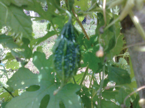
Photograph of M. charantia fruit used in this study.
2.2 Methods
The fresh fruits of M. charantia shown in Figure 1 were washed several times with distilled water to remove the dust. The fruits were cut into small pieces, 35 g of properly washed fruits were added in 175 ml of ultrapure water in a 500 ml Erlenmeyer flask and boiled for 10–15 min. Then Whatman filter paper (No. 40) was used for the filtration of the boiled material to prepare the aqueous fruit extract, which was used as such for the metal NPs synthesis.
Aqueous solution (1 mm) of AgNO3 was prepared. For the green synthesis of AgNPs, 1.8 ml of fruit extract was mixed to 50 ml of the prepared silver metal ion solution and continuously stirred for 4 min at room temperature. The reduction took place rapidly, with the formation of a brown-yellow colored solution after 30 min, indicative of the synthesis of the AgNPs. The effects of the reaction conditions such as the M. charantia fruit extract amount, silver ion concentration and reaction time were also studied.
2.3 Optimization study
To optimize the green biosynthesis of AgNPs, the effect of process variables, including reaction time, silver ion concentration and fruit extract amount was studied.
To investigate the effect of contact time on the green biosynthesis process, the aqueous extract of M. charantia (1.8 ml) was mixed with 50 ml of 1 mm AgNO3 for different time intervals (ranging from 15 min to 5 h). The green biosynthesis of AgNPs were carried out at different silver ion concentrations (0.1 mm–5 mm), with 1.8 ml fruit extract solutions without varying the other conditions. Different amounts (0.5, 1, 1.8, 2.8, 3.8 and 4.8 ml) of aqueous extract of M. charantia were tested and mixed with 50 ml of 1 mm AgNO3 solution.
2.4 UV-Vis and FTIR analyses
The biosynthesis of the AgNPs were characterized by using a UV-Vis spectrophotometer and FTIR spectroscopy. UV-Vis spectra were recorded on a double beam spectrophotometer (Perkin-Elmer Lambda 25, MA, USA) from 300 to 700 nm at a resolution of 1 nm. The distilled water was used as a blank. The synthesized AgNPs were subjected to FTIR spectroscopy measurement. In order to determine the functional groups on the M. charantia extract solution and their possible involvement in the synthesis of AgNPs, FTIR analysis was carried out as described earlier [25]. Momordica charantia fruit extract before reaction with AgNO3 (control samples) and M. charantia fruit extract after reaction with the AgNPs solution (test samples) were used for FTIR analysis. The FTIR spectra were collected at a resolution of 4 cm-1 in the transmission mode (4000–500 cm-1) using a Perkin-Elmer Spectrum-65 FTIR spectrometer (MA, USA).
2.5 TEM analysis
The green synthesized AgNPs using the fruit extract of M. charantia structural morphology and crystallinity were further confirmed by TEM micrograph images. An aliquot of the AgNP solution was placed on copper grid, making a thin film of a sample of the grid and kept for drying at room temperature for 15 min, then the extra sample was removed using the cone of a blotting paper and reserved in a grid box.
3 Results and discussion
Reduction of Ag+ into AgNPs during the exposure to the fruit of M. charantia extract could be seen by the color change. The color of fresh suspension of M. charantia extract was gray-brown. However, after the addition of the AgNO3 solution and the incubation for 30 min at room temperature, the mixture turned brown-yellow. Color changes in aqueous solutions are due to the surface plasmon resonance phenomenon. The results showed the fruit of M. charantia have a good potential for synthesizing the AgNPs as a reducing agent. According to the study of Shameli et al. [26], the chemical equations for the biosynthesis of the AgNPs are possible as follows:
After dispersion of silver ions in the M. charantia aqueous extract [Eq. (1)], the formed [Ag (M. charantia)]+ complex reacted with the aldehyde groups in the molecular structure of the extract to obtain [Ag (M. charantia)] due to the reduction of silver ions by the oxidation of the aldehyde to the carboxylic acid groups [Eq. (2)].
The formation of AgNPs from 1 mm solution of AgNO3 was confirmed by using UV-Vis spectral analysis. Metal AgNPs have free electrons, which give rise to a surface plasmon resonance (SPR) absorption band [27], due to the combined vibration of electrons of metal NPs in resonance with the light wave [28]. Surface plasmon spectra were obtained for brown-yellow colored silver solutions in the range of 300–700 nm.
3.1 Optimization study
3.1.1 Effects of contact time
The formation of AgNPs started within 15 min and the spectra were recorded after that at 15 min, 30 min, 1, 2, 3, 4 and 5 h. The effect of the contact time on AgNPs synthesis was evaluated with UV-Vis spectra and it was noted that with the increase in interaction time the SPR peak became sharper (Figure 2). The reaction time found in the development of NPs in this study was found to significantly increase up to 5 h and had a comparatively shorter reaction time than in earlier reports [29].
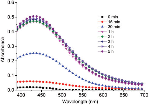
Effect of contact time on AgNPs synthesis.
3.1.2 Effects of silver ion concentration
The effect of AgNO3 concentration on the formation of AgNPs was analyzed using UV-Vis spectrophotometer (Figure 3). It is clear from Figure 3 that the formation of AgNPs depends on the AgNO3 concentration. Plasmon resonance spectra for AgNPs was obtained in 440 nm with brown-yellow colors, at different metal ion concentration. Moreover, it was found that the peak absorbance value increases with the increase in AgNO3 concentration (0.1 mm to 5 mm) which means that the rate of formation of AgNPs is higher for higher concentration of AgNO3 than for lower concentration.
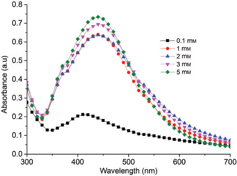
Effect of silver ion concentration on AgNPs synthesis.
3.1.3 Effects of fruit quantity
Different quantities of M. charantia fruit were used for the synthesis of AgNPs. The fruit extract quantity was varied from 0.5, 1.0, 1.8, 2.8, 3.8, 4.8 ml in 50 ml of 1 mm AgNO3 solution which was used in the synthesis of AgNPs. Based on the UV-Vis spectra, the sharpness of the absorption peak depends on the concentration of the fruit extract, which gets further sharpened at a higher concentration (Figure 4). With an increase in fruit extract quantities from 0.5 to 4.8 ml, a consistent increase in the peak absorbance was found (Figure 4). Here the results show the increase in the formation of AgNPs was maximum for the higher fruit extract quantity as well. Similarly, visual examination of the solutions revealed color changes from light yellow to deep yellow on silver solutions with an increase of fruit extract quantity in the reaction solution.
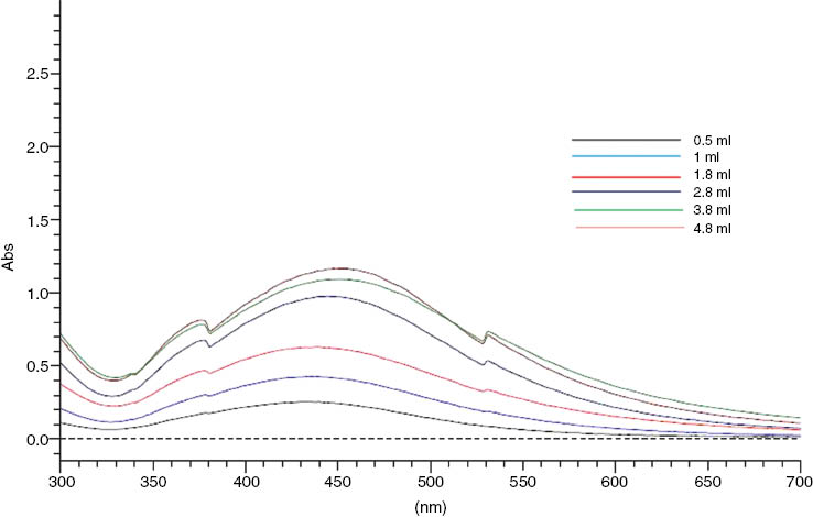
Effect of fruit extract quantity on AgNPs synthesis.
3.1.4 FTIR studies
FTIR has emerged as an important tool for understanding the involvement of surface functional biological groups in metal interaction. FTIR spectroscopy analysis was carried out to identify the possible biomolecules that were responsible for the stabilization of the AgNPs synthesized by M. charantia fruit extract (Figure 5). The FTIR spectra was recorded for the fruit extract and also for the AgNPs. The FTIR illustrates the absorbance bands observed at 3336 cm-1, 1664 cm-1, 1208 cm-1 and 1284 cm-1 in the 4000–500 cm-1 region. A major peak was observed at 3336 cm-1 that could be responsible for O-H stretching [30]. The peak located at 1660 cm-1 indicates the presence of C=O stretching in carboxyl or C=N bending in the amide group [31]. The band with a peak at 1208 cm-1 and 1284 cm-1 corresponds to C-O stretching of esters, ethers and phenols [32].

FTIR spectra of samples before and after the treatment producing AgNPs.
M. charantia fruits contain a variety of flavonoids and phenolic compounds which may be involved in the biosynthesis of AgNPs and act as a reducing agent for the reduction of Ag+ to Ag0. Phenolic compounds possess carboxyl and hydroxyl groups, which are capable to bind to metal [33]. Flavonoids can also directly strip molecular species of active oxygen. Their antioxidant activity resides mainly in their capability to provide electron or hydrogen atoms [34]. We conclude from the overall observations that the synthesized NPs were encircled by the different functional groups, such as carboxyl, carbonyl, amide, ester, ether and phenol. From the investigation of FTIR studies, we observed that these functional groups have stronger ability to bind metal NPs to prevent aggregation and provide higher stability. It is clear from the above discussion that the biological molecules can probably perform the dual functions of formation and stabilization of AgNPs in the aqueous medium [35].
3.1.5 TEM images
The structure and morphology of the green synthesized AgNPs using the extract of M. charantia were further confirmed by the TEM (Tecnai G2 200 kV TEM, Hillsboro, Oregon, USA) micrograph images. Figure 6A and B show various TEM images with different magnifications, depicting that the AgNPs were spherical in shape, with a particle size distribution between 8 and 47 nm.

TEM images of AgNPs using M. charantia fruit extract at different nanometers.
4 Conclusion
In this work, the AgNPs were synthesized using an aqueous extract of M. charantia fruit. The AgNPs were characterized by UV-visible, FTIR spectroscopy, and TEM analysis. The biosynthesis of AgNPs using green resources like M. charantia is a good method over chemical synthesis because this method is environmently-friendly. M. charantia extract was prepared and successfully employed for the development of AgNPs. The results showed that the formation of AgNPs was strongly dependent on the process parameters such as M. charantia extract concentration, silver ion concentration and the reaction time of the solution. This simple, low cost and greener method for the development of AgNPs may be valuable in biotechnological, biomedical and environmental applications.
About the authors

Mst Kamrun Nahar is currently pursuing her PhD degree at the Institute of Nano Electronic Engineering (INEE), Universiti Malaysia Perlis (UniMAP). Her research interests include slaughter and non-slaughter meat, the green biosynthesis of nanoparticles, the functional and physicochemical properties of proteins.

Zarina Zakaria earned her Bachelor’s degree in Botany (1995) from University Malaya and her Master’s degree (1998) from Universiti Putra Malaysia. She obtained her PhD in Biotechnology in 2004 from Universiti Sains Malaysia. Currently, she is a senior lecturer and program chairman of the Faculty of Engineering Technology (FETech), Universiti Malaysia Perlis (UniMAP). Her research interests include biotechnology and tissue culture.

Uda Hashim earned his Bachelor’s degree in Physical and Applied Physics (1987) from Universiti Kebangsaan Malaysia (UKM). He obtained his PhD in Microelectronic from Universiti Kebangsaan Malaysia in 2001. Currently, he is a director of the Institute of Nano Electronic Engineering (INEE), Universiti Malaysia Perlis (UniMAP). His research interests include semiconductor devices, CMOS based sensor, nanoelectronic and nano biochips.

Md Fazlul Bari earned his Bachelor’s (1990) and Master’s degree (1993) in Applied Chemistry from the university of Rajshahi, Bangladesh. He obtained his PhD in Analytical Chemistry from Universiti Sains Malaysia in 2004. Currently, he is an Associate Professor at the School of Materials Engineering, Universiti Malaysia Perlis (UniMAP). His research interests include hydrometallurgy and food chemistry.
Acknowledgments
The authors acknowledge the Universiti Malaysia Perlis (UniMAP) for their financial support for the Graduate Assistantship (GA).
Conflicts of interest: The authors declare that there is no conflict of interests regarding the publication of this article.
References
[1] Albrecht MA, Evans CW, Raston CL. Green Chem. 2006, 8, 417–432.Search in Google Scholar
[2] Yu D-G. Colloids Surf. B 2007, 59, 171–178.10.1016/j.colsurfb.2007.05.007Search in Google Scholar PubMed
[3] Mallick K, Witcomb MJ, Scurrell MS. Mater. Chem. Phys. 2005, 90, 221–224.Search in Google Scholar
[4] Kowshik M, Ashtaputr S, Kharrazi S, Vogel W, Urban J, Kulkarni SK, Paknikar KM. Nanotechnology 2003, 14, 95.10.1088/0957-4484/14/1/321Search in Google Scholar
[5] Shahverdi AR, Minaeian S, Shahverdi HR, Jamalifar H, Nohi A-A. Process Biochem. 2007, 42, 919–923.Search in Google Scholar
[6] Chandran SP, Chaudhary M, Pasricha R, Ahmad A, Sastry M. Biotechnol. Prog. 2006, 22, 577–583.Search in Google Scholar
[7] Dubey SP, Lahtinen M, Särkkä H, Sillanpää M. Colloids Surf. B 2010, 80, 26–33.10.1016/j.colsurfb.2010.05.024Search in Google Scholar PubMed
[8] Nazeruddin GM, Prasad NR, Waghmare SR, Garadkar KM, Mulla IS. J. Alloys Compd. 2014, 583, 272–277.Search in Google Scholar
[9] Dubey SP, Lahtinen M, Sillanpää M. Process Biochem. 2010, 45, 1065–1071.Search in Google Scholar
[10] Yang N, Li W-H. Ind. Crop. Prod. 2013, 48, 81–88.Search in Google Scholar
[11] Iravani S, Zolfaghari B. BioMed. Res. Int. 2013, 2013, 1.Search in Google Scholar
[12] Jagtap UB, Bapat VA. Ind. Crop. Prod. 2013, 46, 132–137.Search in Google Scholar
[13] Pavani K, Gayathramma K, Banerjee A, Suresh S. Am. J. Nanomater 2013, 1, 5–8.Search in Google Scholar
[14] Korbekandi H, Chitsazi MR, Asghari G, Najafi RB, Badii A, Iravani S. Green Process Synth. 2014, 3, 365–373.Search in Google Scholar
[15] Korbekandi H, Jouneghani RM, Mohseni S, Pourhossein M, Iravani S. Green Process Synth. 2014, 3, 271–277.Search in Google Scholar
[16] Schultz S, Smith DR, Mock JJ, Schultz DA. Proc. Natl. Acad. Sci. 2000, 97, 996–1001.Search in Google Scholar
[17] Sharma VK, Yngard RA, Lin Y. Adv. Colloid. Interface Sci. 2009, 145, 83–96.Search in Google Scholar
[18] Krutyakov YA, Kudrinskiy AA, Olenin AY, Lisichkin GV. Russ. Chem. Rev. 2008, 77, 233–257.Search in Google Scholar
[19] Kameswara Rao B, Kesavulu M, Apparao C. Jethnopharmacol. 2001, 78, 67–71.Search in Google Scholar
[20] Kubola J, Siriamornpun S. Food Chem. 2008, 110, 881–890.Search in Google Scholar
[21] Joseph B, Jini D. Asian Pac. J. Trop. Dis. 2013, 3, 93–102.Search in Google Scholar
[22] Fang FE, Ng TB. Curr. Mol. Med. 2011, 11, 417–436.Search in Google Scholar
[23] Limtrakul P, Khantamat O, Pintha K. Cancer Chemoth. Pharmacol. 2004, 54, 525–530.Search in Google Scholar
[24] Ross, IA, Eds., Medicinal Plants of the World: Chemical Constituents, Traditional and Modern Medicinal Uses. v. 1–3. Springer: Vol. 3, 2005.Search in Google Scholar
[25] Bankar AV, Kumar AR, Zinjarde SS. J. Hazard Mater 2009, 170, 487–494.10.1016/j.jhazmat.2009.04.070Search in Google Scholar PubMed
[26] Shameli K, Ahmad MB, Al-Mulla EAJ, Ibrahim NA, Shabanzadeh P, Rustaiyan A, Abdollahi Y, Bagheri S, Abdolmohammadi S, Usman MS, Zidan M. Molecules 2012, 17, 8506–8517.10.3390/molecules17078506Search in Google Scholar PubMed PubMed Central
[27] Noginov MA, Zhu G, Bahoura M, Adegoke J, Small C, Ritzo BA, Drachev VP, Shalaev VM. Appl. Phys. B 2007, 86, 455–460.10.1007/s00340-006-2401-0Search in Google Scholar
[28] Nath S, Chakdar D, Gope G. J. Nanotechnol. Appl. 2007, 2.Search in Google Scholar
[29] Mohammed Fayaz A, Balaji K, Kalaichelvan PT, Venkatesan R. Colloids Surf. B 2009, 74, 123–126.10.1016/j.colsurfb.2009.07.002Search in Google Scholar PubMed
[30] Prathna TC, Chandrasekaran N, Raichur AM, Mukherjeea A. Colloids Surf. B 2011, 82, 152–159.10.1016/j.colsurfb.2010.08.036Search in Google Scholar PubMed
[31] Bankar A, Joshi B, Kumar AR, Zinjarde S. Colloids surf. A 2010, 368, 58–63.10.1016/j.colsurfa.2010.07.024Search in Google Scholar
[32] Chang Chien SW, Wang MC, Huang CC, Seshaiah K. J. Agric. Food Chem. 2007, 55, 4820–4827.Search in Google Scholar
[33] Harborne, JB, Eds., Biochemistry of Phenolic Compounds, Academic Press: London, 1964.Search in Google Scholar
[34] Ahmad N, Sharmab S, Alama MK, Singhc VN, Shamsid SF, Mehtac BR, Fatmae A. Colloids Surf. B 2010, 81, 81–86.10.1016/j.colsurfb.2010.06.029Search in Google Scholar PubMed
[35] Tripathi R, Kumar N, Shrivastav A, Singh P, Shrivastav B. J. Mol. CatalB. Enzym. 2013, 96, 75–80.Search in Google Scholar
©2015 by De Gruyter
This article is distributed under the terms of the Creative Commons Attribution Non-Commercial License, which permits unrestricted non-commercial use, distribution, and reproduction in any medium, provided the original work is properly cited.
Articles in the same Issue
- Frontmatter
- In this issue
- Editorial
- Has GPS landed with precision?
- Original articles
- Solar production of WO3: a green approach
- Sesbania sesban L. biomass as a novel adsorbent for removal of Pb(II) ions from aqueous solution: non-linear and error analysis
- Green methacrylated lignin model compounds as reactive monomers with low VOC emission for thermosetting resins
- Deposition behavior of TiB2 by microwave heating chemical vapor deposition (CVD)
- Interaction and thermodynamics of methylene blue adsorption on oxidized multi-walled carbon nanotubes
- Alkaline leaching of zinc from low-grade oxide zinc ore using ammonium citrate as complexing agent
- Heat shock and titanium dioxide nanoparticles decrease superoxide dismutase and glutathione enzymes activities in Saccharomyces cerevisiae
- Momordica charantia fruit mediated green synthesis of silver nanoparticles
- The continuous synthesis and application of graphene supported palladium nanoparticles: a highly effective catalyst for Suzuki-Miyaura cross-coupling reactions
- Conference announcements
- 5th Flow Chemistry Congress (San Diego, CA, USA, September 15–16, 2015)
- Conferences 2015–2017
- Book reviews
- Flow chemistry
- Biomass as a sustainable energy source for the future: fundamentals of conversion processes