Expression of OX40 Gene and its Serum Levels in Neuromyelitis Optica Patients
-
Parya Alidadiani
 ,
Vahid Shaygannejad
,
Vahid Shaygannejad

Abstract
Neuromyelitis optica (NMO), also known as Devic’s disease, is an autoimmune disorder of the central nervous system (CNS) in which immune system cells and antibodies primarily attack the optic nerves and the spinal cord. OX40 (CD134) is a tumor necrosis factor (TNF)-receptor family member expressed primarily on activated CD4+ and CD8+ T-cells. In an autoimmune disease, OX40 is typically up-regulated at sites of inflammation, and increases in the number of peripheral CD4+ T-cells expressing OX40. OX40 and its ligand OX40L are key TNF members that augment T-cell expansion, cytokine production, and promote T-cell survival. The aim of this study was to evaluate and compare of OX40 gene expression and its serum levels in patients with NMO and healthy controls. Twenty sex-/age-matched healthy controls (HC) (median age = 32 years, 15 females/5 males) were engaged for the present study. Expression of OX40 at the transcript level and serum protein levels were measured by quantitative real-time polymerase chain reaction (qRT-PCR) and enzyme-linked immunosorbent assays, respectively. The results indicated OX40 expression in patients was significantly lower than in healthy controls (p = 0.001). However, the serum level of OX40 was not significantly different between groups (p = 0.37). In addition, the results indicated that CD134 expression was not age-related (p = 0.041). Overall, this study suggests to us that OX40 levels are not a suitable marker for diagnosis or treatment of NMO.
Introduction
Neuromyelitis optica (NMO), also known as Devic’s disease, is an autoimmune disorder of the central nervous system (CNS) in which immune system cells and antibodies attack the optic nerves and the spinal cord. Like multiple sclerosis (MS), NMO is a relapsing-remitting disease [1, 2]. During any relapse, new damage to the optic nerves and/or spinal cord can lead to accumulating disability. Unlike MS, there is no progressive phase of this disease. Therefore, preventing attacks is critical to a good long-term outcome.
Among NMO and NMO spectrum disorder (NMOSD) patients, > 70% are characterized by a presence of serum autoantibodies against astrocyte water channel aquaporin 4 (AQP4) on cell membranes [3, 4]. These pathogenic IgG1 antibodies are important as they illustrate how AQP4-specific T-cells likely participate in the pathogenesis of NMO [5]. Among these, there is an IgG1 autoantibody that is also found in patients with two related neurologic conditions (two-sided optic neuritis (ON) and longitudinal wide transverse myelitis (LETM)) that fall in the NMO range of diseases, [5]. Thus, there is the potential to develop regimens to counteract these and other antibodies - once the latter have been identified - as a means to help prevent/mitigate NMO attacks.
OX40 (CD134) is a tumor necrosis factor (TNF)-receptor family member expressed primarily on activated CD4+ and, CD8+ and regulatory T-cells (Treg cells) [6]. Normally, OX40 is activated by the ligand OX40L (CD134L), and thus functions as a T-cell co-stimulatory molecule. It is known that OX40-OX40L interactions can promote T-cell survival, including that of effector and memory T-cells, as
well as enhance the production of several cytokines [7, 8, 9]. Studies have shown that use of anti-OX40 in the treatment of some autoimmune diseases imparts a potent anti-inflammatory activity in a manner that is dependent on both CD4+ and CD8+ T-cells [10, 11]. Immunization models have also shown that the use of anti-OX40 increases T-cell proliferation and differentiation into expanded populations of effector T-cells, with concomitant changes in cytokine production, T-cell-mediated cytotoxicity, and a decrease in activation-induced cell death [11, 12].
In several autoimmune diseases (e.g., Systemic lupus erythmathosus [SLE], colitis), the presence of OX40 is up-regulated at sites of inflammation, and there are increases in the number of peripheral CD4+ T-cells expressing OX40 [13, 14]. In SLE, the expression of OX40 on CD4+ T-lymphocytes and the serum level of OX40L may act as markers of lupus nephritis and it suggests there is a correlation between the presence of OX40 expression and disease severity[15]. As OX40 also promotes the colitogenic T-helper cell type 1 (TH1) response, its expression on Treg cells may be required for effective competition with OX40-dependent effector responses [14, 16]. In addition, stimulation of OX40-expressing cells after the induction of colitis exaggerates intestinal inflammation - presumably by enhancing effector lymphocyte function. Thus, it seems that stimulation of a host with an agonistic OX40 antibody could increase the number of functional Treg cells that, in turn, can impact on any subsequent developing/ongoing immune response [13, 16, 17].
Soluble OX40 (sOX40) may be produced through cleavage from cell surface OX40 and/or alternative splicing [18, 19, 20]. Soluble forms of TNF receptors have antagonistic effects on membrane-bound receptors. Therefore, it is possible that abnormal activation of T-cells may induce alternative splicing of sOX40 in patients with NMO [9, 12, 21]. However, the function of sOX40 in NMO remains unknown.
The OX40 expression level in peripheral white blood cells of NMO patients may provide insight into immunologic mechanisms related to NMO progression. Accordingly, this study was designed to evaluate OX40 gene expression and serum levels (as sOX40) of the protein for potential use in helping in the eventual development of more effective targeted treatments against this pathology.
Materials and Methods
Patients and controls
Twenty NMO patients were recruited for the study (median age = 31 years, 15 female/5 male; mean disease duration = 2.4 years) from the Kashani Hospital of Isfahan (Iran). Diagnosis of NMO was confirmed by clinical and neurophysiological parameters. All patients met research criteria used to define NMO [3], as verified by a hospital neurologist. For all patients, both the longitudinally-extensive transverse myelitis (LETM) as annualized relapse rate and the Expanded Disability Status Scale (EDSS) were assessed. Cerebral and spinal cord magnetic resonance imaging (MRI) were used to evaluate the presence of specific NMO-like lesions. As controls, 20 healthy sex-/age-matched individuals (HC; median age = 32 years, 15 female/5 male) were also recruited for study. Table 1 presents the demographic and clinical characteristics of all study subjects.
Characteristics of NMO patient and control subjects.
| NMO | Controls | |
|---|---|---|
| Females/Males | 15/5 | 15/5 |
| Average age at onset | 31 | 31 |
| AQ-P4 Ab positive (%) | 30 | --- |
| LETM (%) | 60 | --- |
| Normal brain (MRI) (%) | 80 | --- |
| EDSS (n) | 2 | --- |
Values are presented as frequencies (percentages)
LETM: Longitudinally-extensive transverse myelitis, annualized relapse rate.
EDSS: Expanded Disability Status Scale.
A total of 5 ml fasting venous blood was taken from each individual. Two ml was collected into EDTA-containing tubes for subsequent use in white blood cell RNA extraction. The other 3 ml was collected into a tube without anti-coagulant; this material was immediately centrifuged (4000 rpm, 6 min, 4°C), and the resultant serum was harvested and stored at -20°C for later analysis.
Informed consent: Informed consent has been obtained from all individuals included in this study.
Ethical approval: The research related to human use has been complied with all the relevant national regulations, institutional policies and in accordance the tenets of the Helsinki Declaration, and has been approved by the ethical committee of Isfahan University of Medical Sciences.
RNA extraction and RT-PCR
From each whole blood sample, total RNA was extracted from white blood cells using a YTA kit Total RNA Purification Mini kit (Yekta Tajhiz, Tehran, Iran), according to manufacturer’s protocol. Concentration and integrity of total isolated RNA from each sample was assessed using a Nanophotometer Pipette System (Helmholtz, Nauenberg, Germany) and the material was then stored at -70°C (mean concentration was ≈ 80 μg/ml).
From each sample, cDNA was prepared using a Thermo Scientific Revert Aid RT Kit containing oligo dT primer mixture (Bio-Rad, Watford, UK) according to manufacturer protocol. In brief, 11 μl (≈ 0.9 μg) total RNA was mixed with 1 μl oligo dT primer, and then incubated at 65°C for 5 min). Thereafter, 8 μl Reverse Transcription Master Mix (5X Reaction Buffer) and 2X dNTP Mix (both Applied Biosystems, Cheshire, UK) was added (on ice), and the mixture was incubated for 1 hr at 42°C and then 10 min at 70°C.
Real-time PCR
qRT-PCR was performed using Syber Green PCR Master Mix (Applied Biosystems). Forward and reverse primer sequences used for the qRT-PCR reactions (Table 2) were designed using Allele ID version 7. Each qPCR was performed in a Step One Plus Real-Time PCR system (Applied Biosystems). Thermocycler conditions used in each case were 95°C for 15 min (initial denaturation) followed by 40 cycles of 95°C for 15 sec, 54°C for 60 sec, and 72°C for 20 sec, with a final extension of 72°C for 1 min. All results were evaluated using the 2-DDCt method [22, 23].
Real-time quantitative PCR primers used in the study.
| Primer | Sequence |
|---|---|
| GAPDH Forward | AAGCTCATTTCCTGGTATG |
| GAPDH Reverse | CTTCCTCTTGTGCTCTTG |
| OX40 Forward | TGGTGTAACCCAGAAGTG |
| OX40 Reverse | GTCAACTCCAGGCTTGTA |
ELISA
sOX40 levels in sera were measured (in duplicate) using a human sCD134 (OX40) platinum ELISA kit (eBioscience, Vienna, Austria), following manufacturer protocols. Optical density (OD) at 450 nm wavelength was recorded for each sample using a Hiperion Microplate Reader (Rodermark, Germany). To establish seropositivity, serum from each NMO patient was also evaluated for anti-Aquaporin 4 (AQP4) antibody using an Indirect Immunofluorescence Test (IIFT) cell-based assay (EUROIMMUN IIFT, Luebeck, Germany).
Statistical analysis
All results are expressed as mean ± SD. Normal distribution of all data was determined using a Kolmogorov-Smirnov test. A non-parametric Mann-Whitney U-test was applied for comparing values between groups. A p-value < 0·05 was accepted as significant. All analyses were conducted using an SPSS 20 package (SPSS Inc., Chicago, IL).
Results
OX40 mRNA expression
OX40 mRNA in the white blood cells of NMO patients was found to be significantly (p = 0.001) lower than in control individuals’ cells. The average Ct values were 32.1 and 29.7, respectively. Within-gender analyses revealed that females with NMO had levels significantly (p = 0.041) lower than in female control counterparts; average values were 32.8 and 30.4, respectively. There were no significant differences between male NMO and male controls (p = 0.70). In total, there appeared to be no correlation between OX40 mRNA and age in this group of NMO patients.
Serum sOX40 levels
The ELISA data indicated there were no significant differences between sOX40 levels in the serum of NMO patients compared with those from healthy controls (p = 0.37) (Figure 1). The average values were 12.7 and 14.9 ng/ml, respectively. The current analyses found that median sOX40 levels in AQP4 seropositive subjects were higher than in seronegative NMO patients, but this difference was not significant (p = 0.45) (Figure 2).
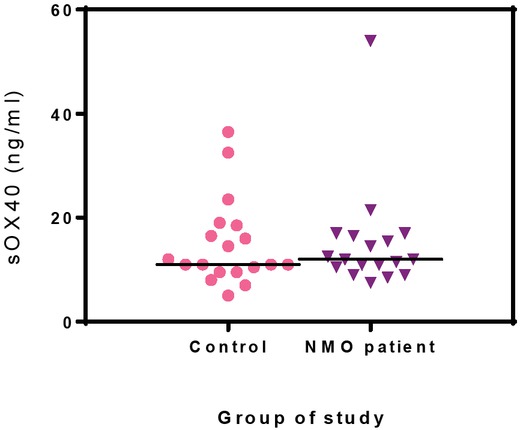
Serum sOX40 levels. There were no significant differences in sOX40 serum levels when comparing NMO patients with controls (p = 0.37). Values shown are means ± SD of 20 subjects/group.

sOX40 serum levels in AQP4 seropositive and -negative NMO patients. There were no significant differences between the two groups (p = 0.044.). Values shown are means ± SD from 6 seropositive and 12 seronegative NHO patients.
Discussion
Several studies have provided evidence for an important role of effector T-cells in various steps during the pathogenesis of neuromyelitis optica (NMO). Levels of interleukin (IL)-17-secreting CD4+ T-cells (TH17 cells) are known to be significantly elevated in NMO patients, suggesting these autoantigen-specific T-cells might help drive some of the pathologies associated with NMO [24]. Interestingly, IL-6 is not only elevated in the cerebral-spinal fluid (CSF), but also in the sera, of patients with NMO. This is important in that IL-6 is involved in development of TH17 cells [25]. Serum IL-22 and IL-21 levels are also elevated during NMO [26] and so it is not surprising that TH22 cell levels have been found to significantly correlate with those of TH17 cells in these patients. Certain types of T-cells are also found in the lesions that occur in NMO patients, though their antigen specificity and function have not been characterized [27]. Thus, a variety of effector T-cells seem to be potentially required for overall NMO development. Whether these are specifically needed for disease-related lesion development, or for disrupting blood-brain or blood-CSF barriers, or just perhaps for creating an inflammatory milieu in situ for anti-AQP4 antibodies to be operational remains to be determined.
At the factor level, a variety of co-stimulatory molecules (such as CD69) have been shown to play important roles in the NMO pathogenesis. It is plausible to assume that the presence - or absence - of other factors may also likely contribute to T-cell activation in NMO [5]. OX40 is essential for optimal function of T-cells. For example, a role for OX40 signaling in activation of cytolytic T-lymphocytes is well established [14]. Expression of OX40 is also believed to be a factor involved in the ability of T-cells to leave the general circulation and enter the CNS [28]. In addition, OX40 has an important role in preventing T-cell apoptosis by inducing expression of anti-apoptotic molecules (such as Bcl-X). When taken together, it is clear that OX40 can impact on the size of the effector T-cell pool in a host. Accordingly, as critical components of T-cell responses, OX40 is likely to be important in the context of autoimmunity [29, 30, 31].
In this study, it was seen that gene expression of OX40 was significantly decreased in the peripheral white blood cells of NMO patients. At the same time, circulating levels of the soluble form of the OX40 protein (sOX40) was unaltered by disease status. This observation contrasts with that of another investigations wherein sOX40 levels in sera of patients with ALS were decreased compared to control values [32]. Other reports have shown that active human NMO lesions contained increased numbers of CD4+ T-cells expressing the OX40 activation marker, and that expression was more profound compared to that seen in MS lesions of comparable activity [33]. Recently, it was shown that T-cells isolated from the spinal cords of Lewis rats with actively-induced EAE (experimental allergic encephalomyelitis) expressed OX40 [28]. As such, it is not clear why systemic levels of OX40 (specifically, as soluble sOX40 form) in NMO patients here were the same as in the control subjects. Similarly, it is not readily apparent why expression levels of OX40 gene among circulating leukocytes in the NMO patients here were decreased in comparison to in control subject cells.
The present study indicated that OX40 gene expression in circulating white blood cells was significantly lower in NMO females than in cells of corresponding female controls. Interestingly, there was no similar male-related differential in outcome. While it is known sex hormones (including estrogens and androgens) have crucial roles in autoimmune diseases [34], it is not clear why there was such a bifurcation in outcomes between the male and female NMO patients (vs. controls) here.
Lastly, the present study showed OX40/CD134 expression (serum) was not age-related among the NMO patients. It is known that CD134 surface density on CD4+ cells was decreased in spinal cords of aging rats and on in vitro-stimulated naïve CD4+ splenocytes taken from aged rats vs. cells from younger rats [35]. This invites the question of if expression of the marker is impacted by localization to a given tissue, as opposed to any similar influence on T-cells while they remain in the general circulation. This specific issue warrants further study.
Acknowledgments
This study was supported by the Isfahan University of Medical Sciences. The authors would like to thank to all employees of the Central Laboratory of IUMS for their help in the performance of this study.
Conflict of interest: Authors state no conflict of interest.
Declaration: The authors alone are responsible for the content of this manuscript.
References
1 Jacob A, McKeon A, Nakashima I, Sato DK, Elsone L, Fujihara K, et al. Current concept of neuromyelitis optica (NMO) and NMO spectrum disorders. J Neurol Neurosurg Psychiatry. 2013;84(8):922-30.10.1136/jnnp-2012-302310Suche in Google Scholar PubMed
2 Matiello M, Jacob A, Wingerchuk DM, Weinshenker BG. Neuromyelitis optica. Current opinion in neurology. 2007;20(3):255-60.10.1097/WCO.0b013e32814f1c6bSuche in Google Scholar PubMed
3 Morrow MJ, Wingerchuk D. Neuromyelitis optica. Journal of Neuro-Ophthalmology. 2012;32(2):154-66.10.1097/WNO.0b013e31825662f1Suche in Google Scholar
4 Trebst C, Jarius S, Berthele A, Paul F, Schippling S, Wildemann B, et al. Update on the diagnosis and treatment of neuromyelitis optica: recommendations of the Neuromyelitis Optica Study Group (NEMOS). Journal of neurology. 2014;261(1):1-16.10.1007/s00415-013-7169-7Suche in Google Scholar
5 Drori T, Chapman J. Diagnosis and classification of neuromyelitis optica (Devic’s syndrome). Autoimmunity reviews. 2014;13(4-5):531-3.10.1016/j.autrev.2014.01.034Suche in Google Scholar PubMed
6 Akiba H, Oshima H, Takeda K, Atsuta M, Nakano H, Nakajima A, et al. CD28-independent costimulation of T cells by OX40 ligand and CD70 on activated B cells. The Journal of Immunology. 1999;162(12):7058-66.10.4049/jimmunol.162.12.7058Suche in Google Scholar
7 Ishii N, Takahashi T, Soroosh P, Sugamura K. OX40– OX40 ligand interaction in T-cell-mediated immunity and immunopathology. Advances in immunology. 105: Elsevier; 2010. p. 63-98.10.1016/S0065-2776(10)05003-0Suche in Google Scholar
8 Kaur D, Brightling C. OX40/OX40 ligand interactions in T-cell regulation and asthma. Chest. 2012;141(2):494-9.10.1378/chest.11-1730Suche in Google Scholar PubMed PubMed Central
9 Webb GJ, Hirschfield GM, Lane PJ. OX40, OX40L and autoimmunity: a comprehensive review. Clinical reviews in allergy & immunology. 2016;50(3):312-32.10.1007/s12016-015-8498-3Suche in Google Scholar PubMed
10 Sand IK. Neuromyelitis optica spectrum disorders. CONTINUUM: Lifelong Learning in Neurology. 2016;22(3, Multiple Sclerosis and Other Demyelinating Diseases):864-96.10.1212/CON.0000000000000337Suche in Google Scholar PubMed
11 Sato DK, Lana‐Peixoto MA, Fujihara K, de Seze J. Clinical spectrum and treatment of neuromyelitis optica spectrum disorders: evolution and current status. Brain Pathology. 2013;23(6):647-60.10.1111/bpa.12087Suche in Google Scholar PubMed PubMed Central
12 Linch SN, McNamara MJ, Redmond WL. OX40 agonists and combination immunotherapy: putting the pedal to the metal. Frontiers in oncology. 2015;5:34.10.3389/fonc.2015.00034Suche in Google Scholar PubMed PubMed Central
13 Griseri T, Asquith M, Thompson C, Powrie F. OX40 is required for regulatory T cell–mediated control of colitis. Journal of Experimental Medicine. 2010;207(4):699-709.10.1084/jem.20091618Suche in Google Scholar PubMed PubMed Central
14 Sitrin J, Suto E, Wuster A, Eastham-Anderson J, Kim JM, Austin CD, et al. The Ox40/Ox40 Ligand Pathway Promotes Pathogenic Th Cell Responses, Plasmablast Accumulation, and Lupus Nephritis in NZB/W F1 Mice. Journal of immunology (Baltimore, Md : 1950). 2017;199(4):1238-49.10.4049/jimmunol.1700608Suche in Google Scholar PubMed PubMed Central
15 Patschan S, Dolff S, Kribben A, Dürig J, Patschan D, Wilde B, et al. CD134 expression on CD4+ T cells is associated with nephritis and disease activity in patients with systemic lupus erythematosus. Clinical & Experimental Immunology. 2006;145(2):235-42.10.1111/j.1365-2249.2006.03141.xSuche in Google Scholar PubMed PubMed Central
16 Zander RA, Obeng-Adjei N, Guthmiller JJ, Kulu DI, Li J, Ongoiba A, et al. PD-1 co-inhibitory and OX40 co-stimulatory crosstalk regulates helper T cell differentiation and anti-Plasmodium humoral immunity. Cell host & microbe. 2015;17(5):628-41.10.1016/j.chom.2015.03.007Suche in Google Scholar PubMed PubMed Central
17 Kroemer A, Xiao X, Vu MD, Gao W, Minamimura K, Chen M, et al. OX40 controls functionally different T cell subsets and their resistance to depletion therapy. The Journal of Immunology. 2007;179(8):5584-91.10.4049/jimmunol.179.8.5584Suche in Google Scholar PubMed
18 Compaan DM, Hymowitz SG. The crystal structure of the costimulatory OX40-OX40L complex. Structure. 2006;14(8):1321-30.10.1016/j.str.2006.06.015Suche in Google Scholar PubMed
19 Jenkins SJ, Perona-Wright G, Worsley AG, Ishii N, MacDonald AS. Dendritic cell expression of OX40 ligand acts as a costimulatory, not polarizing, signal for optimal Th2 priming and memory induction in vivo. The Journal of Immunology. 2007;179(6):3515-23.10.4049/jimmunol.179.6.3515Suche in Google Scholar PubMed
20 Lei W, Zeng D-X, Zhu C-H, Liu G-Q, Zhang X-Q, Wang C-G, et al. The upregulated expression of OX40/OX40L and their promotion of T cells proliferation in the murine model of asthma. Journal of thoracic disease. 2014;6(7):979.Suche in Google Scholar PubMed
21 Montler R, Bell RB, Thalhofer C, Leidner R, Feng Z, Fox BA, et al. OX40, PD‐1 and CTLA‐4 are selectively expressed on tumor‐infiltrating T cells in head and neck cancer. Clinical & translational immunology. 2016;5(4):e70.10.1038/cti.2016.16Suche in Google Scholar PubMed PubMed Central
22 Livak KJ, Schmittgen TD. Analysis of relative gene expression data using real-time quantitative PCR and the 2− ΔΔCT method. methods. 2001;25(4):402-8.10.1006/meth.2001.1262Suche in Google Scholar PubMed
23 Wong ML, Medrano JF. Real-time PCR for mRNA quantitation. Biotechniques. 2005;39(1):75-85.10.2144/05391RV01Suche in Google Scholar PubMed
24 Blanc F, Noblet V, Jung B, Rousseau F, Renard F, Bourre B, et al. White matter atrophy and cognitive dysfunctions in neuromyelitis optica. PLoS One. 2012;7(4):e33878.10.1371/journal.pone.0033878Suche in Google Scholar PubMed PubMed Central
25 Kimura A, Kishimoto T. IL-6: regulator of Treg/Th17 balance. European journal of immunology. 2010;40(7):1830-5.10.1002/eji.201040391Suche in Google Scholar PubMed
26 Wang H, Dai Y, Qiu W, Lu Z, Peng F, Wang Y, et al. Interleukin-17-secreting T cells in neuromyelitis optica and multiple sclerosis during relapse. Journal of Clinical Neuroscience. 2011;18(10):1313-7.10.1016/j.jocn.2011.01.031Suche in Google Scholar PubMed
27 Lin J, Li X, Xia J. Th17 cells in neuromyelitis optica spectrum disorder: a review. The International journal of neuroscience. 2016;126(12):1051-60.10.3109/00207454.2016.1163550Suche in Google Scholar PubMed
28 Carboni S, Aboul-Enein F, Waltzinger C, Killeen N, Lassmann H, Peña-Rossi C. CD134 plays a crucial role in the pathogenesis of EAE and is upregulated in the CNS of patients with multiple sclerosis. Journal of neuroimmunology. 2003;145(1-2):1-11.10.1016/j.jneuroim.2003.07.001Suche in Google Scholar PubMed
29 Balashov KE, Rottman JB, Weiner HL, Hancock WW. CCR5+ and CXCR3+ T cells are increased in multiple sclerosis and their ligands MIP-1α and IP-10 are expressed in demyelinating brain lesions. Proceedings of the National Academy of Sciences. 1999;96(12):6873-8.10.1073/pnas.96.12.6873Suche in Google Scholar PubMed PubMed Central
30 Chen L, Flies DB. Molecular mechanisms of T cell co-stimulation and co-inhibition. Nature Reviews Immunology. 2013;13(4):227.10.1038/nri3405Suche in Google Scholar PubMed PubMed Central
31 Gerondakis S, Siebenlist U. Roles of the NF-κB pathway in lymphocyte development and function. Cold Spring Harbor perspectives in biology. 2010;2(5):a000182.10.1101/cshperspect.a000182Suche in Google Scholar PubMed PubMed Central
32 Iłżecka J. Serum soluble OX40 in patients with amyotrophic lateral sclerosis. Acta Clinica Croatica. 2012;51(1):3-6.Suche in Google Scholar PubMed
33 Pohl M, Kawakami N, Kitic M, Bauer J, Martins R, Fischer M-T, et al. T cell-activation in neuromyelitis optica lesions plays a role in their formation. Acta neuropathologica communications. 2013;1(1):85.10.1186/2051-5960-1-85Suche in Google Scholar PubMed PubMed Central
34 González DA, Díaz BB, Pérez MdCR, Hernández AG, Chico BND, de León AC. Sex hormones and autoimmunity. Immunology letters. 2010;133(1):6-13.10.1016/j.imlet.2010.07.001Suche in Google Scholar PubMed
35 Djikić J, Nacka-Aleksić M, Pilipović I, Stojić-Vukanić Z, Bufan B, Kosec D, et al. Age-associated changes in rat immune system: lessons learned from experimental autoimmune encephalomyelitis. Experimental gerontology. 2014;58:179-97.10.1016/j.exger.2014.08.005Suche in Google Scholar PubMed
© 2019 Parya Alidadiani et al., published by De Gruyter
This work is licensed under the Creative Commons Attribution 4.0 Public License.
Artikel in diesem Heft
- Research Article
- ATP Synthase: Structure, Function and Inhibition
- Insulin Promotes Wound Healing by Inactivating NFkβP50/P65 and Activating Protein and Lipid Biosynthesis and alternating Pro/Anti-inflammatory Cytokines Dynamics
- Mini review
- The disordered boundary of the cell: emerging properties of membrane-bound intrinsically disordered proteins
- Research Article
- Expression of OX40 Gene and its Serum Levels in Neuromyelitis Optica Patients
- Advances in Molecular biomarker for early diagnosis of Osteoarthritis
- Identification of BRCA1/2 p.Ser1613Gly, p.Pro871Leu, p.Lys1183Arg, p.Glu1038Gly, p.Ser1140Gly, p.Ala2466Val, p.His2440Arg variants in women under 45 years old with breast nodules suspected of having breast cancer in Burkina Faso
- Endometriosis Pathoetiology and Pathophysiology: Roles of Vitamin A, Estrogen, Immunity, Adipocytes, Gut Microbiome and Melatonergic Pathway on Mitochondria Regulation
- Integral membrane protein expression of human CD25 on the cell surface of HEK293 cell line: the available cellular model of CD25 positive to facilitate in vitro developing assays
- Review Article
- Role of Nanomedicine in Redox Mediated Healing at Molecular Level
- Research Article
- Glutathione S-transferase M1 (GSTM1) and T1 (GSTT1) variants and breast cancer risk in Burkina Faso
- Incidence of viruses infecting pepper in Thailand
- Mechanochemistry of von Willebrand factor
- Schizophrenia phenomenology comprises a bifactorial general severity and a single-group factor, which are differently associated with neurotoxic immune and immune-regulatory pathways
- Role of Killer cell immunoglobulin-like receptors (KIR) genes in stages of HIV-1 infection among patients from Burkina Faso
- The effect of rs9930506 FTO gene polymorphism on obesity risk: a meta-analysis
- Special Issue: Recent Advances in Basic and Clinical Medicine
- Antihyperglycemic and antihyperlipidemic activities of Nannochloropsis oculata microalgae in Streptozotocin-induced diabetic rats
- Comparison of the effect of vitamin D on osteoporosis and osteoporotic patients with healthy individuals referred to the Bone Density Measurement Center
- Zinc enhances the expression of morphine-induced conditioned place preference through dopaminergic and serotonergic systems
- Pre-operative laparoscopic staging of gastric cancer in patients who are candidates for neo-adjuvant chemotherapy: A Cross Sectional Study
- Neurotoxic Effects of Stanozolol on Male Rats‘ Hippocampi: Does Stanozolol cause apoptosis?
- Only serum pepsinogen I and pepsinogen I/II ratio are specific and sensitive biomarkers for screening of gastric cancer
- Evaluation of Quality of Life in Terms of Sinonasal Symptoms in Children with Cystic Fibrosis
- The Effect of Fine needle aspiration on Detecting Malignancy in Thyroid Nodule
- Diagnosis of Primary Hydatid Cyst of Thyroid Gland: A Case Report
- Ultrasonography in the diagnosis of lung adhesion before surgery