Incidence of viruses infecting pepper in Thailand
-


Abstract
This study was conducted to determine the incidence, diversity and distribution of viruses infecting pepper (Capsicum spp.) in the central, northern and northeastern parts of Thailand. During a survey in 2016 - 2019, a total of 2,149 leaf samples from symptomatic and asymptomatic peppers were collected randomly from farmer’s fields, and preliminary tested by an enzyme-linked immunosorbent assay (ELISA) using 7 antibodies specific for cucumber mosaic virus (CMV), chilli veinal mottle virus (ChiVMV), tomato necrotic ringspot virus (TNRV), tobacco mosaic virus (TMV), potato virus Y (PVY), tomato spotted wilt virus (TSWV), and begomoviruses. Our data revealed that the incidence of the viruses infecting pepper in Thailand was high, accounting for nearly 70% (1,482 infected samples). The highest viral incidence was found in the central part (96%), followed by the north (74.4%) and the northeastern (52.8%), respectively. Begomoviruses, CMV, ChiVMV, and TNRV were detected in the samples at varying rates, whereas PVY, TMV, and TSWV were not detected. Of these, the most frequently found virus was Begomoviruses accounting for nearly 33%, with the highest rate (ca. 82%) in the central Provinces of Thailand. In addition, of the 1,482 infected samples, mixed infections among the four viruses were also found in 616 samples (ca. 42%), and CMV + ChiVMV (approximately 11%) was the most common mixed infection. This is the first report describing an occurrence of viruses in pepper of Thailand, and the results obtained have revealed that viruses infecting pepper are widespread, which may pose a threat to pepper production in Thailand.
Introduction
Hot pepper (Capsicum spp.) native to Central and South America presently dominates the world’s hot spice trade, and due to its popularity, the pepper plants have been cultivated worldwide with a large annual production rate [1]. Due to its pungent taste and aroma, pepper becomes an important cash crop for farmers in many developing countries such as China, India, Pakistan, Indonesia, and Thailand.
Peppers in Thailand can be classified into two types based on fruit sizes: long and short. The length of the long peppers ranges from 9 - 15 cm and its pungency is from low to medium-hot, whereas that of the short ones varies from 2 - 7 cm and the pungency ranging from medium to very hot [2]. Two major Capsicum species are cultivated in Thailand: C. annuum and C. frutescens, although it should be noted that C. annuum is predominant and widely grown throughout the country [3]. Distinct morphology mainly by corolla color and number of flowers per node allow easy discrimination of these two species [4]. Peppers are grown on nearly 23,000 ha in Thailand with an annual production of about 170,000 t in 2016 [5]. Due to traditional practices, various factors are found to affect the pepper production yield. It has been estimated that pests and diseases were the major cause accounting for 40% of the yield loss of pepper production in Thailand in 2012 [6].
One of the most significant problems affecting pepper production is its susceptibility to various microbial pathogens (e.g., bacteria, fungi, and virus), leading to severe diseases and significant yield losses [7]. Viruses in particular can cause heavy production losses. To date, 65 viruses have been reported infecting pepper throughout the world [8]. These include the Genera of Potyvirus, Tobamovirus, Tospovirus, Cucumovirus and Begomovirus [9]. Begomoviruses causing chilli leaf curl virus disease (ChiLCVD) is one of the most destructive viruses in terms of incidence and yield loss. In severe scenarios, 100 percent losses of pepper fruit have been reported [10,11]. The whitefly Bemisia tabaci is a key factor of begomovirus distribution and has been recognized as a cryptic species consisting of more than 110 species in the genera [12]. Several begomoviruses have also been reported infecting many vegetable crops, ornamental plant and weed species including tomato, eggplant, pepper, cassava, bean, chrysanthemum, okra and ageratum [13, 14, 15, 16, 17]. Mixed infection of two different begomovirus species found in pepper plants appears to cause more severe symptoms than single infection [18]. Recently Pepper yellow leaf curl Thailand virus (PepYLCTHV) has been identified as a causative agent of Yellow leaf curl disease (YLCD) of pepper in Thailand with the yellow leaf curl symptoms [19]. Investigations on the incidence and distribution of viral diseases are a crucial step in developing diagnostic tools and suitable control means. The present study was carried out to determine the incidence and distribution of viruses, infecting pepper in Thailand.
Materials and methods
Sample collection
Field surveys were conducted during the pepper planting season of 2016-2019 (March 2016-March 2019) in three major production areas of Thailand (central, northern and northeastern regions). The central part encompassed Kanchanaburi, Ratchaburi, and Suphan Buri; the northern areas were Chiang Mai, Lampang, Phrae, Tak, and Nan; whereas the north-eastern region included Chaiyaphum, Khon Kaen, Sisaket, Sakol Nakhon and Ubon Ratchathani (Figure 1). Pepper cultivars grown in Thailand were short erect fruit type of hot peppers including both Capsicum annuum and C. frutescens species. For the areas used in our study, C. annuum was the predominant species widely grown in nearly all the field, whereas C. frutescens was only found in Kanchanaburi (approximately 50%). For this survey, the pepper samples were randomly collected throughout the plantation area of each study site. In general, the pepper plants in the field of all study areas were grown in a row-pattern with a distance of 70 cm between the rows (see Figure 2). The pepper plants were then cultivated with a distance of 40 cm within the same row. The sampling point was random covering three pepper planting rows, and the pepper samples were then collected from the left of the first row (position a, Figure 2) and from the right of the third row (position b, Figure 2), with a distance of 5 m from one point to another (position a to b). The sample collection was then performed in a similar manner to the end of the rows, and this collection pattern was performed throughout the plantation area. In this present study, a total of 2,149 pepper samples with and without diseased symptoms were eventually collected and transferred to polyethylene bags. The samples were kept in refrigerated containers and brought to the laboratory where they were kept at -20oC until further analysis.

Geographical map of Thailand showing the surveyed areas.

Pepper plantation area where the pepper samples used in this study were collected. The diagram shows how the pepper samples were randomly collected from the field as shown, for example, of the collecting points a, b, and c.
Enzyme-linked immunosorbent assay (ELISA)
For initial screening, pepper leaf samples were tested using direct antigen coating ELISA (DAC-ELISA) for presence of cucumber mosaic virus (CMV), chilli veinal mottle virus (ChiVMV), tomato necrotic ringspot virus (TNRV), tobacco mosaic virus (TMV) with a specific polyclonal antiserum from Plant Health Clinic (Kasetsart University, Thailand) and tomato spotted wilt virus (TSWV) from BIOTECH (NSTDA, Thailand). Double and triple antibody sandwich ELISA (DAS and TAS-ELISA) were also performed to detect the presence of potato virus Y (PVY) (Agdia, USA), and begomovirus (NSTDA, Thailand), respectively. Leaves of virus-infected pepper plants were used as a positive control, and healthy pepper leaf samples were used as negative control.
For DAC-ELISA, about 0.2 g of fresh leaf samples were ground in 2 ml of carbonate coating buffer (15 mM Na2CO3 and 34.9 mM NaHCO3 pH 9.6, 0.2% sodium diethyldithiocarbamate trihydrate). 100 μl of the prepared sap were transferred into flat bottom 96-well EIA/RIA microtiter plate (Costar, USA). The plates were incubated at 37°C for 1 h, and washed three times with 200 μl of phosphate buffer saline plus 0.05% Tween 20 (PBST). The plates were blocked with 100 μl of blocking buffer (2% (w/v) skim milk in PBST) overnight at 4°C. When the incubation was complete, the plates were washed with PBST as described above. Polyclonal antiserum was then added to each well (100 μl) and incubated for 1 h at 37°C. After the incubation, the plates were washed with PBST, and the primary antibodies were detected by addition of 100 μl goat anti-rabbit alkaline phosphatase (GAR) (Sigma-Aldrich, USA). The plates were then incubated for 1 h at 37°C and washed with PBST. Substrate for alkaline phosphatase (100 μl of p-nitrophenyl phosphate (1 mg/ml); Life Technologies, NY, USA) was added, and after a 1 h incubation at 37°C, the reactions were qualitatively indicated by development of yellow color, and quantitatively measured by the absorbance at 405 nm (OD405) using a microplate reader (TECAN, USA). The reaction was considered positive when the OD405 was at least two times of the healthy samples cut-offs value.
The DAS-ELISA test was performed in accordance with manufacturer’s protocol. EIA/RIA microtiter plates were coated with capture antibody in coating buffer and incubated at for 4 h at room temperature. Leaf tissue was extracted in the extracting buffer (GEB; 0.13% sodium sulfite, 2% polyvinylpyrrolidone MW 24-40,000, 0.02% sodium azide, 0.2% powder egg albumin, 2% tween-20, pH 7.4). 100 μl of the prepared sample were transferred into the plate, followed by addition of enzyme conjugate, prepared in ECI buffer (0.2% bovine serum albumin, 2% polyvinylpyrrolidone MW 24-40,000, 0.02% sodium azide, pH 7.4). The assay method was carried out as those described above.
All leaf samples were also tested using TAS-ELISA protocol as reported previously [20]. Plates were coated with rabbit polyclonal antibody to begomovirus diluted 1:5,000 in coating buffer and incubated for 2 h at 37°C. When the incubation was complete, the plates were blocked with 2% (w/v) BSA in PBST for overnight at 4°C. Sap extracts were prepared by grinding leaf tissues (1 g) in 5 ml of extraction buffer (0.05 M Tris-HCl; 0.06 M sodium sulfite, pH 8.5), and incubated for 1 h at 37°C. Monoclonal antisera M1 (diluted 1:200) and D2 (diluted 1:800) in 0.5% (w/v) BSA in PBST was then added separately, and the plates were incubated for 1 h at 37°C. After an incubation, the plates were washed with PBST, and the primary antibodies were detected by addition of 100 μl goat anti-mouse alkaline phosphatase (GAM) (Sigma-Aldrich, USA). The plates were then incubated for 1 h at 37°C and washed with PBST. 100 µl of p-nitrophenyl phosphate (1 mg/ml) (Life Technologies, NY, USA), a substrate for alkaline phosphatase was subsequently added, and incubated at 37°C for 1 h. The detection steps were then performed as previously described.
DNA extraction and PCR assay
Total genomic DNA was prepared from pepper leaf sample following following Dellaporta’s procedure [21] with the following modifications. Small amount (0.1 g) of fresh leaf tissue was ground using polypropylene pellet pestle in a microfuge tube containing 500 μl of extraction buffer (0.1 M Tris-HCl pH 8, 0.05 M EDTA, 0.5 M NaCl) and 33 μl of 20% SDS was then added. The tube was vortexed for 2 min and incubated in water bath at 65°C for 10 min. 160 μl of 5 M potassium acetate was added, and the suspension was mixed by vortexing for 2 min and centrifuged at 12,000 rpm for 10 min. The supernatant was transferred to a new 1.5 ml clean tube. The DNA was then precipitated with 0.5 volume of isopropanol. The liquid was mixed gently, centrifuged at 12,000 rpm for 10 minutes and then was discarded flow through. The DNA pellets were washed with 500 μl of 70% ethanol, centrifuged at 12,000 rpm for 5 min and dried in room temperature after discarding the flow through. Finally, the dried DNA pellets were resuspended in 100 μl of nuclease free water.
The presence of the begomovirus DNA-A genomic component was then conducted using the primers PAL1v1978 (5’-GCATCTGCAGGCCCACATYGTCTTYCCNGT-3’) and PAR1c496 (5-’AATACTGCAGGCTTYCTRTACATRGG-3’) [22]. The PCR mixture was set up in a volume of 25 μl containing DreamTaq Green PCR Master Mix (Thermo Fisher Scientific, USA), 1.0 μM each of primer PAL1v1978 and PAR1c496, and 1 μg of DNA. The DNA was amplified in a thermalcycler GeneAmp PCR System 2700 (Applied Biosystems, USA) with 2 min at 95°C for pre-heating, followed by 30 cycles of denaturation (95°C for 30 s), annealing (55°C for 30 s), and extension (72°C for 60 s). The last step was carried out at 72°C for 10 min, and decreased at 4oC. The amplified products were then visualized by electrophoresis in 1% agarose gel in 0.5X Tris-buffer EDTA (40 mM Tris, 20 mM acetic acid, and 1 mM EDTA).
Results and discussion
Symptoms in diseased plants
Fieldwork was conducted at local farmer’s fields of central (3 Provinces), north (5 Provinces), and northeastern (5 Provinces) regions of Thailand (Table 1 and Figure 1). Approximately 5,000 to 30,000 pepper plants were grown at each field in which its plantation area ranged from 0.16 – 0.96 ha. Based on general observation, all the farms surveyed seemed to have varying degree of viral disease incidence. Some typical characteristics of the viral symptoms are shown in Figure 3. Peppers with and without the diseased symptoms were then collected to determine the presence of the viruses. The numbers of the pepper samples collected were different depending on the size of the plantation area (i.e., the greater the plantation area, the more the samples collected). Typical virus-like diseased characteristics of the pepper samples collected were i) mosaic ii) yellowing; iii) leaf curling; iv) mottle; v) ring spot; vi) necrosis (Figure 3). The first three features have been frequently found in most fields which is typically caused by Pepper yellow leaf curl virus (PepYLCV) [19]. Similar findings of this yellow leaf curl disease have also been detected in pepper grown in India [23], Indonesia [24], and Pakistan [25]. These types of symptoms have also been diagnosed as typical characteristics of the yellow mosaic disease in various plants including yard long bean [16], and yellowing, curling and crumpling symptoms on tomato [26]. In Thailand, pepper begomoviruses were detected associated to yellow leaf curl disease with striking symptoms; these included yellow mosaic of leaves, leaf distortion, small leaves, pale green or yellow fruits, and deformed fruits [19].
Detection of Begomovirus (BG), Cucumber mosaic virus (CMV), Chilli veinal mottle virus (ChiVMV), and Tomato necrotic ringspot virus (TNRV) from diseased pepper leaf samples using ELISA technique.
| Location | Number of samples | BG | CMV | ChiVMV | TNRV | Negative samples |
|---|---|---|---|---|---|---|
| Northeast | ||||||
| Chaiyaphum | 311 | 21 (6.8) | 63 (20.3) | 48 (15,4) | 57 (18.3) | 164 (52.7) |
| Khonkaen | 252 | 51 (20.2) | 32 (12.7) | 50 (20.0) | 33 (13.1) | 159 (63.1) |
| Sisaket | 225 | 49 (21.8) | 129 (57.3) | 98 (43.6) | 6 (2.7) | 58 (25.8) |
| Ubon Ratchathani | 157 | 27 (17.2) | 29 (18.5) | 38 (24.2) | 16 (10.2) | 84 (53.5) |
| Sakol Nakhon | 63 | 18 (28.6) | 3 (4.8) | 2 (3.2) | 38 (60.3) | 11 (17.5) |
| Total | 1,008 | 166 (16.5) | 256 (25.4) | 236 (23.4) | 150 (14.9) | 476 (47.2) |
| Central | ||||||
| Kanchanaburi | 263 | 224 (85.2) | 45 (17.1) | 21 (8.0) | 56 (21.3) | 12 (4.6) |
| Ratchaburi | 94 | 87 (92.6) | 22 (23.4) | 18 (19.2) | 25 (26.6) | - |
| Suphan Buri | 113 | 76 (67.3) | 18 (15.9) | 16 (14.2) | 62 (54.9) | 7 (6.2) |
| Total | 470 | 387 (82.3) | 85 (18.1) | 55 (11.7) | 143 (30.4) | 19 (4.0) |
| North | ||||||
| Chiang Mai | 277 | 139 (50.2) | 22 (8.0) | 26 (9.4) | 50 (18.1) | 118 (42.6) |
| Lampang | 164 | - | 91 (55.5) | 114 (69.5) | 10 (6.1) | 5 (3.1) |
| Phrae | 94 | 11 (11.7) | 76 (80.9) | 2 (2.1) | - | 12 (12.8) |
| Tak | 93 | - | - | 8 (8.6) | 55 (59.1) | 32 (34.4) |
| Nan | 43 | - | 22 (51.2) | 20 (46.5) | 10 (23.3) | 5 (11.6) |
| Total | 671 | 150 (22.4) | 211 (31.5) | 170 (25.3) | 125 (18.6) | 172 (25.6) |
| Total (all areas) | 2,149 | 703 (32.7) | 552 (25.7) | 461 (21.5) | 418 (19.5) | 667 (31.0) |
Notes:
1. The data shown were the numbers of the pepper samples in which each virus group was detected. The numbers in parentheses indicate the percentage distribution.
2. Negative samples did not show positive reactions with all antisera tested suggesting that the pepper samples were healthy.
3. Capsicum annuum was the predominant variety cultivated in most of the areas selected in the present study. There were only the local farms in Kanchanaburi in which both C. annuum and C. frutescens were grown, and both varieties were collected equally.
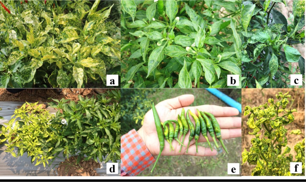
Diseased symptoms of peppers observed in this study. (a) yellow mosaic; (b) green vein mottle; (c) necrotic and ring spot; (d) stunted pepper plant with severe symptom compared with normal plant; (e) yellow mosaic and deformed fruits compared with nomal fruit; (f) yellowing and leaf curling.
The characters of mosaic and mottle is also known to be caused by CMV and ChiVMV [27], whereas necrosis and ring spot are caused by tospoviruses [28]. Severe infection can lead to a stunt of plant growth and the pepper fruits are reduced size (Figure 3). Severe symptoms were clearly observed in Kanchanaburi area (data not shown) which was in agreement with highest infection rate (Table 1). High incidence rate of the CMV and ChiVMV from the pepper fields has also been reported in northern Benin [29], northeastern region of India [30]. In Thailand, the occurrence of TNRV infecting field crops (e.g., tomato and pepper) was previously described [28,31]. Susceptible pepper plant to TNRV typically showed necrotic ringspots on leaves, stems, and fruits; other features were apical leaf distortion, mosaic in older leaves, and severe stunting [28,32]. In this present work, the characters similar to those mentioned were also found in the peppers infected with the TNRV in pepper (see Figure 3c). However, it should be noted that the diseased characteristics of the peppers infected by the viruses are often ambiguous, and these observed features are thus difficult to be used for identifying the disease-causing virus species. This is particularly obvious especially for the case of mixed viral infections.
Incidence of viruses
By using ELISA technique, the presence of the selected viruses was examined in Thai peppers. Table 1 shows an incidence and distribution of these viruses. Our data revealed that the peppers tested were infected with Begomovirus, CMV, ChiVMV, and TNRV. However, no reaction occurred with the antibodies of TMV, TSWV, and PVY, suggesting the absence of these viral groups in the peppers tested. According to Table 1, the incidence of the viruses infecting pepper in Thailand was quite high accounting for 69% (1,482 infected samples). This value was also in agreement with the same number of the pepper samples showing viral-diseased characteristics (1,482 diseased samples). The remaining 667 samples (31%) were not infected by the viruses as shown by their morphological characteristics and this was further confirmed by their negative ELISA reaction (Table 1).
The incidence and distribution of the infected viruses were also varied based on the region of the collecting sites. Our data revealed that the highest viral incidence was found in the central part (96%), followed by the north (74.4%) and the northeastern (52.8%), respectively. Begomoviruses, CMV, ChiVMV, and TNRV were detected in the samples at varying rates, whereas PVY, TMV, and TSWV were not detected. In the central part, 96% of the peppers were virus-infected in which 82.3% were from Begomovirus, 30.4% with TNRV, 18.1% with CMV, and 11.7% with ChiVMV. For the north area, there were 74.4% of the viral incidence; of these, 31.5% were infected with CMV, 25.3% with ChiVMV, 22.4% with Begomovirus, and 18.6% with TNRV. In the northeast region, of 52.8% virus incidence, the high rate of the CMV was found (25.4%), followed by ChiVMV (23.4%), Begomovirus (16.5%), and TNRV (14.9%). In summary, of 2,149 tested samples, 703 (32.7%), 552 (25.7%), 461 (21.5%), and 418 (19.5%) were found to be infected with Begomovirus, CMV, ChiVMV, and TNRV, respectively (Table 1). As a result, Begomoviruses seemed to be the predominant ones accounting for nearly 33% of the total incidence with the highest rate of ca. 93% in Ratchaburi (central part). CMV was the second group accounting for 25.7% with the greatest proportion of 80.9% in Phrae. The incidence of ChiVMV and TNRV was 21.5 and 19.5% with their highest rate in Lampang (69.5%), and in Sakol Nakhon (60.3%), respectively. This variation may be derived from many factors (e.g., geographic parameters, cultivation conditions, farm management, and viral vectors) [13]. Virus incidence was found in all Provinces of the central and northeastern regions, whereas no detection of some viral groups was observed in some Provinces of the northern area. For example, Begomoviruses were not found in Lampang, Tak, and Nan. CMV was absent in Tak, and there was no TNRV in Phrae. This is probable due to the fact that efficient vectors are not present, or to the fact that primary source of viral inoculum was absent [33]. Indeed, based on our observations, the weed hosts that may act as the viral sources were absent in the plantation areas of these northern Provinces, and thus may be the cause of this finding (data not shown). It is worth nothing that the high incidence of the begomoviruses found in this study was probably due to the antisera M1 and D2 which were specific to at least four begomovirus species [20]. As a result, more than one begomovirus species may be present in the infected pepper samples tested, and further identification of which species responsible for the infection would be a future task.
In addition, the present study also revealed mixed viral infections in these diseased pepper plants. It was found that the pepper samples could be infected by two, three, and four different viral groups (Figure 4 and 5). Of the 1,482 infected samples, mixed infections among the four viruses were found in 616 samples (41.6%). The remaining 866 pepper samples (58.4%) were infected singly by either one of the four viruses: 515 samples (34.8%) infected by Begomovirus, 130 samples (8.8%) by TNRV, 123 samples (8.3%) by CMV, and 98 samples (6.6%) by ChiVMV (Figure 4). Mixed double-infections (CMV + ChiVMV, CMV + TNRV, ChiVMV + TNRV, Begomovirus + CMV, Begomovirus + ChiVMV, and Begomovirus + TNRV), triple-infections (CMV + ChiVMV + TNRV, Begomovirus + CMV + ChiVMV, and Begomovirus + CMV + TNRV), and all four viral infections were detected at varying degree (Figure 4). Of all these, CMV + ChiVMV infection was mostly found with an incidence rate of 11.4%. Previous work has showed that mixed infections are common in the field, and can cause a serious problem in plant production [34,35]. Figure 5 shows representatives of diseased characteristic of peppers cause by mixed viral infection. Mixed infection of viral disease on solanaceous crops are typical in the fields as shown by several studies [29,36]. A multiple infection of 5 different viruses has been described in the infected pepper samples analyzed by DAS-ELISA [37]. At least 11 viruses have also been found co-infected in pepper plants [9]. Thirteen begomoviruses have been identified in diseased tomato, pepper and eggplant from different countries of Southeast and East Asia [13]. It should also be noted that this mixed infection may provide a chance for genetic recombination among the viruses present [38].

Incidence of the viral infections (both single and mixed infections) on pepper plants. BG = Begomovirus; CMV = Cucumber mosaic virus, ChiVMV = Chilli veinal mottle virus; and TNRV = Tomato necrotic ringspot virus. The percentage values of the virus incidence were calculated from the total infected samples of 1,482, and shown in parentheses.

Diseased symptoms of the peppers possibly caused by mixed infections of the viruses. (a) yellow leaf curl and mosaic induced by ChiVMV and Begomovirus; (b) veinal mottle by CMV and ChiVMV; (c) chlorotic flecks and necrosis by CMV, ChiVMV and TNRV; (d) yellow mosaic, leaf curling and stunted by CMV, ChiVMV, TNRV and Begomovirus.
Detection of Begomoviruses by PCR
Due to high incidence of Begomovirus as mentioned above, further work was then performed to confirm the presence of Begomoviruses. The total number of pepper samples showing yellow leaf curl pattern was 796 (possibly occurred by Begomoviruses). However, based on ELISA technique, 703 samples (88.3%) positively reacted with the antibody of the Begomoviruses (Table 1). It should be noted then that there were 93 samples (11.7%), showing negative for begomovirus incidence by using this antibody, although they also exhibited the yellow mosaic diseased symptoms. The ELISA technique used to detect the Begomoviruses was developed by Seepiban et al. [20]. For this, the two developed monoclonal antibodies (M1 and D2) were specific to only 4 begomovirus species (Tomato yellow leaf curl Thailand virus (TYLCTHV), Tobacco leaf curl Yunnan virus (TbLCYnV), Tomato leaf curl New Delhi virus (ToLCNDV) and Squash leaf curl China virus (SLCCNV) [20]. This restriction may be one of the drawbacks to detect other begomoviral species as appeared in the present study. To determine if the Begomoviruses were present in the negatively ELISA-reacted pepper samples, a PCR assay was introduced. For this, the degenerate primers PAL1v1978 and PAR1c496 previously shown to be specific to the begomovirus group [22], were selected and used to confirm this. Based on the PCR analysis, the DNA fragments of approximately 1.3 kb were amplified from total DNAs extracted from 93 ELISA-negative samples (Figure 6). A pair of these primers have been reported to use successfully in detecting the presence of the DNA region encoding coat protein, common region, and replicase of the begomovirus DNA-A component with an expected size of 1.1-1.4 kb [22]. Successful amplification of this specific DNA sequence of the begomovirus group has also been described and identified Squash leaf curl virus (SLCV) from symptomatic Cucurbita pepo in Jordan [39], Tomato leaf curl virus (TLCV) in viruliferous whiteflies [40], sweet pepper in Oman [41].

Detection of Begomovirus in diseased pepper leaf samples in this study. The agarose gel shows the amplified products obtained from the begomovirus-specific primers PAL1v1978 and PAR1c496 with an estimated size of 1,300 bp (lane 1-9, 11, and 13). Lane M, 100 bp DNA marker; Lane 10 and 12, negative control.
Conclusion
The present study was carried out aiming to determine the virus incidence of the pepper plants in Thailand. We initially screened the virus incidence using the ELISA technique. Our data showed that Begomoviruses were common, following by CMV, ChiVMV, and TNRV. The viruses can be found singly and in mixed infections (either two, three, or four). For Begomovirus detection, subsequent confirmation using PCR is needed to confirm its presence in the ELISA-negative samples, as demonstrated by a successful amplification of the DNA target. However, the serotyping means seems to be practical for initial screening of the virus infection. Our present study suggests the need for regular survey to monitor the virus infection. If this is planned, it would help the Thai farmers to plan for viral disease control on pepper effectively and sustainably.
Acknowledgements
This research was supported by Bayer Crop Science and Mae Fah Luang University.
Conflict of interest: Authors state no conflict of interest.
References
1 Arimboor R, Natarajan RB, Menon KR, Chandrasekhar LP, Moorkoth V. Red pepper Capsicum annuum carotenoids as a source of natural food colors: analysis and stability-a review. J Food Sci Tech. 2014;52:1258-71.10.1007/s13197-014-1260-7Search in Google Scholar PubMed PubMed Central
2 Kraikruan W, Sukprakarn S, Mongkolporn O, Wasee S. Capsaicin and dihydrocapsaicin contents of Thai chili cultivars. Kasetsart J (Nat Sci). 2008;42:611-6.Search in Google Scholar
3 Jomthaisong J. Pepper. Department of Agriculture Extension. Bangkok. 2008. (in Thai)Search in Google Scholar
4 Csillery G. Pepper taxonomy and the botanical description of the species. Acta Agron Hung. 2006;54:151-66.10.1556/AAgr.54.2006.2.5Search in Google Scholar
5 DOAE. Department of Agriculture Extension. 2006. Available from http://www.agriinfo.doae.go.th/year60/plant/rortor/veget/56.pdfhttp://www.agriinfo.doae.go.th/year60/plant/rortor/veget/57.pdfSearch in Google Scholar
6 Schreinemachers P, Balasubramaniam S, Boopathi NM, Ha CV, Kenyon L, Praneetvatakul S, Sirijinda A, Le NT, Srinivasan R, Wu MH. Farmers’ perceptions and management of plant viruses in vegetables and legumes in tropical and subtropical Asia. Crop Prot. 2015;75:115-23.10.1016/j.cropro.2015.05.012Search in Google Scholar
7 Roberts PD, Adkins S, Pernezny K, Jones JB. Diseases of pepper and their management. In: Naqvi SAMH, editor. Diseases of Fruits and Vegetables. Kluwer Academic Publishers. Dordrecht, The Netherlands; 2004,pp.333-87.10.1007/1-4020-2607-2_10Search in Google Scholar
8 Nigam K, Suhail S, Verma Y, Singhand V, Gupta S. Molecular characterization of begomovirus associated with leaf curl diseasein chilli. World J Pharm Res. 2015;4:1579-92.Search in Google Scholar
9 Jo Y, Choi H, Kim SM, Kim SL, Lee BC, Cho WK. The pepper virome: natural co-infection of diverse viruses and their quasispecies. BMC Genomics. 2017;18:453.10.1186/s12864-017-3838-8Search in Google Scholar PubMed PubMed Central
10 Kumar S, Kumar R, Kumar S, Singh AK, Singh M, Rai AB, Rai M. Incidence of leaf curl disease on capsicum germplasm under field conditions. Indian J Agric Sci. 2011;8:187-9.Search in Google Scholar
11 Senanayake DMJB, Varma A, Mandal BJ. Virus-vector relationships, host range, detection and sequence comparison of chilli leaf curl virus associated with an epidemic of leaf curl disease of chilli in Jodhpur. Indian Phytopathol. 2012;160:146-5510.1111/j.1439-0434.2011.01876.xSearch in Google Scholar
12 Jones DR. Plant viruses transmitted by whiteflies. Eur J Plant Pathol. 2003;109:195-219.10.1023/A:1022846630513Search in Google Scholar
13 Kenyon L, Kumar S, Tsai WS, Hughes J. Virus diseases of peppers Capsicum spp.) and their control. Adv Virus Res. 2014;90:297-354.10.1016/B978-0-12-801246-8.00006-8Search in Google Scholar PubMed
14 Leke WN, Mignouna DB, Brown JK, Kvarnheden A. Begomovirus disease complex: emerging threat to vegetable production systems of west and central Africa. J Agr Food Secur. 2015;4:1.10.1186/s40066-014-0020-2Search in Google Scholar
15 Marwal A, Sahu A, Gaur R. First report on the association of a begomovirus with Chrysanthemum indicum exhibiting yellowing of leaf vein disease characterized by molecular studies. J Hortic Res. 2013;21:17-21.10.2478/johr-2013-0017Search in Google Scholar
16 Nurulita S, Hidayat S, Mutaqin KH. Molecular characterization of begomovirus infecting yard long bean Vigna unguiculata subsp. sesquipedalis L.) in Java, Indonesia. Biotropia. 2015;22:53-60.10.11598/btb.2015.22.1.401Search in Google Scholar
17 Sakata JJ, Shibuya Y, Sharma P, Ikegami M. Strains of a new bipartite begomovirus, Pepper yellow leaf curl Indonesia virus in leaf-curl-diseased tomato and yellow-vein-diseased ageratum in Indonesia. Arch Virol. 2008;153:2307-13.10.1007/s00705-008-0254-zSearch in Google Scholar PubMed
18 Rentería-Canett I, Xoconostle-Cázares B, Ruiz-Medrano R, Rivera-Bustamante RF. Geminivirus mixed infection on pepper plants: Synergistic interaction between PHYVV and PepGMV. Virol J. 2011;8:104.10.1186/1743-422X-8-104Search in Google Scholar PubMed PubMed Central
19 Chiemsombat P, Yule S, Srikamphung B. Begomoviruses associated to pepper yellow leaf curl disease in Thailand. J Agric Res. 2018;3:1-11.10.23880/OAJAR-16000183Search in Google Scholar
20 Seepiban C, Charoenvilaisiri S, Warin N, Bhunchoth A, Phironrit N, Phuangrat B, Chatchawankanphanich O, Attathom S, Gajanandana O. Development and application of triple antibody sandwich enzyme-linked immunosorbent assays for begomovirus detection using monoclonal antibodies against Tomato yellow leaf curl Thailand virus. Virol J. 2017;14:99.10.1186/s12985-017-0763-zSearch in Google Scholar PubMed PubMed Central
21 Dellaporta SL, Wood J, Hicks JB. A plant DNA minipreparation: Version II. Plant Mol Biol Rep. 1983;1:19-21.10.1007/BF02712670Search in Google Scholar
22 Rojas MR, Gilbertson RL, Russell DR, Maxwell DP. Use of degenerate primers in the polymerase chain reaction to detect whitefly-transmitted geminiviruses. Plant Dis. 1993;77:340-7.10.1094/PD-77-0340Search in Google Scholar
23 Khan MS, Raj SK, Singh R. First report of Tomato leaf curl New Delhi virus infecting chilli in India. Plant Pathol. 2006;55:289.10.1111/j.1365-3059.2006.01324.xSearch in Google Scholar
24 Tsai WS, Shih SL, Green SK, Rauf A, Hidayat SH, Jan FJ. Molecular characterization of Pepper yellow leaf curl Indonesia virus in leaf curl and yellowing diseased tomato and pepper in Indonesia. Plant Dis. 2006;90:247.10.1094/PD-90-0247BSearch in Google Scholar PubMed
25 Shih SL, Tsai WS, Green SK. Molecular characterization of tomato and chilli leaf curl begomoviruses from Pakistan. Plant Dis. 2003;87:200.10.1094/PDIS.2003.87.2.200ASearch in Google Scholar PubMed
26 Kumar Y, Hallan V, Zaidi AA. Molecular characterization of a distinct bipartite begomovirus species infecting tomato in India. Virus Genes. 2008;37:425-31.10.1007/s11262-008-0286-1Search in Google Scholar PubMed
27 Loebenstein G, Katis N. Advances in Virus Research: Control of plant virus diseases of seed-propagated crops. 1st ed. Academic Press. London; 2014.10.1016/bs.aivir.2014.10.005Search in Google Scholar PubMed
28 Chiemsombat P, Sharman M, Srivilai K, Campbell P, Persley D, Attathom S. A new tospovirus species infecting Solanum esculentum and Capsicum annuum in Thailand. Australas Plant Dis Notes. 2010;5:75-8.10.1071/DN10027Search in Google Scholar
29 Afouda LAC, Kotchofa R, Sare R, Zinsou V, Winter S. Occurrence and distribution of viruses infecting tomato and pepper in Alibori in northern Benin. Phytoparasitica. 2013;41:271-6.10.1007/s12600-013-0287-zSearch in Google Scholar
30 Chanu NT, Singh YH, Sumitra P, Singh S, Singh SR, Roy SS, Prakash N, Sharma SK. Molecular based indexing of viral disease complex of king chilli Capsicum Chinense J.) in north eastern region of India. J. Pharmacogn. Phytochem. 2017;6:2004-8.Search in Google Scholar
31 Seepiban C, Gajanandana O, Attathom T, Attathom S. Tomato ring spot virus, a new of tospovirus isolated in Thailand. Arch Virol. 2011;156:263-74.10.1007/s00705-010-0856-0Search in Google Scholar PubMed
32 Puangmalai P, Potapohn N, Akarapisarn A, Pascha HJ. Inheritance of Tomato necrotic ring virus resistance in Capsicum annuum J Agri Sci. 2013;5:129-33.10.5539/jas.v5n2p129Search in Google Scholar
33 Arli-Sokmen M, Mennan H, Sevik MA, Ecevit O. Occurrence of viruses in field-grown pepper crops and some of their reservoir weed hosts in Samsun, Turkey. Phytoparasitica. 2005;33:347-58.10.1007/BF02981301Search in Google Scholar
34 Koeda S, Kesumawati E, Tanaka Y, Hosokawa M, Doi M, Kitajima A. Mixed infection of begomoviruses on pepper plants at northern Sumatra, Indonesia. Trop Agr Dev. 2016;60:59-64.Search in Google Scholar
35 Nsa IY, Kareem KT. Additive interactions of unrelated viruses in mixed infections of cowpea Vigna unguiculata L. Walp). Front Plant Sci. 2015;6:812.10.3389/fpls.2015.00812Search in Google Scholar PubMed PubMed Central
36 Olawale A, Samuel BO, Solomon ASO, Kumar PL. Surveys of virus diseases on pepper Capsicum spp.) in South-west Nigeria. Afr J Biotechnol. 2015;14:3198-205.10.5897/AJB2015.14803Search in Google Scholar
37 Soleimani P, Hosseini S, Hosseini A. Distribution of some viral Disease on Pepper Capsicum annuum plants in Dezful fields from Iran. Bull Environ Pharmacol Life Sci. 2014;3:111-4.Search in Google Scholar
38 Webster CG, Frantz G, Reitz SR, Funderburk JE, Mellinger HC, McAvoy E, Turechek WW, Marshall SH, Tantiwanich Y, McGrath MT, Daughtrey ML, Adkins S. Emergence of Groundnut ringspot virus and Tomato chlorotic spot virus in vegetables in Florida and the southeastern United States. Phytopathology. 2015;105:388-98.10.1094/PHYTO-06-14-0172-RSearch in Google Scholar PubMed
39 Almusa A, Anfoka G, Misbeh S, Abhary M, Ahmad FH. Detection and molecular characterization of Squash leaf curl virus (SLCV) in Jordan. Phytopathology. 2008;156:311-6.10.1111/j.1439-0434.2007.01389.xSearch in Google Scholar
40 Khan JA. Detection of tomato leaf curl geminivirus in its vector Bemisia tabaci Indian J Exp Biol. 2000;38:512-5.Search in Google Scholar PubMed
41 Khan JA, Al-Saady NA, Al-Mahruki MS, Al-Oufi M, Al-Subhi AM. Molecular characterization of begomovirus infecting sweet pepper in Oman. Indian J Biotechnol. 2007;6:45-51.Search in Google Scholar
© 2019 Amisa Laprom et al., published by De Gruyter
This work is licensed under the Creative Commons Attribution 4.0 Public License.
Articles in the same Issue
- Research Article
- ATP Synthase: Structure, Function and Inhibition
- Insulin Promotes Wound Healing by Inactivating NFkβP50/P65 and Activating Protein and Lipid Biosynthesis and alternating Pro/Anti-inflammatory Cytokines Dynamics
- Mini review
- The disordered boundary of the cell: emerging properties of membrane-bound intrinsically disordered proteins
- Research Article
- Expression of OX40 Gene and its Serum Levels in Neuromyelitis Optica Patients
- Advances in Molecular biomarker for early diagnosis of Osteoarthritis
- Identification of BRCA1/2 p.Ser1613Gly, p.Pro871Leu, p.Lys1183Arg, p.Glu1038Gly, p.Ser1140Gly, p.Ala2466Val, p.His2440Arg variants in women under 45 years old with breast nodules suspected of having breast cancer in Burkina Faso
- Endometriosis Pathoetiology and Pathophysiology: Roles of Vitamin A, Estrogen, Immunity, Adipocytes, Gut Microbiome and Melatonergic Pathway on Mitochondria Regulation
- Integral membrane protein expression of human CD25 on the cell surface of HEK293 cell line: the available cellular model of CD25 positive to facilitate in vitro developing assays
- Review Article
- Role of Nanomedicine in Redox Mediated Healing at Molecular Level
- Research Article
- Glutathione S-transferase M1 (GSTM1) and T1 (GSTT1) variants and breast cancer risk in Burkina Faso
- Incidence of viruses infecting pepper in Thailand
- Mechanochemistry of von Willebrand factor
- Schizophrenia phenomenology comprises a bifactorial general severity and a single-group factor, which are differently associated with neurotoxic immune and immune-regulatory pathways
- Role of Killer cell immunoglobulin-like receptors (KIR) genes in stages of HIV-1 infection among patients from Burkina Faso
- The effect of rs9930506 FTO gene polymorphism on obesity risk: a meta-analysis
- Special Issue: Recent Advances in Basic and Clinical Medicine
- Antihyperglycemic and antihyperlipidemic activities of Nannochloropsis oculata microalgae in Streptozotocin-induced diabetic rats
- Comparison of the effect of vitamin D on osteoporosis and osteoporotic patients with healthy individuals referred to the Bone Density Measurement Center
- Zinc enhances the expression of morphine-induced conditioned place preference through dopaminergic and serotonergic systems
- Pre-operative laparoscopic staging of gastric cancer in patients who are candidates for neo-adjuvant chemotherapy: A Cross Sectional Study
- Neurotoxic Effects of Stanozolol on Male Rats‘ Hippocampi: Does Stanozolol cause apoptosis?
- Only serum pepsinogen I and pepsinogen I/II ratio are specific and sensitive biomarkers for screening of gastric cancer
- Evaluation of Quality of Life in Terms of Sinonasal Symptoms in Children with Cystic Fibrosis
- The Effect of Fine needle aspiration on Detecting Malignancy in Thyroid Nodule
- Diagnosis of Primary Hydatid Cyst of Thyroid Gland: A Case Report
- Ultrasonography in the diagnosis of lung adhesion before surgery