Ultrasonography in the diagnosis of lung adhesion before surgery
-
Mohsen Eshraghi
 ,
Ahmad Kachoie
,
Ahmad Kachoie

Abstract
Background
The presence of pleural adhesions may render video-assisted thoracoscopic surgery difficult or impossible. The aim of this study was to assess the value of chest ultrasonography in the detection of pleural adhesions prior to thoracotomy.
Methods
Between 2013 and 2014, 42 consecutive patients undergoing thoracotomies (including video-assisted thoracicsurgery) were evaluated with chest ultrasonography. These patients underwent a preoperative ultrasonic examination of the chestwall using a 7.5-10-MHz linear ultrasound probe at 7 points along the chest wall. We measured the movement of the visceral pleuralslide.
Results
In the upper thoracic wall,ultrasonography demonstrated a sensitivity of 63.0%, a specificity of 66%, a negative predictive value of 77%, a positive predictive evalue of 50.0%, and an overall accuracy of 65.0%. And for the lower thoracic wall, ultrasonography demonstrated a sensitivity of 81.0%, a specificity of 59.0%,a negative predictive value of 89.0%, a positive predictivevalue of 44.0%, and an overall accuracy of 65.0%.
Conclusion
Chest ultrasonography is moderately accurate in detecting the presence and location of pleural adhesions. The use of preoperative chest sonographic findings to plan trocar placement and to determine the need for an open approach is valuable in helping prevent visceral injury and facilitating video-assisted thoracoscopic surgery.
Introduction
For thoracic surgeons, Video-Assisted Thoracoscopic surgery (VATS) is an important diagnostic tool that is widely used in the diagnosis and treatment of pulmonary pathologies and pleural adhesions [1, 2]. In the adhesions between the visceral and pleural walls for prevent lungs collapse at the onset of thoracoscopy, open thoracotomy can prevent the increased risk of lung damaged by the video-telescope in severe adhesions [3, 4]. Investigating the adhesions between the visceral wall and the pleural effusion is even more difficult by the CT scan, even based on the thickness of the lung [5]. In other studies, it has been reported that abdominal ultrasonography is performed prior to laparoscopic surgery and determination of peritoneal adhesions is based on visceral slides. If visceral slides show a distance of less than 2 centimeters, it is helpful in detecting adhesion [6, 7, 8, 9, 10]. Today ultrasonography is a modern, easy and accessible method. About 30 years ago, the first ultrasonography units were designed for medical examination. Some types of ultrasonography were controlled on the basis of Defectoscopy and some were based on Doppler. In recent years, ultrasonography devices with two-dimensional and dynamic B-mode images, usually Doppler-type, are most commonly used. Three-dimensional and four-dimensional ultrasonography devices are also available [11]. A study reported that the CT scan had a moderate sensitivity and specificity in the preoperative examination of patients to show pleural lesions as compared to VATS [5]. Another study reported that ultrasonography is the most useful technique for conducting and operating Trocar in laparoscopic surgery. Today, ultrasonography is the gold standard for diagnosing some diseases. In recent years, ultrasonography has been used in all surgical fields, including thoracic surgery [12].
Two studies conducted so far have shown that ultrasonography can be used to determine pre-surgical pleural adhesions [10, 11, 12, 13]. Considering the limited studies in this field, preventing unnecessary thoracotomy, performing thoracoscopy, pleural adhesion in preventing open surgery, use of ultrasonography, reducing the
physical and psychological effects on patients are the importance actions that needs to attention. The aim of this study was to determine the sensitivity, specificity and usefulness of ultrasonography in the diagnosis of preoperative lung adhesion in patients referred to Shahid Beheshti Hospital in the years 2009-2013, and thus determine the false positive and false positive rates.
Methods
This study was performed on the basis of process evaluations among patients undergoing thoracotomy and thoracoscopy surgery, of both genders and aged between 20 and 70 years, who were referred to the Thoracic Surgery, Department of Shahid Beheshti Hospital from October 2009 to May 2010. All subjects were selected to complete the required sample size of admission criteria. Among the inclusion criteria for the project, the consent to participate in the research project, the patients undergoing thoracotomy and thoracoscopy surgery, lack of heart disease such as Dilated Cardiomyopathy which affects the building, a BMI less than 35, lack of Gynecomastia, and lack of mastectomy in the past can be mentioned.In this preoperative study, a single radiologist unit was performed by an ultrasonography device manufactured by Voluson, a US-based company with 7-10-5 MHz probe for 7 points of intercostal ultrasonography, for all patients. Meanwhile, during the ultrasonography, the patient were asked to breathe deeply. The rate of lung movement and slider viscosity of the lung was measured. Ultrasonography was performed in the upper thoracic wall at three points. These points include the second point of the intercostal space in the midclavicular, the third point in the midaxillary and the third point in the paravertebral, defined by the upper thoracic wall. Four points in the lower thoracic wall were include and took place at the seventh point of intercostal space in the midaxillary, the fifth point in the scapula, the ninth point in the scapula, and the seventh point in the midclavicular, defined as the lower thoracic wall (Fig. 1).
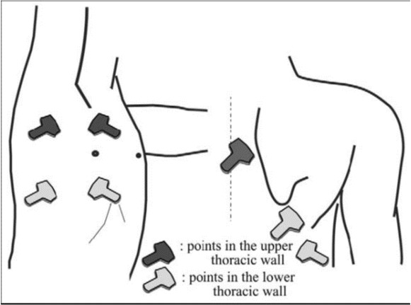
7-point schematic diagram for chest ultrasonography.
The distance between the thoracic wall and the pulmonary movement was measured. The amount of lung movement during operation was also recorded. It should be noted that in order to reduce the experimental error, the surgeon was unaware of the ultrasonography results of the patient. The obtained information was coded and the findings were considered as the gold standard and ultrasonography results were considered. All information was provided by the statistical program V. 12 SPSS were coded and then sensitivity, specificity, positive predictive value, negative predictive value and diagnostic accuracy were calculated.
Ethical approval: The research related to human use has been complied with all the relevant national regulations, institutional policies and in accordance the tenets of the Helsinki Declaration, and has been approved by the Ethics Committee of Qom University of Medical Sciences, Qom, Iran (Ethics Code: IR.MUQ.REC.1393.100 ).
Informed consent: Informed consent has been obtained from all individuals included in this study.
Results
The mean age of the subjects was 45.4 ± 13.8 years (23 up 63). In this study, 82 (85.4%) were male and 14 (14.6%) were female.In the upper thoracic wall, the mean pleural motion on the adhesion site was 0.78 ± 0.8 cm and in the place where the adhesion was not 1.55 cm / 1 cm.The difference in motion, was statistically significant (P <0.05).In the lower thoracic wall, the mean pleural motion in the adhesion site was 1.5 ± 0.19 cm and in the site with no adhesion, it was 2.4 ± 0.89 cm. This difference was also statistically significant (p <0.001).
Comparison of pleural motion between two groups with adhesion and non-adhesion is presented in figure 2.
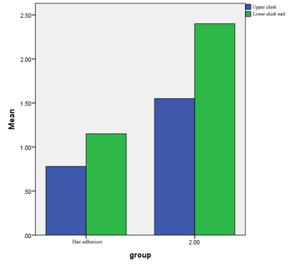
Comparison of pleural motion based on adhesion.
There was no relationship between gender and pleural motion in the upper thoracic wall (P = 0.13), as well as between gender and pleural motion in the lower thoracic wall (P = 0/14).
There was no relationship between age and pleural motion in the upper thoracic wall (P = 0.07), and between age and pleural upper thoracic wall in the loerthoracic wall (P = 0.09).
The findings showed that in the ultrasonography of the upper thoracic wall produced were 21 cases of true positives, 42 true negative cases, 21 cases of false positive and 12 cases of false negative. Thus, the technique had a sensitivity of 63%, a specificity of 66%, a positive predictive value of 50%, a negative predictive value of 77%, and a diagnostic accuracy of 65%. In the examination of ultrasonography in the lower thoracic wall, 22 positive cases were true, 41 cases were true negative, false positives in 28, and false negative in 5 cases. Thus, sensitivity there was a sentivitiy of81%, a specificity of 59%, a positive predictive value of 44%, a negative predictive value of 89%, and a diagnostic accuracy of 65%.
Discussion and conclusion
Thoracoscopy in thoracic surgery leads to a reduction in pain and suffering associated with surgery and a reduction in recovery time. Plan trocar placementin pleural adhesion leads to visceral injuries.Therefore, the determination of pre-operative pleural adhesion reduces the risk of incident events during thoracoscopy.Several studies have been perfomed exploring the benefits of using ultrasonography to evaluate the peritoneal adhesion before laparoscopic surgery [14, 15, 16, 17]. In the study, the benefits of ultrasound in determining the site of abdominal wall adhesion were reported before laparoscopy or laparotomy [14]. Ultrasonography of the upper thoracic wall demonstrated 21 true positive cases, 42 true negative cases, 21 false positive cases, and 12 false negative cases. Therefore, a sensitivity of 63%, a specificity of 66%, apositive predictive value of 50%, a negative predictive value of 77% and an accuracy of 65% were recorded. In a study by Sasaki et al., the sensitivity was close to the same value, but the specificity of using ultrasound in its study was estimated to be around 80%. This difference may be due to the smaller size of sample or the technique and skill of the radiologist. The positive and negative predictive value of the study by Sasaki, as well as its diagnostic accuracy regarding the use of ultrasound to find adhesion in the upper thoracic wall, was also close to those of our study [13].
Ultrasonography of the lower thoracic wall demonstrated 22 true positive cases, 41 true negative cases, 28 false positive cases, and 5 false negative cases. Therefore, a sensitivity of 81%, a specificity of 59%, a positive predictive value of 44%, a negative predictive value of 89% and an accuracy of 65% was reported.In this case, just like in the upper thoracic wall, the only difference with their study was a greater specificity of their research, which can be caused by the reasons given [13].
In a study by Kolecki et al., sensitivity and specificity of ultrasound were found to be 90% and 92%, respectively [15]. This difference could be due to the fact that they included people with a history of abdominal surgery and peritonitis.These interventions can lead to more obvious diagnostic differences whose finding using ultrasound is more feasible.
Tateishi and Mason in the two separate studies in this case, found a sensitivity of 75% and 72%, and a specificity of 93% and 71% respectively [18, 19]. The sensitivity obtained in their studies was close to that of our study, but the estimated specificity was higher. These differences can be due to the sample size or the difference in the diagnostic power of specialists.
It is worth noting that in very limited studies, the ultrasonography diagnostic power in pleural adhesions has been investigated.In one study, ultrasonography of the upper thoracic wall demonstrated, a sensitivity of 63.6%, a specificity of 79.4%, a negative predictive value of 87.7%, a positive predictive value of 50% and a diagnostic accuracy of 75.6% and ultrasonography of the lower thoracic wall demonstrated a sensitivity of 81.5%, a specificity of 81.0%, a negative predictive value of 96%, a positive predictive value of 44% and a diagnostic accuracy of 81.0% [13]. In a study, the sensitivity and specificity of ultrasonography in indicating visceral motion in patients with a history of peritonitis and abdominal surgery were 90% and 92% [15]. In another study, which evaluated pleural adhesion by sonography, the sensitivity of sonography was 75% and its specificity was 93% [18].
Another study to demonstrate pleural adhesion with CT scan, sensitivity was 72% and specificity was 71% [19]. In this study, ultrasonography had a moderate positive sensitivity, diagnostic accuracy, and prognostic value, which may be due to a misinterpretation of ultrasonography for the presence of adhesions and inadequate radiologist skills and the presence of such symptoms as emphysema in the patient. Therefore, the proficiency and skill of the radiologist and the high accuracy of the diagnosis of ultrasound are important in determining the pleural adhesions.
Conclusion
Chest ultrasonography is moderately accurate in detecting the presence and location of pleural adhesions. The use of preoperative chest sonographic findings to plan trocar placement and to determine the need for an open approach is valuable in helping prevent visceral injury and facilitating video-assisted thoracoscopic surgery. In addition, it is recommeded to use two or more radiologists to examine the diagnostic power of ultrasound, so that individual diagnostic differences have less effect on the study outcome.
Acknowledgment
We are grateful to all the staff of Shahid Beheshti Hospital, including physicians, nurses, and pathologists, for their help and support in carrying out this research.
Conflict of interest: Authors state no conflict of interest
References
1 Krasna MJ, Mack MJ, eds. Atlas of thoracoscopic surgery. St. Louis: Quality Medical Publishing, 1994.Search in Google Scholar
2 Landreneau, RJ, Mack, MJ, Keenan, RJ, et al. Strategic planning for video-assisted thoracic surgery. Ann ThoracSurg 1993;56:615-61910.1016/0003-4975(93)90930-GSearch in Google Scholar
3 Wolfer RS, Krasna MJ, Hasnain JU, et al. Hemodynamic effects of carbon dioxide insufflation during thoracoscopy. Ann ThoracSurg 1994;58:404-408.10.1016/0003-4975(94)92215-2Search in Google Scholar
4 Mason AC, Krasna MJ, White CS. The role of radiologic imaging in diagnosing complications of video-assisted thoracoscopic surgery. Chest 1998;113:820-825.10.1378/chest.113.3.820Search in Google Scholar PubMed
5 Mason AC, Miller BH, Krasna MJ, White CS. The accuracy of CT for the detection of pleural adhesions: correlation with video-assisted thoracoscopic surgery. Chest 1999;115:423–7.10.1378/chest.115.2.423Search in Google Scholar PubMed
6 Kodama I, Loiacono LA, Sigel B, et al. Ultrasonic detection of the visceral slide as an indicator of abdominal wall adhesions.J Clin Ultrasound 1992;20:375– 80.10.1002/jcu.1870200603Search in Google Scholar
7 Kolecki RV, Golub RM, Sigel B, et al. Accuracy of visceral slide detection of abdominal wall adhesions by ultrasound. SurgEndosc 1994;8:871– 4.Search in Google Scholar
8 Borzellino G, De Manzoni G, Ricci F, Guglielmi A, Laterza E. Ultrasonography mapping of peritoneal adhesions. RadiolMed (Torino) 1996;92:390 –3.Search in Google Scholar
9 Borzellino G, De Manzoni G, Ricci F. Detection of abdominaladhesions in laparoscopic surgery. A controlled study of 130 cases. SurgLaparoscEndosc 1998;8:273– 6.Search in Google Scholar
10 Tateishi U, Morikawa T, Miyasaka K. Detection of pleural adhesions with sonography. J Clin Ultrasound 2001;29:61–2.10.1002/1097-0096(200101)29:1<61::AID-JCU12>3.0.CO;2-KSearch in Google Scholar PubMed
11 Hrazdira I. Concise repetitorium of ultrasonography .Audioscan 2003;5–15.Search in Google Scholar
12 Peštál A,Veverková L, Jedlička V, Procházková I, Doležel J. The use of ultrasound in thoracic surgery. Scripta Medica (BRNO) 2006;79(2):105–114.Search in Google Scholar
13 Sasaki M, Kawabe M, Hirai S, Yamada N, Morioka K, Ihaya A. Preoperative Detection of Pleural Adhesions by Chest Ultrasonography. Ann ThoracSurg 2005;80:439–42.10.1016/j.athoracsur.2005.03.021Search in Google Scholar
14 Banazadeh M, Eshraghi M, Rahim MB, Alavi AA, Valeshabad AK. Successful management of acute necrotizing mediastinitis with trans-cervical drainage. Annals of Thoracic and Cardiovascular Surgery. 2011;17(5):498-500.10.5761/atcs.cr.10.01620Search in Google Scholar
15 Qureshi NR, Rahman NM, Gleeson FV. Thoracic ultrasound in the diagnosis of malignant pleural effusion. Thorax. 2009;64(2):139-43.10.1136/thx.2008.100545Search in Google Scholar
16 Schaffler GJ, Wolf G, Schoellnast H, Groell R, Maier A, Smolle-Jüttner FM, et al. Non-small cell lung cancer: evaluation of pleural abnormalities on CT scans with 18F FDG PET. Radiology. 2004;231(3):858-65.10.1148/radiol.2313030785Search in Google Scholar
17 Marcun R, Sustic A. Sonographic evaluation of unexplained pleural exudate: a prospective case series. Wien KlinWochenschr. 2009;121(9-10):334-8.10.1007/s00508-009-1188-5Search in Google Scholar
18 Mathis G. Thoraxsonography--Part 1: Chest wall and pleura Praxis (Bern 1994). 2004 Apr 7; 93 (15): 615-21.10.1024/0369-8394.93.15.615Search in Google Scholar
19 Matsumoto S, Hirata T, Ogawa E, Fukuse T, Ueda H, Koyama T, et al. Ultrasonographic evaluation of small nodules in the peripheral lung during video-assisted thoracic surgery (VATS). Eur J Cardiothorac Surg. 2004;26(3):469-73.10.1016/j.ejcts.2004.05.013Search in Google Scholar PubMed
20 Yilmaz U, Polat G, Sahin N, Soy O, Gülay U. CT in differential diagnosis of benign and malignant pleural disease. Monaldi Arch Chest Dis. 2005;63(1):17-22.10.4081/monaldi.2005.653Search in Google Scholar PubMed
21 Piolanti M, Coppola F, Papa S, Pilotti V, Mattioli S, Gavelli G. Ultrasonographic localization of occult pulmonary nodules during video-assisted thoracic surgery. EurRadiol. 2003;13(10):2358-64.10.1007/s00330-003-1916-6Search in Google Scholar
22 Kocijancic I, Vidmar K, Ivanovi-Herceg Z. Chest sonography versus lateral decubitus radiography in the diagnosis of small pleural effusions. J Clin Ultrasound. 2003;31(2):69-74.10.1002/jcu.10141Search in Google Scholar
23 Kodama I, Loiacono LA, Sigel B, et al. Ultrasonic detection of visceral slide as an indicator of abdominal wall adhesions J Clin Ultrasound 1992;20:375-380.10.1002/jcu.1870200603Search in Google Scholar
24 Kolecki RV, Golub RM, Sigel B, et al. Accuracy of visceral slide detection of abdominal wall adhesions by ultrasound SurgEndosc 1994;8:871-874.10.1007/BF00843457Search in Google Scholar
25 Borzellino G, De Manzoni G, Ricci F, Guglielmi A, Laterza E. Ultrasonography mapping of peritoneal adhesions. Radiol Med (Torino) 1996;92:390-393.Search in Google Scholar
26 Borzellino G, De Manzoni G, Ricci F. Detection of abdominal adhesions in laparoscopic surgery. A controlled study of 130 cases SurgLaparoscEndosc 1998;8:273-276.Search in Google Scholar
27 Tateishi U, Morikawa T, Miyasaka K. Detection of pleural adhesions with sonography J Clin Ultrasound 2001;29:61-62.10.1002/1097-0096(200101)29:1<61::AID-JCU12>3.0.CO;2-KSearch in Google Scholar PubMed
28 Mason AC, Miller BH, Krasna MJ, White CS. Accuracy of CT for the detection of pleural adhesionscorrelation with video-assisted thoracoscopic surgery. Chest 1999;115:423-427.10.1378/chest.115.2.423Search in Google Scholar PubMed
© 2019 M. Eshraghi et al., published by De Gruyter
This work is licensed under the Creative Commons Attribution 4.0 Public License.
Articles in the same Issue
- Research Article
- ATP Synthase: Structure, Function and Inhibition
- Insulin Promotes Wound Healing by Inactivating NFkβP50/P65 and Activating Protein and Lipid Biosynthesis and alternating Pro/Anti-inflammatory Cytokines Dynamics
- Mini review
- The disordered boundary of the cell: emerging properties of membrane-bound intrinsically disordered proteins
- Research Article
- Expression of OX40 Gene and its Serum Levels in Neuromyelitis Optica Patients
- Advances in Molecular biomarker for early diagnosis of Osteoarthritis
- Identification of BRCA1/2 p.Ser1613Gly, p.Pro871Leu, p.Lys1183Arg, p.Glu1038Gly, p.Ser1140Gly, p.Ala2466Val, p.His2440Arg variants in women under 45 years old with breast nodules suspected of having breast cancer in Burkina Faso
- Endometriosis Pathoetiology and Pathophysiology: Roles of Vitamin A, Estrogen, Immunity, Adipocytes, Gut Microbiome and Melatonergic Pathway on Mitochondria Regulation
- Integral membrane protein expression of human CD25 on the cell surface of HEK293 cell line: the available cellular model of CD25 positive to facilitate in vitro developing assays
- Review Article
- Role of Nanomedicine in Redox Mediated Healing at Molecular Level
- Research Article
- Glutathione S-transferase M1 (GSTM1) and T1 (GSTT1) variants and breast cancer risk in Burkina Faso
- Incidence of viruses infecting pepper in Thailand
- Mechanochemistry of von Willebrand factor
- Schizophrenia phenomenology comprises a bifactorial general severity and a single-group factor, which are differently associated with neurotoxic immune and immune-regulatory pathways
- Role of Killer cell immunoglobulin-like receptors (KIR) genes in stages of HIV-1 infection among patients from Burkina Faso
- The effect of rs9930506 FTO gene polymorphism on obesity risk: a meta-analysis
- Special Issue: Recent Advances in Basic and Clinical Medicine
- Antihyperglycemic and antihyperlipidemic activities of Nannochloropsis oculata microalgae in Streptozotocin-induced diabetic rats
- Comparison of the effect of vitamin D on osteoporosis and osteoporotic patients with healthy individuals referred to the Bone Density Measurement Center
- Zinc enhances the expression of morphine-induced conditioned place preference through dopaminergic and serotonergic systems
- Pre-operative laparoscopic staging of gastric cancer in patients who are candidates for neo-adjuvant chemotherapy: A Cross Sectional Study
- Neurotoxic Effects of Stanozolol on Male Rats‘ Hippocampi: Does Stanozolol cause apoptosis?
- Only serum pepsinogen I and pepsinogen I/II ratio are specific and sensitive biomarkers for screening of gastric cancer
- Evaluation of Quality of Life in Terms of Sinonasal Symptoms in Children with Cystic Fibrosis
- The Effect of Fine needle aspiration on Detecting Malignancy in Thyroid Nodule
- Diagnosis of Primary Hydatid Cyst of Thyroid Gland: A Case Report
- Ultrasonography in the diagnosis of lung adhesion before surgery