Green synthesis and characterisations of antibacterial silver-polyvinyl alcohol nanocomposite films for wound dressing
-
Sayed M. Badawy
Sayed M. Badawy studied Chemistry at Cairo University and obtained his PhD in the field of Physical Chemistry in 2001 from Ain Shams University, Cairo. He has been employed as a fellow of Chemistry at Cairo University since 2008. He currently works as an Assistant Professor of Physical Chemistry at Aljouf University. His main research interests are polymer and environmental chemistry. He is the author or co-author of about 20 peer-reviewed publications.


Abstract
Green synthesis and characterisations of silver-polyvinyl alcohol nanocomposite films for possible environmental and biomedical applications such as wound dressing have been investigated. The synthesis was carried out in water, in an environmentally-friendly solvent using β-D-glucose as a reducing agent and in polyvinyl alcohol (PVA) as a bio-friendly polymer. The green synthesis approaches have advantages over conventional methods involving chemical agents associated with environmental toxicity. The formation of silver nanoparticles (AgNPs) was confirmed by X-ray diffraction (XRD), energy dispersive X-ray spectroscopy (EDX) and UV-visible spectroscopy. The antimicrobial activity of the synthesised Ag-PVA nanocomposite materials against strains of different bacteria such as Escherichia coli, Enterobacter cloacae, Klebsiella spp., Salmonella typhi and Staphylococcus aureus was studied.
1 Introduction
It is well known that silver ions and silver-based compounds are highly toxic to microorganisms showing strong biocidal effects on as many as 16 species of bacteria including Escherichia coli [1, 2]. Thus, silver ions, as an antibacterial component, have been used in the formulation of dental resin composites [3, 4], ion exchange fibres [5] and in the coatings on medical devices [6]. The possible use of silver nanoparticles (AgNPs) as an antibacterial agent has, therefore, been investigated as a mean of stopping increasing bacterial resistance to conventional bactericides and antibiotics [7]. Numerous approaches have been used to prepare AgNPs for a rapidly growing list of catalysis, electronics, non-linear optics, and biomaterial applications [8].
Polymer materials reinforced with nanoparticles are adding new dimensions to composite materials and providing major improvements in functional and structural properties. Polymer metal nanocomposites are blends of different polymer matrices with metal nanoparticles. The properties of such nanocomposites are remarkably different compared to classical polymers. These properties improvements concern mechanical properties, surface properties, dimensional stability, thermal stability, chemical stability, antimicrobial properties, photocatalytic, optical and electrical properties. The incorporation of only a few percent of nanosized particles can make dramatic property changes [9].
Many techniques of synthesising AgNPs, such as chemical reduction of silver ions in aqueous solutions have been reported in the literature. Most of these methods are extremely expensive and also involve the use of toxic, hazardous chemicals, which may pose potential environmental and biological risks [10].
As noble metal nanoparticles are widely applied to areas of human contact, there is a growing need to develop environmentally friendly processes for nanoparticle synthesis that do not use toxic chemicals. In a recent published work, a green method was used for the successful synthesis of polyvinyl alcohol (PVA) reinforced with AgNPs for electromagnetic wave shielding at microwave frequency [11].
PVA is a bio-friendly polymer as it is water soluble and has extremely low cytotoxicity. In the present work, the stable Ag-PVA nanocomposite films are synthesised using the green method using β-D-glucose as the reducing agent in water solvent and investigated against strains of different bacteria. The polymer nanocomposites combine the excellent functional properties of NPs with the desired properties of host polymers.
2 Materials and methods
The green synthesis of silver nanocomposite films involves three main steps, based on green chemistry, including: (1) selection of environmentally-friendly solvent medium, (2) selection of environmentally benign reducing agent and (3) selection of nontoxic hydrogel matrices.
2.1 Method of preparation
The materials used for this study are: PVA (99–100% hydrolyzed), silver nitrate (AVONVHEM, UK), β-D-glucose (INTERCHEM, UK) and distilled water. The PVA bulk solution was prepared by dissolving PVA powder (5 wt%) in distilled water under controlled temperature at 90°C and continuous stirring for 3 h. After the solution had cooled down to ambient temperature, AgNO3 solution was added with stirring and then a solution of β-D-glucose as the reducing agent was added dropwise and stirred. The final ratio of Ag to glucose was 1/2 mole. The blend solution was poured into Petri dishes, 15 ml/dish, and allowed to dry to form films by casting under ambient temperature for 1 day in a dark room.
2.2 Characterisations
X-ray diffraction was recorded in the continuous scanning mode at room temperature on an EMPYREAN Diffractometer system, UK, operated at 45 kV and a current of 30 mA using copper (Cu) tube with Cu Kα radiation (λ=1.5406 Å). The diffraction intensities were recorded from 4° to 80°, in 2θ angles in steps of 0.026° with scan step time 19 s. Energy dispersive X-ray analysis (EDX) was recorded using an dispersive X-ray fluorescence (EDX) spectrometer Model (Oxford) attached with an SEM Model JEOL-JSM-5600 operating at accelerating voltage 25.00 kV. The UV-VIS spectroscopy measurement of Ag-PVA nanocomposite films was recorded on APEL spectrophotometer (model PD-303 UV) operated with wavelength accuracy of ±2 nm. The Ag-PVA nanocomposite was measured with UV-VIS spectroscopy in a wavelength ranging from 300 to 800 nm. The photometric range varied from 0 to 2.5Å.
2.3 Antimicrobial activity of silver nanocomposites
The synthesised silver nanocomposites were used in bactericidal study against different bacterial strains. The bactericidal tests were performed with bacterial strains as Bacillus cereus, Escherichia coli, Staphylococcus aureus, Enterobacter cloacae, Klebsiella spp. and Salmonella typhi. Nutrient agar and Muller Hinton agar were used as media to grow bacteria. The bacterial solution was prepared in 0.86% saline. The antibacterial activity of silver nanoparticle sample was assayed by following the standard disk diffusion technique. The bacterial suspension was spread on nutrient agar in Petri plate to create a confluent lawn of bacterial growth. The disks of 6 mm were loaded. These plates were incubated for 24 h at 35°C. The susceptibility of test organisms was determined after 24 h by measuring the zone of inhibition around each disk.
3 Results and discussion
3.1 Mechanism for the formation of silver nano-composites
In the green method, Ag-PVA nanocomposites are prepared using water as an environmentally benign solvent and glucose as a reducing agent. PVA is appropriate as a stabiliser and polymeric media for reducing the AgNO3 using β-D-glucose as a green reducing agent. PVA is a bio-friendly polymer as it is water soluble and has extremely low cytotoxicity [12].
In the present work, silver nanocomposites were synthesised by the reduction of aqueous PVA silver ions using glucose as shown in equations (1) and (2). The basic method for the synthesis of NPs in PVA is to disperse metal ion solution in the polymer and reduce them to a zero valent state.
The colour of silver solutions in PVA-glucose solutions gradually changed from colourless to yellow and light brown and then to brown, and finally dark brown. The distinctive colours of nano silver are due to a phenomenon known as plasmon absorbance. Incident light creates oscillations in conduction electrons on the surface of the NPs and electromagnetic radiation is absorbed [13]. This indicates the formation of AgNPs. With an increase in reaction time, particle size and aggregation of silver nanocrystal gradually increased together.
3.2 Characterisations of Ag-PVA nanocomposites
X-ray diffraction (XRD) has a good potential for the analysis of nanostructures, because the width and shape of reflections yield information about the substructure of the materials such as crystal structure, crystallite size, and strain [14]. Figures 1 and 2 show the XRD patterns of pure PVA and Ag-PVA nanocomposite, respectively. The XRD of pure PVA sample exhibits a broad diffraction peak due to the amorphous nature of the polymer. The XRD of the Ag-PVA nanocomposite shows amorphous phase indicating that AgNPs are well dispersed and incorporated in the amorphous phase of PVA matrix.
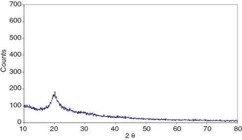
X-ray diffraction of polyvinyl alcohol.
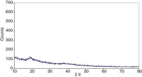
X-ray diffraction of silver-polyvinyl alcohol nanocomposite.
EDX microanalysis is a technique used for identification of the elemental composition of a specimen. Atoms of each element release X-rays with unique amounts of energy during the transfer process. The “fingerprint” energies of the emitted X-rays can then be used to identify an element. Also, EDX microanalysis is capable of generating a map of one or more chemical elements of interest [15]. Analysis of Ag-PVA nanocomposite through EDX spectrometer confirmed the presence of elemental silver signals of the AgNPs (Figure 3). The peaks around 3.40 keV correspond to the binding energies of Ag L while the peaks situated at binding energies of 0.2 kV and 0.5 keV belong to carbon and oxygen, respectively. The EDX analysis of the Ag-PVA nanocomposite represents the element percentage (%) of the Ag as 10.80 (Table 1 and Figure 3). The carbon and oxygen in the examined samples are attributed to the matrix of hydrogel PVA. The Si signal in EDX spectra was expected from the silicon substrate itself, as the EDX analysis of Ag-PVA was carried out on Si substrate. No obvious peak belong to impurity is detected.
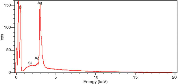
Energy dispersive X-ray analysis of silver-polyvinyl alcohol nanocomposite.
Atomic% and element% of Ag-PVA nanocomposite.
| Elements | Atomic % | Element % |
|---|---|---|
| C K | 32.50 | 24.11 |
| Ag L | 1.62 | 10.80 |
| O K | 65.86 | 65.06 |
Figure 4 shows UV-Vis spectra of Ag-PVA nanocomposite prepared with different silver concentrations of 0.03 M and 0.10 M in the prepared PVA solution. A characteristic absorbance peak of a well-defined surface plasmon resonance (SPR) appears at 430 nm. The peak of absorption spectrum of silver concentration 0.10 M was manually extended because the maximum photometric range of the spectrophotometer is stopped at 2.5Å.
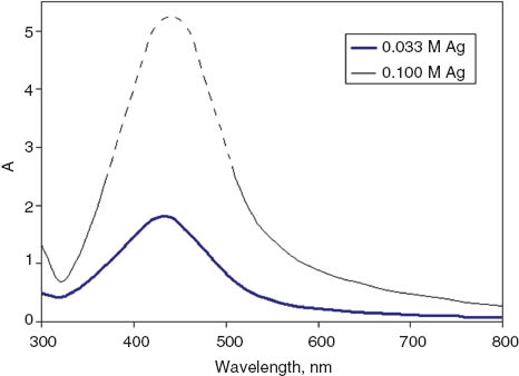
UV-Vis spectra of silver-polyvinyl alcohol nanocomposite.
The appearance of a sharp plasmon peak at 430 nm, due to surface SPR phenomena of the electrons in the conduction bands of silver, indicated the formation of silver with nanometer-sized dimensions in the PVA. Ag-NPs are synthesised in situ inside the host polymers PVA by a one-step procedure. Reduction of Ag+ ions in hydrogel matrix yielded the typical SPR of Ag particles. This value is in good agreement with the literature reports on the spectrum of AgNPs in aqueous solutions. In most experiments AgNPs show SPR in the interval 400–430 nm [13, 16–19]. The observed SPR centred at 430 nm is attributed to the excitation of the dipolar resonance and dipolar distribution of the polarisation charge [16].
The intensity of this absorption peak significantly increase as a result of increasing concentration of silver. The difference in the peak intensity is due to the concentration of the silver inside the host which can also be observed through the colours of the films. This observation indicates the formation of Ag-NPs within the PVA matrix and also the occurrence of quantum size effect as a result of increasing silver concentration. Noble metal nanoparticles, are well known for their strong interactions with visible light through the resonant excitations of the collective oscillations of the conduction electrons within the particles [13, 20–24].
The full width half maximum (FWHM) of the respective absorption patterns of Ag NPs in PVA prepared from solution of 0.01 M and 0.03 M silver concentration are found to be 110 nm and 140 nm, respectively. It is important to note that although the silver concentration is increased, the particle size does not significantly increase, possibly due to the capping action of PVA. It is important to note that PVA as a protective agent plays a decisive part in controlling size distribution of SNPs.
3.3 Antimicrobial activities
Silver is known for its antimicrobial properties and has been used for years in the medical field for antimicrobial applications. Additionally, silver has been used in water and air filtration to eliminate microorganisms [25]. Inhibition zone values were obtained from the synthesised silver nanocomposites tested against bacteria; Escherichia coli (E. coli), Enterobacter clocae (E. cloacae), Klebsiella spp. (K. spp.), Salmonella typhi (S. typhi) and Staphylococcus aureus (S. aureus).
Results of the inhibition zones for samples with concentrations of 0.1 M and 0.5 M Ag are presented as average values in mm in the Table 2. The maximum antibacterial activity was recorded for E. coli. Due to their particle size, AgNPs can easily reach the nuclear content of bacteria by disrupting the membranes of bacteria, and finally killing them.
The results of antibacterial activity with zone of inhibition.
| No. | Bacterial strains | Zone of inhibition | |
|---|---|---|---|
| 0.01 M Ag | 0.10 M Ag | ||
| 12 | Escherichia coli | 14.15 mm | 14.16 mm |
| 15 | Enterobacter cloacae | 10 mm | 11.11 mm |
| 19 | Klebsiella spp. | 10.11 mm | 10.11 mm |
| 20 | Salmonella typhi | 10.8 mm | 11.12 mm |
| 13 | Staphylococcus aureus | -ve | -ve |
Table 2 shows that the nanocomposites had high antibacterial activity against gram-positive bacteria; E. coli, E. cloacae, Klebsiella spp., and S. typhi. Different mechanisms for bacterial action of AgNPs were suggested [19]: (i) Ag+ ions are supposed to bind to sulfhydryl groups, which leads to protein denaturation by the reduction of disulfide bonds; (ii) Ag+ can complex with electron donor groups containing sulfur, oxygen, or nitrogen that are normally present as thiols or phosphates on amino acids and nucleic acids. Also, SNPs have been found to attach to the surface of the cell membrane and disturb its function, penetrate into bacteria, and release Ag0; (iii) SNPs target the bacterial membrane, leading to a dissipation of the proton motive force. Thus, a decrease in the NPs’ size can lead to an increase in the specific surface of a bactericidal specimen, inducing an increase in their ability to penetrate the cell membrane, and thus improving antibacterial activity [19].
Table 2 shows that the silver nanocomposites samples are negative against Gram-negative S. aureus bacteria. The cell wall of Gram-negative bacteria consists of lipids, proteins and lipopolysaccharides (LPS) that ensure more effective defense against biocides in comparison to Gram-positive bacteria, where the cell wall of Gram-positive bacteria does not contain outer membrane of LPS.
4 Conclusion
Characterisations of antibacterial metal nanocomposite materials (Ag-PVA) prepared by the green method have been investigated. The green synthesis was carried out in water using β-D-glucose as reducing agent and PVA as a bio-friendly polymer. The polymer nanocomposites combine the excellent functional properties of nanoparticles with desired properties of host polymers. XRD confirmed that the silver (Ag) nanoparticles incorporated into the same phase of PVA matrix. Analysis of Ag-PVA nanocomposite through EDX spectrometer confirmed the presence of elemental silver signals of the silver nanoparticles. UV-Vis spectra of Ag-PVA nanocomposite showed a characteristic absorbance peak of well-defined plasmon resonance (SPR) at 430 nm. The antimicrobial activity of synthesised Ag-PVA nanocomposite materials against strains of different bacteria showed that the nanocomposites had high antibacterial activity against Gram-positive bacteria: E. coli, E. cloacae, Klebsiella spp. and S. typhi.
About the author

Sayed M. Badawy studied Chemistry at Cairo University and obtained his PhD in the field of Physical Chemistry in 2001 from Ain Shams University, Cairo. He has been employed as a fellow of Chemistry at Cairo University since 2008. He currently works as an Assistant Professor of Physical Chemistry at Aljouf University. His main research interests are polymer and environmental chemistry. He is the author or co-author of about 20 peer-reviewed publications.
Acknowledgments
Sayed M. Badawy wishes to thank A. Hassan for performing the bactericidal tests at the Faculty of Applied Medical Science, Aljouf University. Financial support by Aljouf University (Research Project Number 232/34) is gratefully acknowledged.
References
[1] Zhao GJ, Stevens SE. Biomaterials 1998, 11, 27–32.10.1023/A:1009253223055Search in Google Scholar
[2] Spadaro JA, Berger TJ, Barranco SD, Chapin SE, Becker RO. Antimicrob. Agents Ch. 1974, 6, 637–642.Search in Google Scholar
[3] Yoshida K, Tanagawa M, Atsuta M. J. Biomed. Mater. Res. 1999, 47, 516–522.Search in Google Scholar
[4] Herrera M, Carrion P, Baca P, Liebana J, Castillo A. Microbios. 2001, 104, 141–148.Search in Google Scholar
[5] Nonaka T, Noda E, Kurihara S. J. Appl. Polym. Sci. 2000, 77, 1077–1086.Search in Google Scholar
[6] Schierholz JM, Beuth J, Pulverer G. Am. J. Med. 1999, 107, 101–102.Search in Google Scholar
[7] Gogoi SK, Gopinath P, Paul A, Ramesh A, Ghosh SS, Chattopadhyay A. Langmuir 2006, 22, 9322–9328.10.1021/la060661vSearch in Google Scholar PubMed
[8] Nigam N, Kumar S, Dutta PK, Ghosh T. Asian Chitin J. 2009, 5, 97–100.Search in Google Scholar
[9] Koo JH, Ed., Polymer Nanocomposites-Processing, Characterization, and Applications, McGraw-Hill: New York 2006.Search in Google Scholar
[10] Prabhu S, Poulose EK. Int. Nano Lett. 2012, 2, 32.Search in Google Scholar
[11] Al-Ghamdi AA, Al-Hartomy OA, El-Tantawy F, Yakuphanoglu F. Microsyst. Technol. 2014 (in press).Search in Google Scholar
[12] Razzak MT, Zainuddin E, Dewi S, Lely H, Taty S. Radiat. Phys. Chem. 1999, 55, 153–165.Search in Google Scholar
[13] Solomon SD, Bahadory M, Jeyarajasingam AV, Rutkowsky SA, Boritz C. J. Chem. Educ. 2007, 84, 2.Search in Google Scholar
[14] Dorofeeva G, Streletskiib A, Povstugara I, Protasova A, Elsukova E. Colloid J. 2012, 74, 675–685.Search in Google Scholar
[15] McNeil SC, Ed., Characterization of Nanoparticles Intended for Drug Delivery, Springer Science+Business Media, LLC, Humana Press: London, 2011.10.1007/978-1-60327-198-1Search in Google Scholar
[16] Alsawafta M, Badilescu S, Paneri A, Truong V and Packirisamy M. Polymers 2011, 3, 1833–1848.10.3390/polym3041833Search in Google Scholar
[17] Dawy M, Rifaat HM, Moustafa SA and Mousa HA. Aust. J. Basic Appl. Sci. 2012, 6, 257–262.Search in Google Scholar
[18] Ananth AN, Umapathy S, Sophia J, Mathavan T, Mangalaraj D. Appl. Nanosci. 2011, 1, 87–96.Search in Google Scholar
[19] Patil RS, Kokate MR, Jambhale CL, Pawar SM, Han SH, Kolekar SS. Adv. Nat. Sci.: Nanosci. Nanotechnol. 2012, 3, 015013 (7pp).10.1088/2043-6262/3/1/015013Search in Google Scholar
[20] Dmitruk NL, Malynych SZ, Moroz IE, Kurlyak VY. Semicond. Phys.Quantum Electron. Optoelectron. 2010, 13, 369–373.Search in Google Scholar
[21] Yao H, Jin L, Sue H, Sumic Y, Nishimurac R. J. Mater. Chem. 2013, 1, 10783–10789.Search in Google Scholar
[22] Mock JJ, Smith DR, and Schultz S. Nano Lett. 2003, 3, 8.Search in Google Scholar
[23] Moores A, Goettmann F. New J.Chem 2006, 30, 1121–1132.10.1039/b604038cSearch in Google Scholar
[24] Noguez C. J. Phys. Chem. 2007, 111, 3806–3819.Search in Google Scholar
[25] Sharma VK, Yngard RA, Lin Y. Adv. Colloid Interface Sci. 2009, 145, 83–96.Search in Google Scholar
©2014 by Walter de Gruyter Berlin/Boston
This article is distributed under the terms of the Creative Commons Attribution Non-Commercial License, which permits unrestricted non-commercial use, distribution, and reproduction in any medium, provided the original work is properly cited.
Articles in the same Issue
- Frontmatter
- In this issue
- Editorial
- Megatrends – megascience? Part 2
- Feature
- Novel sustainable industrial processes: from idea to commercial scale implementation
- Original articles
- Application of environmental and economic metrics to guide the development of biocatalytic processes
- Synthesis of nanostructured MgO powders with photoluminescence by plasma-intensified pyrohydrolysis process of bischofite from brine
- A solvent free approach for Knoevenagel condensation: facile synthesis of 3-cyano and 3-carbethoxycoumarins
- Green synthesis and characterisations of antibacterial silver-polyvinyl alcohol nanocomposite films for wound dressing
- Company profiles
- AVA Biochem: commercialising renewable platform chemical 5-HMF
- Invenios: micro process technology – chemical process technology of tomorrow
- Conference announcements
- International Conference on Green Chemistry and Sustainable Engineering International (Barcelona, Spain, July 29–31, 2014)
- 7th Annual European Forum for Industrial Biotechnology and the Biobased Economy (Reims, France, September 30–October 2, 2014)
- 19th International Symposium on Homogeneous Catalysis (ISHC-XIX; Ottawa, ON, Canada, July 6–11, 2014)
- 248th American Chemical Society National Meeting and Exposition: Chemistry and Global Stewardship (San Francisco, CA, USA, August 10–14, 2014)
- Conferences 2014–2017
- Book reviews
- Green materials for sustainable water remediation and treatment
- Engineering catalysis