Binding domain characterization of growth hormone secretagogue receptor
-
 ,
,

Abstract
Background and Objectives
Activation of ghrelin receptor growth hormone secretagogue receptor (GHS-R) by endogenous or synthetic ligands amplifies pulsatile release of growth hormone (GH) and enhances food intake, very relevant to development and growth. GHS-R is a G-protein coupled receptor that has great druggable potential. Understanding the precise ligand and receptor interactions is crucial to advance the application of GHS-R.
Materials and Methods
We used radiolabeled ligand-binding assay and growth hormone release assay to assess the binding and functional characteristics of GHS-R to synthetic agonists MK-0677 and GHS-25, as well as to endogenous peptide ligand ghrelin. We analyzed the ligand-dependent activity of GHS-R by measuring aequorin-based [Ca++]i responses. To define a ligand-binding pocket of GHS-R, we generated a series of human/puffer fish GHS-R chimeras by domain swapping, as well as a series of mutants by site-directed mutagenesis.
Results
We found that the synthetic ligands have high binding affinity to GHS-R in the in vitro competitive binding assay. Remarkably, the in vivo GH secretagogue activity is higher with the synthetic agonists MK-0677 and GHS-25 than that of ghrelin. Importantly, the activity was completely abolished in GHS-R knockout mice. In GHS-R chimera analysis, we identified the C-terminal region, particularly the transmembrane domain 6 (TM6), to be critical for the ligand-dependent activity. Our site-directed mutagenesis study further revealed that amino acid residues D99 and W276 in GHS-R are essential for ligand binding. Interestingly, critical residues distinctively interact with different ligands, MK-0677 activation depends on E124, while ghrelin and GHS-25 preferentially interact with F279.
Conclusion
The ligand-binding pocket of human GHS-R is mainly defined by interactive residues in TM6 and the adjacent region of the receptor. This novel finding in GHS-R binding domains advances the structural/ functional understanding of GHS-R, which will help to select/design better GHS-R agonists/ antagonists for future therapeutic applications.
Introduction
The growth hormone (GH) axis, including the endocrine signaling of GH and insulin-like growth factor-1 (IGF-1), plays important roles in human development and growth. Aging is associated with declined activity of the GH–IGF-1 axis. In order to develop drugs for the treatment of hormone deficiencies in the elderly, a reverse pharmacology approach was taken to identify small molecules that can restore the amplitude of GH pulsatility.[1,2] We elucidated the action of a class of small synthetic growth hormone releasing peptides (GHRPs) and used this knowledge to develop non-peptide mimetics,[3, 4, 5] such as Merck compounds MK-0677 and GH secretagogue (GHS)-25.[6,7] Daily treatment with MK-0677 reversed the GH–IGF-1 axis of 70–90-year-old subjects and produced GH and IGF-1 levels typical to those of young adults.[8,9] In 1996, we expression-cloned an orphan G-protein coupled receptor (GPCR), which we named the GHS receptor (GHS-R) from a pituitary DNA library based on specific activation by the synthetic GH-releasing ligands MK-0677 and GHRP-6.[10] GHS-R1a with all seven transmembrane domains (TMs) was proven to be the functional receptor of ghrelin, and its ligands potentially offer an oral form of alternative therapy instead of injectable GH for GH deficiency.[11,12]
In 2000, we cloned the puffer fish GHS-R gene, which shares 58% identity to human ortholog. In common with the human GHS-R1a, it is activated by the structurally distinct ligands GHRP-6 and MK-0677.[13,14] The ligand activation domain of GHS-R1a has been evolutionarily conserved from puffer fish to humans, in support of the notion that the GHS-R1a and its natural ligand play a fundamentally important role in biology.
The physiological relevance of GHS-R1a has been confirmed by identification of two natural ligands, ghrelin and adenosine, by fractionating and assaying animal tissue extracts in cell lines engineered to express GHS-R1a.[15, 16, 17] Ghrelin plays an important role in modulating feeding patterns, insulin secretion, and glucose metabolism.[18,19] In contrast to ghrelin, a 28-amino acid octanoylated peptide,[15] adenosine is a purinergic nucleotide and it activates GHS-R1a through a binding domain distinct from that of ghrelin and MK-0677.[16,17] Even though they both activate GHS-R1a, when administering ghrelin and adenosine to rats, only ghrelin stimulates GH release.[16,20] The structure of synthetic agonist MK-0677 is different from that of the endogenous peptide ghrelin. Although molecular modeling studies developed from proton nuclear magnetic resonance (NMR) that compared the structural features of MK-0677 and other synthetic GHS-R1a ligands illustrated certain similarities with ghrelin, these studies did not precisely predict the ligand–receptor binding sites.[21] Our previous studies showed that ghrelin, not adenosine, functions as a competitor of [35S]MK-0677 binding,[17] suggesting ghrelin and MK-0677 share similar binding sites on GHS-R1a.
GHSs increase the activity of hypothalamic arcuate nucleus neurons involved in controlling the release of GH. GHS-25 is another small molecule mimetic similar to MK-0677. In common with other GHSs, GHS-25 induces significant amounts of Fos immunoreactivity in the arcuate nucleus of conscious male rats.[7] However, unlike other GHSs, GHS-25 also induces Fos immunoreactivity in the supraoptic nucleus, which suggests that in addition to its actions on the GH axis, GHS-25 may also regulate the release of neurohypophysial hormone.[7] Therefore, it raised the question whether there is a different subtype receptor for GHS-25.
Ghrelin analogs, as either agonists or antagonists, have been suggested to have important clinical impact. Several studies have been done on ghrelin and other GHSs,[2] but the binding pocket of GHS-R1a has not been fully defined. To determine the ligand-binding domains on GHS-R1a, we used receptor-binding assay, site-directed mutagenesis, and chimeric receptor approaches to study the binding and activity of GHS-R1a in response to ghrelin, MK-0677, and GHS-25.
Materials and methods
Materials
MK-0677, [35S]MK-0677, and GHS-25 were kindly provided by Merck Research Laboratories. Human ghrelin was purchased from Phoenix Pharmaceuticals. Glutathione was from Sigma, and coelenterazine was from Molecular Probes. The rest of the chemicals used in culture were all from Invitrogen.
Construction of wild-type GHS-R1a expression clones
An EcoRI and NotI fragment (1.1 kb) of human and puffer fish (Spheroides nephelus) GHS-R cDNA, which includes the complete open reading frame, was generated as described previously.[10,13] This fragment was cloned in the mammalian expression vector pcDNA3 (Invitrogen).
Mutagenesis of human GHS-R clones
Point mutants (double nucleotide changes) were constructed using QuickChange™ Site-Directed Mutagenesis Kit (Stratagene) according to the manufacturer’s instructions. Nucleotide sequence analysis of all mutants verified that nucleotide sequence errors had not occurred.
Transfection of 293-AEQ17 cells
The lipofectamine procedure (Invitrogen) was used for transient transfections according to the manufacturer’s instructions. Transfections were performed in 60-mm dishes (80% confluent cells) with 2 μg GHS-R1a plasmid DNA. Receptor expression was allowed to proceed for 48 h.
Ligand binding assay
Binding of [35S]MK-0677 to crude membranes prepared from HEK293 cells that stably express GHS-R was performed as previously described.[10,22] Twenty-five micrograms of membrane protein was used to incubate with [35S]MK-0677 (0.1 nmol/ L) + ligands at 20℃ for 60 min. The membrane was then isolated and counted for [35S] MK-0677 incorporation. Specific binding (>90% of the total) was defined as the difference between total binding and nonspecific binding conducted in the presence of 50 nmol/L unlabeled MK-0677.
GH release assay
To measure GH, mice were injected intraperitoneally (i.p.) with pentobarbital (50 mg/kg body weight); 15 min later, 100 mL saline with or without 10 μg of different ligands (ghrelin, MK-0677, or GHS-25) was i.p. injected. Blood was collected by retro-orbital bleeding at 0, 5, and 15 min after the injection. GH was measured in plasma samples using a rat GH EIA kit (ALPCO diagnostics).
Aequorin bioluminescence assay
Measurement of GHS-R1a activity in the aequorin-expressing stable reporter cell line 293-AEQ17 was performed as previously described.[23,24] All testing constructs were transiently transfected into the cells for 48 h. Cells were charged for 2 h with coelenterazine under reduced conditions (30 μmol/L reduced glutathione) in ECB buffer to generate Ca2+-dependent luminescence.
Statistical analysis
GraphPad Prism version 6.0 software (GraphPad Software, San Diego, CA, USA) was used. Two-way analysis of variance (ANOVA) with repeated measures or one-way ANOVA was used. Data are represented as mean ± standard error of the mean (SEM), and P < 0.05 was considered statistically significant.
Results
Ghrelin, MK-0677, and GHS-25 all bind to GHS-R1a
Ghrelin is an octanoylated peptide, whereas MK-0677 and GHS-25 are non-peptide synthetic mimetics. The structure of ghrelin is quite different from the small synthetic mimetics MK-0677 and GHS-25 (Figure 1A), but all activate GHS-R1a. Previously, using a receptor binding assay, we showed that the binding affinity of structurally diverse secretagogues is tightly correlated with GH secretory activity.[22] Using competition binding assay, we showed that MK-0677[13] and GHS-25[7] are potent synthetic agonists of GHS-R, and Kojima et al.[15] showed that ghrelin is the endogenous ligand of GHS-R1a. The binding affinity of ghrelin and GHS-25 is similar to that of MK-0677. Our binding assays using [35S]MK-0677 showed that MK-0677 and GHS-25 are competitive inhibitors, binding to human GHS-R1a with IC50 values at 0.3 and 5.6 nmol/L, respectively, which is consistent with the modestly lower functional activity of MK-0677 (EC50 at 1.1 nmol/L) compared to GHS-25 (EC50 at 7.7 nmol/ L) in cells stably expressing hGHS-R1a and aequorin.[7]

Structure diagrams of MK-0677, GHS-25, and ghrelin. MK-0677 and GHS-25 are non-peptide small synthetic molecules, and ghrelin is an octanoylated peptide. GHS: growth hormone secretagogue.
GHS-25’s GHS activity is mediated by GHS-R1a
The functional relationship between GHS-R1a, ghrelin, and MK-0677 has been well established.[10,25] Our previous study showed that besides stimulating GH release, in comparison to ghrelin and MK-0677, GHS-25 induced Fos immunoreactivity in hypothalamic regions beyond the arcuate nucleus.[7] To investigate whether there is a different receptor for GHS-25 other than GHS-R1a, we compared GHS-25–induced GH release in wild-type and Ghsr null mice. Similar to the effects of MK-0677 and ghrelin we reported previously,[25] GHS-25’s GHS effect was only detected in wild-type mice, but not in Ghsr null mice (Figure 2), indicating that GHS-25’s GHS action is mediated by GHS-R1a.

The GHS activity of GHS-25 in Ghsr wild-type (+/+) and Ghsr null (-/-) mice. GH was measured in plasma samples collected at 5 and 15 min after i.p. injection of 10 μg GHS-25 dissolved in 100 μL saline. n = 9, *P < 0.001. Ghsr wild-type (+/+) and Ghsr null (-/-). GH: growth hormone; GHS: growth hormone secretagogue; i.p.: intraperitoneal.
Construction of chimeric human and puffer fish GHS-R clones
GHS-R is a member of the GPCR family, which has seven TM domains and extracellular/ intracellular domains. The seven TM helixes contain recognizable motifs that help to identify conserved structural elements used in the two-dimensional sequence alignments. Humans and puffer fish share a 58% identity in protein sequence homology.[13] We identified two restriction enzyme sites, ApaI and ScaI, which are in equivalent locations (at the same region of the receptor with same codon usage) in humans and puffer fish GHS-R cDNA clones, but not in the expression vector pcDNA3 itself. By using these restriction sites, we were able to swap equivalent fragments between human and puffer fish clones to generate two sets of chimeric clones: ApaI set and ScaI set (Figure 3). By further digestion of ApaI and ScaI, we were able swap fragments between sets of ApaI and ScaI clones to generate the ApaI–ScaI clones, either human clone with puffer fish insert or puffer fish clone with human insert.

Schematic diagram of cloning strategy for generating human and puffer fish GHS-R chimeras. The rectangles show GHS-R TMs. Human GHS-R TMs are in orange color, and puffer fish GHS-R TMs are in blue color. The forward and reverse arrows indicate the PCR primers used. GHS-R: growth hormone secretagogue receptor; PCR: polymerase chain reaction; TM: transmembrane.
To precisely define the binding pockets on GHS-R1a, we also used polymerase chain reaction (PCR)-based and oligonucleotide-directed mutagenesis to introduce unique restriction enzyme sites into the cDNAs to enable further fragment swapping. SfiI site is present in puffer fish receptor, but not in human receptor pcDNA3 construct. By using a forward primer with SfiI binding sequences at the 5' end and a short sequence complementary to human GHS-R TM5 at the 3' end, a reverse primer complementary to SP6 in pcDNA3, and the fish/human/fish ApaI–ScaI clone as template, we are able to amplify a PCR fragment with the third intracellular loop, TM6/the third extracellular loop (ECL3) of human, and TM7 of puffer fish with a newly introduced SfiI site at the 5' end. The PCR fragment was further restriction enzyme digested with SfiI and NotI (with a pcDNA3 polylinker site at the 3' end of the cDNA), then swapped into an equivalent place of SfiI- and NotI-digested wild-type puffer fish clone to generate the fish/ human/fish SfiI clone. The XcmI clone was generated similar to the SfiI clone. All chimeric receptor clones were sequenced to make sure that the cloning sites were accurate with no mutation introduced by PCR and had the correct replacement of equivalent coding sequences.
Identification of ghrelin domain using human/ puffer fish GHS-R chimeras
GHS-R1a activation leads to generation of inositol trisphosphate (IP3) and Ca2+ release through activation of the G protein subunit Gα11.[10,23] It has been shown that mammalian cells which stably expressed jellyfish aequorin can be used to assess the activation of cell surface receptor-induced Ca2+ mobilization.[24] Thus, the aequorin bioluminescence assay was carried out in aequorin-expressing stable reporter cell line HEK 293 AEQ-17.
To determine the optimal concentrations of the ligands, we conducted a titration study for human GHS-R1a with ghrelin at concentrations of 1 nmol/ L, 10 nmol/L, 100 nmol/ L, 1 mmol/L, 10 mmol/L, and 100 mmol/L. We found that 10 nmol/L ghrelin can activate wild-type human GHS-R1a, and the receptor activation reaches plateau with 100 nmol/L ghrelin (Figure 4A). However, on the other hand, while 1 mmol/L MK-0677 is sufficient to activate human GHS-R, 100 mmol/L MK-0677 is required to activate fish GHS-R (Figure 4B). Because the human–fish chimeras are made of partially human and partially fish GHS-R, we used 1 and 100 mmol/L to assess the activity of these human–fish chimera.

The relative bioluminescence activity of WT human and fish GHS-R with the ligands, ghrelin and MK-0677. (A) Titration study of human GHS-R activation with various concentrations of ghrelin. (B) Required concentration of MK-077 to activate WT human and fish GHS-R. Each data point was measured in triplicate and the experiment was repeated at least 3 times. *P < 0.05, **P < 0.01, ***P < 0.001. Control versus treatment of ghrelin or MK-677. GHS-R: growth hormone secretagogue receptor; WT: wild type.
The relative activity of all the chimeras with MK-0677, ghrelin, and GHS-25 is shown in Table 1. Wild-type human and puffer fish receptors were used as controls for respective chimeric constructs. All ligands can bind on the wild-type human receptor, but only high concentration of MK-0677 can activate the wild-type puffer fish receptor. Ghrelin and GHS-25 are inactive ligands for the puffer fish receptor. The ApaI set of receptors showed that TM5, 6, and 7 regions define the specificity of binding pockets for MK-0677, ghrelin, and GHS-25, respectively. Interestingly, as shown by the ScaI set of receptors, TM7 appears to distinguish the activation between human and puffer fish for GHS-25. As shown with the ApaI–ScaI chimera set, ghrelin’s binding pockets are mainly defined by the residues proximate to TM5 and TM6. SfiI chimera further narrowed the binding domains to the residues proximate to human TM6. More interestingly, the XcmI chimera, containing only TM6 and the ECL3, was proven still functional, suggesting that ghrelin’s binding pockets can be further restricted to TM6 and ECL3.
Activity of human/puffer fish growth hormone secretagogue receptor chimeras in response to various ligands§
| GHS-R1a | Structure | Activation score |
|||
|---|---|---|---|---|---|
| MK-0677 | Ghrelin | GHS-25 | Ligand dose (μmol/L) | ||
| Human (H) wt |  |
++++ | ++++ | ++++ | 1 |
| Fish (F) wt | 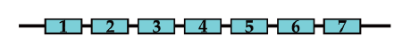 |
++ | – | – | 100 |
| H/F ApaI chimera |  |
– | – | – | 100 |
| F/H ApaI chimera |  |
++++ | +++++ | ++++ | 1 |
| H/F ScaI chimera |  |
++ | ++ | ++ | 1 |
| F/H ScaI chimera |  |
++ | – | +++ | 100 |
| H/F/H ApaI–ScaI chimera |  |
+++ | – | +++ | 100 |
| F/H/F ApaI–ScaI chimera |  |
+++ +++ | +++ ++++ | +++ +++ | 1 100 |
| F/H/F SfiI chimera |  |
+++ +++++ | +++ ++++++ | +++ ++++ | 1 100 |
| F/H/F XcmI chimera |  |
+++ ++++ | +++ +++++ | ++ +++ | 1 100 |
- §
Human TMs are colored in orange and puffer fish TMs are in blue. The relative bioluminescence activity of different chimeras under different concentrations of ligands is scored by the number of +s. Our score system is defined as: no activity as “-” and activity equals to 100% of WT human receptor activity as “++++.” Thus “+” = 25% activity, “++” = 50% of activity, “+++” = 75% of activity of WT human receptor. In every assay, the activity of each construct was measured in triplicate, and each construct was tested at least 3 times. TM: transmembrane; WT: wild type.
The activity of human GHS-R1a mutants and the identification of unique binding sites of different ligands on GHS-R1a
Diversity in the TM region (especially the presence of a charge in the hydrophobic helixes) can give clues to the location sites specifically tailored to interact with ligands.[26,27] MK-0677 and ghrelin activate human GHS-R1a through overlapping binding domains, as we illustrated with GHS-R1a human/puffer fish chimeras. To further identify the key amino acid residues that are critical for binding and activation by ligands, we have identified a number of amino acids which are distinctively different in human and puffer fish. Using site-directed mutagenesis, we first generated a whole series of human GHS-R1a mutants at the potential key residues and assessed their activity by aequorin bioluminescence assay. To further interrogate the specific binding site(s) of human GHS-R1a, we used structurally distinctive GHS-R1a agonists, ghrelin, MK-0677, and GHS-25, to assess the activation of the mutant human GHS-R1a generated by site-directed mutagenesis. A summary of the activity of all GHS-R1a mutants is shown in Table 2. The results revealed the specificity of the mutants for MK-0677, ghrelin, and GHS-25. D99N and E124Q damped the activation of MK-0677. C116A and W276A eliminated the activation of both ghrelin and GH-25, but remained responsive to MK-0677. We also found the activity of MK-0677, ghrelin, and GHS-25; all required the amino acid residues D99 and W276. In addition, our data show that MK-0677 activity requires interaction with E124 and H280, ghrelin activity requires C116, M213, and F279, whereas GHS-25 activity is sensitive to changes in many residues in GHS-R1a. Overall, our findings suggest that each ligand has its unique requirement for the binding pockets, and the unique ligand-binding domains share at least two critical residues of D99 in TM2 and W276 in TM6.
Summary of mutant human growth hormone secretagogue receptor-1a activity in response to various ligands¶
| Domain | Mutation | Activity score |
||
|---|---|---|---|---|
| MK-0677 | Ghrelin | GHS-25 | ||
| FL | Wild type | +++ | +++ | +++ |
| TM2 | D99N | - | ++ | - |
| TM2 | R102H | +++ | +++ | ++ |
| TM3 | C116A | +++ | - | - |
| TM3 | Q120H | +++ | +++ | +++ |
| TM3 | S123A | ++ | +++ | ++ |
| TM3 | E124Q | - | +++ | +++ |
| ICL2 | Y142A | +++ | ++ | ++ |
| ECL2 | D191N | +++ | +++ | + |
| ECL2 | D194N | +++ | +++ | + |
| TM5 | M213K | +++ | + | ++ |
| TM5 | S217A | +++ | ++ | ++ |
| TM5 | S218A | +++ | +++ | + |
| TM6 | W276A | ++ | - | - |
| TM6 | F279L | +++ | + | + |
| TM6 | H280F | ++ | +++ | ++ |
| TM6 | H280Q | +++ | +++ | ++ |
| TM6 | R283H | +++ | +++ | + |
| TM6 | Y284F | +++ | +++ | +++ |
- ¶
For each mutant, the number indicates the amino acid location of the point mutation, the letter before the number indicates the original amino acid, and the letter after the number indicates what the amino acid was changed into. The relative bioluminescence activity of different mutants activated by different ligands was scored by the number of +s. The activity equivalent to full-length WT human receptor activity was denoted as “+++”, each "+" is equivalent to 33% of WT activity, and no activity was denoted as "–". Also, 100 nmol/L of MK-677, Ghrelin, or GH-25 was used. ECL2: extracellular loop 2; FL: full length; GHS: growth hormone secretagogue; ICL2: intracellular loop 2; TM2: transmembrane domain 2; TM3: transmembrane domain 3; TM5: transmembrane domain 5; TM6: transmembrane domain 6.
Discussion
The ghrelin receptor GHS-R1a is a promising alternative pharmacologic target for treatment of GH deficiency, which has a significant impact on children’s development and growth, as well as a great relevance to adults’ lean mass and bone mineral density that have an influence on fracture risk and quality of life.[28,29] GHS-R1a is an important drug target for metabolic disorders including diabetes, obesity, and cachexia. The endogenous ghrelin level is dependent on circadian rhythm and fasting state. It has been shown that physical exercise can transiently increase ghrelin secretion.[30] Therefore, development of synthetic agonists or antagonists of GHS-R1a as pharmacological agents has been vigorously pursued, and some candidates have been tested in clinical trials.[6,31, 32, 33, 34] While evaluation of these compounds or peptides are still ongoing, understanding of the molecular basis of GHS-R1a and ligand interactions, particularly the ligand-binding pocket structure, is essential for the design and development of new GHS-R mimetics.
In this study, we compared the binding characteristics of GHS-R1a peptide ligand ghrelin and two synthetic GHS-R1a agonists MK-0677 and GHS-25. We found that ghrelin and GHS-25 are competitors of MK-0677, although they are structurally different (Figure 1). Importantly, the GHS activity of all of them was completely abolished in Ghsr knockout mice (as GHS-25 shown in Figure 2 and MK-0677 shown in reference [25]), suggesting they all are specific GHS-R1a ligands. It is also noteworthy that while 10 mg of GHS-25 induced 70 ng/mL of GH release 5 min after its administration (Figure 2), 10 mg of ghrelin or MK-677 induced 24 or 48 ng/mL of GH release, respectively,[25] suggesting that GHS-25 is a more potent GHS than ghrelin and MK-677. The potent in vivo effect of GHS-25 could be attributed to my many factors. In addition to its biding efficiency to GHS-R, it could also be due to its pharmacological half-life and/ or its ability to cross the blood–brain barrier.
GHS-R1a protein sequence is evolutionarily conserved. Puffer fish GHS-R1a shares 58% amino acid identity with humans and is activated by the structurally distinct ligand peptide GHRP-6 and non-peptide MK-0677.[13,14] Surprisingly, in contrast to GHRP-6 and MK-0677, ghrelin is unable to activate the puffer fish GHS-R1a (Table 1). GPCRs exhibit considerable variation in the structural determinants of ligand recognitions. Agonists and antagonists may bind to GHS-R1a in vastly different manners, as they are competitive ligands for a common receptor. Chimeric receptor has been used as a major tool to study the structure–function relationship of the seven-helix GPCR family.[35] Hence, we used the human and fish GHS-R1a chimeric receptors to assess the entire structure–function relationship of GHS-R1a and to identify its specific recognition sites for various ligands.
Given the very high degree of sequence identity between the human and puffer fish receptors, especially the TMs, it is reasonable to predict that both human and puffer fish GHS-R1a receptors would bind to its common core in a consistent manner. However, surprisingly, ghrelin and GHS-25 were not able to activate the puffer fish receptor (Table 1). The human and puffer fish chimera allowed us to mix and match different binding pockets to identify the unique binding sites that distinguish puffer fish from human sequences. The ScaI and ApaI–ScaI sets of chimeras suggested that the selectivity of the ligands is mainly achieved through the favorable interactions with the structural elements proximate to TM5 and TM6 (Table 1), which distinguish the peptide ligand ghrelin from small synthetic ligands MK-0677 and GHS-25. With the XcmI chimera, the finding further narrowed the binding domain into a region of 40 amino acids between XcmI and ScaI that constitutes human GHS-R TM6 and ECL3 (Figure 5A), and this replacement is sufficient to rescue ghrelin activation in the puffer fish receptor backbone. Amazingly, with only 40 humanized amino acid residues, we were able to transform the inactive puffer fish receptor to an active “humanized” receptor with activity comparable to that of wild-type human receptor in ghrelin activation. However, it should be noted that any result from chimeric studies may not fully reflect the true structure–function relationship of the wild-type receptors. Nevertheless, our data offer a valuable insight for addressing the question of the structural basis of ligand binding and ligand selectivity.
![Figure 5 The predicted topology and the alternative splitting forms of GHS-R. (A) The amino acid sequence homology between human and puffer fish GHS-R (modified from reference [13] with permission). The receptor structure is illustrated by a schematic representation of TM domains, ECL, and ICL. Conserved amino acid residues are shown in white circles, and the amino acid residues of puffer fish GHS-R different from those in humans are shown in red circles. The empty circles are the amino acid residues present in humans, but absent in puffer fish. The restriction enzyme sites used to construct the chimeras are pointed by the arrows. (B) The diagram of two GHS-R alternative transcripts. The receptor isoform GHS-R1a contains TM 1–7, and GHS-R1b only contains TM 1–5. ECL: extracellular loops; GHS-R: growth hormone secretagogue receptor; ICL: intracellular loops; TM: transmembrane.](/document/doi/10.2478/jtim-2022-0033/asset/graphic/j_jtim-2022-0033_fig_005.jpg)
The predicted topology and the alternative splitting forms of GHS-R. (A) The amino acid sequence homology between human and puffer fish GHS-R (modified from reference [13] with permission). The receptor structure is illustrated by a schematic representation of TM domains, ECL, and ICL. Conserved amino acid residues are shown in white circles, and the amino acid residues of puffer fish GHS-R different from those in humans are shown in red circles. The empty circles are the amino acid residues present in humans, but absent in puffer fish. The restriction enzyme sites used to construct the chimeras are pointed by the arrows. (B) The diagram of two GHS-R alternative transcripts. The receptor isoform GHS-R1a contains TM 1–7, and GHS-R1b only contains TM 1–5. ECL: extracellular loops; GHS-R: growth hormone secretagogue receptor; ICL: intracellular loops; TM: transmembrane.
Among GPCR ligands, the interaction of a small ligand with the receptor is very different from that of a peptide ligand. Small molecules primarily interact with the amino acid residues in the TMs, but peptide ligands have also been shown to interact with extracellular domains.[35] Our data showed that swapping GHS-R C-terminal regions affects the activity of ghrelin more than the activity of MK-0677 and GHS-25 (Table 1). As shown in Figure 5A, the amino acid sequences of human and puffer fish receptors are highly conserved in TM6, but the residues on ECL3 are quite different between humans and puffer fish. Ghrelin has a very unique octanoylated modification of a hydroxyl group on its third serine residue different from small synthetic molecules, which is essential for ghrelin’s binding to GHS-R1a.[15] It is known that human GHS-R gene genomic sequence contains an intron that divides the open reading frame into two exons, encoding TM 1–5 and TM 6–7, which generates two GHS-R subtypes: 1a and 1b.[10] GHS-R1a encodes a full-length isoform and is a functional receptor; in contrast, GHS-R1b is a shorter isoform with only TM 1–5 (Figure 5B), which is an inactive receptor.
Our site-directed mutagenesis data further demonstrated that ghrelin, MK-0677, and GHS-25 bind to distinct, but partially overlapped pockets on GHS-R1a (Table 2). Particularly, the findings showed that W276A and F279L mutations in TM6 abolished ghrelin activity. Interestingly, a naturally occurring mutant allele of F279L has been identified in a family with obesity and short stature,[36] confirming that TM6 residues are critical to ghrelin’s GH-stimulating function. Another identified human mutant GHS-R1a variant is A204E, which has been reported to be associated with short stature in two families.[37] Recently, a mouse model of GHS-R A203E mutation that ablates constitutive GHS-R activity and blunts GH release has been reported, and the phenotype showed short body and femur length in aged mice.[38] In addition to the important binding residues in GHS-R1a C-terminal region, we also observed the mutation D99N in TM2 that abolished the activity of all three ligands. Although our domain swapping study did not specifically check TM2, the assay of SfiI and XcmI chimeras showed a lower activity than wild-type human GHS-R1a at a ligand dose of 1 μmol/L (Table 1), suggesting absence of human TM2 could partially affect the receptor activity in response to ligands.
Our finding collectively supports that TM6 and ECL3 are essential for the optimal activity of GHS-R1a in response to agonists in general, and specificity to natural ligand ghrelin.[13,39] This is also well in line with the recent elegant work from Kojima’s group that elucidated the crystal structure of the ghrelin receptor bound to an antagonist.[40] The crystal structure of GHS-R bound to an antagonist confirmed that the binding pocket of GHS-R1a is bifurcated into two cavities by a salt bridge between E124 and R283.[39,40] A unique characteristic feature is a gap structure between TM6 and TM7. The extracellular side of the TM7 curves outward to a greater degree than that of corresponding structures of other class A GPCRs. In agreement with this observation, our finding revealed that humanizing region of ECL3 plays a major role in converting the puffer fish GHS-R1a to a structure that is no longer inert toward activation by ghrelin. In fact, recent multiple studies of GHS-R protein structure demonstrated that important ECL3 amino acid residues at the junction to TM6 contribute to form ligand-binding pocket, particularly the phenylalanine residues F286 and F290, which are located near the extracellular surface of the receptor and play a critical role in the entry of ghrelin into the receptor core.[40] Structure showed that F286 and F290 interact with ghrelin’s fifth leucine residue[41] and F286 is also in close proximity to ghrelin’s third serine side chain.[42] Mutating F286 and F290 to alanine or polar and charged amino acids displays dramatically diminished receptor activity.[41, 42, 43]
In conclusion, GHS-R1a is capable of binding different ligands in a variety of ways, depending on the selectivity and chemical structure of ligands. The residues on TM6 and ECL3 are essential for the natural ligand ghrelin-dependent activation. Understanding of GHS-R1a and ligand interactions will facilitate the development of better ghrelin receptor agonists/antagonists for treatment of GH deficiency and neuronal developmental disorders.
Acknowledgment
We greatly appreciate the institutional support given to us by Baylor College of Medicine to conduct this study.
-
Author Contributions
Conceptualization: Sun Y and Smith RG; methodology and analysis: Sun Y, Kennedy H, and Smith AGA; writing of the original draft preparation: Sun Y and Smith RG; writing of revision and editing: Ye X and Sun Y; supervision and funding acquisition: Smith RG. All authors have read and agreed to the published version of the manuscript.
-
Conflict of Interest
The authors declare no conflict of interest.
References
1 Smith RG, Van der Ploeg LHT, Howard AD, Feighner SD, Cheng K, Hickey GJ, et al. Peptidomimetic Regulation of Growth Hormone Secretion. Endocrine Reviews 1997;18:621-45.10.1210/edrv.18.5.0316Search in Google Scholar
2 Smith RG, Sun Y, Betancourt L, Asnicar M. Growth hormone secretagogues: prospects and potential pitfalls. Best Pract Res Clin Endocrinol Metab 2004;18:333-47.10.1016/j.beem.2004.04.001Search in Google Scholar
3 Momany FA, Bowers CY, Reynolds GA, Chang D, Hong A, Newlander K. Design Synthesis, and Biological Activity of Peptides which Release Growth Hormone in Vitro. Endocrinology 1981;108:31-9.10.1210/endo-108-1-31Search in Google Scholar
4 Smith RG, Cheng K, Schoen WR, Pong SS, Hickey G, Jacks T, et al. A nonpeptidyl growth hormone secretagogue. Science 1993;260:1640-3.10.1126/science.8503009Search in Google Scholar
5 Smith RG, Pong SS, Hickey G, Jacks T, Cheng K, Leonard R, et al. Modulation of pulsatile GH release through a novel receptor in hypothalamus and pituitary gland. Recent Prog Horm Res 1996;51:26185; discussion 285-66.Search in Google Scholar
6 Patchett AA, Nargund RP, Tata JR, Chen MH, Barakat KJ, Johnston DB, et al. Design and biological activities of L-163,191 (MK-0677): a potent, orally active growth hormone secretagogue. Proc Natl Acad Sci U S A 1995;92:7001-5.10.1073/pnas.92.15.7001Search in Google Scholar
7 Bailey AR, Gilliver L, Leng G, Smith RG. Central actions of the nonpeptide growth hormone secretagogue GHS-25. Endocrine 2001;14:15-9.10.1385/ENDO:14:1:015Search in Google Scholar
8 Chapman IM, Bach MA, Van Cauter E, Farmer M, Krupa D, Taylor AM, et al. Stimulation of the growth hormone (GH)-insulin-like growth factor I axis by daily oral administration of a GH secretogogue (MK-677) in healthy elderly subjects. J Clin Endocrinol Metab 1996;81:4249-57.10.1210/jc.81.12.4249Search in Google Scholar
9 Svensson J, Lonn L, Jansson JO, Murphy G, Wyss D, Krupa D, et al. Two-month treatment of obese subjects with the oral growth hormone (GH) secretagogue MK-677 increases GH secretion, fat-free mass, and energy expenditure. J Clin Endocrinol Metab 1998;83:362-9.10.1210/jc.83.2.362Search in Google Scholar
10 Howard AD, Feighner SD, Cully DF, Arena JP, Liberator PA, Rosenblum CI, et al. A receptor in pituitary and hypothalamus that functions in growth hormone release. Science 1996;273:974-7.10.1126/science.273.5277.974Search in Google Scholar PubMed
11 Smith RG, Betancourt L, Sun Y. Molecular endocrinology and physiology of the aging central nervous system. Endocr Rev 2005;26:203-50.10.1210/er.2002-0017Search in Google Scholar PubMed
12 Smith RG, Jiang H, Sun Y. Developments in ghrelin biology and potential clinical relevance. Trends Endocrinol Metab 2005;16:436-42.10.1016/j.tem.2005.09.004Search in Google Scholar PubMed
13 Palyha OC, Feighner SD, Tan CP, McKee KK, Hreniuk DL, Gao YD, et al. Ligand activation domain of human orphan growth hormone (GH) secretagogue receptor (GHS-R) conserved from Pufferfish to humans. Mol Endocrinol 2000;14:160-9.10.1210/mend.14.1.0412Search in Google Scholar PubMed
14 Smith RG, Feighner S, Prendergast K, Guan X, Howard A. A New Orphan Receptor Involved in Pulsatile Growth Hormone Release. Trends Endocrinol Metab 1999;10:128-35.10.1016/S1043-2760(98)00132-5Search in Google Scholar
15 Kojima M, Hosoda H, Date Y, Nakazato M, Matsuo H, Kangawa K. Ghrelin is a growth-hormone-releasing acylated peptide from stomach. Nature 1999;402:656-60.10.1038/45230Search in Google Scholar
16 Tullin S, Hansen BS, Ankersen M, Møller J, von Cappelen KA, Thim L. Adenosine Is an Agonist of the Growth Hormone Secretagogue Receptor. Endocrinology 2000;141:3397-402.10.1210/endo.141.9.7631Search in Google Scholar
17 Smith RG, Griffin PR, Xu Y, Smith AG, Liu K, Calacay J, et al. Adenosine: A partial agonist of the growth hormone secretagogue receptor. Biochem Biophys Res Commun 2000;276:1306-13.10.1006/bbrc.2000.3610Search in Google Scholar
18 Delhanty PJ, van der Lely AJ. Ghrelin and glucose homeostasis. Peptides 2011;32:2309-18.10.1016/j.peptides.2011.03.001Search in Google Scholar
19 Tilston TW, Brown RD, Wateridge MJ, Arms-Williams B, Walker JJ, Sun Y, et al. A Novel Automated System Yields Reproducible Temporal Feeding Patterns in Laboratory Rodents. J Nutr 2019;149:1674-84.10.1093/jn/nxz116Search in Google Scholar
20 Nakazato M, Murakami N, Date Y, Kojima M, Matsuo H, Kangawa K, et al. A role for ghrelin in the central regulation of feeding. Nature 2001;409:194-8.10.1038/35051587Search in Google Scholar
21 Silva Elipe MV, Bednarek MA, Gao YD. 1H NMR structural analysis of human ghrelin and its six truncated analogs. Biopolymers 2001;59:489-501.10.1002/1097-0282(200112)59:7<489::AID-BIP1054>3.0.CO;2-SSearch in Google Scholar
22 Pong SS, Chaung LY, Dean DC, Nargund RP, Patchett AA, Smith RG. Identification of a new G-protein-linked receptor for growth hormone secretagogues. Mol Endocrinol 1996;10:57-61.10.1210/mend.10.1.8838145Search in Google Scholar
23 McKee KK, Palyha OC, Feighner SD, Hreniuk DL, Tan CP, Phillips MS, et al. Molecular analysis of rat pituitary and hypothalamic growth hormone secretagogue receptors. Mol Endocrinol 1997;11:415-23.10.1210/mend.11.4.9908Search in Google Scholar
24 Button D, Brownstein M. Aequorin-expressing mammalian cell lines used to report Ca2+ mobilization. Cell Calcium 1993;14:663-71.10.1016/0143-4160(93)90091-JSearch in Google Scholar
25 Sun Y, Wang P, Zheng H, Smith RG. Ghrelin stimulation of growth hormone release and appetite is mediated through the growth hormone secretagogue receptor. Proc Natl Acad Sci U S A 2004;101:4679-84.10.1073/pnas.0305930101Search in Google Scholar PubMed PubMed Central
26 Probst WC, Snyder LA, Schuster DI, Brosius J, Sealfon SC. Sequence alignment of the G-protein coupled receptor superfamily. DNA Cell Biol 1992;11:1-20.10.1089/dna.1992.11.1Search in Google Scholar PubMed
27 Schertler G.F, Villa C, Henderson R. Projection structure of rhodopsin. Nature 1993;362:770-2.10.1038/362770a0Search in Google Scholar PubMed
28 Tritos NA, Biller BMK. Current concepts of the diagnosis of adult growth hormone deficiency. Rev Endocr Metab Disord 2021;22:109-16.10.1007/s11154-020-09594-1Search in Google Scholar PubMed
29 Lewinski A, Karbownik-Lewinska M, Wieczorek-Szukala K, Stasiak M, Stawerska R. Contribution of Ghrelin to the Pathogenesis of Growth Hormone Deficiency. Int J Mol Sci 2021;22:9066.10.3390/ijms22169066Search in Google Scholar PubMed PubMed Central
30 Mani BK, Castorena CM, Osborne-Lawrence S, Vijayaraghavan P, Metzger NP, Elmquist JK, et al. Ghrelin mediates exercise endurance and the feeding response post-exercise. Mol Metab 2018;9:114-30.10.1016/j.molmet.2018.01.006Search in Google Scholar PubMed PubMed Central
31 Adunsky A, Chandler J, Heyden N, Lutkiewicz J, Scott BB, Berd Y, et al. MK-0677 (ibutamoren mesylate) for the treatment of patients recovering from hip fracture: a multicenter, randomized, placebo-controlled phase IIb study. Arch Gerontol Geriatr 2011;53:183-9.10.1016/j.archger.2010.10.004Search in Google Scholar PubMed
32 Bright GM, Do MT, McKew JC, Blum WF, Thorner MO. Development of a Predictive Enrichment Marker for the Oral GH Secretagogue LUM-201 in Pediatric Growth Hormone Deficiency. J Endocr Soc 2021;5:bvab030.10.1210/jendso/bvab030Search in Google Scholar PubMed PubMed Central
33 Shin A, Camilleri M, Busciglio I, Burton D, Stoner E, Noonan P, et al. Randomized controlled phase Ib study of ghrelin agonist, RM-131, in type 2 diabetic women with delayed gastric emptying: pharmacokinetics and pharmacodynamics. Diabetes Care 2013;36:41-8.10.2337/dc12-1128Search in Google Scholar PubMed PubMed Central
34 Zhang H, Garcia JM. Anamorelin hydrochloride for the treatment of cancer-anorexia-cachexia in NSCLC. Expert Opin Pharmacother 2015;16:1245-53.10.1517/14656566.2015.1041500Search in Google Scholar PubMed PubMed Central
35 Schwartz TW. Locating ligand-binding sites in 7TM receptors by protein engineering. Curr Opin Biotechnol 1994;5:434-44.10.1016/0958-1669(94)90054-XSearch in Google Scholar
36 Wang HJ, Geller F, Dempfle A, Schauble N, Friedel S, Lichtner P, et al. Ghrelin receptor gene: identification of several sequence variants in extremely obese children and adolescents, healthy normal-weight and underweight students, and children with short normal stature. J Clin Endocrinol Metab 2004;89:157-62.10.1210/jc.2003-031395Search in Google Scholar PubMed
37 Pantel J, Legendre M, Cabrol S, Hilal L, Hajaji Y, Morisset S, et al. Loss of constitutive activity of the growth hormone secretagogue receptor in familial short stature. J Clin Invest 2006;116:760-8.10.1172/JCI25303Search in Google Scholar PubMed PubMed Central
38 Torz LJ, Osborne-Lawrence S, Rodriguez J, He Z, Cornejo MP, Mustafa ER, et al. Metabolic insights from a GHSR-A203E mutant mouse model. Mol Metab 2020;39:101004.10.1016/j.molmet.2020.101004Search in Google Scholar PubMed PubMed Central
39 Feighner SD, Howard AD, Prendergast K, Palyha OC, Hreniuk DL, Nargund R, et al. Structural requirements for the activation of the human growth hormone secretagogue receptor by peptide and nonpeptide secretagogues. Mol Endocrinol 1998;12:137-45.10.1210/mend.12.1.0051Search in Google Scholar PubMed
40 Shiimura Y, Horita S, Hamamoto A, Asada H, Hirata K, Tanaka M, et al. Structure of an antagonist-bound ghrelin receptor reveals possible ghrelin recognition mode. Nat Commun 2020;11:4160.10.1038/s41467-020-17554-1Search in Google Scholar PubMed PubMed Central
41 Liu H, Sun D, Myasnikov A, Damian M, Baneres JL, Sun J, et al. Structural basis of human ghrelin receptor signaling by ghrelin and the synthetic agonist ibutamoren. Nat Commun 2021;12:6410.10.1038/s41467-021-26735-5Search in Google Scholar PubMed PubMed Central
42 Wang Y, Guo S, Zhuang Y, Yun Y, Xu P, He X, et al. Molecular recognition of an acyl-peptide hormone and activation of ghrelin receptor. Nat Commun 2021;12:5064.10.1038/s41467-021-25364-2Search in Google Scholar PubMed PubMed Central
43 Qin J, Cai Y, Xu Z, Ming Q, Ji SY, Wu C, et al. Molecular mechanism of agonism and inverse agonism in ghrelin receptor. Nat Commun 2022;13:300.10.1038/s41467-022-27975-9Search in Google Scholar PubMed PubMed Central
© 2022 Yuxiang Sun, Xiangcang Ye, Hilda Kennedy, Alexander G. A. Smith, Roy G. Smith, published by Sciendo
This work is licensed under the Creative Commons Attribution-NonCommercial-NoDerivatives 3.0 License.
Articles in the same Issue
- Editorial
- Research progress on the role of probiotics in acute liver failure
- Commentary
- A bifunctional enzyme of Legionella that distinctly regulates phosphoribosyl ubiquitination of the SidE family effectors
- The potential role of the brain–gut axis in the development and progression of Alzheimer's disease
- Review Article
- Novel insights into alcoholic liver disease: Iron overload, iron sensing and hemolysis
- Right ventricle remodeling in chronic thromboembolic pulmonary hypertension
- Original Article
- Prevalence and risk factors of hyperuricemia and gout: a cross-sectional survey from 31 provinces in mainland China
- Binding domain characterization of growth hormone secretagogue receptor
- Pan-cancer landscape of the RUNX protein family reveals their potential as carcinogenic biomarkers and the mechanisms underlying their action
- Letter to Editor
- Extracorporeal membrane oxygenation using a modified cardiopulmonary bypass system
- A Case of abdominal pain and diarrhea post immunotherapy: Hypophysitis associated with immune checkpoint inhibitors