Corrigendum to: Carpal tunnel dimensions following osteopathic manipulation utilizing dorsal carpal arch muscle energy: a pilot study
-
Lilian Zhan
 ,
Juanita Brown
,
Sharon Gustowski
,
Patrick Davis
and
Mario Loomis
,
Juanita Brown
,
Sharon Gustowski
,
Patrick Davis
and
Mario Loomis


The authors regret errors that appeared in the following article:
Zhan L, Brown J, Gustowski S, Davis P, Loomis M. Carpal tunnel dimensions following osteopathic manipulation utilizing dorsal carpal arch muscle energy: a pilot study. J Osteopath Med. 2025. doi: 10.1515/jom-2024-0167.
The errors relate to units were reported as millimeters instead of centimeters, and the vertical axis of the carpal tunnel ellipse was reported as the tunnel’s depth, when, in fact, it is one half of the total tunnel depth.
The first two sentences of the abstract’s Result’s section should be listed as:
Results: Comparison of the OMT and control groups revealed a mean increase in carpal tunnel depth from 0.89 cm ± 0.27 cm pre-OMT to 0.95 cm ± 0.28 cm post-OMT (p=0.0146, Cohen’s d=0.214, 95 % CI 0.013 to 0.103). There was also a mean increase in cross-sectional area from 1.83 cm2 ± 0.56 cm2 pre-OMT to 1.98 cm2 ± 0.59 cm2 post-OMT (p=0.0138, Cohen’s d=0.260, 95 % CI 0.06 to 0.48).
The first paragraph of the article’s Results section should be listed as:
Twenty-five participants, mean age of 26 ± 2.18 (male=6, female=19) completed the study. Comparison of the OMT and control groups revealed a mean increase in carpal tunnel depth from 0.89 cm ± 0.27 cm pre-OMT to 0.95 cm ± 0.28 cm post-OMT (p=0.0146, Cohen’s d=0.214, 95 % CI 0.013 to 0.103). There was also a mean increase in cross-sectional area from 1.83 cm2 ± 0.56 cm2 pre-OMT to 1.98 cm2 ± 0.59 cm2 post-OMT (p=0.0138, Cohen’s d=0.260, 95 % CI 0.06 to 0.48). There was no significant difference in canal width (p=0.5973) or transverse carpal ligament length (p=0.2673) following OMT intervention. The control group, which received the sham procedure, demonstrated no significant differences in the transverse carpal ligament length 3.14 cm ± 0.24 cm vs. 3.14 cm ± 0.32 cm (p=0.8977), carpal tunnel width 2.43 cm ± 0.29 cm vs. 2.48 cm ± 0.35 cm (p=0.2517), depth 0.97 cm ± 0.42 cm vs. 0.95 cm ± 0.42 cm (p=0.3952), or cross-sectional area 1.84 cm2 ± 0.81 cm2 vs. 1.83 cm2 ± 0.81 cm2 (p=0.8559) before and after the sham intervention (Figure 4). A post-hoc power analysis was conducted utilizing G’Power 3.1.9.7 and showed that the achieved power was low at 0.351, likely due do the small effect size and sample size in this pilot study. The effect sizes were calculated and the 95 % confidence interval (CI) of the difference are presented with significant p values.
Figure 4 and its legend should be listed as:
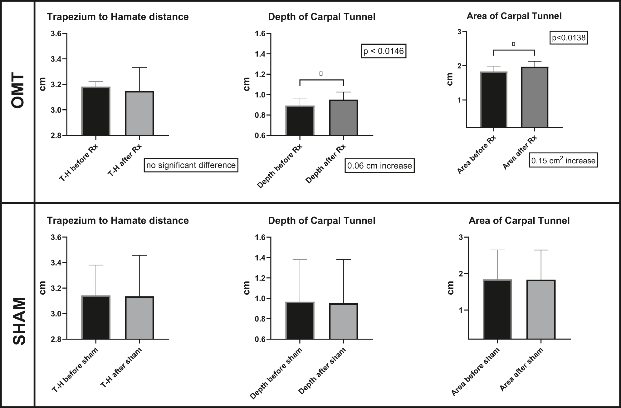
Comparing the means of measurements made before and after manipulation. The top row shows the results following DCA-ME OMT with no significant difference in the distance from trapezium to hamate (the length of the transverse carpal ligament), a 0.06 cm increase in depth of the carpal tunnel, and a 0.15 cm2 increase in carpal tunnel area. The bottom row shows the results following the sham manipulation with no significant difference between any of the measurements made before and after.
© 2025 the author(s), published by De Gruyter, Berlin/Boston
This work is licensed under the Creative Commons Attribution 4.0 International License.
Articles in the same Issue
- Frontmatter
- Behavioral Health
- Original Article
- Bridging the gap: associations of provider enrollment in OKCAPMAP with social deprivation, child abuse, and barriers to access in the state of Oklahoma, USA
- Cardiopulmonary Medicine
- Original Article
- Impact of osteopathic tests on heart rate and heart rate variability: an observational study on osteopathic students
- General
- Review Article
- Comparing intubation techniques of Klippel–Feil syndrome patients in the last 10 years: a systematic review
- Medical Education
- Original Article
- Understanding COMLEX-USA Level-1 as a Pass/Fail examination: impact and opportunities
- Musculoskeletal Medicine and Pain
- Original Article
- Longitudinal outcomes among patients with fibromyalgia, chronic widespread pain, or localized chronic low back pain
- Neuromusculoskeletal Medicine (OMT)
- Original Article
- Carpal tunnel dimensions following osteopathic manipulation utilizing dorsal carpal arch muscle energy: a pilot study
- Corrigendum
- Corrigendum to: Carpal tunnel dimensions following osteopathic manipulation utilizing dorsal carpal arch muscle energy: a pilot study