Atypical cells parameter in sysmex UN automated urine analyzer: a single center study
-
Ozgur Aydin
 ,
Selcuk Sarikaya
,
Emel Caglar
,
Ramazan Ayas
and
Taner Ozgurtas
,
Selcuk Sarikaya
,
Emel Caglar
,
Ramazan Ayas
and
Taner Ozgurtas


Abstract
Objectives
The literature about atypical cells in automated urine analyzers begins with the introduction of Sysmex UN. While modern urinalysis instruments report various kinds of epithelial cells, presence of atypical cells has not been considered. This study aims to observe the efficiency of the atypical cells parameter on Sysmex UN Automated Urine Analyzer with respect to customary manual urine cytology.
Methods
The patients with any indication for a cystoscopy examination were enrolled for the study between March 2022 and October 2022. Voided urine samples were gathered before the cystoscopy and bladder washing sampling was performed afterwards. Bladder washing samples were divided into two; one part was sent for urine cytology and the other was sent for urinalysis. Bladder biopsy was performed if cystoscopy evaluation was suspicious for malignancy.
Results
Thirty three patients (4 females and 29 males) underwent cystoscopy examination for suspicious bladder lesions and 15 of these patients were diagnosed as bladder cancer: nine were high-grade and six were low-grade bladder cancer due to their pathologic test results. Cytology examination results were negative for all patients. Using follow-up histologic diagnosis of urothelial carcinoma as a reference standard, the sensitivity of the atypical cells parameter on Sysmex UN was 27 %, specificity 78 %, positive predictive value 50 %, negative predictive value 56 %.
Conclusions
The results of this study confirmed the ability of Sysmex UN on detecting atypical cells in urine. Yet, the performance of the instrument needs to be improved before the parameter is presented for routine application.
Introduction
It has been quite a while since Sysmex Corporation (Kobe, Japan) has introduced Sysmex UN-Series automated urine analyzer. Besides ordinary parameters of urinalysis, the instrument reports some research parameters including “Atypical cells”. Atypical cells suggest the probability of neoplastic growth, not only in urine but also in general. Particularly considering the urine as the medium, detecting atypical cells favors urothelial carcinoma of the bladder.
Bladder cancer is the eighth most common cancer in the World. Its pathogenesis is complex and multifactorial [1]. Bladder cancer detection and follow-up is based on cystoscopy and/or urine cytology, but remains inaccurate and invasive. As urothelial neoplasms tend to grow into the bladder lumen as papillary structures, there is a likelihood of atypical cells becoming detached from these highly delicate structures to be discharged into the urine and detected in urine samples sent to the laboratory.
Urine cytology and urinalysis are two common laboratory procedures to investigate urine samples in such patients. Urine samples collected from patients with suspected bladder cancer are sent to the laboratory for urine cytology. The test is largely a manual microscopic examination and almost entirely focusses on the presence or absence of atypical cells. Urine samples collected for other indications are sent to the laboratory for urinalysis, which is performed by fully automated devices such as Sysmex UN. The urinalysis report may include more than 30 chemical and particle parameters. While modern urinalysis instruments report various kinds of epithelial cells like tubular, urothelial or squamous types, the presence of atypia has not been included. Although a research parameter on Sysmex UN test menu, reporting atypical cells by an automated urine analyzer is a revolution to converge urinalysis and urine cytology in the laboratory.
It’s exciting to see urinalysis technology evolve [2]. On one side, high technology hardware like new generation lasers or fluorescent dyes strengthen the capability of the instruments while advanced software systems sharpen their intelligence on the other. Besides the impressive progress, it is odd to observe the ignorance of cellular atypia of the epithelial cells. Actually the issue has never been a matter of concern including the age before automation when urinalysis was performed by manual microscopy [3]. However, flow cytometry technology used in automated urine analyzers has the ability to detect atypical cells. This study aims to observe the efficiency of the atypical cells parameter on Sysmex UN Automated Urine Analyzer with respect to customary manual urine cytology.
Materials and methods
Patients and samples
The research has been conducted between March 2022 and October 2022, in the Urology and Biochemistry Departments. The patients with any indication for a cystoscopy examination were enrolled for the study. Before their cystoscopy evaluation, every patient gave a voided urine sample (VU). The same urologist performed the cystoscopy and findings were recorded. After the cystoscopy, bladder washing samples (BW) were gathered. Finally, every patient was examined through a VU and a BW by Sysmex UN. The BW samples were divided into two; one part was sent for urine cytology and the other was sent for urinalysis by Sysmex UN. The samples sent for urine cytology were stained with Papanicolaou stain. The atypical cytology cases were reported according to The Paris System which indicates a standardized terminology for reporting urine cytology. Samples were classified under seven diagnostic categories as previously described by Barkan et al. [4] depending on N/C ratio (<0.5, 0.5–0.7 range, or >0.7) as the major criterion, and three minor criteria including hyperchromasia (present or absent), nuclear irregularity (present or absent) and chromatin pattern (fine or coarse). Diagnostic categories included: 1) Nondiagnostic or Unsatisfactory, 2) Negative for high-grade urothelial carcinoma (NHGUC), 3) Atypical urothelial cells (AUC), 4) Suspicious for high-grade urothelial carcinoma (SHGUC), 5) High-grade urothelial carcinoma (HGUC), 6) Low-grade urothelial neoplasm (LGUN), and 7) Other (primary and secondary malignancies and miscellaneous lesions. Patients with suspicious bladder lesions on cystoscopy evaluation underwent transurethral biopsies. Final pathology reports were accepted as the gold standard for statistical comparison.
Sysmex UN
VU specimens were prepared within 2 h after voiding and BW specimens were prepared within 2 h after cystoscopy. Both were studied by Sysmex-UN instrument. The modular Sysmex UN-Series offers variable combinations of instruments. The UC-3500 uses test strips for chemical analysis of the urine. Sysmex UF-4000/5000 uses fluorescence flow cytometry technology and hydrodynamic focusing for urine sediment analysis, where particles are stained by specific fluorochromes for nucleic acids and surface structures and then sent through the semi-conductor laser beam. Counting and classification is based on signals of scattered light and fluorescence to determine the characteristics of the particles. Atypical cells show side fluorescence and scattered light properties indicating their enlarged nuclei and increased nucleus/cytoplasm ratio. If more investigation of certain particles is requested Sysmex UD-10 presents images captured by an internal camera. Sysmex UN-Series reports a series of research parameters besides routine parameters. These parameters are reported for every sample presented to the instrument but are not validated or sent to the laboratory information system. While evaluating the Sysmex UF-5000 results, values over 0.1/µL were accepted as positive and the values were compared to pathology results [5].
Data were analyzed using IBM SPSS 21 and Microsoft Excel 2013 to calculate results. Kolmogorov-Smirnov test was used to determine normality in parametric variables. The groups were found to be abnormally distributed. The significance of the differences between these groups was determined by Mann-Whitney test. p-Values equal or less than 0.05 were considered statistically significant. The kappa statistic was used to test interrater reliability.
Results
Thirty three patients that admitted to Urology Department were involved in our study. All patients underwent cystoscopy evaluation. Four patients were females and 29 patients were males. Their mean age was 62 years. Fifteen of 33 patients who underwent cystoscopy examination showed suspicious bladder lesions and biopsied for pathological evaluation. All of these 15 patients were diagnosed as bladder cancer according to the final pathology reports. Nine pathology results revealed high-grade bladder cancer and six results revealed low-grade bladder cancer. 18 patients were without suspicious lesions on cystoscopic evaluation and did not undergo biopsies. BW samples from all 33 patients were sent for cytology examination. Samples were evaluated by different pathologists. All 33 samples were reported as “negative for high-grade urothelial carcinoma (Paris 2020)”. BW and VU of patients were evaluated with Sysmex UF-5000 and the average atypical cells results was 0.16/µL for BW samples and 0.17/µL for VU samples. There were no statistically significant difference of atypical cell counts between BW and VU samples (p=0.678). While the number of atypical cells detected by the instrument varied between BW and UV samples of the same patient, there were no discrepancies in means of positivity and negativity (the instrument did not give positive to a negative or negative to a positive for the BW and VU samples of the same patient). The samples were also examined microscopically with Sysmex UD-10 and atypical cells were identified by a pathologist (Figure 1). Atypical cells were observed in samples of eight patients. Three of these patients had high-grade bladder cancer, one patient had low-grade bladder cancer by their pathology reports (true positives). Other four patients were without a biopsy and lacked a confirmative pathology report as no suspicious lesions were detected on cystoscopy. Atypical cells were not observed in 25 patients. 11 of 25 negative results belonged to patients who were diagnosed for bladder carcinoma by their pathology reports (false negatives). Six of them were high-grade, five of them were low-grade urothelial carcinoma by their pathology reports. 14 of 25 negative results were also negative by cystoscopy or pathology (true negatives). The sensitivity of the atypical cells parameter was 27 %, specificity 78 %, positive predictive value 50 % and negative predictive value was 56 %. Cohen’s kappa was 0.046 showing poor agreement between Sysmex UN atypical cells results and final pathology diagnosis.
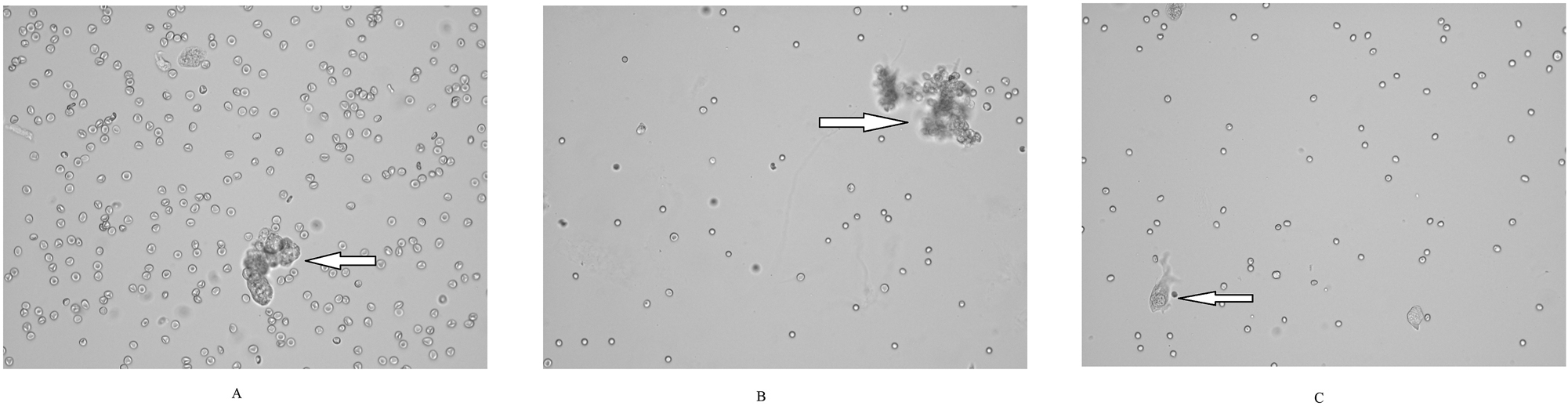
Most samples were rich in red blood cells. Epithelial cells were seen in groups (A, B) or as single cells (C). Large epithelial cell groups were a feature indicating a neoplastic proliferation. Although unstained, digital images were satisfactory in evaluating cytological features of epithelial cells. It was mostly the single cells that presented cytological features of atypia (c). (High power field images, original sysmex UD-10 images. (A, C) High grade urothelial carcinoma; (B) low grade urothelial carcinoma by pathology reports).
Discussion
The most remarkable finding in our study was urine cytology results which were all negative (negative for high grade urothelial carcinoma-Paris System-2020) including in patients with a high grade urothelial carcinoma. Cystoscopy of the bladder has been the recommended investigation when evaluating patients with haematuria to identify bladder cancer and urinary cytology has been a frequently used laboratory test to aid in the diagnosis. However, the role of urine cytology in this respect has been questioned for some time. In their study which they defined as ‘real-world’, Tan et al. suggested that routine urinary cytology had no added benefit for the assessment of haematuria [6]. Finally, 2020 American Urological Association and the Society of Urodynamics (AUA/SUFU) Microhematuria Guideline indicated urine cytology out of the initial diagnostic algorithm [7].
The literature about atypical cells in automated urine analyzers improves with the introduction Sysmex UN which is the single instrument in the market reporting the atypical cells parameter [8]. In a study in 2015 by Anderlini et al. digital images of AIM iQ200 Analyzer (software version 6.4, Iris Diagnostics Inc., Chatsworth, California, USA) were retrospectively evaluated by expert pathologists in search for atypical cells [9]. Whereby a fully automated urine analyzer functioning similar to the Sysmex UN-Series was evaluated the atypical cells were searched in a bulk of images qualified as being “unclassifiable” by the instrument. The experts examined 1,635,287 routine urine samples taken over 5 years and detected atypical cells in 198 samples. The study poses an adoring effort to conclude that atypical cells can be detected only if they are looked for. Recently, Ren et al. analyzed 163 urine specimens, including voided urine samples, bladder washings, and catheterized urine samples, from 128 patients with suspected urothelial carcinoma using Sysmex UF-5000 [10]. They reported the sensitivity of the atypical cells parameter as 59.0 %, specificity 82.1 %, positive predictive value 75.0 %, negative predictive value 68.8 %. With urinary cytopathology as the reference standard, UF-5000 exhibited an agreement of 73.0 % compared to urine cytology. In a similar way we studied patients with a suspicion of urethelial carcinoma. We report the sensitivity of the atypical cells parameter as 27 %, specificity 78 %, positive predictive value 50 %, negative predictive value 56 %. In general, VU samples contained less cells compared to the BW in cytology examination by Sysmex UD-10. In positive for malignancy cases (true positives) atypical cells were much more easily found in BW samples compared to VU samples of the same patient (observational data, no total cell count/sample was measured). Past and future studies may contradict in sensitivity and specificity due to the sample they investigate [11]. A major difference of our study from the one by Ren et al. was Sysmex UD-10 digital imaging module which was used to confirm the results by Sysmex-UF module. Sysmex UF-5000 uses flow-cytometry to detect and the software to decide on the particles. However, Sysmex UD-10 presents up to 80 high quality digital images of the specimen that enables the user to directly observe the particles in the specimen. A positive sample for atypical cells by UF-5000 might need a cytological confirmation before further investigations [12]. It means to run the urine cytology procedure which is found to be non-cost effective. However, UD-10 presents simultaneous up to 80 high power field digital images of the sample. The quality of the images was quite satisfactory. The epithelial nature and structural characteristics of atypical cells could be observed except some that could only be observed in stained samples like hyperchoromasia of the nuclei. At this point we depend on UF-5000 that intensity of signaling is equivalent of dense staining. Finally, the pathologist (using Sysmex UD-10) and Sysmex UF-5000 instrument were expected to compose a common decision on the presence or absence of atypical cells. The pathologist evaluated every patient with the knowledge of the atypical cells count of Sysmex UF-5000. Of the 33 patients there were no discrepancies between Sysmex UD-10 inspection and Sysmex UF-5000, which means that the pathologists observed atypical cells on the digital images of every sample with an atypical cells count higher than 0.1/µL and did not observe any atypical cells on digital images with an atypical cells count 0/µL or 0.1/µL.
Atypical cells have already been measured by almost all instruments in the market in the field of hematology. From the point of cytology, neoplastic cells share common features of atypia like high nucleus/cytoplasm ratio, enlarged and hyperchoromatic nuclei. Particularly considering the Sysmex Corporation, instruments of hematology (Sysmex XN Series) and instruments of urinalysis (Sysmex UN Series) use very similar methodology. They both count cells with the fluorescence flow cytometry method. Considering the struggle on the atypical cells (e.g.: blast, atypical lymphocytes) parameter in hematology, it is too early for a clear cut judgment for atypical cells in urinalysis [13].
In a practical way, automated urine analyzers act analogous to the human who is performing manual microscopy to report urine samples. They both ‘look’ at the sample and ‘see’ the particles thorough in-built microscopes or flow-cytometry and they ‘decide’ on the nature of the particle they see through their software systems. So, suggestions for a better instrument should focus on developing better strategies on sample presentation, improving the act of vision and sharpening the intelligence of instruments by new versions of software writings. Actually, Oyaert et al. report improved diagnostic performance of atypical cells on Sysmex UF-5000 after an application of intelligent verification criteria [14]. In some patients with relatively large lesions, some BW samples failed to present enough atypical cells, indicating that sampling is still a field to be improved.
A major limitation of this study was the small number of patients. Only the BW samples were sent for cytology examination and patients without suspicious lesions did not undergo biopsy, so the absence of malignancy was not pathologically approved. We should emphasize that when investigating the performance of cystoscopy and particularly atypical cells parameter, the gold standard was histological confirmation. True and false positives were simply classified depending on pathological confirmation of malignant bladder tissue obtained by biopsy. In contrast, defining true and false negatives was challenging in patients with a negative cystoscopy as no further examinations including biopsy were planned. Samples were evaluated by different pathologists with different experience in urine cytology. An important point to be mentioned was the dependence of the instrument on sampling and sample processing. By using Sysmex UD-10 we were able to view the sample processed by Sysmex UF-5000. All false negative samples were almost acellular which means that Sysmex UN did not miss the atypical cells but atypical cells were absent in these samples. A recent study by Shukuya et al. showed that atypical cells could be detected using the UF-5000 when the number of atypical cells was high [15]. On the contrary, Sysmex UN misdiagnosed non-neoplastic cells as atypical in false positives. Sysmex UD-10 showed that these samples (both BW and VU) were crowded with non-neoplastic cell clusters which were deceptive for both Sysmex UF-5000 and the pathologist using Sysmex UD-10. Although behind the scope of our study, our results showed the high performance of cystoscopy in diagnosis of bladder cancer, in line with the literature where sensitivity of cystoscopy range from 87 to 100 % and specificity from 64 to 100 % [16].
Finally, although in a small number of patients, we present hopeful results of atypical cells parameter in Sysmex-UN. Apparently, time and further work is needed to improve the performance of the parameter. Of outmost importance, considering the fall of urine cytology from clinical flow charts were due to imbalance of cost and effectiveness, automated urine analyzers offer a cost-effective choice.
-
Research ethics: Ethics approval was obtained from Gulhane Military Medical Academy, 2021/373.
-
Informed consent: Informed consent was obtained from all individuals included in this study.
-
Author contributions: All authors have accepted responsibility for the entire content of this manuscript and approved its submission.
-
Use of Large Language Models, AI and Machine Learning Tools: None declared.
-
Conflict of interest: The authors state no conflict of interest.
-
Research funding: None declared.
-
Data availability: Not applicable.
References
1. Miyazaki, J, Nishiyama, H. Epidemiology of urothelial carcinoma. Int J Urol 2017;24:730–4. https://doi.org/10.1111/iju.13376.Search in Google Scholar PubMed
2. Oyaert, M, Delanghe, J. Progress in automated urinalysis. Ann Lab Med 2019;39:15–22. https://doi.org/10.3343/alm.2019.39.1.15.Search in Google Scholar PubMed PubMed Central
3. Fogazzi, GB, Pallotti, F, Garigali, G. Atypical/malignant urothelial cells in routine urinary sediment: worth knowing and reporting. Clin Chim Acta 2015;439:107–11. https://doi.org/10.1016/j.cca.2014.10.021.Search in Google Scholar PubMed
4. Barkan, GA, Wojcik, EM, Nayar, R, Savic-Prince, S, Quek, ML, Kurtycz, DF, et al.. The Paris System for reporting urinary cytology: the quest to develop a standardized terminology. Acta Cytol 2016;60:185–97. https://doi.org/10.1159/000446270.Search in Google Scholar PubMed
5. Karaburun, MC, Özkaya, MF, Ergüder, Bİ, Süer, E. Investigation of atypical cell parameter in the surveillance of patients with NMIBC; Initial outcomes of a single center prospective study. J Med Syst 2023;47:41. https://doi.org/10.1007/s10916-023-01929-0.Search in Google Scholar PubMed
6. Tan, WS, Sarpong, R, Khetrapal, P, Rodney, S, Mostafid, H, Cresswell, J, et al.. Does urinary cytology have a role in haematuria investigations? BJU Int 2019;123:74–81. https://doi.org/10.1111/bju.14459.Search in Google Scholar PubMed PubMed Central
7. Barocas, DA, Boorjian, SA, Alvarez, RD, Downs, TM, Gross, CP, Hamilton, BD, et al.. Microhematuria: AUA/SUFU guideline. J Urol 2020;204:778–86. https://doi.org/10.1097/ju.0000000000001297.Search in Google Scholar
8. Aydin, O, Yapıcı, O, Copuroglu, R. Atypical cells in Sysmex UN automated urine particle analyzer: a case report and pitfalls for future studies. Turk J Biochem 2020;45:617–19. https://doi.org/10.1515/tjb-2019-0418.Search in Google Scholar
9. Anderlini, R, Manieri, G, Lucchi, C, Raisi, O, Soliera, AR, Torricelli, F, et al.. Automated urinalysis with expert review for incidental identification of atypical urothelial cells: an anticipated bladder carcinoma diagnosis. Clin Chim Acta 2015;451:252–6. https://doi.org/10.1016/j.cca.2015.10.005.Search in Google Scholar PubMed
10. Ren, C, Wang, X, Yang, C, Li, S, Liu, S, Cao, H. Investigation of Atyp.C using UF-5000 flow cytometer in patients with a suspected diagnosis of urothelial carcinoma: a single-center study. Diagn Pathol 2020;15:77. https://doi.org/10.1186/s13000-020-00993-1.Search in Google Scholar PubMed PubMed Central
11. Aydin, O. Atypical cells parameter in Sysmex UN automated urine analyzer: feedback from the field. Diagn Pathol 2021;16:9. https://doi.org/10.1186/s13000-021-01068-5.Search in Google Scholar PubMed PubMed Central
12. Aydin, O. Sysmex UD-10 should accompany UF-5000 in managing atypical cells in urine. J Bioanal Methods Tech 2020;1:102.Search in Google Scholar
13. Aydin, O. Atypical cells parameter in an automated urine analyzer: does it have a future? Anal Biochem 2020;600:113763. https://doi.org/10.1016/j.ab.2020.113763.Search in Google Scholar PubMed
14. Oyaert, M, Maghari, S, Speeckaert, M, Delanghe, J. Improving clinical performance of urine sediment analysis by implementation of intelligent verification criteria. Clin Chem Lab Med 2022;60:1772–9. https://doi.org/10.1515/cclm-2022-0617.Search in Google Scholar PubMed
15. Shukuya, K, Morita, Y, Hisasue, T, Ono, Y, Tomiyasu, S, Kurano, M, et al.. Comparison of the clinical performance of the Atyp.C parameter of the UF-5000 fully automated urine particle analyzer with that of microscopic urine sediment analysis. Pract Lab Med 2023;36:e00328. https://doi.org/10.1016/j.plabm.2023.e00328.Search in Google Scholar PubMed PubMed Central
16. Devlies, W, de Jong, JJ, Hofmann, F, Bruins, HM, Zuiverloon, TCM, Smith, EJ, et al.. The diagnostic accuracy of cystoscopy for detecting bladder cancer in adults presenting with haematuria: a Systematic Review from the European Association of Urology Guidelines Office. Eur Urol Focus 2023;24:2405–4569. (23)00184-0.Search in Google Scholar
© 2024 the author(s), published by De Gruyter, Berlin/Boston
This work is licensed under the Creative Commons Attribution 4.0 International License.
Articles in the same Issue
- Frontmatter
- Review
- Targeting oxidative stress, iron overload and ferroptosis in bone-degenerative conditions
- Research Articles
- Assessing medical biochemistry professionals’ knowledge, attitudes, and behaviors regarding green and sustainable medical laboratory practices in Türkiye
- The efficacy of high pressure liquid chromatography (HPLC) in detecting congenital glycosylation disorders (CDG)
- Atypical cells parameter in sysmex UN automated urine analyzer: a single center study
- The frequency of single specific immunoglobulin E and allergen mixes with a MAST (multiple-antigen simultaneous test) technique
- Differences in second trimester risk estimates for trisomy 21 between Maglumi X3/Preaccu and Immulite/Prisca systems
- Comparison of classical and flowcytometric osmotic fragility and flowcytometric eosin-5-maleimide binding tests in diagnosis of hereditary spherocytosis
- Casticin inhibits the hedgehog signaling and leads to apoptosis in AML stem-like KG1a and mature KG1 cells
- Trimethylamine N-oxide, S-equol, and indoxyl sulfate inflammatory microbiota players in ocular Behçet’s disease
- Genomic profiling of interferon signaling pathway gene mutations in type 2 diabetic individuals with COVID-19
- CDR1as/miR-7-5p/IGF1R axis contributes to the suppression of cell viability in prostate cancer
- Role of interferon regulatory factors in predicting the prognosis of Crimean-Congo hemorrhagic fever
- The significance of taurine for patients with Crimean-Congo hemorrhagic fever and COVID-19 diseases: a cross-sectional study
- Gene mining, recombinant expression and enzymatic characterization of N-acetylglucosamine deacetylase
- Ethanol inhibited growth hormone receptor-mediated endocytosis in primary mouse hepatocytes
- Gypsophila eriocalyx roots inhibit proliferation, migration, and TGF-β signaling in melanoma cells
- The role of kynurenine and kynurenine metabolites in psoriasis
- Tobacco induces abnormal metabolism of tryptophan via the kynurenine pathway
- Effect of vitamin D and omega-3 on the expression of endoplasmic reticulum-associated protein degradation and autophagic proteins in rat brain