Posterior cortical atrophy
-
Melvin Parasram
 ,
Michelle Roytman
,
Michelle Roytman

A 64-year-old man presented to the neurology clinic in June 2021 after he developed progressive visual and cognitive decline over 2 years, including errors at work, peripheral vision loss, and dependence on family members for daily activities. Examination revealed disorientation to location and time; impaired reading, memory, and calculation; and bitemporal hemianopsia. Brain magnetic resonance imaging, reversible dementia work-up, and neuro-ophthalmologic evaluation were unremarkable. Fluorodeoxyglucose (FDG) positron emission tomography (PET) revealed bilateral parietooccipital hypometabolism with otherwise preserved lobar FDG avidity, suspicious for posterior cortical atrophy (PCA) (Figure 1, arrows).
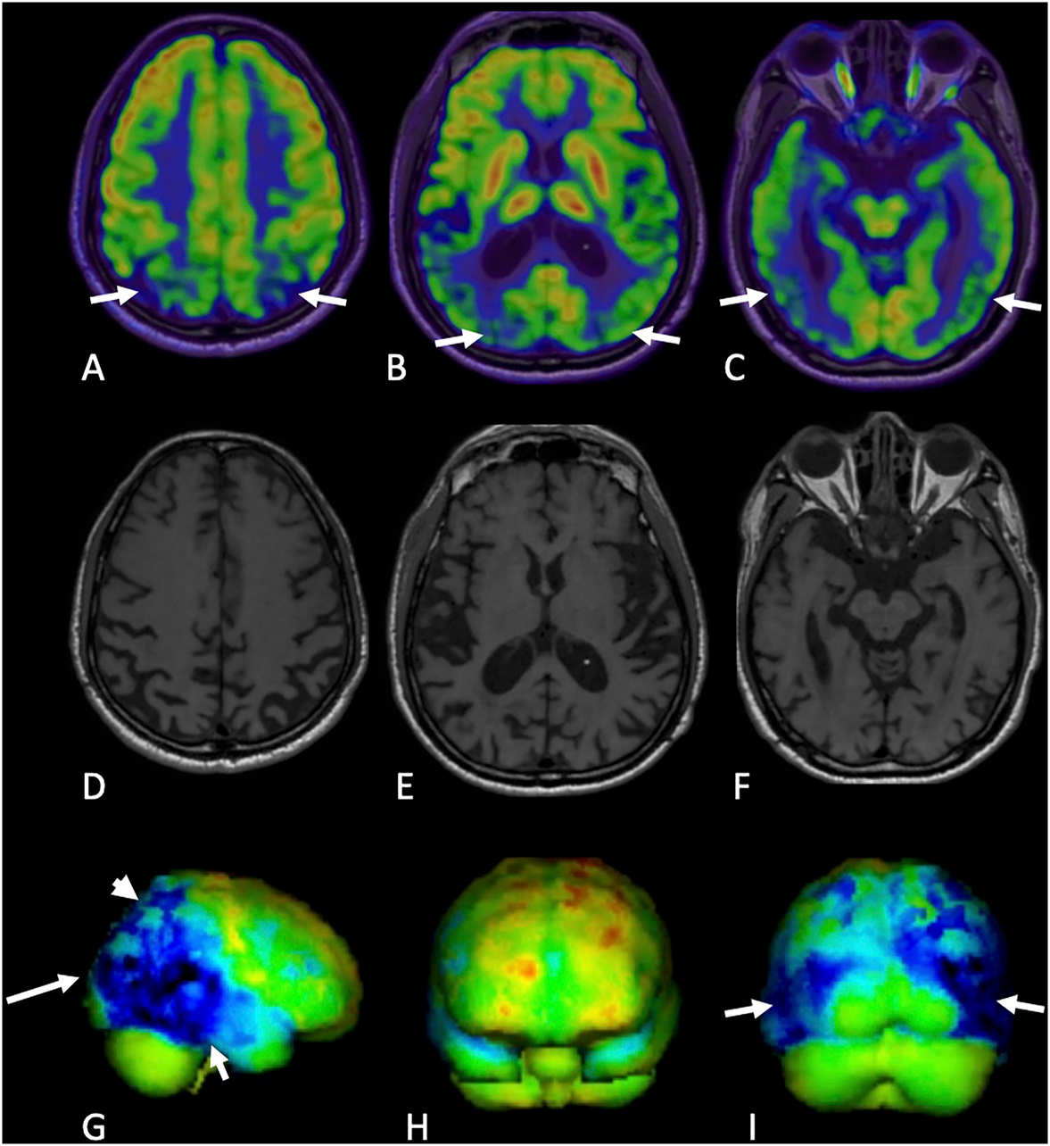
FDG-PET demonstrates decreased FDG avidity in the right greater than left lateral occipital cortex (A–C, arrows), inferior and lateral parietal cortex (G, arrows), and lateral temporal convexity (H and I, arrows) with relative preservation of the frontal cortices, calcarine cortex, and basal ganglia. Axial T1-weighted MRI images are shown in portions D–F.
PCA is a neurodegenerative syndrome that is characterized by progressive cognitive, visuospatial, and visuoperceptual decline [1]. The pathophysiology in PCA is attributed to neuritic plaques and neurofibrillary tangles deposition in the brain, which is seen in Alzheimer’s disease (AD). Thus, PCA is often referred to as “visual variant AD” [2]. Neuroimaging is often employed for the diagnosis of PCA and the key imaging feature is regional atrophy or hypometabolism involving the occipitoparietal and occipitotemporal regions [1, 3], [4], [5]. Currently, there are no treatment options which improve or prevent further decline in symptoms in patients with PCA.
-
Research funding: None reported.
-
Author contributions: M.P. and J.I. provided substantial contributions to conception and design, acquisition of data, or analysis and interpretation of data; all authors drafted the article or revised it critically for important intellectual contact; all authors gave final approval of the version of the article to be published; all authors agree to be accountable for all aspects of the work in ensuring that questions related to the accuracy or integrity of any part of the work are appropriately investigated and resolved.
-
Competing interests: None reported.
References
1. Roytman, M, Ivanidze, J. Posterior cortical atrophy. In: Franceschi, AM, Franceschi, D, editors. Hybrid PET/MRI neuroimaging: a comprehensive approach. New York, NY: Springer Nature; 2021:283–9 pp.10.1007/978-3-030-82367-2_24Search in Google Scholar
2. Levine, DN, Lee, JM, Fisher, CM. The visual variant of Alzheimer’s disease: a clinicopathologic case study. Neurology 1993;43:305–13. https://doi.org/10.1212/wnl.43.2.305.Search in Google Scholar PubMed
3. Bokde, ALW, Pietrini, P, Ibáñez, V, Furey, ML, Alexander, GE, Graff-Radford, NR, et al.. The effect of brain atrophy on cerebral hypometabolism in the visual variant of Alzheimer disease. Arch Neurol 2001;58:480–6. https://doi.org/10.1001/archneur.58.3.480.Search in Google Scholar PubMed
4. Whitwell, JL, Jack, CRJr, Kantarci, K, Weigand, SD, Boeve, BF, Knopman, DS, et al.. Imaging correlates of posterior cortical atrophy. Neurobiol Aging 2007;28:1051–61. https://doi.org/10.1016/j.neurobiolaging.2006.05.026.Search in Google Scholar PubMed PubMed Central
5. Singh, TD, Josephs, KA, Machulda, MM, Drubach, DA, Apostolova, LG, Lowe, VJ, et al.. Clinical, FDG and amyloid PET imaging in posterior cortical atrophy. J Neurol 2015;262:1483–92. https://doi.org/10.1007/s00415-015-7732-5.Search in Google Scholar PubMed PubMed Central
© 2022 the author(s), published by De Gruyter, Berlin/Boston
This work is licensed under the Creative Commons Attribution 4.0 International License.
Articles in the same Issue
- Frontmatter
- Behavioral Health
- Case Report
- Successful buprenorphine transition while overlapping with a full opioid agonist to treat chronic pain: a case report
- General
- Original Article
- How did the dietary habits of patients with chronic medical conditions change during COVID-19?
- Medical Education
- Original Article
- Is cadaveric dissection essential in medical education? A qualitative survey comparing pre-and post-COVID-19 anatomy courses
- Musculoskeletal Medicine and Pain
- Commentary
- Advancing care and research for traumatic brain injury: a roadmap
- Neuromusculoskeletal Medicine (OMT)
- Original Article
- Concussion-related visual memory and reaction time impairment in college athletes improved after osteopathic manipulative medicine: a randomized clinical trial
- Public Health and Primary Care
- Review Article
- A narrative review of nine commercial point of care influenza tests: an overview of methods, benefits, and drawbacks to rapid influenza diagnostic testing
- Original Article
- Disparities in seasonal influenza vaccine uptake and language preference among Hispanic US adults: an analysis of the 2017–2020 BRFSS
- Clinical Images
- Posterior cortical atrophy
- Leukoderma with perifollicular sparing: a diagnostic clue of cutaneous onchocerciasis