Study of ATO nanoparticles by the solvothermal method for thermal insulated coated glass: a green energy application
-
Trinh Xuan Anh
Trinh Xuan Anh is a lecturer at the School of Chemical Engineering, Hanoi University of Science and Technology (HUST). He received his PhD from Kyoto University in 2008. His research interests are photoluminescence, transparent conductive oxide (TCO) materials, and polymer materials.
Duong Thanh Tung received his PhD in Material Science from Chungnam National University, Korea in 2015. He became a lecturer at HUST in 2015. His current research interests include new generation of solar cells, new phosphors and metal oxides for use in high-efficiency lighting devices, and transparent conductive oxide materials.
Do Quy Nhan received his Bachelor’s degree in organic chemistry at HUST. Currently, he is a Master’s student doing research on TCO nanomaterials at HUST.
Tran Vinh Hoang is a lecturer/researcher at School of Chemical Engineering, HUST. He obtained his PhD from University Diderot – Paris 7, France in 2013. His research interests include nanomaterials, nanocomposites, hybrid materials and chemosensor/biosensors for environmental screening and clinical diagnosis. He has published around 20 papers in different international journals or presented at conferences.
Do Quang Trung received his PhD from HUST in 2015 in material science. Presently, he is a lecturer at Quang Ninh University of Industry. His research interest is optoelectronic materials.
Le Dieu Thu received her MS at HUST in 2007. Currently, she is a PhD student at the School of Chemical Engineering, HUST. Her research topic is “Study on luminescent materials applied in agriculture”.
Phan Huy Hoang received his PhD in Chemical Engineering from Chungnam National University, Korea in 2012. He became a lecturer at the School of Chemical Engineering, HUST in 2005. His current research interests include synthesis of nanomaterials and their application as catalysts for organic reactions, “green” chemical synthesis from biomass and lignocelluloses materials, and solar cells.
Nguyen Duy Cuong received his PhD degree in Materials Science and Engineering at Chungnam National University, Korea in 2007. From 2009 to 2013, he worked as a postdoc at Hyogo University, Japan. Since 2013, he has been a lecturer at HUST. His research interests include thin film technology, TCO nanomaterials, and solar cells being fabricated by solution methods such as Cu(In,Ga)Se2 and Cu(Zn,Sn)Se2.


Abstract
Antimony-doped tin oxide (ATO) nanoparticles (NPs) (Sb-doped content 3%, 10%, and 15%) were synthesized by the (2 l autoclave, medium-scale) solvothermal method followed by sintering at various temperatures (500°C, 800°C, 900°C and 1000°C) so they would crystallize. The particle size increased from several to tens of nanometers with the increase of sintered temperature from 500°C to 1000°C, sharply from 800°C to 1000 °C; ~30 g of final product was received for each experiment. More interestingly, the crystallinity of the as-synthesized ATO was also increased with the increasing Sb doped content from 3% to 15%. The ATO NPs were coated onto glass substrates and then sintered at 500°C, which effectively prevented transmittance of infrared (IR) wavelengths (>800 nm) with 10% wt Sb-doped content, which is useful for thermal insulated glass coating application.
1 Introduction
Antimony-doped tin oxide (ATO) is a transparent conducting material which has both high transmittance in the visible region (Vis) and high electrical conductivity which reflects and/or absorbs near infrared radiations (NIR) [1]. These characteristics are comparable with conventional Indium Tin Oxide, which was recently introduced as an alternative thermal insulating coating for building windows, but cheaper [2], [3].
ATO nanoparticles (NPs) were fabricated using various methods including the conventional solid-state method [4], aqueous and non-aqueous methods such as the co-precipitation method [5], the sol-gel method, the hydrothermal method [6], [7], [8] and so on. For both aqueous and non-aqueous procedures, most of the precursors produce nanosized crystalline nuclei at a temperature of 200–300°C; further NPs crystallization was controlled by reactive time or annealing at various temperatures [9], [10]. To synthesis the metal oxide NPs with an industrial scale, solvothermal method is used more commonly than other methods, such as sol-gel [11].
In the present study, we introduce the (2 L autoclave, medium-scale) solvothermal method for fabrication of ATO NPs, which produces nanosized crystalline nuclei at a temperature of 200°C followed by annealing at various temperatures. The as-synthesized powders were applied for thermal insulated glass coating application; structural, morphology, and UV-Vis-NIR transmitting properties of ATO NPs were investigated in detail.
2 Materials and methods
The reagents used in the preparation of the sol-gel mixture were antimony chloride (SbCl3, 99%, Sigma-Aldrich) and tin chloride (SnCl4, 98%, Sigma-Aldrich), which were utilized as Sb3+ and Sn4+ ion precursors. The precursors were mixed with the ethanol solvent (1 l, 0.2 m) with various Sb/Sn+Sb mol ratio of 3%, 10% and 15% and the pH of the solution was fixed at 10 with ammoniac solution; after a 24 h stirring, the solution was transferred to an autoclave (2 l) which was maintained at 200oC, 40 atm, and 200 rpm for 24 h. After cooling down, the powders obtained were washed in distilled water and dried at 100oC for 24 h. To achieve fine crystallinity, the as-synthesized ATO NPs were sintered at various temperatures; ~30 g of final product was received for each experiment.
In order to investigate the UV-Vis-NIR transmitting properties, the ATO film was coated on glass substrate by doctor blade method using the mixed paste of ATO powders and ethylene glycol binder (ratio of 1:1 wt.) followed by annealing in 500oC for 1 h. Due to the aggregation of NPs during sintering interfering with the dispersed issues, only as-synthesized ATO with Sb 3%, 10% and 15% doped concentration was used.
The structure and surface morphology of NPs were investigated by scanning electron microscopy (SEM) (JSM-7600F, Jeol). The Sb doping concentration was confirmed by energy dispersive X-ray spectroscopy (EDX; JSM-7600F, Jeol) at an accelerating voltage of 10 kV. The preferred orientation and crystallinity were characterized by X-ray diffraction (XRD; D8 Advance, Bruker) using Cu Kα radiation. The UV-Vis-NIR transmitting properties of ATO NPs thin films were investigated by Agilent Cary 5000 UV-Vis-NIR equipment.
3 Results and discussion
To observe the changes in the size and morphology of ATO NPs with annealing temperatures, we conducted SEM measurements. At low Sb content (3% Sb, see Figure 1), the as-synthesized ATO NPs have very small particle size, which is difficult to observe by SEM images. However, after treatment at a temperature of 500°C, the ATO crystallites were seen more clearly, as shown in Figure 1B; the size of the particles is below 3 nm. The size of the NPs strongly increased from 7 nm ATO to 15 nm with the increase of annealing temperature from 800°C to 1000°C (see Figure 1C and D); these results are equivalent to previous works [11].
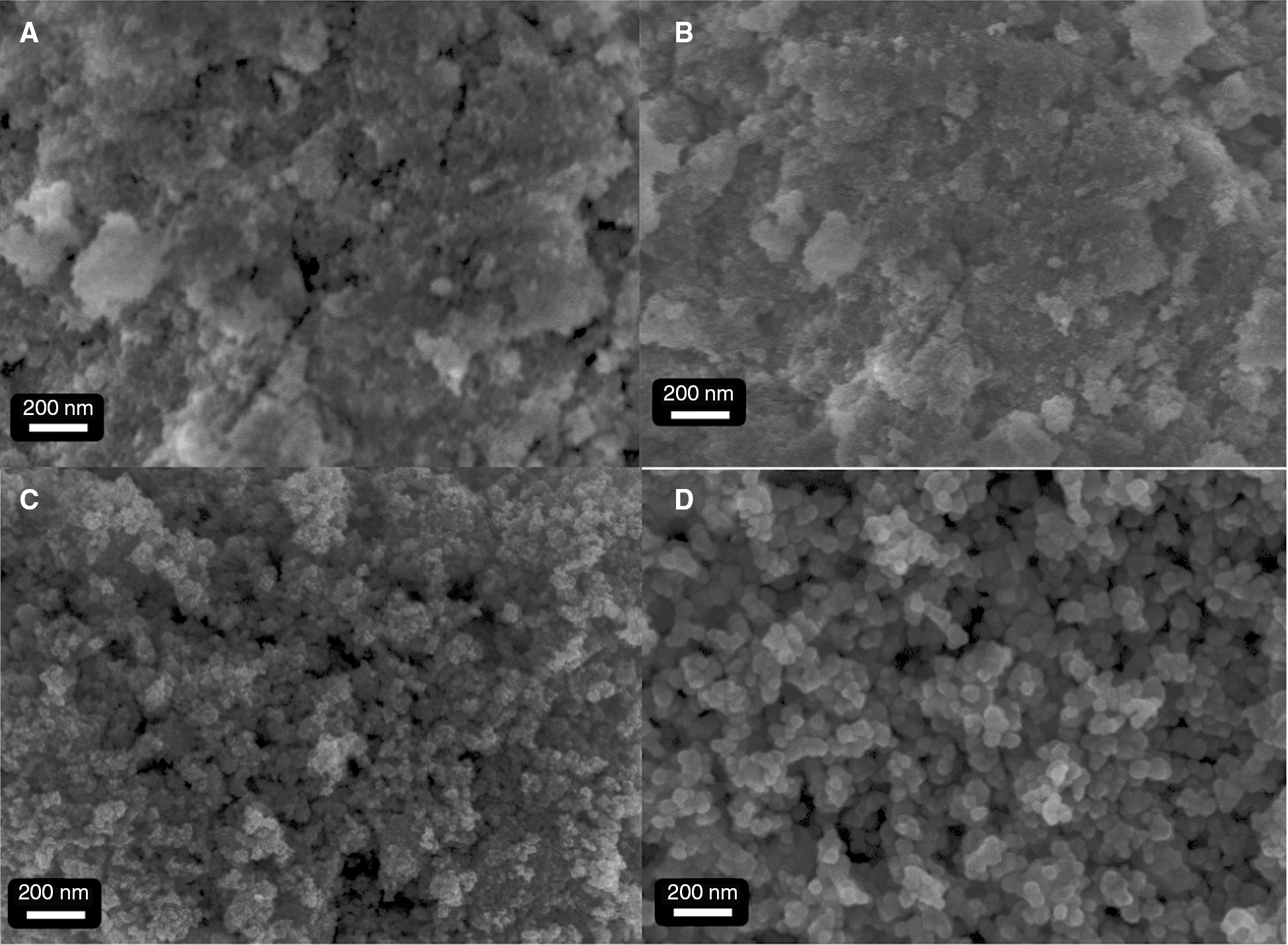
The field emission scanning electron microscopy (FESEM) images of the antimony-doped tin oxide (ATO) nanoparticles (NPs) (Sn/Sn+Sb=3% at.) obtained with calcination temperatures: (A) as-synthesized, (B) 500°C, (C) 800°C, and (D) 1000°C.
In the case of the ATO samples with 10% Sb, we also annealed at different temperatures and SEM results are shown in Figure 2. Here we can see that formation of NPs in the as-synthesized sample is clearly observed (Figure 2A). With annealing at high temperature, the particle size also increased strongly, especially in the range of 800–1000°C (see Figure 2B–D).

The FESEM images of the antimony-doped tin oxide (ATO) nanoparticles (NPs) (Sn/Sn+Sb=10% at.) obtained with calcination temperatures: (A) as-synthesized, (B) 500°C, (C) 800°C, and (D) 1000°C.
Figure 3A–D show SEM images of the ATO with 15% Sb. The variation of the particle size of ATO NPs is similar to that of 10% Sb-ATO, as discussed above (Figure 2).

The FESEM images of the antimony-doped tin oxide (ATO) nanoparticles (NPs) (Sn/Sn+Sb=15% at.) obtained with calcination temperatures: (A) as-synthesized, (B) 500°C, (C) 800°C, and (D) 1000°C.
To investigate the composition of NPs, we performed EDX measurements and the results are shown in Figure 4. The EDX results indicate that the actual atomic concentration of Sb is 5.5% and 16.7% for the samples with 3% and 15% Sb content, respectively.

Energy dispersive X-ray spectroscopy (EDX) spectra of as-synthesized antimony-doped tin oxide (ATO) nanoparticles (NPs) with (A) 3% Sb and (B) 15% Sb.
In order to analyze the phase structure, crystallinity and the preferred orientations, we carried out XRD measurements and the results are shown in Figure 5. The diffraction peaks were observed at the positions of 26.5, 33.8, 37.9, 51.7, 54.8, and 57.8°; these peaks correspond to the crystal orientations of (110), (101), (200), (211), (220), and (002); ATO (JCPDS card, file-no: 41–1445). In the case of sample ATO with 3% Sb, diffraction peak of the sample annealed at 500°C is very weak and wide, this indicates that nanocrystals have a very small size and poor crystallinity. However, diffraction peaks become strong and sharp after being annealed at temperatures in the range of 800–1000°C; this shows that the size of the particles and crystallinity have increased significantly. This is quite consistent with the results of the SEM image as described in Figure 2. However, the XRD peak intensity decreased with the ATO with 15% Sb, 1000°C. This may be due to the low melting temperature of the Sb element; the high Sb content (>15%) decomposes the ATO crystal at a high temperature. From XRD data, the crystal sizes of the ATO NPs were calculated via the Scherrer equation, Eq. (1):

X-ray diffraction (XRD) pattern of antimony-doped tin oxide (ATO) nanoparticles (NPs) 3% and 15% as a function of sintering temperature (A) and (B) the {101} shifted peak of sintered at 1000°C ATO 3% and 15% and (C) crystallite size vs. sintering temperature of ATO 3% and 15%.
where τ is the mean size of the ordered (crystalline) domains, which may be smaller or equal to the grain size, and K is a dimensionless shape factor, with a value close to unity. The shape factor has a typical value of about 0.9, λ is the X-ray wavelength, and β is the line broadening at half the maximum intensity (FWHM). This quantity is also sometimes denoted as Δ(2θ); θ is the Bragg angle. Particle size strongly varied in the annealing temperature range of 800–1000°C, as shown in Figure 5C and D. The biggest particles observed at the annealing temperature of 1000°C are 22 nm and 15 nm for 3% Sb-ATO and 15% Sb-ATO, respectively.
To study the optical properties of ATO NPs, we conducted measurements of the transmission spectra for as-synthesized ATO NPs with different Sb concentrations followed by sintering at 500°C for 1 h, and the results are described in Figure 6. The ATO NPs with 3% Sb seem to be transparent from the Vis to IR region, which are similar transmittance properties of SnO2 NPs, because the concentrations of Sb in this sample are too low, only 3%. However, when the concentration of Sb was increased up to 10%, transmittance (or absorption) edge fell sharply at the wavelengths >817 nm, implying that it could be used in IR reflective applications. For ATO NPs with 15% Sb, transmittance edge red-shifted strongly at far IR wavelengths (>1250 nm). Several works have been reported where the carrier concentration of ATO NPs thin films optimized with Sb doped content of about 10% wt. It is worth noting that plasma wavelength moves toward shorter wavelengths by the increase of carrier concentration according to the Drude theory; consequently, the IR reflection begins from shorter wavelengths [12]. The present results show that the ATO NPs films effectively prevent transmittance of IR wavelengths with 10% wt Sb doped content.

UV-visible-near infrared radiation (UV-Vis-NIR) normalized transmittance spectra of as-sintered antimony-doped tin oxide (ATO) nanoparticles (NPs) thin films at various Sb doping concentrations of 3%, 10% and 15%.
4 Conclusion
In summary, we have established a simple process for synthesizing ATO NPs at a medium scale. With this process, we successfully fabricated ATO NPs with different Sb concentration. The particle size of ATO NPs strongly varied at annealing temperatures in the range of 500–1000°C. The crystallinity of ATO NPs was improved significantly after annealing at the temperatures in the range of 800–1000°C; ~30 g of the final product was received for each experiment. Moreover, the ATO NPs films, which are coated onto glass substrates and then sintered at 500°C, effectively prevent transmittance of IR wavelengths (>800 nm ) with 10% wt Sb doped content, which is useful for thermal insulated glass coating application.
About the authors

Trinh Xuan Anh is a lecturer at the School of Chemical Engineering, Hanoi University of Science and Technology (HUST). He received his PhD from Kyoto University in 2008. His research interests are photoluminescence, transparent conductive oxide (TCO) materials, and polymer materials.

Duong Thanh Tung received his PhD in Material Science from Chungnam National University, Korea in 2015. He became a lecturer at HUST in 2015. His current research interests include new generation of solar cells, new phosphors and metal oxides for use in high-efficiency lighting devices, and transparent conductive oxide materials.

Do Quy Nhan received his Bachelor’s degree in organic chemistry at HUST. Currently, he is a Master’s student doing research on TCO nanomaterials at HUST.

Tran Vinh Hoang is a lecturer/researcher at School of Chemical Engineering, HUST. He obtained his PhD from University Diderot – Paris 7, France in 2013. His research interests include nanomaterials, nanocomposites, hybrid materials and chemosensor/biosensors for environmental screening and clinical diagnosis. He has published around 20 papers in different international journals or presented at conferences.

Do Quang Trung received his PhD from HUST in 2015 in material science. Presently, he is a lecturer at Quang Ninh University of Industry. His research interest is optoelectronic materials.

Le Dieu Thu received her MS at HUST in 2007. Currently, she is a PhD student at the School of Chemical Engineering, HUST. Her research topic is “Study on luminescent materials applied in agriculture”.

Phan Huy Hoang received his PhD in Chemical Engineering from Chungnam National University, Korea in 2012. He became a lecturer at the School of Chemical Engineering, HUST in 2005. His current research interests include synthesis of nanomaterials and their application as catalysts for organic reactions, “green” chemical synthesis from biomass and lignocelluloses materials, and solar cells.

Nguyen Duy Cuong received his PhD degree in Materials Science and Engineering at Chungnam National University, Korea in 2007. From 2009 to 2013, he worked as a postdoc at Hyogo University, Japan. Since 2013, he has been a lecturer at HUST. His research interests include thin film technology, TCO nanomaterials, and solar cells being fabricated by solution methods such as Cu(In,Ga)Se2 and Cu(Zn,Sn)Se2.
Acknowledgements
This work was supported by the Vietnam Ministry of Education and Training (MOET) through research project B2015.01.101 (2015–2016).
References
[1] Coleman JP, Lynch AT, Madhukar P, Wagenknecht JH. Sol. Energy Mater. Sol. Cells 1999, 56, 395.10.1016/S0927-0248(98)00144-5Search in Google Scholar
[2] Mei S, Ma W, Zhang G, Wang J, Yang J, Li Y. Micro Nano Lett. 2012, 7, 12–14.10.1049/mnl.2011.0558Search in Google Scholar
[3] Giovannetti F, Föste S, Ehrmann N, Rockendorf G. Energy Procedia 2012, 30, 106–115.10.1016/j.egypro.2012.11.014Search in Google Scholar
[4] Bernardi MIB, Cava S, Paiva-Santos CO, Leite ER, Paskocimas CA, Longo E. J. Eur. Ceram. Soc. 2002, 22, 2911.10.1016/S0955-2219(02)00057-2Search in Google Scholar
[5] Zhang J, Gao L. Inorg. Chem. Commun. 2004, 7, 91–93.10.1016/j.inoche.2003.10.012Search in Google Scholar
[6] Koivula R, Harjula R, Lehto J. Micropor. Mesopor. Mater. 2002, 55, 231.10.1016/S1387-1811(02)00411-0Search in Google Scholar
[7] Zhang J, Gao L. Mater. Chem. Phys. 2004, 87, 10.10.1016/j.matchemphys.2004.06.004Search in Google Scholar
[8] Pena JS, Brouse T, Sanchez L, Morales J, Schleich DM. J. Power Sources 2001, 97/98, 232.10.1016/S0378-7753(01)00620-6Search in Google Scholar
[9] Duong T, Do Q, Pham A, Nguyen D. J. Alloys & Compounds 2016, 686, 854–858.10.1016/j.jallcom.2016.06.204Search in Google Scholar
[10] Niederberger M, Pinna N. Metal Oxide Nanoparticles in Organic Solvents: Synthesis, Formation, Assembly and Application, Springer: Dordrecht, Heidelberg, London, New York, 2009.10.1007/978-1-84882-671-7Search in Google Scholar
[11] Jeon H, Jeon M, Kang M, Lee S, Lee Y, Hong Y, Choi B. Mater. Lett. 2005, 59, 1801–1810.10.1016/j.matlet.2005.01.070Search in Google Scholar
[12] Khusayfan N, El-Nahas M. Adv. Cond. Matter Phys. 2013, ID 408182, 8.10.1155/2013/408182Search in Google Scholar
©2016 Walter de Gruyter GmbH, Berlin/Boston
This article is distributed under the terms of the Creative Commons Attribution Non-Commercial License, which permits unrestricted non-commercial use, distribution, and reproduction in any medium, provided the original work is properly cited.
Articles in the same Issue
- Frontmatter
- In this issue
- Green processing of thermosensitive nanocurcumin-encapsulated chitosan hydrogel towards biomedical application
- Supramolecular chemistry at interfaces: host-guest interactions for attaching PEG and 5-fluorouracil to the surface of porous nanosilica
- Study of ATO nanoparticles by the solvothermal method for thermal insulated coated glass: a green energy application
- Synthesis of Cu-BTC, from Cu and benzene-1,3,5-tricarboxylic acid (H3BTC), by a green electrochemical method
- Improving the electrochemical behavior of sustainable polyaniline titanium dioxide composite by intercalation of carbon nanotubes
- Sustainable composite materials based on ethylene-vinylacetate copolymer and organo-modified silica
- Conference announcement
- 7th International Conference of the Flow Chemistry Society (Cambridge, UK, February 7–8, 2017)