Ocular pharmacoscintigraphic and aqueous humoral drug availability of ganciclovir-loaded mucoadhesive nanoparticles in rabbits
-
Sohail Akhter
Sohail Akhter is currently working as postdoctoral research associate at the Department of Pharmaceutics, Utrecht University, The Netherlands. His work has focused on the pharmaceutical development, pharmacokinetic and pharmacodynamic evaluation of antipsychotics polymeric particles for overcoming non-adherence in psychotic disorders. He did his Master’s and PhD in Pharmaceutical Sciences (Pharmaceutics) on the development of lipid vesicular and polymeric nanoparticulate system for therapeutic targeting and bioavailability enhancement. During his PhD, he was awarded with senior research fellowship of Council of Scientific and Industrial Research (CSIR), Department of Biotechnology (DBT) and University Grant Commission (UGC). In addition, he was awarded with travel grants for scientific presentations under young scientist category by Department of Science and Technology (DST) and Indian Council of Medical Research (ICMR). His research interests involve rational development of nanoparticulates and vesicular systems for effective therapeutic targeting, pharmacokinetics and bioanalysis. E-mail:
sohailakhtermph@gmail.com Farshad Ramazani obtained his Pharmacy Degree (Pharm.D) in 2007 from Tabriz School of Pharmacy, Tabriz, Iran. After been awarded a full scholarship from Iranian Health Ministry in 2010, Farshad joined the Department of Pharmaceutics at Utrecht University, Utrecht, The Netherlands. His research is focused on the local delivery of kinase inhibitors for the treatment of cancer under the supervision of Prof. dr. ir. W.E. Hennink and Dr. Robbert J. Kok.
Mohammad Zaki Ahmad is currently a Faculty in Department of Pharmaceutics, College of Pharmacy, Najran University, Saudi Arabia. He instructs pharmaceutics courses to Pharm.D. students. He did major in Pharmaceutics from Dibrugarh University Assam, India. Prior to joining academic, he worked as Junior Research Fellow under University Grant Commission. His research focused primarily in the area of novel drug delivery, drug targeting and nanoparticles as drug carrier.
Farhan Jalees Ahmad is an Associate Professor at Faculty of Pharmacy, Hamdard University, New Delhi, India and Director of Nanomedicine research Lab in the same institution. His work interest is multi-disciplinary research focus on development of oral and parenteral controlled drug delivery system, drug targeting and novel nanotechnologies for medical applications. He received his PhD in in Pharmaceutical Sciences (Pharmaceutics) from Hamdard University. Currently, he is the President of Indian Pharmaceutical Association (Delhi) and also serving as the director of food technology program at Hamdard University. After his PhD, He worked as Research Scientist in Ranbaxy Research Labs for 6 years before coming into the academic research. So far, Dr. Ahmad successfully accomplished 28 projects from government and industry related to nano-therapeutics, bioavailable delivery of herbal drugs, product development, scale-up, technology transfer and validation. He is credited with 2 US and 6 Indian patents. Moreover, Dr. Ahmad published 6 books and more than 200 research and review papers in peered reviewed journals. He is member of the editorial (advisory) board of a variety of scientific journals. He was awarded with
Young Scientist award from Department of Science & Technology, Ministry of Science India (2003),FIP Development (Grant 2001 ), Netherlands, Scientist of the year award (2005) by National Environment Science Academy and Best Publication Award (2012) from Hamdard University, Delhi, India.Ziyaur Rahman is currently a Faculty in Irma Lerma Rangel College of Pharmacy, Texas A&M Health Science Center, Kingsville, Texas. He instructs pharmaceutics courses to Pharm.D. students. He did major in Pharmaceutics from Hamdard University, New Delhi, India. Prior to joining academic, he worked as ORISE fellow in Center for Drug Evaluation and Research, Food and Drug Administration, Maryland, USA. He served on the editorial board of American Journal of Analytical Chemistry, Scientia Pharmaceutica and Journal of Pharmaceutical Investigation. His research focused primarily in the area of QbD and Process Analytical Technologies (PAT), controlled drug delivery of challenging molecules, oral delivery of macromolecules, and nanoparticles.
Aseem Bhatnagar, MD, DRM, PhD (Toxicology) is the head of the Department and Director of Nuclear medicine in Institute of Nuclear Medicine & Allied Sciences (INMAS) Defence Research and Development Organization (DRDO), Delhi, India. His work interest is multi-disciplinary research activity focused on Nuclear medicine, Thyroidology, Drug development, Clinical trials including pharmacoscintigraphy and Nanomedicine. He is basically a clinician (MD) and received his PhD as well in Toxicology. He is credited with more than patents granted/filed and successful development of 50 biomedical products including 30 approved formulations from Drug Controller General of India, mainly for the clinical high altitude related medical problems. He is serving as an IAEA consultant on radiopharmaceutical clinical trials. Moreover, Dr. Bhatnagar published 3 books and more than 150 research and review papers in peered reviewed journals. He is member of the editorial (advisory) board of a variety of scientific journals in the field of clinical and drug development research.
Gert Storm obtained his PhD degree in 1987 at the Department of Pharmaceutics of the Utrecht University. His research interests are in the fields of biopharmaceutics and drug targeting. In 1988–1989 he was a visiting scientist at Liposome Technology Inc. in Menlo Park, USA, and visiting Assistant Professor at the School of Pharmacy, UCSF, San Francisco. In September 1991 he took up his position at the Utrecht University. In 1999, he was appointed adjunct professor at the Royal School of Pharmacy, Copenhagen. From July 2009 on, he is Honorary Professor in Biomacromolecular Drug Delivery at the University of Copenhagen. In 2000, he was appointed as professor (Targeted Drug Delivery) at Utrecht University. From 2012 on, he is also professor (Targeted Therapeutics) at the MIRA institute of the University of Twente. Moreover, he is active at the University Medical Center Utrecht (UMCU) within the CBOI institute (Centre for Image-Guided Oncological Interventions). He is author/co-author of more than 400 original articles, reviews and book chapters in the field of advanced drug delivery/drug targeting, and theme (co-)editor of Advanced Drug Delivery Reviews and the book ‘Long Circulating Liposomes. Old Drug, New Therapeutics’. He was coordinator of an Integrated Project (FP6) on targeted nanomedicines (MediTrans) based on the collaboration of 30 European partners and funded by the EC and industry. He is program director of the program Drug Delivery embedded within the recently approved New Nano Initiative (NanoNextNL) strongly sponsored by the Dutch government and industry. He is also principal investigator of a national industry-academia partnership (HIFU-CHEM) studying the clinical application of MRI-guided high-intensity focused ultrasound (HIFU) to improve cancer chemotherapy with temperature-sensitive targeted nanomedicines. He is on the Board of Scientific Advisors of the Controlled Release Society (CRS). He is a member of the editorial (advisory) board of a variety of scientific journals. He was involved in the foundation and is currently on the board of the European Society for Nanomedicine (ESNAM/CLINAM) and The Netherlands Platform for Targeted Nanomedicine (NPTN).


Abstract
The present report describes the improved ocular retention and aqueous humoral drug availability of ganciclovir (GCV) when administered via topical instillation of different kind of nanoparticles onto the rabbit eye. GCV was loaded into PLGA nanoparticles, chitosan-coated nanoparticles and chitosan-coated niosomal nanoparticles. All three formulations contained nanoparticles equally round in shape with a mean particle size in the range of 180–200 nm. The ocular corneal retention property was evaluated by gamma scintigraphy, revealing that the clearance was slowest in the case of the chitosan-containing formulations. GCV in chitosan-coated PLGA nanoparticles and chitosan-coated niosomal nanoparticles showed approx. 6-fold higher aqueous humor drug availability as compared to a GCV solution and nearly 2.5-fold higher as compared to the chitosan-lacking GCV-PLGA nanoparticles. The results indicate that the use of a mucoadhesive chitosan coating can improve the ocular residence time and aqueous humoral availability of GCV when administered topically in nanoparticles.
Introduction
Ganciclovir (GCV) is a synthetic acyclic nucleoside analog of 2′-deoxyguanosine, exhibiting antiviral activity against herpes simplex virus and cytomegalovirus at relatively low inhibitory concentrations [(IC50 of ∼50 to 500 ng/mL, respectively] (1, 2). GCV plays an important role in the treatment of ocular viral infections. Conventional treatment involves the oral administration of GCV at a dose of 3 g/day. However, this relatively high dose regimen is associated with the occurrence of side effects including bone marrow suppression and neutropenia (1, 3). Compared to systemic treatment, intravitreal injection of GCV provides higher intraocular drug concentrations but repeated intravitreal injections are poorly tolerated (4). Topical ocular delivery of GCV is also an option, but its hydrophilic character and rapid elimination will result in poor intraocular availability of the drug. The tear film as well as the corneal and conjunctival epithelia represents a compact barrier hindering the absorption of topically applied hydrophilic drugs into the intraocular region. Therefore, topical delivery of GCV is associated with a low aqueous humor bioavailability (5–8). This limitation encourages the development of mucoadhesive GCV nanoformulations for topical ocular delivery with the aim to attain a higher aqueous humoral availability allowing the administration of lower topical doses and reduction of dosing frequency. Thus, the objective of this work was to evaluate the topical ocular retention and increased intraocular delivery of GCV when administered topically entrapped in mucoadhesive nanoparticles as compared to topical ocular delivery of a GCV solution and nanoparticles without mucoadhesive coating. In this study, PLGA nanoparticles and niosomes have been prepared and evaluated. PLGA, being a biodegradable, biocompatible and FDA-approved polymer as drug carrier and medical device is an obvious choice for topical ophthalmic formulation. Moreover, niosomes have already been investigated as a potential topical ocular drug delivery system for many drugs categories such as antiglaucoma and antibiotics (9, 10).
These vesicles appear to be similar to liposomes in terms of their physical properties. They are also prepared in the same way and, under a variety of conditions, and form unilamellar or multilamellar structures. Niosomes have been claimed to alleviate some of the disadvantages associated with liposomes, such as relatively high cost (11). They also have the potential for controlled and targeted drug delivery (9, 10).
Materials and methods
Chemicals
A gift sample of GCV was provided by Ranbaxy Laboratories Ltd. (Gurgaon, Haryana, India). Poly (lactide-co-glycolide) (PLGA, Resomer® 503H, lactic acid/glycolic acid, 50:50) was purchased from Boehringer Ingelheim (Germany). Chitosan (CS, high density, deacetylation degree >80%) was received as a gift sample from India Sea Foods, Cochin (India). Span 60 and cholesterol were purchased from S.D. Fine-chem Ltd. (Mumbai, India). Acetonitrile and trifluoroacetic acid (TFA) of HPLC grade were obtained from Qualigens Fine Chemicals (Mumbai, India). Water was produced in the laboratory by a Milli-Q purification system (Millipore, Billerica, MA, USA). All other reagents used were of analytical grade.
Preparation of GCV nanocarriers
Preparation of PLGA nanoparticles (GCV-NPs)
GCV-NPs were prepared by a double emulsion solvent evaporation method (12). Briefly, 100 μL of phosphate buffered saline (PBS; pH 7.4) containing GCV (0.4% w/v) was emulsified in 1 mL dichloromethane containing 100 mg PLGA by sonication. The resulting primary emulsion was added to 20 mL of polyvinyl alcohol (PVA, 1.5% w/v in distilled water) with continuous stirring to form a double emulsion. The emulsion was stirred at 1500 rpm until complete evaporation of dichloromethane was achieved. The resulting nanosuspension was subjected to 3 cycles of homogenization at a pressure of 10,000 bar (EmulsiFlex-C5, Avestin, Inc., Canada). GCV-NPs were collected by centrifugation at 15,000 rpm for 10 min at 4°C, washed three times with distilled water, and Lyophilization was carried out in a freeze-drier (Heto DRYWINNER, Germany) at 0.05 mBar for 24 h. The entrapment efficiency for the GCV in prepared formulation was then determined using the method reported by Duvvuri et al. (13) by UPLC quantification technique.
Preparation of chitosan-coated PLGA nanoparticles (GCV-CSNPs)
GCV-CSNPs were prepared using a water–oil–water (w/o/w) emulsion solvent evaporation method with slight modification (12). 100 μL of PBS (pH 7.4) containing GCV (0.4% w/v) was emulsified in 1 mL dichloromethane containing 100 mg PLGA by sonication. The resulting primary emulsion was added to 20 mL of a mixed solution consisting of chitosan (CS) (0.25% w/v in 0.5 M acetate buffer, pH 4.4) and polyvinyl alcohol (PVA, 1.5% w/v in distilled water) with continuous stirring to form a double emulsion. The emulsion was stirred at 1500 rpm until complete evaporation of dichloromethane was achieved. The resulting nanosuspension was subjected to 3 cycles of homogenization at a pressure of 10,000 bar (EmulsiFlex-C5, Avestin, Inc., Canada). GCV-CSNPs were collected by centrifugation at 15,000 rpm for 10 min at 4°C, washed three times with distilled water, and freeze-dried for 24 h. The entrapment efficiency for the GCV in prepared formulation was then determined using the method reported by Duvvuri et al. (13) by UPLC quantification technique.
Preparation of chitosan-coated niosomal nanoparticles (GCV-NDs)
GCV-NDs were prepared by the reverse-phase evaporation technique (1). Briefly, GCV-loaded niosomes were prepared by dissolving Span 60 and cholesterol in the ratio of 3:2 w/w in diethyl ether and adding 2 mL of PBS (pH 7.4) containing GCV. The mixture was sonicated for 5 min and a thick emulsion was obtained which was stirred at 1500 rpm to remove any residual ether. To this emulsion 3 mL of PBS was added in order to hydrate the vesicles. The resulting vesicles were incubated for 2 h with a 0.2% w/w chitosan solution (in glacial acetic acid, 0.5% v/v, pH 5.5). GCV entrapment (%) was then estimated as per the method reported by Akhter et al. (1).
GCV quantification
GCV analysis was performed using a Waters Acquity ultra performance liquid chromatography (UPLC) system (Milford, MA, USA) equipped with a binary solvent manager, an autosampler, column manager composed of a column oven, a precolumn heater and a photo diode array detector. 5 μL of the final analytical solution was injected into a Waters Acquity second generation bridged ethyl hybrid (BEH) C18 (50 mm×2.1 mm, 1.7 μm) UPLC column kept at 50°C. The mobile phase consisted of a mixture of 0.1% TFA in water (adjusted to pH 3.5 using 5.0% dilute ammonia) and acetonitrile (95:5, v/v). The flow rate was 0.45 mL/min and detection was performed at a wavelength of 254 nm with total run time of 3 min. Data acquisition, data handling and instrument control were performed by Empower Software v1.0.
Characterization of GCV nanocarriers
Transmission electron microscopy
The surface morphology of the GCV nanoformulations was studied by transmission electron microscopy (TEM). Formulations treated on copper grids (Polysciences, Warrington, PA) with 1% uranyl acetate for negative staining, followed by sample drying, were analyzed by TEM (Philips CM 10, The Netherlands) at an accelerating voltage of 100 kV.
Particle size distribution and zeta potential
The mean particle size of the GCV nanoformulations was determined by photon correlation spectroscopy using a Zetasizer Nano ZS (Malvern Instruments, Malvern, UK). For size determination, a sample of the dispersion was diluted to the appropriate concentration with Milli-Q water and analysis was performed at 25°C with an angle detection of 90°. Zeta potential was measured using a disposable zeta cuvette with the same instrument.
In-vivo study
Ocular pharmacoscintigraphic and intraocular drug availability experiments were carried out using New Zealand Albino rabbits (2.25±0.25 kg). The study was carried out under the guidelines of CPCSEA (Committee for the Purpose of Control and Supervision of Experiments on Animals, Ministry of Culture, Government of India). The protocol was approved by the Institutional Animal Ethics Committee, Jamia Hamdard, New Delhi (approval no. 822) and the ARVO guidelines for animal usage were followed. Utmost care was taken to ensure that animals were treated in the most humane and ethically acceptable manner.
Ocular retention study: gamma-scintigraphy
The precorneal retention of nanoparticles after topical administration was assessed by γ-scintigraphy. The radiolabeling of GCV loaded formulations were achieved by using 99mTc as per the protocol developed by Institute of Nuclear Medicine and Allied Sciences (INMAS), New Delhi, India (14). Labeling efficiency was determined using instantaneous thin layer chromatography (ITLC) and was found to be >98%. Corneal retention of chitosan-containing 99mTc-labeled GCV-CSNPs and GCV-NDs were compared with chitosan-lacking 99mTc labeled GCV-NPs. A total of 20 μL of the labeled formulations were instilled into the cul-de-sac of the left eye of the rabbit, and the eye was manually blinked three times to distribute the formulation over the cornea. The right eye of each rabbit served as a negative control. A gamma camera (Millenium VG, Milwaukee, Wisconsin), autotuned to detect the 140 KeV radiation of Tc-99m, was used for the scintigraphy study. Rabbits were anesthetized by using a ketamine HCl injection given intramuscularly at a dose of 15 mg/kg body weight and positioned 5 cm in front of the probe before the radio imaging. Recording was started 5 s after instillation and continued for 30 min using 128×128 pixel matrix. Sixty individual frames (60×30 s) were captured by dynamic imaging process. The region of interest (ROI) was selected on one frame of the image, and the time activity curve was plotted to calculate the rate of disappearance from the eye surface. ROI activity of all the images were recorded on a computer system assisted with the software Entegra Version-2.
Ocular drug delivery study: aqueous humoral drug concentration profile
Four groups, each with seven New Zealand Albino rabbits (2.25±0.25 kg), were used. The protocol was approved by the Institutional Animal Ethics Committee, Jamia Hamdard, New Delhi, India and corresponding guidelines were followed. Each group received, in both the eyes, a single topical instillation (50 μL) of GCV-solution, GCV-NPs, GCV-CSNPs and GCV-NDs at a GCV dose equivalent to 0.3% w/v of GCV (3 mg/mL). The eyes were anesthetized after the treatment using topical application of 4% xylocaine sterile solution (AstraZeneca LP); 50 μL of the aqueous humor was collected using 30 gauze needles pre- and post-treatment at 0.5, 1, 2, 4, 6, 8 and 12 h. All aqueous humor samples were collected in pre-labeled eppendorf tubes for UPLC analysis, sealed and stored at –20°C until UPLC analysis. The aqueous humor samples were prepared as mentioned above. Pharmacokinetic parameters (PK) were calculated by non-compartmental analysis (model independent analysis using WinNonLin version 4.0 (Pharsight Corp., Mountain View, CA, USA).
Sample preparation
All the rabbit aqueous humor samples were stored at –20°C and allowed to thaw at room temperature prior to sample preparation. A 50 μL aliquot of rabbit aqueous humor was pipetted into a 1.0 mL eppendorf tube, followed by the addition of 100 μL of acetonitrile. The samples were vortexed for 5 min followed by 5 min of centrifugation at 10,000 rpm. The samples were filtered through a 0.22 μm nylon filter and 5 μL of the filtrate was directly injected into the UPLC system
Ocular tolerance test
The potential ocular irritancy of GCV-NPs, GCV-CSNPs and GCV-NDs were evaluated according to a modified Draize test (15) using a slit-lamp. The congestion, redness and swelling, and tear discharge of the conjunctiva were graded on a scale from 0 to 3, 0 to 4, and 0 to 3, respectively. Nanoparticles (50 μL) were topically applied in the right eye (every 10 min for 1 h) and the left eyes were used as controls. Observation of the ocular tissue condition was performed at 12 h and 24 h after the end of the experiments.
Results and discussion
Preparation and characterization of GCV nanocarriers
We designed PLGA- and Span-based nanoparticles (niosomes) coated with a chitosan coating to increase the mucoadhesivness and as a result the retention time after instillation onto the eye cornea was improved. The mean of GCV entrapment (%) in GCV-NPs, GCV-CSNPs and GCV-NDs were 49.3%, 46.7% and 47.2%, respectively. The TEM and the particle size distribution of the GCV nanoformulations are shown in Figures 1 and 2, respectively. The micrographs of the chitosan-coated GCV-CSNPs and GCV-NDs demonstrated a very different appearance as compared to the reference GCV-NPs (Figure 1B and C). Spherical-shaped particles with a double layer structure were observed in case of chitosan-coated nanoformulations indicating the existence of chitosan surrounding the GCV-CSNPs and GCV-NDs. The hydrophilic polymer coating of PLGA and surfactant based nanoparticles would depend on the capability of the polymer to adhere to the hydrophobic layer. The interaction between chitosan and such particles surface area due to a combination of physical adsorption, hydrophobic interaction and bridging between them. In general, all of the three formulations, GCV-NPs, GCV-NDs, and GCV-CSNPs were equally round in shape with a mean particle size in the range of 180–200 nm (Figure 2). The polydispersity index (PI), which is a measure of uniformity of size within the formulation (1, 16), was also measured. The GCV-NDs exhibited a more narrow size distribution (PI, 0.181) compared to GCV-CSNPs (PI, 0.65) and GCV-NPs (PI, 1.15), with a mean size of 190 nm, 200 nm and 190 nm, respectively. The zeta potential of GCV-NPs, GCV-NDs, and GCV-CSNPs were –23.4 mV, +41.8 mV and +37.2 mV, respectively. The zeta potential is an important particle characteristic indicating the surface charge that can influence the nano-suspension as well as the mucoadhesion into the ocular surface. The electrostatic charge repulsion between the similar charge (either positive or negative) particles prevents the aggregation and thus ensure the dispersed state of the nanosuspension. On the other hand, positive surface charge (illustrate by positive zeta potential value) on the particles is responsible for mucoadhesion with mucin layer. In the present study, positive value zeta potential of GCV-NDs, and GCV-CSNPs provided the proof of successful chitosan adsorbed cationic surface modification of niosomes and GCV-NPs. Moreover, all the three formulations showed good dispersion stability.

Transmission electron microphotographs of GCV nanoparticles: (A) GCV-NPs (reference), (B) chitosan-coated GCV-CSNPs, and (C) chitosan-coated GCV-NDs.
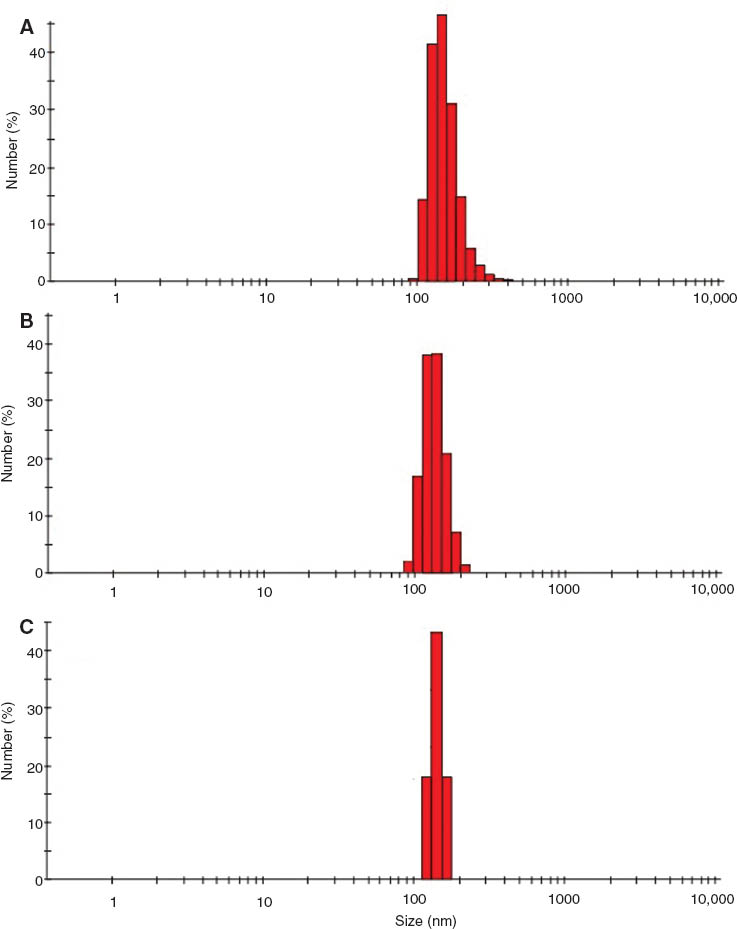
Particle size distribution of GCV nanoparticles: (A) reference GCV-NPs, (B) chitosan- coated GCV-CSNPs, and (C) chitosan-coated GCV-NDs.
Rabbit studies
Ocular retention
Gamma-scintigraphy is a well-established technique for the in-vivo monitoring of the fate of nanocarriers (14). After administration of the radiolabeled ophthalmic formulation, a good spreading was observed over the entire precorneal area. The quantification of radioactivity in ROIs enabled the assessment of the remaining activity at different time points after instillation. Radioactivity values counted in the ROI on gamma scintigraphic dynamic whole body images collected for the first 30 min after administration of the reference (chitosan lacking) and chitosan-coated formulations (radioactivity versus time profile) is shown in Figure 3. As expected, the clearance of the reference (chitosan lacking) PLGA nanoparticles (GCV-NPs) from the ocular surface was most rapid. This can be explained by the lack of mucoadhesion with the cornea. In contrast, the chitosan-containing GCV-CSNPs and GCV-NDs were retained on the ocular surface significantly longer (p<0.05) than GCV-NPs (Figure 3A–C). The AUC0–30 min for the retained activity of GCV-CSNPs and GCV-NDs were 47,505 and 39,431, respectively, which is about 4-fold higher as compared to the reference GCV-NPs with a value of 11,032. These findings advocate that the presence of chitosan in the formulations serves to prolong the retention of the nanoparticles on the corneal surface. Particularly, due to the interaction of positively charged chitosan with the cornea surface which is negatively charged by the presence of mucin and other components (4, 7). Moreover, the uniform and spherical large surface area increased the spreading and contact time of the mucoadhesive nanoparticles over the corneal surface thereby promoting the corneal retention (17).

Pre-corneal retention of 99mTc- labeled formulations after single topical instillation of: (A) reference GCV-NPs, (B) chitosan-coated GCV-CSNPs, and (c) chitosan-coated GCV-NDs.
Intraocular aqueous humoral drug delivery
Aqueous humor GCV concentration-time profiles upon topical instillation of GCV solution, GCV-CSNPs, GCV-NPs and GCV-NDs to the rabbit eye are shown in Figure 4. Calculated kinetic parameters are shown in Table 1. In the group treated with the GCV solution, a relatively low ocular bioavailability (AUC0-t, 495±11 ng·h/mL) was observed; the aqueous humor levels of the drug were undetectable after 2 h, which can be attributed to rapid pre-corneal loss. Administration of the reference, chitosan-lacking GCV-NP formulation yielded an about 3-fold higher value (1361.4±12.0 ng·h/mL) compared to the GCV solution indicating that incorporation in nanosized PLGA particles can improve the permeation by improving corneal retention (12, 17). Topical instillation of GCV-CSNPs and GCV-NDs even more enhanced the humoral drug concentration with nearly similar availability of 3991±20 and 3428±29 ng·h/mL, respectively. The humoral drug availability was approx. 7- to 8-fold higher as compared to the GCV solution and approx 2.5-fold higher as compared to the reference GCV-NPs. Our results indicate that both chitosan-coated GCV nanoparticle formulations provided higher aqueous humoral drug availability as compared to a GCV solution and the chitosan-lacking reference nanoparticles. This can be attributed to an increased corneal retention of the GCV nanoformulations, providing a larger time frame for sustained release of the entrapped drug. Furthermore, the large surface area provided by nanosized particles increased the spreading and contact time over the corneal surface which favours the corneal retention. Clearly, the significantly increased corneal retention mediated by the mucoadhesiveness conferred by chitosan contributes strongly to the improvement of the transcorneal drug permeation (7, 17).
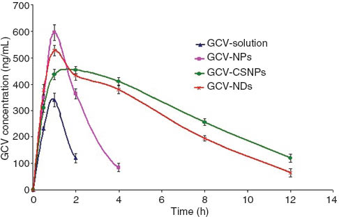
Aqueous humor concentration–time profile of GCV after topical instillation of GCV- solution, reference GCV-NPs, chitosan-coated GCV-CSNPs and chitosan-coated GCV-NDs onto the rabbit eye.
Topical instillation of GCV solution and GCV nanoparticles onto the rabbit eye: pharmacokinetic parameters.
| Parameter | GCV-sol | GCV-NPs | GCV-CSNPs | GCV-NDs |
|---|---|---|---|---|
| atmax (h) | 0.9±0.03 | 1.0±0.05 | 1.4±0.07 | 1.0±0.04 |
| aCmax (ng/mL) | 325±5.1 | 589±8.9 | 449±6.5 | 523±8.2 |
| aKe (1/h) | 1.50±0.11 | 0.67±0.08 | 0.08±0.01 | 0.13±0.02 |
| aAUC0–t (ng·h/mL) | 495.1±11.3 | 1361.4±12.0 | 3991.3±20.1 | 3428.1±29.4 |
| aAUC0–∞ (ng·h/mL) | 720.5±19.6 | 1528.3±20.70 | 5622.1±39.5 | 4160.7±46.9 |
aMean values±SD (n=14; both eyes of each rabbit out of the seven rabbits were used).
Ocular tolerance study
For such a nanometric delivery system to be proposed as an ophthalmic nanomedicine, it is important not only to examine the biopharmaceutical properties but also their non-irritant nature. Therefore, in-vivo ocular irritation test for GCV-NP, GCV-CSNPs, GCV-NDs were evaluated by using modified Draize test. The in-vivo result of ocular irritation test in rabbits showed no visual sign of irritation or other tissue damaging effect to anterior ocular tissues (data were not given). Moreover, there were no excessive tear formation and the clinical scores for conjunctival swelling, corneal opacity and discharge were always zero in the case of treatment group of all the formulation during the study phase. The results confirmed that the studied formulations were non-irritant and well tolerated.
Conclusion
Entrapment of GCV in PLGA nanoparticles and niosomes increases the GCV concentration in the aqueous humor. Coating of the surface of the nanoparticles with chitosan further promotes the corneal drug absorption process by virtue of an increased retention on the ocular surface. Therefore, we propose that chitosan-coated nanoparticles are promising carriers for GCV in novel topical ophthalmic nanoformulations.
Conflict of interest statement:
Authors do not have any conflict of interest related to this work.
About the authors

Sohail Akhter is currently working as postdoctoral research associate at the Department of Pharmaceutics, Utrecht University, The Netherlands. His work has focused on the pharmaceutical development, pharmacokinetic and pharmacodynamic evaluation of antipsychotics polymeric particles for overcoming non-adherence in psychotic disorders. He did his Master’s and PhD in Pharmaceutical Sciences (Pharmaceutics) on the development of lipid vesicular and polymeric nanoparticulate system for therapeutic targeting and bioavailability enhancement. During his PhD, he was awarded with senior research fellowship of Council of Scientific and Industrial Research (CSIR), Department of Biotechnology (DBT) and University Grant Commission (UGC). In addition, he was awarded with travel grants for scientific presentations under young scientist category by Department of Science and Technology (DST) and Indian Council of Medical Research (ICMR). His research interests involve rational development of nanoparticulates and vesicular systems for effective therapeutic targeting, pharmacokinetics and bioanalysis. E-mail: sohailakhtermph@gmail.com
Farshad Ramazani obtained his Pharmacy Degree (Pharm.D) in 2007 from Tabriz School of Pharmacy, Tabriz, Iran. After been awarded a full scholarship from Iranian Health Ministry in 2010, Farshad joined the Department of Pharmaceutics at Utrecht University, Utrecht, The Netherlands. His research is focused on the local delivery of kinase inhibitors for the treatment of cancer under the supervision of Prof. dr. ir. W.E. Hennink and Dr. Robbert J. Kok.

Mohammad Zaki Ahmad is currently a Faculty in Department of Pharmaceutics, College of Pharmacy, Najran University, Saudi Arabia. He instructs pharmaceutics courses to Pharm.D. students. He did major in Pharmaceutics from Dibrugarh University Assam, India. Prior to joining academic, he worked as Junior Research Fellow under University Grant Commission. His research focused primarily in the area of novel drug delivery, drug targeting and nanoparticles as drug carrier.

Farhan Jalees Ahmad is an Associate Professor at Faculty of Pharmacy, Hamdard University, New Delhi, India and Director of Nanomedicine research Lab in the same institution. His work interest is multi-disciplinary research focus on development of oral and parenteral controlled drug delivery system, drug targeting and novel nanotechnologies for medical applications. He received his PhD in in Pharmaceutical Sciences (Pharmaceutics) from Hamdard University. Currently, he is the President of Indian Pharmaceutical Association (Delhi) and also serving as the director of food technology program at Hamdard University. After his PhD, He worked as Research Scientist in Ranbaxy Research Labs for 6 years before coming into the academic research. So far, Dr. Ahmad successfully accomplished 28 projects from government and industry related to nano-therapeutics, bioavailable delivery of herbal drugs, product development, scale-up, technology transfer and validation. He is credited with 2 US and 6 Indian patents. Moreover, Dr. Ahmad published 6 books and more than 200 research and review papers in peered reviewed journals. He is member of the editorial (advisory) board of a variety of scientific journals. He was awarded with Young Scientist award from Department of Science & Technology, Ministry of Science India (2003), FIP Development (Grant 2001), Netherlands, Scientist of the year award (2005) by National Environment Science Academy and Best Publication Award (2012) from Hamdard University, Delhi, India.
Ziyaur Rahman is currently a Faculty in Irma Lerma Rangel College of Pharmacy, Texas A&M Health Science Center, Kingsville, Texas. He instructs pharmaceutics courses to Pharm.D. students. He did major in Pharmaceutics from Hamdard University, New Delhi, India. Prior to joining academic, he worked as ORISE fellow in Center for Drug Evaluation and Research, Food and Drug Administration, Maryland, USA. He served on the editorial board of American Journal of Analytical Chemistry, Scientia Pharmaceutica and Journal of Pharmaceutical Investigation. His research focused primarily in the area of QbD and Process Analytical Technologies (PAT), controlled drug delivery of challenging molecules, oral delivery of macromolecules, and nanoparticles.

Aseem Bhatnagar, MD, DRM, PhD (Toxicology) is the head of the Department and Director of Nuclear medicine in Institute of Nuclear Medicine & Allied Sciences (INMAS) Defence Research and Development Organization (DRDO), Delhi, India. His work interest is multi-disciplinary research activity focused on Nuclear medicine, Thyroidology, Drug development, Clinical trials including pharmacoscintigraphy and Nanomedicine. He is basically a clinician (MD) and received his PhD as well in Toxicology. He is credited with more than patents granted/filed and successful development of 50 biomedical products including 30 approved formulations from Drug Controller General of India, mainly for the clinical high altitude related medical problems. He is serving as an IAEA consultant on radiopharmaceutical clinical trials. Moreover, Dr. Bhatnagar published 3 books and more than 150 research and review papers in peered reviewed journals. He is member of the editorial (advisory) board of a variety of scientific journals in the field of clinical and drug development research.
Gert Storm obtained his PhD degree in 1987 at the Department of Pharmaceutics of the Utrecht University. His research interests are in the fields of biopharmaceutics and drug targeting. In 1988–1989 he was a visiting scientist at Liposome Technology Inc. in Menlo Park, USA, and visiting Assistant Professor at the School of Pharmacy, UCSF, San Francisco. In September 1991 he took up his position at the Utrecht University. In 1999, he was appointed adjunct professor at the Royal School of Pharmacy, Copenhagen. From July 2009 on, he is Honorary Professor in Biomacromolecular Drug Delivery at the University of Copenhagen. In 2000, he was appointed as professor (Targeted Drug Delivery) at Utrecht University. From 2012 on, he is also professor (Targeted Therapeutics) at the MIRA institute of the University of Twente. Moreover, he is active at the University Medical Center Utrecht (UMCU) within the CBOI institute (Centre for Image-Guided Oncological Interventions). He is author/co-author of more than 400 original articles, reviews and book chapters in the field of advanced drug delivery/drug targeting, and theme (co-)editor of Advanced Drug Delivery Reviews and the book ‘Long Circulating Liposomes. Old Drug, New Therapeutics’. He was coordinator of an Integrated Project (FP6) on targeted nanomedicines (MediTrans) based on the collaboration of 30 European partners and funded by the EC and industry. He is program director of the program Drug Delivery embedded within the recently approved New Nano Initiative (NanoNextNL) strongly sponsored by the Dutch government and industry. He is also principal investigator of a national industry-academia partnership (HIFU-CHEM) studying the clinical application of MRI-guided high-intensity focused ultrasound (HIFU) to improve cancer chemotherapy with temperature-sensitive targeted nanomedicines. He is on the Board of Scientific Advisors of the Controlled Release Society (CRS). He is a member of the editorial (advisory) board of a variety of scientific journals. He was involved in the foundation and is currently on the board of the European Society for Nanomedicine (ESNAM/CLINAM) and The Netherlands Platform for Targeted Nanomedicine (NPTN).
References
1. Akhter S, Kushwaha S, Warsi MH, Anwar M, Ahmad MZ, Ahmad I, et al. Development and evaluation of nanosized niosomal nanoparticles for oral delivery of Ganciclovir. Drug Dev Ind Pharm 2012;38:84–92.10.3109/03639045.2011.592529Suche in Google Scholar PubMed
2. Cheng L, Hostetler KY, Lee J, Koh HJ, Beadle JR, Bessho K, et al. Characterization of a novel intraocular drug-delivery system using crystalline lipid antiviral prodrugs of ganciclovir and cyclic cidofovir. Invest Ophthalmol Vis Sci 2004;45: 4138–44.10.1167/iovs.04-0064Suche in Google Scholar PubMed PubMed Central
3. Shen Y, Tu J. Preparation and ocular pharmacokinetics of ganciclovir liposomes. AAPS J 2007;9:E371–7.10.1208/aapsj0903044Suche in Google Scholar PubMed PubMed Central
4. Paolicelli P, de la Fuente M, Sánchez A, Seijo B, Alonso MJ. Chitosan nanoparticles for drug delivery to the eye. Expert Opin Drug Deliv 2009;6:239–53.10.1517/17425240902762818Suche in Google Scholar PubMed
5. Du Toit LC, Pillay V, Choonara YE, Govender T, Carmichael T. Ocular drug delivery-a look towards nanobioadhesives. Expert Opin Drug Deliv 2011;8:71–94.10.1517/17425247.2011.542142Suche in Google Scholar PubMed
6. Calderón L, Harris R, Cordoba-Diaz M, Elorza M, Elorza B, Lenoir J, et al. Nano and microparticulate chitosan-based systems for antiviral topical delivery. Eur J Pharm Sci 2013;48:216–22.10.1016/j.ejps.2012.11.002Suche in Google Scholar PubMed
7. Pahuja P, Arora S, Pawar P. Ocular drug delivery system: a reference to natural polymers. Expert Opin Drug Deliv 2012;9:837–61.10.1517/17425247.2012.690733Suche in Google Scholar PubMed
8. Lallemand F, Daull P, Benita S, Buggage R, Garrigue JS. Successfully improving ocular drug delivery using the cationic nanoemulsion, novasorb. J Drug Deliv 2012;60:42–4.10.1155/2012/604204Suche in Google Scholar PubMed PubMed Central
9. Aggarwal D, Kaur IP. Improved pharmacodynamics of timolol maleate from a mucoadhesive niosomal ophthalmic drug delivery system. Int J Pharm 2005;290:155–9.10.1016/j.ijpharm.2004.10.026Suche in Google Scholar PubMed
10. Abdelbary G, El-gendy N. Niosome-encapsulated gentamicin for ophthalmic controlled delivery. AAPS PharmSciTech 2008;9:740–7.10.1208/s12249-008-9105-1Suche in Google Scholar PubMed PubMed Central
11. Akhter S, Ahmad I, Ahmad MZ, Ramazani F, Singh A, Rahman Z, et al. Nanomedicines as cancer therapeutics: current status. Curr Cancer Drug Targets 2013;13:362–78.10.2174/1568009611313040002Suche in Google Scholar PubMed
12. Dillen K, Vandervoort J, Van den Mooter G, Verheyden L, Ludwig A. Factorial design, physicochemical characterisation and activity of ciprofloxacin PLGA nanoparticles. Int J Pharm 2004;275:171–87.10.1016/j.ijpharm.2004.01.033Suche in Google Scholar PubMed
13. Duvvuri S, Janoria KG, Mitra AK. Effect of polymer blending on the release of ganciclovir from PLGA microspheres. Pharm Res 2006;23:215–23.10.1007/s11095-005-9042-6Suche in Google Scholar PubMed
14. Abul Kalam M, Sultana Y, Ali A, Aqil M, Mishra AK, Chuttani K, et al. Part II: enhancement of transcorneal delivery of gatifloxacin by solid lipid nanoparticles in comparison to commercial aqueous eye drops. J Biomed Mater Res A 2012. doi: 10.1002/jbm.a.34467.10.1002/jbm.a.34467Suche in Google Scholar PubMed
15. Bottari F, Giannaccini B, Cristofori B, Saettone MF, Tellini N. Semisolid ophthalmic vehicles I. A study of eye irritation in albino rabbits of a series of gel type aqueous bases. Il Farmaco Ed. Pratica 1978;10:434–46.Suche in Google Scholar
16. Akhter S, Jain GK, Ahmad FJ, Khar RK, Jain N, Khan ZI, et al. Investigation of nanoemulsion system for transdermal delivery of domperidone: ex-vivo and in vivo Studies. Current Nanoscience 2008;4:381–90.10.2174/157341308786306071Suche in Google Scholar
17. Jain GK, Pathan SA, Akhter S, Jayabalan N, Talegaonkar S, Khar RK, et al. Microscopic and spectroscopic evaluation of novel PLGA-chitosan Nanoplexes as an ocular delivery system. Colloids Surf B Biointerfaces 2011;82:397–403.10.1016/j.colsurfb.2010.09.010Suche in Google Scholar PubMed
©2013 by Walter de Gruyter Berlin Boston
Artikel in diesem Heft
- Masthead
- Masthead
- What’s up in nanomedicine?
- Reviews
- Self-assembled liposomal nanoparticles in photodynamic therapy
- Tumor photothermolysis: using carbon nanomaterials for cancer therapy
- Therapeutic delivery using cell-penetrating peptides
- Original Article
- Ocular pharmacoscintigraphic and aqueous humoral drug availability of ganciclovir-loaded mucoadhesive nanoparticles in rabbits