Tumor photothermolysis: using carbon nanomaterials for cancer therapy
-
Alicia Sawdon
Alicia J. Sawdon is currently a PhD student in the Department of Chemical Engineering at Michigan Technological University, with a BS in Biochemistry from Oakland University. Her research interests include design and synthesis of nanomaterials for drug and gene delivery. In particular, her research is aimed at using functionalized polymeric prodrug micelles for cancer therapy.
Ethan J. Weydemeyer received his BS degree in Chemical Engineering at Michigan Technological University. His research interests include the use of gold nanoparticles and carbon nanotubes for photothermal therapy. Currently, his research focuses on the development of perfluorocarbon emulsions incorporated with gold nanoparticles or carbon nanotubes as photoacoustic contrast agents.
Ching-An Peng earned a BS in Chemical Engineering at National Taiwan University; an MS in Chemical Engineering at the University of Notre Dame; and a PhD in Chemical Engineering at the University of Michigan. After earning his doctoral degree, he worked as a joint postdoctoral fellow at the University of British Columbia and the StemCell Technologies. In 1997, he started his assistant professorship at the University of Southern California in Chemical Engineering Department. Since 2003, he has been promoted as an Associate Professor of Chemical Engineering and Materials Science with tenure. In 2006, he joined the Department of Chemical Engineering at the National Taiwan University with the rank of full professor. In 2008, he joined the Department of Chemical Engineering at Michigan Technological University as the first holder of the James and Lorna Mack Endowed Chair in Bioengineering. His research interests include bio-based products, drug/gene delivery, nanomedicine, and tissue engineering.


Abstract
Carbon nanomaterials have unique physicochemical properties based solely on their small size, which makes them ideal for nano-oncology. While there have been tremendous advances in the current treatment of high-risk cancers, conventional treatment still causes harm to the surrounding healthy tissue. Carbon nanomaterials such as carbon nanotubes, carbon nanohorns, and graphenes have been increasingly used in the field of cancer photothermal therapy. Through surface functionalization, carbon nanomaterials can be specifically targeted to the tumorous tissue allowing for an increase in therapeutic potential. The unique photo-electron transfer features of carbon nanomaterials coupled with functional moieties, is proving useful for their use in the photothermolysis of cancer cells.
Introduction
Despite considerable advances in surgical and adjuvant therapies, many forms of cancer still show resistance to treatment. Photothermal therapy (PTT) is a cancer treatment method in which heat is used to increase tumor cells’ sensitivity to cytotoxic damage. In this technique, photothermal agents are excited by laser irradiation, and the resulting emission of thermal energy is used for local cell or tissue destruction (1–4). Due to a tumor cell’s sensitivity to mild temperature increases, an increase of 3–6°C can result in irreparable damage to a tumor cell and only mild damage to surrounding normal cells. Local hyperthermia is therefore a promising approach to cancer therapy. The advantages of thermal ablation of cancer cells over current surgical and radioactive therapies are that it is minimally invasive, has a greater efficacy against resistant cancers, and has higher precision.
Synthesized nanomaterials ideally should have high functionality, deep tissue penetration, enhanced bioavailability, controlled release and low toxicity. With these attributes in mind, nanoparticles engineered with these varying requirements have been synthesized and have shown potential to destroy cancer cells in vivo. Numerous nanoparticles of different morphologies, functionalities and compositions have been investigated for use in cancer PTT (5–9). In recent years, a wide range of near-infrared (NIR) light absorbing nanomaterials have been developed for PTT cancer treatment. With a plethora of nano-particulate systems demonstrating strong optical and electromagnetic absorption, choice of the ideal nanomaterial is critical. Nanoparticles, nanoshells, nanorods and nanotubes have all shown potential as therapeutic agents for cancer therapy (5, 6, 10–12). The small size of nanoconstructs and their unique physical, chemical and biological properties make them attractive for cancer therapy.
Due to their strong absorption in the visible NIR regions, graphitic nanomaterials [carbon nanotubes (CNTs), carbon nanohorns and graphenes], have been considered as multipurpose innovative carriers for photothermal destruction of cancer cells (Figure 1). Compared to other classes of nanomaterials (e.g., gold nanomaterials), carbon nanomaterials require a lower energy and laser intensity for heat induction. In addition, their ability to be surface modified allows for significant enhancement of activity. In this review, we will summarize the advances of carbon nanomaterials for the thermal ablation of tumor cells, as well as address the importance of surface modifications to increase both solubility and targeting of these carbon nanomaterials.
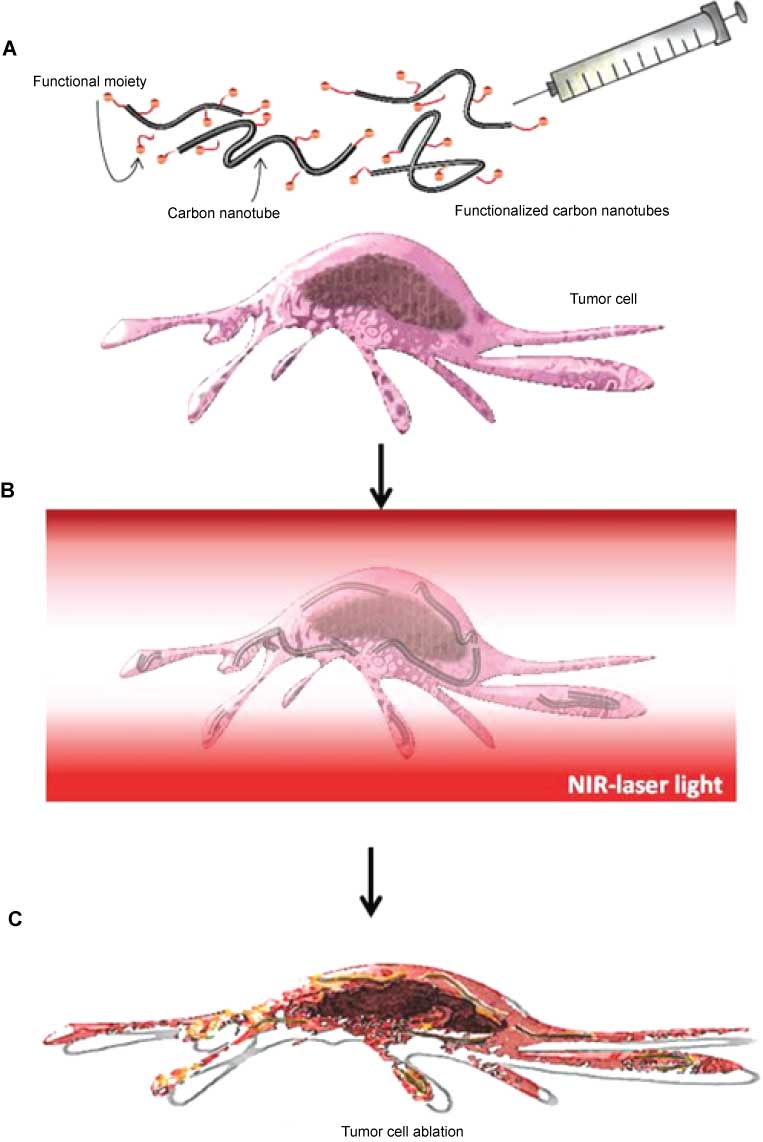
Schematic illustration of anticancer treatment using site-targeted carbon nanotubes for thermal ablation of malignant cells. (A) Intravenous injection of carbon nanotubes into the patient; (B) administration of near-infrared laser light to functionalized carbon nanotubes ingested in targeted cells; (C) irreversible cellular damage of treated tumor cells trigged by locally raised temperature.
Physical and optical properties of carbon nanomaterials
CNTs are theoretically sheets of graphene rolled into cylinders (13). There are two distinct types of CNTs – single-walled carbon nanotubes (SWNTs) and multi-walled carbon nanotubes (MWNTs) (Figure 2). MWNTs consist of two to 30 nanotubes which are concentrically embedded in one another and have an outer diameter ranging from 2 to 100 nm (14). SWNTs have diameters ranging from 0.4 to 2 nm.
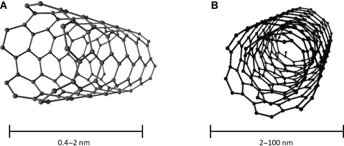
Types of carbon nanotubes: (A) single walled (SWNT) and (B) multi-walled (MWNT).
Like graphene, the atoms of CNTs possess sp2 hybrid orbitals (15). The enhanced mechanical strength of CNTs is caused by the carbon-carbon double bonds formed by the sp2 orbitals, which lead to the formation of a network of conjugated π-bonds (16). This π-conjugation causes electron delocalization, which endows CNTs with excellent thermal and electrical conductivity (17). The nucleophilic π-bonds also serve as reactive sites for surface modifications (18). Additionally, the electron delocalization produces van der Waals forces in CNTs, which provides for surface modification through nonpolar interactions. By tuning surface chemistry, CNTs can be optimized for biomedical applications. While the aforementioned properties of CNTs have been explored intensely, an increasingly important feature of CNTs is their ability to convert NIR radiation to heat (19).
Depending on configuration, the electronic behavior of CNTs can either be semiconducting or metallic (20). CNTs used in biomedical applications are typically a mixture of metallic and semiconducting CNTs. Recently, a comparison between semiconducting SWNTs (s-SWNTs) and metallic SWNTs (m-SWNTs) was conducted by Murakami et al. (21). In addition to the photothermal effect, the photodynamic effect of CNTs was demonstrated to play a role in tumor ablation. Following exposure to NIR radiation, s-SWNTs were found to produce reactive oxygen species via photodynamic effect, which are capable of eliminating malignant cells. Due to the lower band gap of m-SWNTs compared to s-SWNTs, the m-SWNTs demonstrated a higher efficiency in heat production.
In addition to exhibiting both photothermal and photodynamic effects, CNTs themselves serve as high contrast agents for multimodal photoacoustic, photothermal, and fluorescent imaging (22–25). Surface functionalization has not been found to impair the in vivo tracking and thermal lethality of CNTs through photoacoustic methods. It has been shown that CNTs functionalized with indocyanine green (ICG) through noncovalent attachment of arginine-glycine-aspartic acid (RGD) tripeptide resulted in a 20-fold increase in photoacoustic signal intensity, as compared to un-functionalized CNTs (25). The SWNTs conjugated with ICG and RGD selectively bound to αvβ3 integrins on the surface of U87MG cells compared to control particles which showed no binding.
Besides CNTs, it has been suggested that single walled carbon nanohorns (SWNH), graphene and its derivatives, including graphene oxide, offer many advantages as photothermal agents. As with CNTs, SWNHs and graphenes absorb electromagnetic signals from NIR irradiation, which leads to heat generation. While these structures show promise for cancer therapy, their benefit is just beginning to emerge, as new studies are being conducted to explore bioavailability, toxicity and photothermal potential of these newly developed structures.
SWNH aggregates are composed of thousands of graphitic tubules with closed ends and horn-shaped tips. These aggregates assemble to form spherical structures with diameters ranging from 50 to 100 nm (26). One advantage of SWNHs over CNTs is that SWNHs contain no metal catalyst, because they are produced by laser ablation of graphite, which lowers the risk of possible toxicity (27, 28). In addition, SWNHs have a large surface area and hollow interior space which can be tuned through oxidative heating parameters (29). SWNHs are also easily functionalized, making them an exceptional platform for drug delivery (30–32). Moreover, it has been shown previously that SWNHs used in conjunction with NIR lasers can selectively eliminate microbes and viruses (33, 34). Miyako’s group has also used SWNHs and laser irradiation to control gene expression (35). SWNHs complexed with bovine serum albumin (BSA) were introduced into various cells and then treated with a low-power laser (785 nm, 6–16 W/mm2, 30 min) which supplied enough thermal energy to induce heat shock promoter-mediated gene expression.
Graphene is composed of a single-layer of carbon atoms packed onto a two-dimensional honey comb lattice (36). Graphene structures have a high surface-to-volume ratio, which promotes interactions with various biomolecules and has demonstrated usefulness in several fields (37–39). Due to its flexible membrane, the carbon backbone of graphene offers endless possibilities for modification and functionalization. Chemically modified graphene, i.e., graphene oxide (GO), has emerged as a precursor attracting substantial attention for biomedical research (40). Using the intrinsic NIR absorbance of functionalized graphene, GO and reduced GO (rGO), photothermolysis has been achieved without significant toxicity (41).
The biocompatibility of carbon nanomaterials, similar to other therapeutic agents, is dependent upon their physiochemical properties. It is known that size and morphology of particles plays an important role in their biodistribution (42, 43). Similarly, surface modifications of carbon nanomaterials can affect their biocompatibility. While it is not the scope of this review to thoroughly discuss toxicology issues with carbon nanomaterials, it is noteworthy that toxicity data for CNTs is inconsistent and sometimes conflicting (44–46). Conflicting data could be attributed to the variation of synthesis parameters used to make CNTs. It is surmised that because SWNHs are synthesized without a metal catalyst, toxicity will be significantly reduced (27). Despite concern about toxicity, appropriate surface modifications of graphitic nanomaterials have shown to effectively decrease in vivo toxicity and is worth examining (44, 47).
Surface functionalization of carbon nanomaterials
CNTs are insoluble in most organic solvents, as well as have a strong tendency to aggregate in aqueous solvents due to hydrophobic interactions. For CNTs to be useful for biomedical applications, their surface chemistry needs to be modified. Therefore, many investigations have been conducted to improve CNT, and therefore other carbon nanomaterials solubility, for enhanced photothermal applications. Furthermore, functionalization with targeting moieties has yielded site specific delivery of carbon nanomaterials. Table 1 summarizes the studies described below and their importance for photothermal therapy.
Surface coatings used to functionalize carbon nanomaterials.
| Surface coating | Type | Advantage/application | References |
|---|---|---|---|
| Antibodies | SWNT MWNT | Site specific delivery using surface markers over-expressed by cancer cells, including anti-CD133, anti-GD2. | (48, 49) |
| BSA | SWNH | Multifunctional linker. | (50) |
| Collagen | SWNT | Stabilize and disperse CNTs. | (51) |
| DNA/RNA | SWNT | Negatively charged for additional method of functionalization. | (52, 53) |
| FITC | SWNT | Fluorescent tracking of CNTs. | (52, 54) |
| Folic acid | SWNT | Selective targeting of malignant cells with over-expressed folate receptors on their surface. | (52) |
| Glycated Chitosan | SWNT | Promotes immune responsiveness and macrophage uptake, synergy with thermal ablation and chemotherapy. | (54, 55) |
| Kentra | SWNT | Highly stable, does not alter the carbon nanotube electronic structure appreciably. | (5) |
| Poly(citric acid) (PCA) | MWNT | Improves both hydrophilicity and functionality. | (56) |
| Polyethylene Glycol (PEG) | SWNT SWNH | Drastic increase in bloodstream circulation time, easily metabolized, biocompatible. | (52, 57–63) |
| NGS | |||
| GO | |||
| rGO | |||
| Pluronic | SWNH | Improves solubility. | (64, 65) |
Solubility
There are various strategies used to solubilize and disperse CNTs in water as well as to make them biologically compatible. Modifications can be grouped into three main categories: 1) covalent attachment of chemical groups onto the CNT skeleton through π-bonds; 2) noncovalent adsorption or wrapping of functional moieties onto the surface of CNTs; and 3) endohedral filling of the inner cavity of CNTs (66). Due to the non-polarity and high van der Waals forces exuded by CNTs, surface functionalization is necessary for increasing nanotube solubility in aqueous environments (19).
PEGylation, functionalization with poly(ethylene glycol) (PEG), has numerous advantages for drug delivery. Its abilities to prevent agglomeration of nanoparticles and shield drug carriers from recognition by the mononuclear phagocyte system (MPS) are attractive properties for the coating of carbon nanomaterials with PEG. Liu et al. demonstrated that PEGylation of SWNTs leads to increased blood circulation times (57). SWNTs were covalently conjugated with phospholipid-PEG, and then intravenously injected into mice. Raman scattering was utilized to track the transport and accumulation of the SWNTs. It was found that as the molecular weight of PEG increased, the concentration of PEGylated SWNTs in the liver and spleen decreased, suggesting that functionalization can be used to prolong SWNT circulation in the bloodstream by preventing MPS phagocytosis. Moreover, no resistance to SWNT clearance was found. Similarly, SWNTs functionalized with poly(ethylene glycol) methyl ether and poly(maleic anhydride-alt-1-octadecene) were found to possess high tumor uptake – roughly 30% of the injected dose – in addition to a blood circulation time of 30 h (67).
Matsumura et al. synthesized a conjugate composed of a comb-shaped PEG and carbon nanohorn-binding peptide (cPEG-NHBP) (58). The oxidized SWNHs (oxSWNHs) conjugated with cPEG-NHBP revealed higher dispersibility than the ones conjugated with 20PEG-NHBP which is made of linear 20 kDa PEG and NHBP. Absorbance readings at 800 nm showed that within 5 h, untreated oxSWNHs began to agglomerate. 20PEG-NHBP inhibited agglomeration to a degree. However, cPEG-NHBP showed only a slight reduction in absorbance and demonstrated good dispersion. Moreover, in vivo evaluation of oxSWNHs treated with cPEG-NHBP showed suppressed agglomeration in the lungs of mice compared to untreated oxSWNHs.
Yang et al. treated mice with nanographene sheets (NGS) coated with PEG, and the behavior of the sheets was studied (59). Due to the efficient passive tumor targeting of their newly synthesized NGS, in vivo PTT studies were carried out. Mice bearing 4T1 tumors were intravenously injected with 200 μL NGS-PEG (2 mg/mL) and exposed to an 808-nm laser at a power of 2 W/cm2 for 24 h after injection. One day after laser treatment, tumors disappeared and there was no tumor regrowth for up to 40 days. A combined chemotherapy and photothermal therapy system was developed by Zhang et al. (60). Doxorubicin-loaded PEGylated nanographene oxide (NGO-PEG/DOX) was delivered to murine mammary EMT6 tumor cells both in vitro and in vivo. Cytotoxity of NGO-PEG/DOX compared to free DOX (10 μg/mL) showed an inhibition rate of 82.1% and 57.1%, respectively, when treated with NIR radiation (808 nm, 2 W/cm2, 5 min). The trend of a synergistic effect in the inhibition rate of NGO-PEG/DOX compared to free DOX was not obvious at a DOX concentration of 2 and 30 μg/mL, however. In vivo results showed that, free DOX and NGO-PEG and NIR treated tumors began to grow after 10 days, NGO-PEG/DOX and NIR treated tumors however, showed no tumor re-growth up to 30 days. These results suggested that combined chemotherapy and photothermal therapy demonstrated a synergistic effect in vivo.
For photothermal ablation with GO, a high dosage of GO and laser power is needed due to the suboptimal absorption of NIR light by GO. Therefore, Dai’s group developed rGO sheets (61). By chemically reducing covalently PEGylated GO, they were able to partially restore the aromatic and conjugated characteristics of graphene sheets, increasing the NIR absorbance >6-fold. Conjugation with RGD peptides allowed for efficient selection to human glioblastoma cells. Cells were treated with a low concentration of rGO particles (6.6 mg/L) and NIR laser (808 nm, 15 W/cm2) with total cell destruction occurring within 8 min. Liu’s group synthesized a similar PEGylated rGO, and studied how the size and surface coating affects the behavior of graphene in vivo (62). Following treatment with an 808-nm NIR laser at a power density of 0.15 W/cm2 for 5 min, complete tumor elimination in mice was observed from ultra-small rGO with non-covalent PEG coating. This research highlighted the importance of optimizing nano-graphene and its derivatives for effective photothermal treatment.
While PEG is commonly thought to be the “gold standard” for enhancing a therapeutic carrier’s delivery, several notable drawbacks have recently emerged (68). These disadvantages have prompted researchers to use alternative solubilizing agents. Mao et al. has synthesized SWNTs functionalized with collagen (51). Collagen not only helped to stabilize the SWNTs for months in solution but increased cellular uptake and retention time. Sobhani et al. synthesized MWNTs functionalized with poly(citric acid) to improve both hydrophilicity and functionality of the MWNTs and to enhance the delivery of paclitaxel (56). These studies demonstrated that modifications to CNTs can improve their dispersion as well as drug biocompatibility, cell penetration efficacy and targeted delivery capabilities, even if PEG is not used.
Immunologically modified SWNTs were developed using glycated chitosan (GC) by Zhou et al. (54, 55). The synergy between immunological and photothermal effects of SWNT-GC was shown to increase cell destruction. Specifically, 980-nm laser light was used to induce cell destruction and also led to secretion of damage associated molecular pattern molecules (i.e., molecules which can perpetuate immune response). In vivo studies on EMT6 cells injected into a mouse model showed that after treatment with SWNT-GC and laser light (980 nm, 0.74 W/cm2, 10 min), 89.2% of cells showed apoptosis. Mice were monitored 100 days after tumor inoculation and the mice with CNT treatment and NIR laser showed complete tumor regression. Long-term antitumor effects showed that SWNT-GC inside a viable tumor was a prerequisite for the induction of effective immune response.
Whitney et al. synthesized SWNHs coated with Pluronic, a non-ionic surfactant which improves solubility of biological materials and which acts as a thermal enhancer (64). In this study, kidney cancer cells were treated with 1064-nm laser irradiation at 40 W/cm2 for 0–5 min. After 2–3 min of treatment, cell viability showed nearly complete tumor cell death, suggesting SWNHs could be beneficial to treat superficial tumors. Rylander et al. used a fiber optic microneedle device to deliver SWNHs (coated with Pluronic F-127) to the wall of porcine bladders in ex vivo treatment (65). For their study, they wanted to evaluate the distribution of SWNHs as well as if they could control the degree of lateral dispersion and depth in which SWNHs permeate in the tissues. The 1064-nm laser light, at an intensity of 0.95 W/cm2, emitted over 40 s irradiated the tissues and increased tumor cell temperatures. For their hyperthermic results, the SWNH-perfused tissue increased temperature by approximately 19°C, falling just short of the 60°C needed for in vivo treatment. The authors acknowledged that there were several limitations to the study. However, they believe that use of a fiber-optic microneedle device to deliver SWNHs is a versatile technology for local delivery of therapeutic agents, not limited to just SWNHs.
Zhang et al. synthesized zinc phthalocyanine (ZnPc, a photodynamic therapy agent) SWNHs conjugated with BSA (to improve dispersion) for a dual photodynamic and photohyperthermia cancer therapy system (50). The effect of ZnPc-SWNHs-BSA was studied in both in vitro and in vivo studies. Interestingly, they found that the double therapy using ZnPc-SWNHs-BSA had greater anticancer efficiency than ZnPc (phytodynamic therapy) and SWNH-BSA (photohyperthermia) treatment alone. One drawback to their study, however, was the use of 670-nm laser irradiation, which does not penetrate biological tissue as deeply as longer wavelengths of NIR light.
In addition to NIR laser adsorption, CNTs have demonstrated conversion of radiofrequency (RF) waves into heat (5). While ablation of malignancies using CNTs subjected to NIR radiation has been studied extensively, use of RF treatment has its merits for further evaluation because RF fields offer significantly deeper tissue penetration in comparison to NIR light (69). Gannon et al. performed in vitro thermal ablation of HepG2, Hep3B, and Panc-1 cancer cells using an RF field (5). SWNTs were functionalized with Kentera, a polyphenylene ethynylene-based polymer. Kentera was bound to the SWNTs through π-π stacking between the phenyl groups of the polymer and the SWNT surface. This modification did not alter the electronic structure of the CNTs appreciably. Moreover, the use of a high-field RF and low concentrations (5–500 mg/L) of aqueous SWNT suspensions produced thermal effects higher than previously reported studies. One explanation for this was attributed to dynamic self-assembly of SWNTs into longer nano-antennae. In vitro and in vivo studies were conducted and showed successful killing of tumor cells. Specifically, rabbits xenografted with VX2 tumors were directly injected with Kentra-functionalized SWNTs and then treated with an RF field at 13.56 MHz for 2 min. Irradiation of all VX2 tumors in mice treated with SWNTs had complete death after 2 days, while control tumors which were not treated with SWNTs were still viable. It was suggested that further studies be conducted with functionalized SWNTs for targeted delivery by linking antibodies, peptides or pharmacological agents to target molecules over expressed on the surface of cancer cells (5).
Targeting
While dispersibility of carbon nanomaterials is important, for clinical use, selective treatment of tumor cells without collateral damage to healthy cells is crucial. Hence, specific targeting to overexpressed markers on the surface of cancer cells must also be achieved. An early demonstration of using CNTs for photothermal cancer therapy was conducted by Kam et al. (52). In the study, SWNTs conjugated with DNA underwent endocytosis by malignant HeLa cells. HeLa cells containing the SWNT complexes were exposed to 808-nm laser radiation at six 10 s on-and-off pulses (1.4 W/cm2). They revealed that SWNTs can be used to deliver DNA into live cells without harming the cells. However, if they increased the exposure time to 2 min, extensive cell death was observed. It was demonstrated that SWNTs could be selectively internalized by cancer cells, and then irradiated to cause cellular destruction. Moreover, since many cancerous cells overexpress folate receptors, they functionalized SWNTs with phospholipid-PEG-folic acid (PL-PEG-FA). It was found that the SWNT-PL-PEG-FAs were internalized by the cells and that NIR pulses can induce local heating of SWNTs for release of therapeutic agents into the cells without harming them. Additionally, selective delivery of SWNTs into cells via receptor-mediated delivery and NIR-triggered death was achieved following NIR laser light administration for 2 min.
Wang et al. conjugated MWNTs with GD2 monoclonal antibodies to selectively target neuroblastoma cells (48). Significant photothermal ablation of the neuroblastoma cells occurred under an 808-nm NIR laser at a power of 6 W/cm2 for 15 min. The interaction of MWNTs with the neuroblastoma cells was followed by rhodamine B labeling. It was seen that 6 h incubation with the MWNTs was sufficient for antibody-mediated endocytosis of ligand-tagged MWNTs. Uptake of GD2-targeted MWNTs for 12 h led to oversaturation; laser exposure data confirmed oversaturation of MWNTs generated too much heat, which led to collateral damage of non-GD2-expressing cells. Moreover, MWNTs bound with anti-GD2 antibody could specifically target GD2 expressing neuroblastoma cells, but not cells without GD2 expression, proving the importance of surface functionalization for specific targeting and selective ablating MWNT-laden cancer cells.
Wang et al. obliterated CD133+ stem-like cells xenografted to mice using chitosan functionalized SWNTs (49). CD133 has been recognized as a typical stem cell marker which inhabits the surface of cancerous blood, brain, colon, prostate, and liver cells. By conjugating anti-CD133 monoclonal antibody to SWNTs solubilized with chitosan, CD133+ cells were selectively targeted by the functionalized SWNT conjugates. Following the preliminary cell culture studies, mice were subcutaneously injected with a mixture of CD133+ and CD133-cells which had been pretreated with the anti-CD133-SWNTs. Two days following the treatment, 808-nm NIR laser pulses at 2 W/cm2 were directed at the injection sites for 5 min. In addition to complete thermal ablation of the cancerous cells, the treatment prevented the formation of new malignancies.
Synergy between SWNT photothermal therapy and RNA interference was demonstrated by Wang et al. (53). SWNT-polyethylenimine (PEI) was added to siRNA and asparagine-glycine-arginine (NGR) peptides to produce SWNT-PEI-siRNA-NGR. Cell viability studies indicated an absence of cytotoxicity in PC-3 prostate cancer cells at SWNT-PEI concentrations below 30 μg/mL. However, SWNT-PEI-siRNA and SWNT-PEI-siRNA-NGR showed noticeable cytotoxicity in PC-3 cells after 72 h treatment (60 and 55% cell viability, respectively). This study demonstrated that the suppression of PC-3 cells by siRNA is preserved following conjugation of siRNA to SWNT-PEI. In all experimental groups, 3 min of 808-nm NIR laser radiation produced PC-3 cell viabilities below 5%. In vivo trials of SWNT-PEI, SWNT-PEI-siRNA, and SWNT-PEI-siRNA-NGR were conducted in nude mice bearing PC-3 tumors injected subcutaneously. Through molecular marking with quantum dots, it was shown that SWNT-PEI and SWNT-PEI-NGR exhibited higher accumulation in the liver, lung, spleen, and kidney than in the heart and brain which is consistent with localization in organs of the MPS. Importantly, SWNT-PEI-NGR had higher accumulation than SWNT-PEI in PC-3 tumors suggesting that NGR peptide enhanced uptake. These PC-3 tumors, accumulated with SWNT-PEI-siRNA-NGR nanoparticles and concomitantly irradiated with NIR laser, revealed the highest suppression of tumor growth. Without laser treatment, injections of SWNT conjugates containing siRNA led to a decrease in overall tumor volume. SWNT conjugates without siRNA mimicked the behavior of a saline solution and lead to negligible tumor reduction. No difference in tumor size was seen in the saline solutions with and without laser treatment, demonstrating that the conjugated SWNTs photothermally ablated the PC-3 tumor cells. It should be noted that since the NIR laser irradiation was conducted directly on the sites that PC-3 cells were injected subcutaneously, the SWNTs accumulated in organs with low cell toxicity were not exposed to NIR and therefore did not result in photothermolysis of cells.
Cancer stem cells have been proposed to contribute to cancer resistance and tumorigenesis (70). Similar to normal stem cells, cancer stem cells have self-renewal and proliferation capabilities. Due to their resistance to current treatments, cancer stem cells are difficult to kill and may contribute to cancer reoccurrence. For this reason, treatment regimens focused on targeting cancer stems cells are being researched. NIR-stimulated CNTs have been used for the photothermal destruction of breast cancer stem cells (BCSCs) by Torti and coworkers (63). They showed that BCSCs were resistant to traditional hyperthermia treatment and had an increased cancer stem cell fractionation of 1.6–1.9 fold. In contrast, the intense localized heat generated by CNTs treated with an NIR laser (1064 nm, 3 W, 45 s) caused almost complete cell death in both breast cancer cells and BCSCs. Moreover, laser plus CNT treatment diminished the long-term proliferative ability of the BCSCs.
Conclusion
There are an abundant amount of studies which have been conducted on CNTs, and much is known about their unique physical, chemical and physiological properties. SWNHs and graphenes, however, are just beginning to emerge as potential candidates for PTT. While studies have been conducted on their ability to be used for drug delivery, not many studies delve into their potential for photothermolysis.
Owing to their ability to be surface functionalized, graphitic nanomaterials are continually being developed and tested both in vitro and in vivo for biological and biomedical applications. Due to their strong absorption in the NIR range and also broad range of wavelengths that can be used, graphitic nanomaterials are proving beneficial for PTT treatment. Despite advances however, before carbon nanomaterials can enter clinical trials, various issues need to be addressed. For example, biocompatibility of carbon nanomaterials needs to be established. This includes a better standard for determining toxicity of both carbon nanomaterials and the surface coatings being used. Aggregation of carbon nanomaterials under physiological conditions is another challenge which needs to be dealt with via appropriate surface functionalization. Continued research into these issues will ultimately yield success of CNTs, graphenes and SWNHs for cancer diagnosis and therapy.
About the authors

Alicia J. Sawdon is currently a PhD student in the Department of Chemical Engineering at Michigan Technological University, with a BS in Biochemistry from Oakland University. Her research interests include design and synthesis of nanomaterials for drug and gene delivery. In particular, her research is aimed at using functionalized polymeric prodrug micelles for cancer therapy.

Ethan J. Weydemeyer received his BS degree in Chemical Engineering at Michigan Technological University. His research interests include the use of gold nanoparticles and carbon nanotubes for photothermal therapy. Currently, his research focuses on the development of perfluorocarbon emulsions incorporated with gold nanoparticles or carbon nanotubes as photoacoustic contrast agents.

Ching-An Peng earned a BS in Chemical Engineering at National Taiwan University; an MS in Chemical Engineering at the University of Notre Dame; and a PhD in Chemical Engineering at the University of Michigan. After earning his doctoral degree, he worked as a joint postdoctoral fellow at the University of British Columbia and the StemCell Technologies. In 1997, he started his assistant professorship at the University of Southern California in Chemical Engineering Department. Since 2003, he has been promoted as an Associate Professor of Chemical Engineering and Materials Science with tenure. In 2006, he joined the Department of Chemical Engineering at the National Taiwan University with the rank of full professor. In 2008, he joined the Department of Chemical Engineering at Michigan Technological University as the first holder of the James and Lorna Mack Endowed Chair in Bioengineering. His research interests include bio-based products, drug/gene delivery, nanomedicine, and tissue engineering.
References
1. Anderson RR, Parrish JA. Selective photothermolysis: precise microsurgery by selective absorption of pulsed radiation. Science 1983;220:524–7.10.1126/science.6836297Suche in Google Scholar PubMed
2. Camerin M, Rello S, Villanueva A, Ping X, Kenney ME, Rodgers MA, et al. Photothermal sensitisation as a novel therapeutic approach for tumours: studies at the cellular and animal level. Eur J Cancer 2005;41:1203–12.10.1016/j.ejca.2005.02.021Suche in Google Scholar PubMed
3. He X, Bischof JC. Quantification of temperature and injury response in thermal therapy and cryosurgery. Crit Rev Biomed Eng 2003;31:355–422.10.1615/CritRevBiomedEng.v31.i56.10Suche in Google Scholar
4. Brunetaud J, Mordon S, Maunoury V, Beacco C. Non-PDT uses of lasers in oncology. Laser Med Sci 1995;10:3–8.10.1007/BF02133156Suche in Google Scholar
5. Gannon CJ, Cherukuri P, Yakobson BI, Cognet L, Kanzius JS, Kittrell C, et al. Carbon nanotube-enhanced thermal destruction of cancer cells in a noninvasive radiofrequency field. Cancer 2007;110:2654–65.10.1002/cncr.23155Suche in Google Scholar PubMed
6. O’Neal DP, Hirsch LR, Halas NJ, Payne JD, West JL. Photo-thermal tumor ablation in mice using near infrared-absorbing nanoparticles. Cancer Lett 2004;209:171–6.10.1016/j.canlet.2004.02.004Suche in Google Scholar PubMed
7. Xu Y, Mahmood M, Fejleh A, Li Z, Watanabe F, Trigwell S, et al. Carbon-covered magnetic nanomaterials and their application for the thermolysis of cancer cells. Int J Nanomed 2010;5:167–76.10.2147/IJN.S8306Suche in Google Scholar
8. Huang X, El-Sayed MA. Plasmonic photo-thermal therapy (PPTT). Alexandria J Med 2011;47:1–9.10.1016/j.ajme.2011.01.001Suche in Google Scholar
9. Krishnan S, Diagaradjane P, Cho SH. Nanoparticle-mediated thermal therapy: evolving strategies for prostate cancer therapy. Int J Hyperther 2010;26:775–89.10.3109/02656736.2010.485593Suche in Google Scholar PubMed PubMed Central
10. El-Sayed IH, Huang X, El-Sayed MA. Selective laser photo-thermal therapy of epithelial carcinoma using anti-EGFR antibody conjugated gold nanoparticles. Cancer Lett 2006;239:129–35.10.1016/j.canlet.2005.07.035Suche in Google Scholar PubMed
11. Huang X, El-Sayed IH, Qian W, El-Sayed MA. Cancer cell imaging and photothermal therapy in the near-infrared region by using gold nanorods. J Am Chem Soc 2006;128:2115–20.10.1021/ja057254aSuche in Google Scholar
12. Hu M, Chen J, Li ZY, Au L, Hartland GV, Li X, et al. Gold nanostructures: engineering their plasmonic properties for biomedical applications. Chem Soc Rev 2006;35:1084–94.10.1039/b517615hSuche in Google Scholar
13. Iijima S. Helical microtubules of graphitic carbon. Nature 1991;354:56–8.10.1038/354056a0Suche in Google Scholar
14. Saito R, Dresselhaus G, Dresselhaus MS. Physical properties of carbon nanotubes, vol.4. London: Imperial College Press, 1998;4:1–272.Suche in Google Scholar
15. Eklund P, Holden J, Jishi R. Vibrational modes of carbon nanotubes; spectroscopy and theory. Carbon 1995;33:959–72.10.1016/0008-6223(95)00035-CSuche in Google Scholar
16. Falvo MR, Clary GJ, Taylor RM, Chi V, Brooks FP, Washburn S, et al. Bending and buckling of carbon nanotubes under large strain. Nature 1997;389:582–4.10.1038/39282Suche in Google Scholar PubMed
17. Wildoer JW, Venema LC, Rinzler AG, Smalley RE, Dekker C. Electronic structure of atomically resolved carbon nanotubes. Nature 1998;391:59–62.10.1038/34139Suche in Google Scholar
18. Sun YP, Fu K, Lin Y, Huang W. Functionalized carbon nanotubes: properties and applications. Acc Chem Res 2002;35:1096–104.10.1021/ar010160vSuche in Google Scholar PubMed
19. O’connell MJ, Bachilo SM, Huffman CB, Moore VC, Strano MS, Haroz EH, et al. Band gap fluorescence from individual single-walled carbon nanotubes. Science 2002;297:593–6.10.1126/science.1072631Suche in Google Scholar PubMed
20. Tans SJ, Verschueren AR, Dekker C. Room-temperature transistor based on a single carbon nanotube. Nature 1998;393:49–52.10.1038/29954Suche in Google Scholar
21. Murakami T, Nakatsuji H, Inada M, Matoba Y, Umeyama T, Tsujimoto M, et al. Photodynamic and photothermal effects of semiconducting and metallic-enriched single-walled carbon nanotubes. J Am Chem Soc 2012;134:17862–5.10.1021/ja3079972Suche in Google Scholar PubMed
22. de la Zerda A, Zavaleta C, Keren S, Vaithilingam S, Bodapati S, Liu Z, et al. Carbon nanotubes as photoacoustic molecular imaging agents in living mice. Nat Nanotechnol 2008;3: 557–62.10.1038/nnano.2008.231Suche in Google Scholar
23. Kim JW, Shashkov EV, Galanzha EI, Kotagiri N, Zharov VP. Photothermal antimicrobial nanotherapy and nanodiagnostics with self-assembling carbon nanotube clusters. Laser Surg and Med 2007;39:622–34.10.1002/lsm.20534Suche in Google Scholar
24. Robinson JT, Welsher K, Tabakman SM, Sherlock SP, Wang H, Luong R, et al. High performance in vivo near-IR (>1 μm) imaging and photothermal cancer therapy with carbon nanotubes. Nano Res 2010;3:779–93.10.1007/s12274-010-0045-1Suche in Google Scholar
25. de La Zerda A, Liu Z, Zavaleta C, Bodapati S, Teed R, Vaithilingam S, et al. Enhanced sensitivity carbon nanotubes as targeted photoacoustic molecular imaging agents. San Jose, CA: Proc of SPIE, 2009; 7177.10.1117/12.809601Suche in Google Scholar
26. Iijima S, Yudasaka M, Yamada R, Bandow S, Suenaga K, Kokai F, et al. Nano-aggregates of single-walled graphitic carbon nano-horns. Chem Phys Lett 1999;309:165–70.10.1016/S0009-2614(99)00642-9Suche in Google Scholar
27. Miyawaki J, Yudasaka M, Azami T, Kubo Y, Iijima S. Toxicity of single-walled carbon nanohorns. ACS Nano 2008;2:213–26.10.1021/nn700185tSuche in Google Scholar PubMed
28. Tahara Y, Miyawaki J, Zhang M, Yang M, Waga I, Iijima S, et al. Histological assessments for toxicity and functionalization-dependent biodistribution of carbon nanohorns. Nanotechnology 2011;22:265106.10.1088/0957-4484/22/26/265106Suche in Google Scholar PubMed
29. Utsumi S, Miyawaki J, Tanaka H, Hattori Y, Itoi T, Ichikuni N, et al. Opening mechanism of internal nanoporosity of single-wall carbon nanohorn. J Phys Chem B 2005;109:14319–24.10.1021/jp0512661Suche in Google Scholar PubMed
30. Murakami T, Ajima K, Miyawaki J, Yudasaka M, Iijima S, Shiba K. Drug-loaded carbon nanohorns: adsorption and release of dexamethasone in vitro. Mol Pharm 2004;1:399–405.10.1021/mp049928eSuche in Google Scholar PubMed
31. Ajima K, Murakami T, Mizoguchi Y, Tsuchida K, Ichihashi T, Iijima S, et al. Enhancement of in vivo anticancer effects of cisplatin by incorporation inside single-wall carbon nanohorns. ACS Nano 2008;2:2057–64.10.1021/nn800395tSuche in Google Scholar PubMed
32. Ajima K, Yudasaka M, Murakami T, Maigné A, Shiba K, Iijima S. Carbon nanohorns as anticancer drug carriers. Mol Pharm 2005;2:475–80.10.1021/mp0500566Suche in Google Scholar PubMed
33. Miyako E, Nagata H, Hirano K, Makita Y, Nakayama K-i, Hirotsu T. Near-infrared laser-triggered carbon nanohorns for selective elimination of microbes. Nanotechnology 2007;18:475103.10.1088/0957-4484/18/47/475103Suche in Google Scholar
34. Miyako E, Nagata H, Hirano K, Sakamoto K, Makita Y, Nakayama K, et al. Photoinduced antiviral carbon nanohorns. Nanotechnology 2008;19:957–4484.10.1088/0957-4484/19/7/075106Suche in Google Scholar PubMed
35. Miyako E, Deguchi T, Nakajima Y, Yudasaka M, Hagihara Y, Horie M, et al. Photothermic regulation of gene expression triggered by laser-induced carbon nanohorns. Proc Natl Acad Sci 2012;109:7523–8.10.1073/pnas.1204391109Suche in Google Scholar PubMed PubMed Central
36. Novoselov KS, Geim AK, Morozov SV, Jiang D, Zhang Y, Dubonos SV, et al. Electric field effect in atomically thin carbon films. Science 2004;306:666–9.10.1126/science.1102896Suche in Google Scholar PubMed
37. Sun X, Liu Z, Welsher K, Robinson JT, Goodwin A, Zaric S, et al. Nano-graphene oxide for cellular imaging and drug delivery. Nano Res 2008;1:203–12.10.1007/s12274-008-8021-8Suche in Google Scholar PubMed PubMed Central
38. Wang Y, Li Z, Wang J, Li J, Lin Y. Graphene and graphene oxide: biofunctionalization and applications in biotechnology. Trends Biotechnol 2011;29:205–12.10.1016/j.tibtech.2011.01.008Suche in Google Scholar PubMed PubMed Central
39. Yang K, Feng L, Shi X, Liu Z. Nano-graphene in biomedicine: theranostic applications. Chem Soc Rev 2013;42:530–47.10.1039/C2CS35342CSuche in Google Scholar PubMed
40. Shen H, Zhang L, Liu M, Zhang Z. Biomedical applications of graphene. Theranostics 2012;2:283–4.10.7150/thno.3642Suche in Google Scholar PubMed PubMed Central
41. Akhavan O, Ghaderi E. Toxicity of graphene and graphene oxide nanowalls against bacteria. ACS Nano 2010;4:5731–6.10.1021/nn101390xSuche in Google Scholar PubMed
42. Liu Y, Tan J, Thomas A, Ou-Yang D, Muzykantov VR. The shape of things to come: importance of design in nanotechnology for drug delivery. Ther Deliv 2012;3:181–94.10.4155/tde.11.156Suche in Google Scholar PubMed PubMed Central
43. Liang F, Chen B. A review on biomedical applications of single-walled carbon nanotubes. Curr Med Chem 2010;17:10–24.10.2174/092986710789957742Suche in Google Scholar PubMed
44. Smart SK, Cassady AI, Lu GQ, Martin DJ. The biocompatibility of carbon nanotubes. Carbon 2006;44:1034–47.10.1016/j.carbon.2005.10.011Suche in Google Scholar
45. Yang K, Liu Z. In vivo biodistribution, pharmacokinetics, and toxicology of carbon nanotubes. Curr Drug Metab 2012;13:1057–67.10.2174/138920012802850029Suche in Google Scholar PubMed
46. Hurt RH, Monthioux M, Kane A. Toxicology of carbon nanomaterials: status, trends, and perspectives on the special issue. Carbon 2006;44:1028–33.10.1016/j.carbon.2005.12.023Suche in Google Scholar
47. Yang K, Wan J, Zhang S, Zhang Y, Lee ST, Liu Z. In vivo pharmacokinetics, long-term biodistribution, and toxicology of PEGylated graphene in mice. ACS Nano 2010;5: 516–22.10.1021/nn1024303Suche in Google Scholar PubMed
48. Wang CH, Huang YJ, Chang CW, Hsu WM, Peng CA. In vitro photothermal destruction of neuroblastoma cells using carbon nanotubes conjugated with GD2 monoclonal antibody. Nanotechnology 2009;20:315101.10.1088/0957-4484/20/31/315101Suche in Google Scholar PubMed
49. Wang CH, Chiou SH, Chou CP, Chen YC, Huang YJ, Peng CA. Photothermolysis of glioblastoma stem-like cells targeted by carbon nanotubes conjugated with CD133 monoclonal antibody. Nanomedicine 2011;7:69–79.10.1016/j.nano.2010.06.010Suche in Google Scholar PubMed
50. Zhang M, Murakami T, Ajima K, Tsuchida K, Sandanayaka AS, Ito O, et al. Fabrication of ZnPc/protein nanohorns for double photodynamic and hyperthermic cancer phototherapy. Proc Natl Acad Sci 2008;105:14773–8.10.1073/pnas.0801349105Suche in Google Scholar PubMed PubMed Central
51. Mao H, Kawazoe N, Chen G. Uptake and intracellular distribution of collagen-functionalized single-walled carbon nanotubes. Biomaterials 2013;34:2472–9.10.1016/j.biomaterials.2013.01.002Suche in Google Scholar PubMed
52. Kam NW, O’Connell M, Wisdom JA, Dai H. Carbon nanotubes as multifunctional biological transporters and near-infrared agents for selective cancer cell destruction. Proc Natl Acad Sci 2005;102:11600–5.10.1073/pnas.0502680102Suche in Google Scholar PubMed PubMed Central
53. Wang L, Shi J, Zhang H, Li H, Gao Y, Wang Z, et al. Synergistic anticancer effect of RNAi and photothermal therapy mediated by functionalized single-walled carbon nanotubes. Biomaterials 2013;34:262–74.10.1016/j.biomaterials.2012.09.037Suche in Google Scholar PubMed
54. Zhou F, Xing D, Ou Z, Wu B, Resasco DE, Chen WR. Cancer photothermal therapy in the near-infrared region by using single-walled carbon nanotubes. J Biomed Opt 2009;14: 021009.10.1117/1.3078803Suche in Google Scholar PubMed PubMed Central
55. Zhou F, Wu S, Song S, Chen WR, Resasco DE, Xing D. Antitumor immunologically modified carbon nanotubes for photothermal therapy. Biomaterials 2012;33:3235–42.10.1016/j.biomaterials.2011.12.029Suche in Google Scholar PubMed PubMed Central
56. Sobhani Z, Dinarvand R, Atyabi F, Ghahremani M, Adeli M. Increased paclitaxel cytotoxicity against cancer cell lines using a novel functionalized carbon nanotube. Int J Nanomed 2011;6:705–19.Suche in Google Scholar
57. Liu Z, Davis C, Cai W, He L, Chen X, Dai H. Circulation and long-term fate of functionalized, biocompatible single-walled carbon nanotubes in mice probed by Raman spectroscopy. Proc Natl Acad Sci 2008;105:1410–5.10.1073/pnas.0707654105Suche in Google Scholar PubMed PubMed Central
58. Matsumura S, Sato S, Yudasaka M, Tomida A, Tsuruo T, Iijima S, et al. Prevention of carbon nanohorn agglomeration using a conjugate composed of comb-shaped polyethylene glycol and a peptide aptamer. Mol Pharm 2009;6: 441–7.10.1021/mp800141vSuche in Google Scholar PubMed
59. Yang K, Zhang S, Zhang G, Sun X, Lee S-T, Liu Z. Graphene in mice: ultrahigh in vivo tumor uptake and efficient photothermal therapy. Nano Lett 2010;10:3318–23.10.1021/nl100996uSuche in Google Scholar PubMed
60. Zhang W, Guo Z, Huang D, Liu Z, Guo X, Zhong H. Synergistic effect of chemo-photothermal therapy using PEGylated graphene oxide. Biomaterials 2011;32:8555–61.10.1016/j.biomaterials.2011.07.071Suche in Google Scholar PubMed
61. Robinson JT, Tabakman SM, Liang Y, Wang H, Sanchez Casalongue H, Vinh D, et al. Ultrasmall reduced graphene oxide with high near-infrared absorbance for photothermal therapy. J Am Chem Soc 2011;133:6825–31.10.1021/ja2010175Suche in Google Scholar PubMed
62. Yang K, Wan J, Zhang S, Tian B, Zhang Y, Liu Z. The influence of surface chemistry and size of nanoscale graphene oxide on photothermal therapy of cancer using ultra-low laser power. Biomaterials 2012;33:2206–14.10.1016/j.biomaterials.2011.11.064Suche in Google Scholar PubMed
63. Burke AR, Singh RN, Carroll DL, Wood JC, D’Agostino Jr RB, Ajayan PM, et al. The resistance of breast cancer stem cells to conventional hyperthermia and their sensitivity to nanoparticle-mediated photothermal therapy. Biomaterials 2012;33:2961–70.10.1016/j.biomaterials.2011.12.052Suche in Google Scholar PubMed PubMed Central
64. Whitney JR, Sarkar S, Zhang J, Do T, Young T, Manson MK, et al. Single walled carbon nanohorns as photothermal cancer agents. Laser Surg Med 2011;43:43–51.10.1002/lsm.21025Suche in Google Scholar PubMed
65. Hood RL, Carswell W, Rodgers A, Kosoglu M, Rylander M, Grant D, et al. Spatially controlled photothermal heating of bladder tissue through single-walled carbon nanohorns delivered with a fiberoptic microneedle device. Laser Med Sci 2012:1–8. DOI: 10.1007/s10103-012-1202-4. Available at http://link.springer.com/journal/10103/onlineFirst/page/8Suche in Google Scholar
66. Tasis D, Tagmatarchis N, Bianco A, Prato M. Chemistry of carbon nanotubes. Chem Rev 2006;106:1105–36.10.1021/cr050569oSuche in Google Scholar PubMed
67. Robinson JT, Hong G, Liang Y, Zhang B, Yaghi OK, Dai H. In vivo fluorescence imaging in the second near-infrared window with long circulating carbon nanotubes capable of ultrahigh tumor uptake. J Am Chem Soc 2012;134: 10664–9.10.1021/ja303737aSuche in Google Scholar PubMed PubMed Central
68. Garay RP, El-Gewely R, Armstrong JK, Garratty G, Richette P. Antibodies against polyethylene glycol in healthy subjects and in patients treated with PEG-conjugated agents. Expert Opin Drug Deliv 2012;9:1319–23.10.1517/17425247.2012.720969Suche in Google Scholar PubMed
69. Bernardi P, Cavagnaro M, Pisa S, Piuzzi E. Specific absorption rate and temperature elevation in a subject exposed in the far-field of radio-frequency sources operating in the 10-900-MHz range. IEEE Trans Biomed Eng 2003;50:295–304.10.1109/TBME.2003.808809Suche in Google Scholar PubMed
70. Guo W, Lasky JL, Wu H. Cancer stem cells. Pediatr Res 2006;59:59R–64R.10.1203/01.pdr.0000203592.04530.06Suche in Google Scholar PubMed
©2013 by Walter de Gruyter Berlin Boston
Artikel in diesem Heft
- Masthead
- Masthead
- What’s up in nanomedicine?
- Reviews
- Self-assembled liposomal nanoparticles in photodynamic therapy
- Tumor photothermolysis: using carbon nanomaterials for cancer therapy
- Therapeutic delivery using cell-penetrating peptides
- Original Article
- Ocular pharmacoscintigraphic and aqueous humoral drug availability of ganciclovir-loaded mucoadhesive nanoparticles in rabbits