Abstract
Objectives
Isolated fetal ascites carries an uncertain prognosis and broad differential diagnosis. When detected on prenatal sonography, a thorough evaluation is warranted to exclude development of hydrops and search for an underlying condition. While gastrointestinal abnormalities account for approximately 20% of cases of fetal ascites, surgical correction is commonly required postnatally. While there have been reports of isolated fetal ascites resolving in utero, spontaneous resolution of the causative gastrointestinal abnormality is unusual.
Case presentation
We report a case of a multiparous 33-year-old found to have moderate fetal ascites and a complex fetal abdominal mass near the small bowel detected by ultrasound at 32 weeks with spontaneous resolution of both ascites and mass by 37 weeks. Following the delivery of a normal neonate, we suspect the mass and ascites to have been produced by a small bowel rupture resulting in meconium peritonitis.
Conclusions
When fetal ascites with late gestational onset has spontaneous resolution in utero and hydrops never develops, there is generally a favorable prognosis and normal neonatal outcome.
Introduction
Reported cases of isolated fetal ascites determined to be idiopathic have been shown to carry a favorable prognosis with frequent spontaneous resolution in utero [1], [2], [3]. These cases generally reported better fetal outcomes with delivery of normal neonates as compared to cases with associated conditions such as gastrointestinal and genitourinary abnormalities, intrauterine infections, and overlapping cases with hydrops [1]. Additionally, isolated fetal ascites occurring due to a congenital abnormality has a variable prognosis, requiring extensive evaluation and possibly even prenatal or postnatal treatment. In this case, fetal ascites was prenatally diagnosed on ultrasound at 32 weeks secondary to a suspected gastrointestinal abnormality and was found to have spontaneously resolved in utero with a normal neonatal course. No existing reports of antenatally resolving fetal ascites with a discovered gastrointestinal anomaly describe concurrent resolution of the underlying abnormality without surgical correction or postpartum regression with medical management.
Case presentation
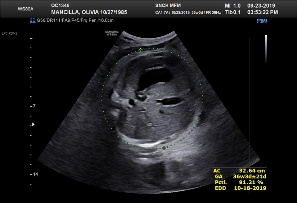
A 33-year-old multiparous G3P1011 female presented for routine visit at 32 weeks and was found to have a diagnosis of moderate fetal ascites by ultrasound examination with a complex mass noted in the fetal abdomen near the small bowel measuring 5.2 × 3.6 cm. See Figure 1A and B. There were no pleural effusions, pericardial effusion or skin edema noted. Both umbilical artery and middle cerebral artery Doppler studies were also noted to be normal. The fetus was noted to be large for gestational age with an estimated fetal weight of 2,600 g (5 lbs 12 oz, >90% for gestational age) however this was largely driven by the fetal abdominal circumference which was measuring five weeks ahead at 33.24 cm due to the ascites. Her prenatal course had been uncomplicated previously with low risk prenatal screening and an unremarkable Level II anatomy ultrasound at 20 weeks. Her only medical history included a liver mass diagnosed in 2012 by a hepatologist. The mass was never biopsied due to location and benign appearance. Her most recent imaging with MRI/MRCP without contrast in 2017 revealed an enlarged, fatty liver with possible Riedel’s lobe and a small heterogeneous lesion in the left hepatic lobe. Her liver enzymes were normal at 26/28 at 34 weeks. She maintains regular follow up with her hepatologist and denies any issues. Her prenatal course otherwise became complicated by gestational diabetes but was controlled on insulin.

Thirty-two week images demonstrating fetal ascites. (A) Ultrasound image of the fetal abdominal circumference at 32 weeks with moderate ascites noted. (B) Ultrasound image of the fetal abdomen illustrating noted gastrointestinal mass.
Due to the concern for possible fetal gastrointestinal (GI) abnormality and potential developing hydrops, she received a full course of betamethasone and lab studies were sent. TORCH titers and Parvovirus titers were negative based on prenatal records. The patient’s blood type was A positive. Fetal cardiac anatomy was noted to be normal. The patient was counseled on possible genetic etiology however she had low risk first and second trimester screening and amniocentesis was deferred due to her advanced gestational age. For further evaluation of the previously noted complex intra-abdominal mass, a fetal MRI was performed which revealed traces of fetal ascites and mild subcutaneous edema. A space-occupying lesion of a loculated area of ascites was noted under the area of the gallbladder on the left side with concern for ruptured gallbladder vs. ruptured bowel.
On follow up ultrasound assessments, the complex mass appeared as an area of echogenic bowel and was noted to be smaller at 5.4 × 2.7 cm at 33 weeks and then 2.2 × 1.97 cm at 34 weeks and only trace ascites was now seen. The liver appeared enlarged and the gallbladder was difficult to visualize. At 35 weeks, the previously noted ascites appeared to have resolved. The complex abdominal mass near the small bowel continued to measure smaller (Figure 2). At 37 weeks, both the fetal abdominal mass and ascites had resolved (Figure 3). During these weekly visits, no fetal hydrops was noted, Doppler studies remained normal, and antenatal fetal testing was reassuring.

Ultrasound image of the fetal abdominal circumference at 35 weeks demonstrating no remarkable fetal ascites.

Ultrasound image of fetal abdominal circumference at 37 weeks demonstrating no remarkable fetal ascites.
The patient presented to labor and delivery at 39 weeks for scheduled repeat cesarean section and subsequently delivered a viable female infant weighing 3,515 g with APGAR scores of 9 and 9 at 1 and 5 min, respectively. A neonatal abdominal ultrasound was unremarkable. The neonate had no issues with feeding and normal urinary and bowel patterns noted and both the patient and neonate were discharged home on hospital day 3. Informed consent of this patient was obtained for the purposes of publication of this case.
Discussion
This case demonstrates fetal ascites secondary to a suspected gastrointestinal etiology without the development of hydrops. Most cases of fetal ascites without hydrops are associated with other conditions, and require thorough workup to rule out infectious, cardiac, renal, gastrointestinal, genitourinary, pulmonary, and metabolic disorders as etiologies [1]. Intra-abdominal disorders secondary to urinary tract obstruction are common causes of isolated ascites, while gastrointestinal abnormalities account for roughly 20% of cases [4].
One of the most commonly reported gastrointestinal abnormalities causing fetal ascites is meconium peritonitis (MP) which develops secondary to bowel obstruction and perforation in utero [5]. Occurring in 3.7 out of 10,000 live births [5], fetal ascites is the most commonly occurring sonographic finding of MP, in some reports as high as 93.3% of cases [6], making it a likely explanation behind the fetal ascites in our case. While there have been reports of MP fetal ascites secondary to gastrointestinal abnormality resolving in utero, it is rare that additional abnormalities resolve in utero as well. Intra-abdominal calcifications, a frequent find in MP, as well as dilated bowel loops have been shown to persist in reported cases in which fetal ascites spontaneously resolve [1]. Our case was shown to have resolution of both the intra-abdominal mass and ascites by 37 weeks.
Since spontaneous resolution is uncommon, antenatal detection of GI abnormalities requires further monitoring for worsening or changing signs that may require intervention prenatally and/or postnatally. Chen et al. [5] reported that 91.9% of cases in a retrospective study of neonates with a prenatal diagnosis of MP (n=37) required surgical correction. Recent case reports with postnatal diagnoses and surgical correction include etiologies of distal small bowel pseudocyst and bowel atresia [7], and one report of a perforated extrahepatic bile duct that was diagnosed postnatally [8].
Meconium peritonitis has a wide range of reported etiologies. In one report of 34 neonates requiring surgical correction, the most frequent anomaly found was volvulus of the gastrointestinal tract [5]. Out of 89 cases reviewed by Corteville et al. [9] of fetal bowel abnormalities suggested by prenatal ultrasound, two of the 12 cases of isolated fetal ascites were found to have small bowel obstruction, also without resolution in utero. Additional causes have been reported such as meconium ileus, intestinal atresia, stenosis, internal hernia, Hirschsprung’s disease, intussusception, extrinsic band and duplication, colonic atresia, and torsion of a fallopian tube cyst. We suspect our case was from the result of a mild small bowel rupture resulting in meconium peritonitis with resolution in utero and fortunately no postnatal intervention required.
Severity, one of the prognostic factors of isolated fetal ascites, has been determined to be negatively correlated to gestational age of onset, the most important factor in predicting outcomes in isolated fetal ascites [10]. In a retrospective review by Nose et al. [10], 86% of patients with ascites detected after 30 weeks had regression without surgical intervention while 80% of those detected before 30 weeks required eventual surgery. However, there have been two cases of isolated fetal ascites without any additional condition reported by Mueller-Heubach et al. [3], with normal neonatal outcomes with ascites detected at 25 and 27 weeks both with spontaneous resolution in utero. In our case, the ascites and associated abnormality was detected at 32 weeks and was subjectively determined to be moderate in severity.
In summary, isolated fetal ascites requires a comprehensive work-up given the broad range of etiologies and variance in possible outcomes. The data supports that late gestational onset is associated with a more mild condition of isolated fetal ascites and carries a more favorable prognosis, while onset before 24 weeks or association with fetal hydrops carries a 77% mortality rate [8]. Isolated fetal ascites even with associated gastrointestinal abnormality can have good neonatal outcomes with late gestational onset and in utero spontaneous resolution.
-
Research funding: None declared.
-
Author contributions: All authors have accepted responsibility for the entire content of this manuscript and approved its submission.
-
Competing interests: Authors state no conflict of interest.
-
Informed consent: Not applicable.
-
Ethical approval: Not applicable.
References
1. Zelop, C, Benacerraf, BR. The causes and natural history of fetal ascites. Prenat Diagn 1994;14:941–6. https://doi.org/10.1002/pd.1970141008.Search in Google Scholar PubMed
2. Kurbet, S, Mahantshetti, N, Patil, P, Patil, M, Singh, D. Isolated fetal ascites: a case report with review of literature. Indian Journal of Health Sciences 2014;7:55. https://doi.org/10.4103/2349-5006.135048.Search in Google Scholar
3. Mueller-Heubach, E, Mazer, J. Sonographically documented disappearance of fetal ascites. Obstet Gynecol 1983;61:253–7.Search in Google Scholar
4. Abdellatif, M, Alsinani, S, Al-Balushi, Z, Al-Dughaishi, T, Abuanza, M, Al-Riyami, N. Spontaneous resolution of fetal and neonatal ascites after birth. Sultan Qaboos Univ Med J 2013;13:175–8. https://doi.org/10.12816/0003216.Search in Google Scholar PubMed PubMed Central
5. Chen, C-W, Peng, C-C, Hsu, C-H, Chang, J-H, Lin, C-Y, Jim, W-T, et al.. Value of prenatal diagnosis of meconium peritoneum. Medicine 2019;98:e17079. https://doi.org/10.1097/md.0000000000017079.Search in Google Scholar
6. Ping, LM, Rajadurai, VS, Saffari, SE, Chandran, S. Meconium peritonitis: correlation of antenatal diagnosis and postnatal outcome - an institutional experience over 10 years. Fetal Diagn Ther 2016;42:57–62. https://doi.org/10.1159/000449380.Search in Google Scholar PubMed
7. Bishry, GE. The outcome of isolated fetal ascites. Eur J Obstet Gynecol Reprod Biol 2008;137:43–6.10.1016/j.ejogrb.2007.05.007Search in Google Scholar PubMed
8. Arikan, I, Barut, A, Harma, M, Harma, M, Dogan, S. Isolated foetal ascites: a case report. J Med Cases 2012;3:110–2.10.4021/jmc475wSearch in Google Scholar
9. Corteville, JE, Gray, DL, Langer, JC. Bowel abnormalities in the fetus - correlation of prenatal ultrasonographic findings with outcome. Am J Obstet Gynecol 1996;175:724–9.https://doi.org/10.1053/ob.1996.v175.a74412.Search in Google Scholar PubMed
10. Nose, S, Usui, N, Soh, H, Kamiyama, M, Tani, G, Kanagawa, T, et al.. The prognostic factors and the outcome of primary isolated fetal ascites. Pediatr Surg Int 2011;27:799–804. https://doi.org/10.1007/s00383-011-2855-y.Search in Google Scholar PubMed
© 2021 Walter de Gruyter GmbH, Berlin/Boston
Articles in the same Issue
- Editorial
- The journal Case Reports in Perinatal Medicine starts with open access
- Case Reports – Obstetrics
- Myomectomy scar pregnancy ‒ a serious, but scarcely reported entity: literature review and an instructive case
- Postpartum ovarian vein thrombosis
- Management of a patient in the state of total occlusion of aorta due to Takayasu arteritis in preconceptional and pregnancy period
- Stress degree demonstrated in mothers with phenylketonuria or hyperphenylalaninemia infant when requested for total or partial breastfeeding replacement
- Successful pregnancy outcome in patient with cardiac transplantation
- Further insights into unusual acrania-exencephaly-anencephaly sequence caused by amniotic band – first trimester fetoscopic correlation with two- and three-dimensional ultrasound
- Elevated fetal middle cerebral artery peak systolic velocity in diabetes type 1 patient: a case report
- Postpartum fibroid degeneration associated with elevated procalcitonin levels
- Case report: The first COVID-19 case among pregnant women at 21-week in Vietnam
- Posterior urethral valves (PUVs): prenatal ultrasound diagnosis and management difficulties: a review of three cases
- Premature fetal closure of the ductus arteriosus of unknown cause – could it be influenced by maternal consumption of large quantities of herbal chamomile tea – a case report?
- Spontaneous resolution of fetal ascites secondary to gastrointestinal abnormality
- A case of severe SARS-CoV-2 infection with negative nasopharyngeal PCR in pregnancy
- Respiratory decompensation due to COVID-19 requiring postpartum extracorporeal membrane oxygenation
- Obstetrical history of a family with combined oxidative phosphorylation deficiency 3 and methylenetetrahydrofolate reductase polymorphisms
- A case of newly diagnosed autoimmune diabetes in pregnancy presenting after acute onset of diabetic ketoacidosis
- Mother and child with osteogenesis imperfecta type III. Pregnancy management, delivery, and outcome
- Early detection of Emanuel syndrome: a case report
- Case Reports – Newborn
- Neonatal cervical lymphatic malformation involving the fetal airway the setting of emergency caesarean section
- Rothia dentocariosa bacteremia in the newborn: causative pathogen or contaminant?
- Severe hypocalcemia and seizures after normalization of pCO2 in a patient with severe bronchopulmonary dysplasia and permissive hypercapnia
- Infrequent association of two rare diseases: amniotic band syndrome and osteogenesis imperfecta
- Transient congenital Horner syndrome and multiple peripheral nerve injury: a scarcely reported combination in birth trauma
- No footprint too small: case of intrauterine herpes simplex virus infection
- Liver laceration presented as intraabdominal bleeding in a newborn with hypoxic-ischemic encephalopathy
- Extremely preterm infant with persistent peeling skin: X-linked ichthyosis imitates prematurity
- Thrombospondin domain1-related congenital chylothorax in an infant with maple syrup urine disease: a challenging case
- Parenteral nutrition extravasation into the abdominal wall mimicking an abscess
- Subcutaneous fat necrosis of the newborn and nephrolithiasis
- Fetal MRI assessment of head & neck vascular malformation in predicting outcome of EXIT-to-airway procedure
- Scimitar syndrome – a case report
- Asymptomatic severe laryngotracheoesophageal cleft (LTEC) in a preterm newborn
- Transient generalized proximal tubular dysfunction in an infant with a urinary tract infection: the effect of maternal infliximab therapy?
- Congenital Lobular Capillary Hemangioma in a 48 hours old neonate: a case report and a literature review
- Neonate born with ischemic limb to a COVID-19 positive mother: management and review of literature

