Orbital hemorrhage as a primary manifestation of disseminated intravascular coagulation (DIC) associated with intrauterine fetal death and placental abruption
-
Karoline Mayer-Pickel
 ,
Manfred Georg Mörtl
,
Manfred Georg Mörtl

Abstract
Introduction: Disseminated intravascular coagulation (DIC) is a serious complication of obstetric emergencies, and its clinical manifestation occurs in various organs and tissues. Ocular and orbital involvement has been reported only rarely.
Presentation of the case: A 15-year-old primigravida complained about loss of vision in the right eye for 3 days. Magnetic resonance imaging showed a retrobulbar hemorrhage. A first diagnosis of pregnancy (estimated gestational age of 23 weeks) was made, and intrauterine fetal death was diagnosed by ultrasound examination. Laboratory workup revealed the diagnosis of DIC. Due to massive vaginal bleeding a cesarean section was performed, and placental abruption was diagnosed intraoperatively.
Discussion: The concomitance of intrauterine fetal death and other obstetric complications such as placental abruption might induce a fulminant coagulopathy with severe consequences even with uncommon organ localization.
Introduction
Obstetric disseminated intravascular coagulation (DIC) is usually a very acute, serious emergency in pregnancy. In obstetric complications such as placental abruption, amniotic fluid embolism and retained dead fetus, the coagulation cascade is activated [5], and hemorrhage as the most common clinical presentation can occur in various organs and tissues. Ocular and orbital involvement in pregnancy has been rarely reported [1–4, 6].
Severe complications arising from DIC due to retained dead fetus are uncommonly observed in modern obstetric practice because labor is induced following diagnosis of fetal death before clinically significant coagulation changes have taken place.
We report a case of a 15-year-old patient with retrobulbar hemorrhage as the primary manifestation of DIC caused by placental abruption and concomitant “dead fetus syndrome”.
Presentation of the case
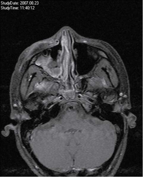
A 15-year-old previously healthy primigravida was admitted to the Department of Ophthalmology with a 3-day history of ocular protrusion and loss of vision in the right eye (Figure 1). When pregnancy was diagnosed, she was transferred to the obstetrics department for further evaluation and management.

Retrobulbar hemorrhage at the right eye.
At admission, she had no vaginal bleeding, her vital signs were stable and her blood pressure was normal. Ultrasound revealed fetal death, and gestational age was estimated by biometry as well as last menstrual period to be about 23 weeks. The laboratory workup at time of admission showed definite signs of DIC [hemoglobin, 8.1 g/dL; activated partial thromboplastin time (aPTT), 44.2 s; platelet count, 55×109/L; and fibrinogen markedly decreased to 80 mg/dL]; renal and hepatic parameters were normal and there were no signs of infection.
We estimated the time period between fetal death and DIC to be <1 week according to the degree of maceration and the last known fetal movement.
Neither clinical nor ultrasonographic examination revealed signs of placental abruption; therefore, an induction of labor with prostaglandins was initiated. Simultaneously, a stabilization of the coagulation status, including coagulation factors (Prothromblex® 1800 IE, Baxter Healthcare GmbH Austria, Vienna, Austria), fibrinogen (Haemocomplettan® 5g, CSL Behring GmbH, Marburg, Germany) and AT III (Atenativ® 3.000 IE, Atenativ and Octaplas, Octapharma AG, Lachen, Switzerland), was started and the patient was closely monitored.
Despite aggressive blood product replacement, massive vaginal bleeding occurred 2 h later. At this time, the laboratory workup showed severe aggravation of the DIC [aPTT, 117.3 s; platelet count, 37×109/L; and fibrinogen <80 mg/dL]. Due to worsening of the maternal vital signs, a cesarean section was performed, and during this intervention a complete detachment of the placenta was noted. The dead fetus showed beginning signs of maceration. Finally, the patient was transferred to the intensive care unit in stable condition. During surgery, the patient received 10 U of packed red blood cells and 3 units of fresh frozen plasma (Octaplas®).
To characterize the ophthalmologic complications, magnetic resonance imaging was performed, which revealed an extraconal intraorbital hemorrhage with elongation of the ocular bulb and stretching of the optic nerve. Therefore, an immediate surgical intervention (i.e., orbital decompression) was performed (Figure 2).
Retrobulbar hemorrhage after surgical intervention.
After 2 days, the patient was transferred to the obstetrics department in stable condition, and she was finally discharged on the 9th postoperative day. Despite all therapeutic efforts, the loss of vision of the right eye was permanent. Screening for thrombophilia 8 weeks postpartum revealed heterozygosity for activated protein C resistance (factor V Leiden).
Discussion
DIC is associated with several obstetric complications, such as preeclampsia and other hypertensive disorders, amniotic fluid embolism, placental abruption, retained dead fetus, obstetric sepsis and uterine rupture [5].
The trigger mechanisms of DIC during pregnancy activate the coagulation cascade at different levels. In cases of placental abruption, amniotic fluid embolism and retained dead fetus, thromboplastin is transferred into the maternal circulation and activates the coagulation cascade [5]. Additionally, DIC develops from localized consumption of platelets, coagulation factors and fibrinogen in the intrauterine retroplacental hematoma.
Hemorrhage is the most common clinical presentation of DIC and affects various organs or tissues (frequently brain and skin), whereas involvement of the eye has seldom been reported in pregnancy [5].
Ocular or orbital hemorrhage as the primary manifestation of DIC in pregnancy is associated with labor or preeclampsia [1–4, 6]. Ocular involvement consists of thrombotic vascular occlusion of the chorioidea and retina as well as hemorrhage in the corpus vitreum, retina, chorioidea, optic nerve sheath and, at worst, retinal detachment in association with preeclampsia, fetal demise and placental abruption. Orbital hemorrhage is divided into the more common intraorbital hemorrhage and the less common subperiosteal orbital hemorrhage. Subperiosteal hemorrhage is mostly associated with labor [1–4, 6].
A retained dead fetus as the most leading cause of DIC was first described in 1950 by Weiner et al. [9]. The author’s explanation for the coagulation disorder was rhesus sensitization and consecutive hemostatic failure due to hypofibrinogenemia. In the following years, it has been reported that DIC in association with fetal death is not an acute process like placental abruption or amniotic fluid embolism, but rather a chronic process that does not occur in <4–5 weeks in 25%–40% of cases, although in rare cases DIC may develop earlier than 4 weeks [7].
The factors responsible for the development of DIC only in some patients as well as the delay in its appearance are still unknown. Reasonable causes are a progressive breakdown of the placental barrier with consecutive transfer of thromboplastic substances from the dead fetus to the maternal circulation and the compensatory maternal capacity [8].
Hoines and Buettner [3] reported a case of retinal detachment and DIC in association with abruptio placentae. In contrast to our case, the condition resolved 4 weeks after cesarean section.
Whereas DIC in retained dead fetus is usually described as a chronic process, in our case it seems to be an acute situation, regarding estimated time period between fetal death and DIC, as well as clinical and laboratory findings.
Retroplacental hematoma, the characteristic feature of premature placental separation, remains controversial as either the cause or sequela of the hemostatic disorder.
In our case, it remains controversial if the DIC is either due to the retained dead fetus or to placental abruption, but it seems that the thromboplastic potency of severe obstetric entities such as placental abruption and fetal death might provoke a severe DIC with permanent damage within a short time. To the best of our knowledge, this is the first case report with retrobulbar hemorrhage as the primary manifestation of DIC caused by retained dead fetus and placental abruption. Additionally, it seems that the dead fetus syndrome might happen in modern obstetric medicine despite optimal management.
Summary
Obstetric DIC in combination with intrauterine fetal demise is a very seldom phenomenon that is uncommonly observed in modern medicine. The clinical manifestation happens in various tissues and organs; the first sign is usually vaginal bleeding.
The corporate thromboplastic potency of severe obstetric entities such as abruptio placentae and fetal death might provoke a foudroyant DIC with permanent damage within a short time. Any obstetrician should be aware of the possibility of severe DIC in cases of fetal death.
Because of the uncommon location of the primary symptom and the acute course in this case, in our opinion it seems worthy of publication.
References
[1] Attala ML, McNab AA, Sullivan TJ, Sloan B. Nontraumatic subperiosteal orbital hemorrhage. Ophthalmology. 2001;108:183–9.10.1016/S0161-6420(00)00482-6Suche in Google Scholar
[2] Bjerknes T, Askvik J, Albrechtsen S, Skulstad SM, Dalaker K. Retinal detachment in association with preeclampsia and abruptio placentae. Eur J Obstet Gynecol. 1995;60:91–3.10.1016/0028-2243(95)80003-4Suche in Google Scholar
[3] Hoines J, Buettner H. Ocular complications of disseminated intravascular coagulation (DIC) in abruptio placentae. Retina. 1989;9:105–9.10.1097/00006982-198909020-00006Suche in Google Scholar
[4] Jacobson DM, Itani K, Digre KB, Ossoinig KC, Varner MW. Maternal orbital hematoma associated with labor. Am J Ophthalmol. 1988;105:547–53.10.1016/0002-9394(88)90249-8Suche in Google Scholar
[5] Letsky EA. Disseminated intravascular coagulation. Best Pract Res Clin Obstet Gynaecol. 2001; 15: 623–44.10.1053/beog.2001.0204Suche in Google Scholar
[6] Oruc S, Sener EC, Akman A, Sanac AS. Bilateral orbital hemorrhage induced by labor. Eur J Ophthalmol. 2001;11:77–9.10.1177/112067210101100115Suche in Google Scholar
[7] Pritchard JA. Fetal death in utero. Obstet Gynecol. 1959; 14:573–80.Suche in Google Scholar
[8] Romero R, Copel JA, Hobbins JC. Intrauterine fetal demise and hemostatic failure: The fetal death syndrome. Clin Obstet Gynecol. 1985;28:24–31.10.1097/00003081-198528010-00004Suche in Google Scholar
[9] Weiner AE, Reid DE, Roby CC, Diamond LK. Coagulation defects with intrauterine death from Rh isosensitization. Am J Obstet Gynecol. 1950;60:1015–22.10.1016/0002-9378(50)90507-2Suche in Google Scholar
-
The authors stated that there are no conflicts of interest regarding the publication of this article.
©2013 by Walter de Gruyter Berlin Boston
Artikel in diesem Heft
- Masthead
- Masthead
- Case reports – Obstetrics
- Orbital hemorrhage as a primary manifestation of disseminated intravascular coagulation (DIC) associated with intrauterine fetal death and placental abruption
- The intrapartum use of antithrombin III in an antithrombin III-deficient patient: a case report and review of the literature
- Cardiac tamponade in a woman with preeclampsia
- Spontaneous hematoma of the rectus abdominal wall in pregnancy
- Budd-Chiari syndrome following vaginal delivery in a patient with Crohn’s disease: a case report and review of the literature
- Postpartum takotsubo cardiomyopathy with reversible cerebral vasoconstriction syndrome: a case report
- Sarcomatoid carcinoma of the oral cavity during pregnancy
- Can peripartum cardiomyopathy be caused by chemotherapy and radiation of breast cancer?
- Case reports – Fetus
- Fetal death associated with diffuse mesangial sclerosis combined with bilateral multicystic kidney
- Prenatal diagnosis of agenesis of the corpus callosum and cerebellar vermian hypoplasia associated with a microdeletion on chromosome 1p32a
- Pulmonary lymphangiomatosis as a cause of first trimester nuchal cysts in a euploid fetus
- Prenatal ultrasound and molecular diagnosis elucidate the prognosis of Pfeiffer syndrome1)
- Prenatal diagnosis of isolated agnathia with two and three-dimensional ultrasound
- Case reports – Newborn
- Sudden death from cardiac tamponade in an extremely low birth weight neonate with an umbilical venous catheter in situ
- Congenital cytomegalovirus infection after maternal persistent immunoglobulin-M antibodies against cytomegalovirus prior to conception
- Aloe vera induced toxic colitis in a breast-feeding baby: a case report
- A rare cause of neonatal hydrocephalus
- Superior vena cava syndrome causing chylothoraces in a preterm neonate: a case report and literature review
- Trisomy 13 with anorectal malformation: an association or an incidental finding?
- Acute neonatal appendicitis: the potential value of laparoscopy as a diagnostic and therapeutic tool