Corrosion, stress corrosion cracking and corrosion fatigue behavior of magnesium alloy bioimplants
-
Priyabrata Das
Priyabrata Das obtained his B. Tech. in Mechanical Engineering from Biju Patnaik University of Technology, Odisha and his M. Tech. degree in Metallurgical and Materials Engineering from the Indian Institute of Technology (Bhubaneswar) in 2017 and 2020, respectively. He is currently pursuing his doctoral research at Indian Institute of Technology Delhi in the area of biomaterials. Priyabrata is recipient of the institute silver medal for academic excellence in the M. Tech. program and has published three peer reviewed papers in international journals.
T. S. Sampath Kumar is head of the Medical Materials Laboratory, Department of Metallurgical and Materials Engineering, Indian Institute of Technology Madras. He received his PhD in Materials Engineering from Indian Institute of Science, Bangalore in 1986. His research interests are in nanostructured implants, multifunctional nanocarriers, injectable bone cements, electrospun 3D scaffolds and biomaterials from eggshell waste. He has published more than 125 papers in peer reviewed journals, 3 book chapters and has 3 patents to his credit.
Dr. Kisor K. Sahu obtained a Master’s degree in Metallurgy from Indian Institute of Science, Bangalore and a PhD from Graduate School of Energy Sciences, Kyoto University with a scholarship by Japan Government (MEXT). He is currently on the faculty at School of Minerals, Metallurgical and Materials Engineering, IIT Bhubaneswar. He is presently an editorial board member of
Scientific Reports . He has published more than 40 peer reviewed papers in journals of international repute.Dr. Srikant Gollapudi is currently on the faculty at the School of Minerals, Metallurgical and Materials Engineering, IIT Bhubaneswar. He obtained his Bachelor’s degree in Metallurgical Engineering from NIT Rourkela, his Master’s degree in Metallurgy from IISc, Bangalore and his PhD in Materials Science and Engineering from NC State University, USA. His research interests are in corrosion, titanium and magnesium. He has more than 30 publications in well-known peer reviewed journals and 7 patent filings (3 granted and 4 under review).


Abstract
The use of magnesium and its alloys as temporary implants has gained interest in the last two decades due to their good mechanical properties and bio-degradability in the in-vivo conditions. However, the issues of higher corrosion rate and stress corrosion cracking persist, which are responsible for the implants’ early failure. This review paper focuses on the challenges involved in the use of magnesium-based implants and the advancements in mitigating the corrosion-related issues for in-vivo use of biodegradable magnesium alloy implants. Herein we review the degradation behavior of three groups of magnesium alloys, i.e., aluminum-containing Mg alloy, rare earth element (REE) containing Mg alloy, and aluminum-free Mg alloy in a variety of testing media. We also review various surface modification techniques such as mechanical methods, physical methods, and chemical methods adopted to address the shortcomings of the Mg alloys. Furthermore, recent developments in Mg based bioimplants such as Mg-based open porous scaffolds, nanostructured Mg alloys and Mg based bulk metallic glasses are reviewed. In the end, recent clinical trials of the Mg-based implant were reported in detail.
1 Introduction
1.1 Introduction to implant materials
Materials occupy a significant position in improving human lives by positively contributing to medical science and technology. Various materials have been used to produce medical instruments for a wide range of applications ranging from cardiovascular applications to bone implants. These specific materials are called biomaterials, which have gained significant attention from researchers from both materials and the medical field. According to Niinomi (2002) and Williams (1976), biomaterial-based implants can be used to heal and stabilize the fractured bones, correct deformities, and replace the damaged part of the anatomy as joint replacements, and improve organ functionalities. The earliest instance of using a material to improve human life was reported in 200 A.D. (Crubzy et al. 1998), where a dental implant made up of iron was used. Further, many medical cases are treated using a variety of materials such as stainless steel (Cahoon and Paxton 1968), titanium alloys (Niinomi 1998; Wang 1996), and polymers like polylactic acid (PLA) (Kulkarni et al. 1971). Based on the predetermined service time and applications, medical implants can be classified as temporary implants and permanent implants (Figure 1). Dental implants and joint replacements are examples of permanent implants where long service time is expected, and implants used to fix broken bone and ligaments are an example of temporary implants. Implants should possess biocompatibility, antimicrobial property, flexibility, proper mechanical strength, and adequate corrosion & fatigue resistance (Nielsen 1987; Niinomi 2008). In the case of permanent implants, very high corrosion and fatigue resistance are expected, whereas, in the case of temporary implants, the corrosion and fatigue resistance have to be maintained until the implant’s purpose is fulfilled in the physiological environment. Table 1 summarizes some traditional metallic implant materials and their particular area of use in medical technology.
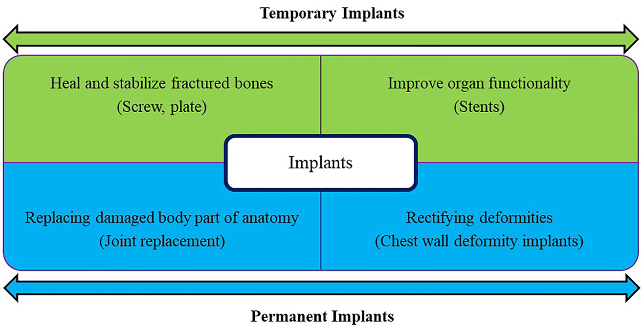
Classification of implants based on their use.
Currently used metallic implant materials and their areas of application.
| Materials | Properties | Applications | References |
|---|---|---|---|
| Stainless steel | Strength, ductility, fatigue resistance | Cardiovascular stents, orthopedic prosthesis, dental implants | Bekmurzayeva et al. (2018) |
| Titanium alloys | Superior biocompatibility, strength and corrosion resistance | Hip joints, dental implants | Niinomi (2003) |
| Cobalt-chromium alloys | High wear resistance | Artificial joints | Hermawan et al. (2011) |
| Gold alloys | Excellent corrosion resistance, ductility | Dental restorations, gold plated stents to support weak blood vessels | Baltzer and Copponnex (2014) |
Though other implant materials categories like ceramic, polymer, and composites are also available, metallic materials are preferred when it comes to strength consideration. Specifically, metallic components made out of stainless steel, titanium alloys, etc., are more favored in orthopedic, dental, and cardiovascular implants. It is due to their excellent mechanical strength, corrosion & fatigue resistance, and outstanding load-bearing capacity. However, when these materials are used as temporary implants, a second operation is needed to remove them after the bone and tissue’s complete healing, which adds up to the cost (Staiger et al. 2006). Stress shielding effect (Inoue et al. 2020; Nagels et al. 2003; Sumner 2015) is another major issue where bone density decreases due to the removal of typical stress from the bone. The effect of stress shielding can be seen in Figure 2, which shows the radiographs of a humeral implant (A) just after the surgery, (B) six months, and (C) one year after the surgery. As most of the load was taken by the implant material, bone loss can be seen at the proximal region (arrow marked) due to stress shielding effect. Similarly, abnormal bone deposition was evident in the distal region due to increased stress in that area (rectangular region). It occurs as the stiffness of implant material is very high compared to bone. The bone density will be less at the proximal part of the implant than the distal region. Additionally, these alloys cause tissue inflammation due to the release of cytotoxic ions (Eliades et al. 2004; Wang et al. 1996) and create an unpleasant experience for the patients by slowing the healing process. New metal and alloys are explored to address all these issues and provide potential materials for temporary implants.
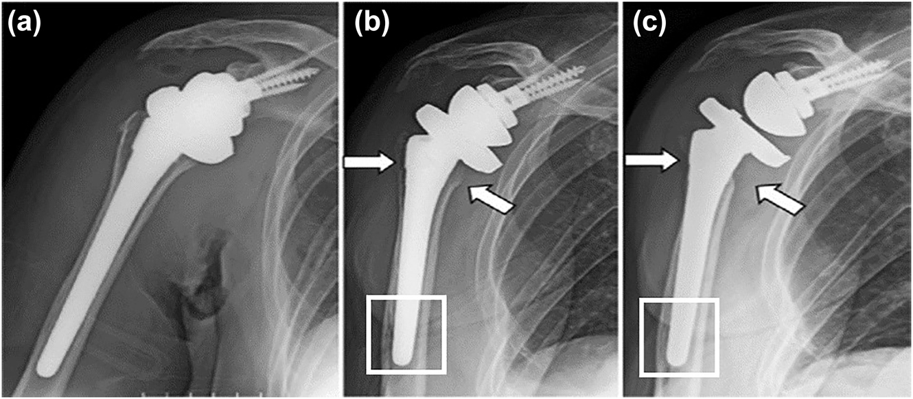
Radiographs of humeral implants: (a) just after surgery, (b) 6 months, and (c) 1 year after surgery showing gradual loss of bone near the implant (arrow marked) and abnormal deposition of bone (rectangle region) far from the implant. Reprinted from (Inoue et al. 2020) with permission from Elsevier.
1.2 Magnesium alloys as potential materials for bio-implant
The use of magnesium as a biomaterial date back to 1878 when Huse reported the use of Mg in ligatures for suturing wounds and blood vessels. After that, many in-vivo and in-vitro studies (Hoffheinz and Dimitroff 1928; Mcbride 1938; Stroganov et al. 1972) have been reported involving Mg and its alloys. These are suitable candidates for temporary bio-implant applications due to their biodegradable nature within the body fluids. It means that the implants made of such alloys will support the broken bones for some time and slowly dissolve within the fluid available in a physiological environment. Besides, these materials have excellent biocompatibility within the body. Other advantages of using Mg and its alloys are their assistance in tissue healing (Jacobs et al. 2003), human metabolism (Hänzi et al. 2010; Seitz et al. 2014), enhanced bone formation (Kraus et al. 2012), and negligible cellular toxicity.
The quest for Mg-based implants (Sikora 2012) is a direct outcome of the similarity between the mechanical properties of natural bone and Mg-based alloys. Table 2 reports the mechanical properties of bone, traditional implant materials, and magnesium alloy bio-implants. From Table 2, we can see that the stainless steel, titanium alloys, and cobalt-chromium alloys have a higher density and strength than natural bone. Also, the significant difference of elastic modulus values between traditional implant materials and bone causes stress shielding effect, which is undesirable. In contrast, Mg and its alloys have a similar density, yield strength, and elastic modulus of the natural bone. Due to this advantage, the stress shielding effect can be mitigated within the physiological environment without compensating for the strength. Our review paper focuses on corrosion, stress corrosion cracking (SCC) and corrosion fatigue (CF) of Mg based bioimplants. We have created an appendix at the end which summarizes the different testing standards for conducting these studies.
Mechanical properties of various implant materials compared to natural bone (Amaral et al. 2002; Kokubo et al. 2003; Niinomi 1998).
| Property | Natural bone | Magnesium alloys | Titanium alloys | Cobalt-chromium alloys | Stainless steel |
|---|---|---|---|---|---|
| Density (g/cm3) | 1.8–2.1 | 1.74–2 | 4.4–4.5 | 8.3–9.2 | 7.9–8.1 |
| Elastic modulus (GPa) | 3–20 | 41–45 | 110–117 | 230 | 189–205 |
| Yield strength (GPa) | 130–180 | 85–190 | 758–1117 | 450–1000 | 170–310 |
| Fracture toughness (MPa m−1/2) | 2–12 | 15–40 | 40–92 | – | 55–95 |
2 Challenges
Although magnesium alloys are potential candidates for degradable implants, they are yet to be widely used for clinical applications. Some of the major challenges involved in using magnesium alloys as bio-implants are listed below.
2.1 High rate of corrosion
Magnesium is a very reactive element as its standard reduction potential is −2.3 V versus SHE. Due to high electronegativity, pure magnesium and its alloys are susceptible to corrosion in aqueous media. The corrosion rate of pure magnesium is very high in the physiological environment, and the integrity of the implant is lost before the fulfillment of the whole purpose of the implant. In other metals and alloys, the thin oxide layer at the surface acts as a passivation layer that protects the material from corrosion. But in magnesium, the oxide layer formed does not act as a good passivating layer (Song et al. 2012). Thus, the corrosion kinetics is much faster, which deteriorates the mechanical properties of the bio-implants, causing failure prematurely. The corrosion products are formed as per the following reactions.
The magnesium hydroxide layer is formed on the magnesium implant surface in an aqueous environment. The formation of the hydroxide layer over the implant surface reduces the metal ion transfer to the surrounding fluids. However, our body fluid contains a significant amount of chloride ions. In the presence of chloride ions, the hydroxide layer starts to dissolve and is prone to the breakdown of the film locally. Due to the reaction between magnesium hydroxide and chloride ions,
Another issue caused by the corrosion of Mg-based implants is the hydrogen gas evolution. Because of this, the hydrogen bubbles cling to the implant surface and cause tissue separation. According to Song (2007), the critical tolerance level for hydrogen is less than 0.01 mL/cm2/day within in-vitro conditions. This value can be used as a screening parameter for magnesium alloy bio-implants. However, Witte et al. (2005) suggested that in in-vivo condition, hydrogen evolution is high in the first week of post-operation, and it starts to disappear with time. So, controlling the hydrogen evolution in the first few weeks of post-surgery is essential to keep the value within 0.01 mL/cm2/day (Chakraborty et al. 2019).
The mechanism of corrosion in Mg alloy bio-implants depends on the type of alloy and simulated body fluid used for studying the alloy. In some cases, Mg alloy implants are used in conjunction with other metals or alloys, which can lead to galvanic corrosion. This type of corrosion also happens in alloy systems containing second phases, intermetallic, etc. (Poinern et al. 2012). The schematic diagram for both cases is depicted in Figure 3.
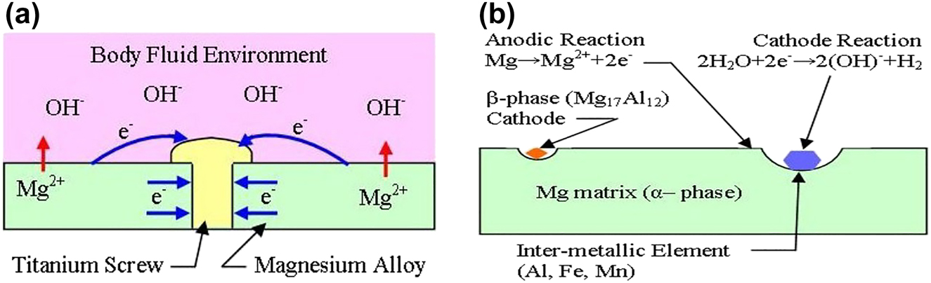
Galvanic corrosion in the physiological environment due to (a) the use of two dissimilar metals, (b) the presence of the second phase and/or intermetallic (Poinern et al. 2012).
Another form of corrosion prevalent in Mg alloys is pitting corrosion, which is a form of localized corrosion. In this corrosion mode, critical crevice solution (CCS) remains static in a micro sized crevice, increasing the pH of the nearby fluid (Kelly 2003). This results in the formation of deep pits due to the dissolution of Mg in a local environment. All these events can initiate cracks that grow over time, and the implants fail prematurely. Besides pitting corrosion, filiform corrosion can also be seen in Mg based implants. In this corrosion mode, a randomly distributed thread-like filament network can be seen. Figure 4(a) shows the instances of severe filiform corrosion over an Mg–5Dy alloy in 0.9 wt% NaCl solution in ambient conditions (Yang et al. 2011b). With increase in Dy content from 5 to 20 wt%, the severity of filiform corrosion reduces and pitting corrosion becomes evident (Figure 4(b)–(d)).
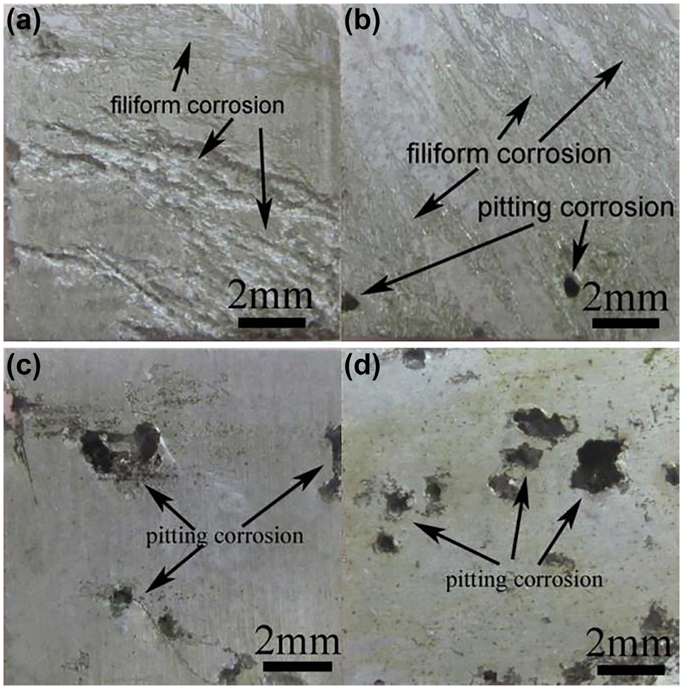
Types of corrosion in Mg-Dy alloy (a) Severe filiform corrosion in Mg-5Dy, (b) reduced filiform corrosion and prominent pitting corrosion in Mg-10Dy, (c) and (d) severe pitting corrosion in Mg-15Dy and Mg-20Dy after 72 h immersion in 0.9 wt% NaCl solution. Reprinted from (Yang et al. 2011b) with permission from Elsevier.
2.2 Stress corrosion cracking (SCC) and corrosion fatigue (CF) in implants
The implant when placed within a living body, experiences cyclic stress due to day to day activity of the patient. Also, the physiological environment contains various salts and enzymes which cause corrosion of the implant material. Due to the cumulative action of cyclic loading and corrosive body fluid, sudden fracture/failure may occur even in a ductile material (Kannan and Raman 2008). The failure mechanisms involved in those cases are called corrosion fatigue (CF) and stress corrosion cracking (SCC) (Harandi and Singh Raman 2017; Jafari et al. 2015). These modes of failure are more evident in the case of magnesium and its alloys as these alloys are more likely to undergo pitting corrosion in
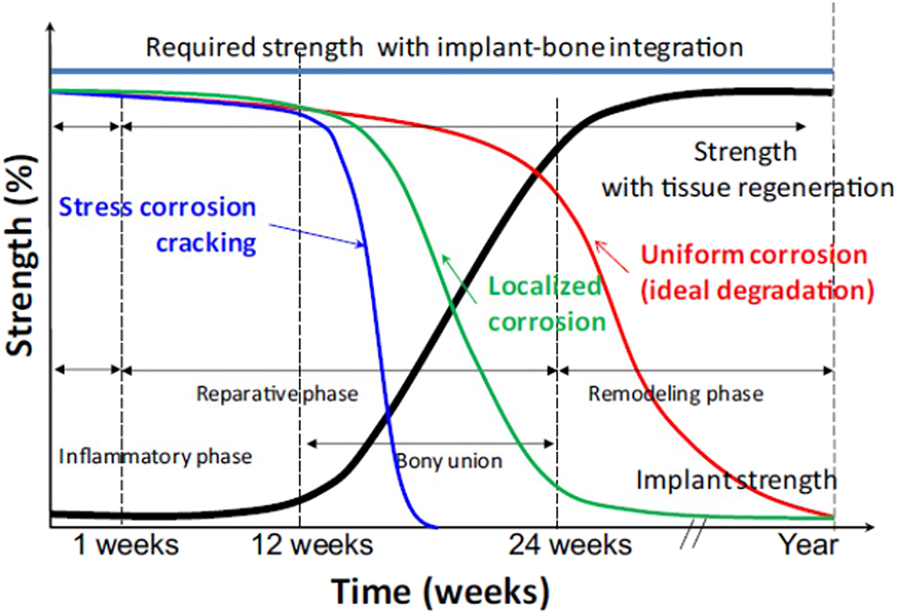
Mechanical integration between implant material and tissue generation over time in a physiological environment. Reprinted from (Koo et al. 2017) with permission from Elsevier.
Koo et al. (2017) also performed a microstructural investigation of AZ31B alloy implants immersed in Hank’s Balanced Salt Solution (HBSS), which were under stress up to 30 and 90 days, respectively. The SEM microstructure (Figure 6) showed the presence of stress corrosion cracks over the surface and severe localized corrosion at the cross-section.
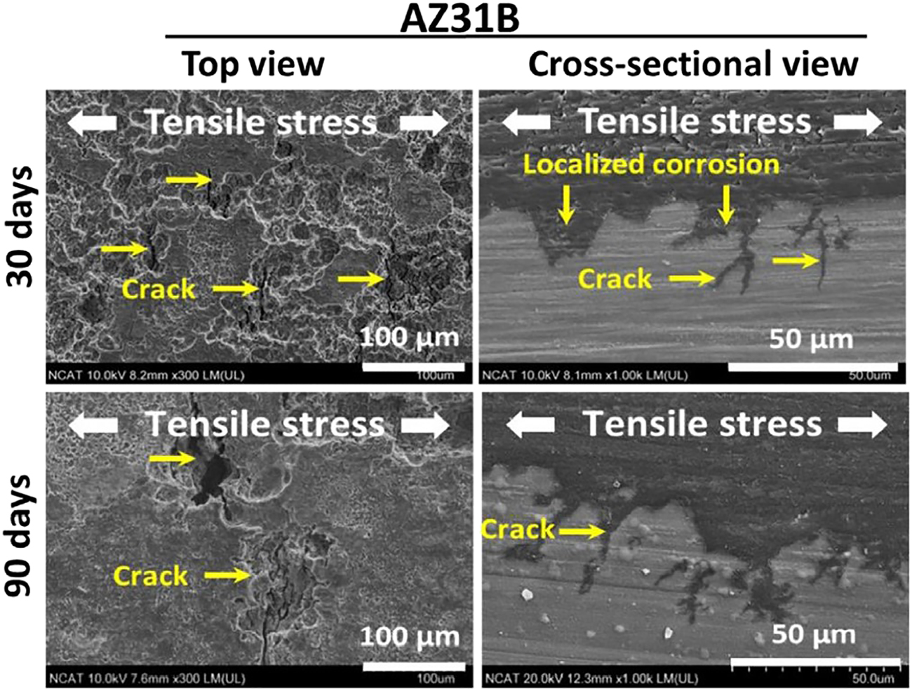
Top view and the cross-sectional view of SCC in AZ31B alloy implants immersed in Hanks balanced salt solution (HBSS) under stress for 30 and 90 days. Reprinted from (Koo et al. 2017) with permission from Elsevier.
Many other studies (Choudhary et al. 2014; Kannan et al. 2008; Logan 1958) have also presented a detailed microstructural investigation to see the failure mechanism in different Mg alloys. One of the studies reported by Choudhary et al. (2014) showed the fracture behavior of WE43 alloy in modified simulated body fluid (m-SBF) at 37 °C. The fractography can be seen in Figure 7. This study showed the SCC behavior of WE43 to be controlled by both intergranular and transgranular fracture, which contrasts with the fact that Mg alloys mostly fail by transgranular fracture during SCC. This observation was attributed to the presence of large precipitates along grain boundaries, which preferentially corroded with respect to the grain, resulting in intergranular SCC. A similar type of failure mode was also reported by Kannan et al. (2008), which can be seen from Figure 8. In this study, the fractured surface of ZE41 alloy was studied in air, distilled water and 0.5 wt% NaCl. After Slow Strain Rate Testing (SSRT) in air, dimple rupture along grain boundaries occurred, which can be seen from Figure 8(a). Similarly, dissolution along grain boundaries is evident from Figure 8(b). In the presence of NaCl, both intergranular and transgranular cracking are observed (Figure 8(c)). The above studies confirmed that the Mg alloys containing the rare-earth element (REE) are likely to fail in a mixed mode of intergranular and transgranular SCC.
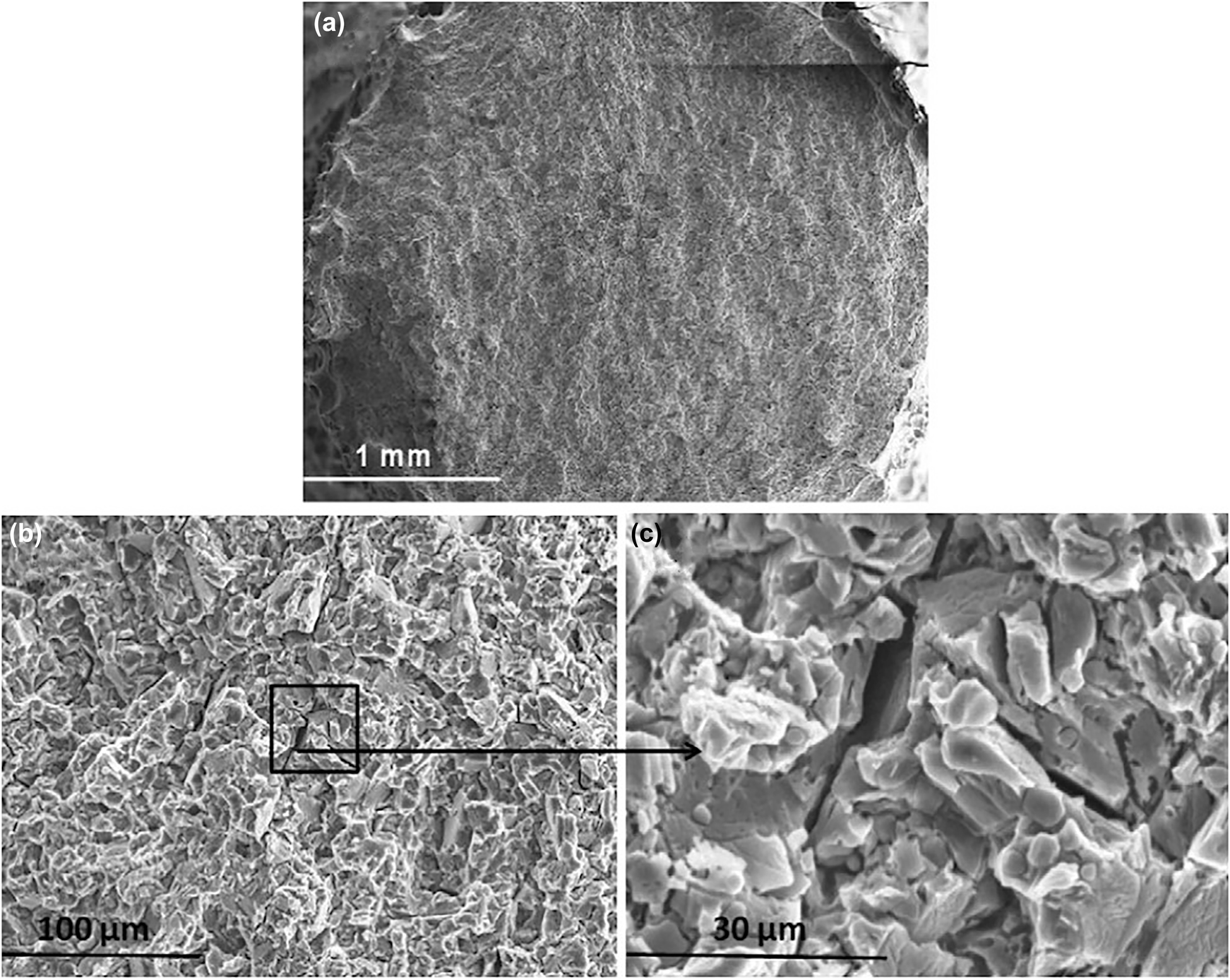
Micrograph of (a) the fractured surface, (b) intergranular and transgranular SCC, (c) fractured surface at higher magnification showing intergranular cracks in WE43 alloy tested in m-SBF. Reprinted from (Choudhary et al. 2014) with permission from Elsevier.
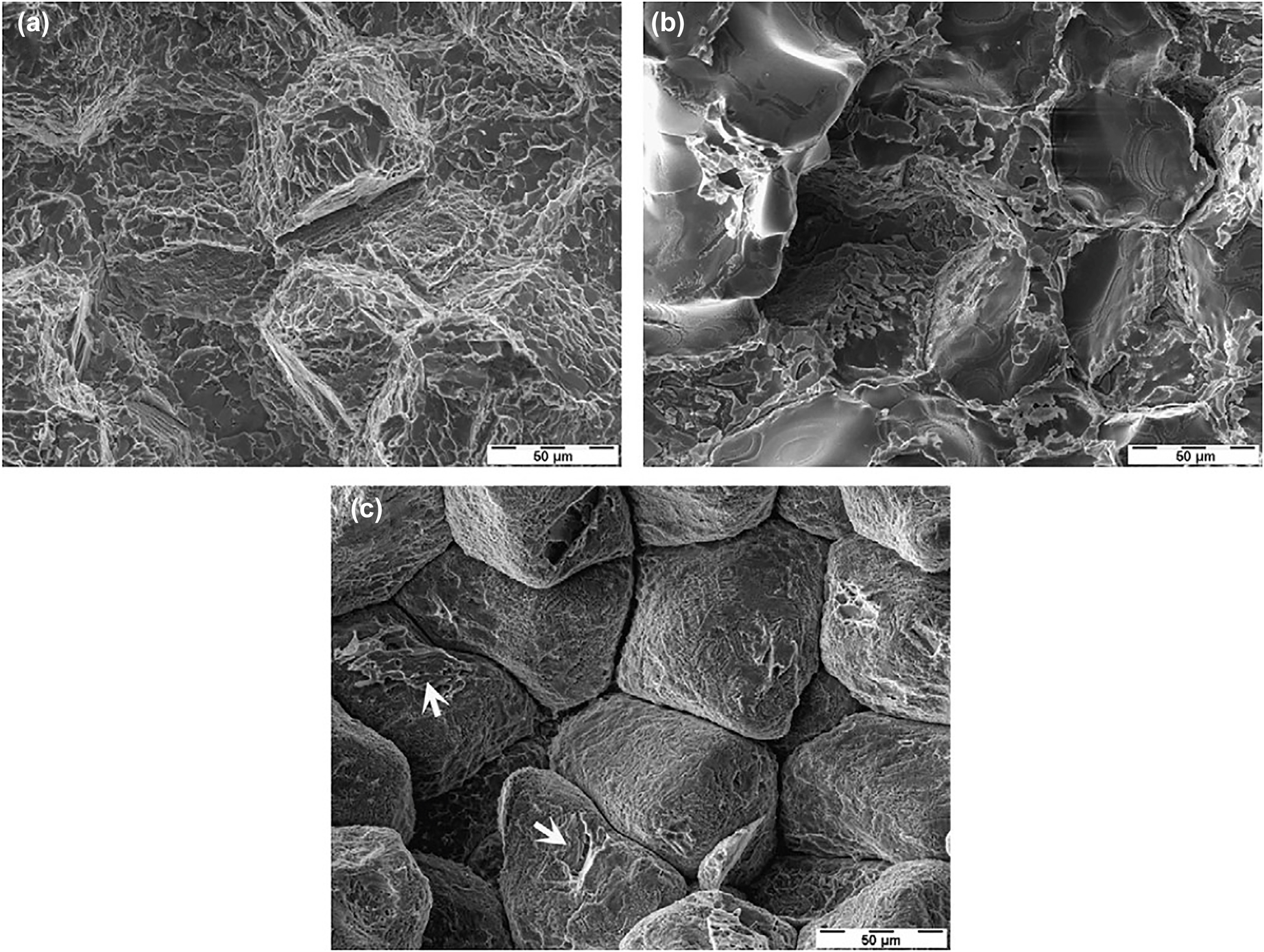
Micrograph of ZE41 after (a) slow strain rate testing in air, (b) immersion in distilled water, (c) immersion in 0.5 wt% NaCl (arrows indicate transgranular cracking). Reprinted from (Kannan et al. 2008) with permission from Elsevier.
The main challenge associated with this failure mode is to properly understand the combination of mechanical stresses and interacting species present in the physiological environment. In short, to mimic the environment for SCC and CF, proper mechano-chemical testing is needed. The importance of selecting proper mechanochemical testing to test an implant material is mentioned by Harandi and Singh Raman (2015). According to them, selecting appropriate mechanochemical testing can significantly reduce the chances of failure of Mg alloy implants in SCC and CF.
2.3 Comparison between the in vivo and in vitro condition
Another challenge in this area is the trial of new alloys for in-vivo testing. In-vivo testing is costly and also poses a risk to the patient. Insufficient understanding of the in-vivo environment might lead to serious health issues. An alloy can perform very well in in-vitro studies and may underperform in an in-vivo environment. One such example can be seen from the in-vivo and intro study of AZ91D alloy. Wen et al. (2009) reported a lower degradation rate and uniform corrosion morphology of AZ91D alloy tested in m-SBF up to 24 days. However, in-vivo testing of AZ91D alloy (Witte et al. 2005) showed severe pit formation over the implant surface. The reasons behind these failures are the use of static loading or no loading conditions for performance evaluation in in-vitro studies, unknown cytotoxicity in the presence of proteins and other constituents of body fluids, etc. While performing in-vitro testing, one should use a testing method that perfectly mimics the stress distribution and physiological environment of the actual application area within our body. Additionally, a material must be evaluated for its cytotoxicity in the physiological environment; otherwise, it may create health risks for the patient either in short or long run. For example, Mg–Al alloys had been seen as a potential bio-implant material as they showed improved mechanical and corrosion properties compared to other existing alloys. However, recently, it was found that Mg–Al alloys may cause various neurological disorders such as Alzheimer’s and Dementia (Gupta et al. 2005; Taïr et al. 2016) in the long run in the physiological environment. So now, researchers are focusing on developing aluminum-free Mg alloys for bio-implant applications.
Developing solutions for in-vitro testing is another crucial area. This step demands a complete understanding of the body fluid. For this purpose, various solutions have been developed to mimic the body fluid environment. Among those solutions, simulated body fluid (SBF) and Hank’s salt solution (HSS) are mostly used for in-vitro studies due to their close resemblance with the actual body fluid. The typical composition for blood plasma, HSS, SBF, modified SBF (m-SBF), revised SBF (r-SBF), Kokubo’s SBF, minimum essential medium (MEM), and Dulbecco’s MEM (DMEM) are reported in Singh Raman et al. (2015) and Phakatkar et al. (2020). However, the effect of organic compounds presents in the blood plasma like proteins and fatty acids, etc., are ignored while using these solutions for in-vitro testing, which is a research gap between in-vivo and in-vitro testing.
3 Advancements to overcome the challenges
3.1 Alloy modification
As Mg is more susceptible to corrosion, various alloying elements have been employed to tune the corrosion rate and CF performance as per the need. A recent in-vivo study done by Kawamura et al. (2020) compared the corrosion behavior of Ti alloy (Ti–6Al–4V), pure Mg, Mg alloy (AZ31) and poly lactic acid (PLA). Figure 9(a–d) shows the degradation of Ti–6Al–4V, AZ31 pure Mg, and PLA, respectively. Ti–6Al–4V showed no degradation over 12 weeks period (Figure 9(a)), and pure Mg showed the maximum degradation, which resulted in a decrease in cross-sectional area (Figure 9(b)). PLA performed better than pure Mg and AZ31, which can be seen from Figure 10, but mechanical properties are way lesser than Mg. On the other hand, AZ31 showed a significant decrease in the corrosion rate in comparison to pure Mg. From Figure 10, it is clear that with the addition of aluminum and zinc to the pure Mg, the degradation rate decreased from 2.5 mm/year to nearly 0.8 mm/year, which is one order of magnitude decrease. The decrease in the degradation rate can be explained with the help of Figure 9(b). We can see that the mechanism of corrosion was different in the case of AZ31 as compared to pure Mg. After two weeks, a nonuniform film formation can be seen over the AZ31 alloy, which protects the alloy from further corrosion, whereas in the case of pure Mg, no such films are observed.
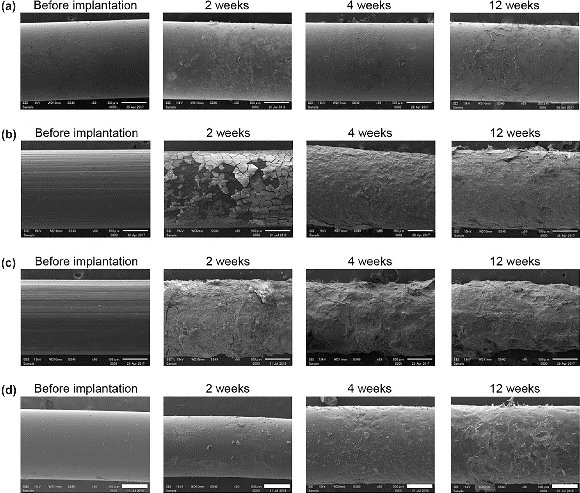
Degradation of (a) TiA (Ti–6Al–4V), (b) MgA (AZ31), (c) pure Mg, and (d) PLA with time in an in-vivo environment (Kawamura et al. 2020).
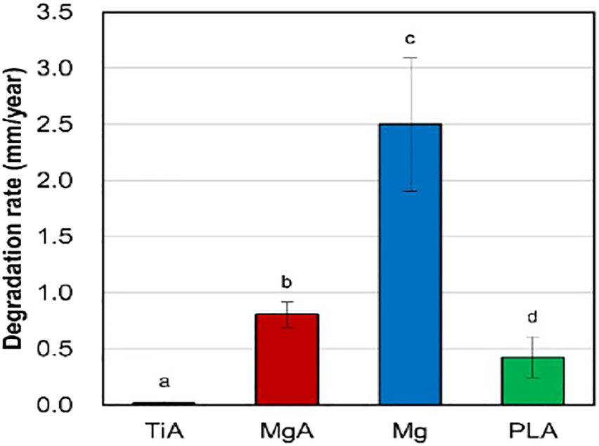
Degradation rate comparison between (a) TiA (Ti-6Al-4V), (b) MgA (AZ31), (c) pure Mg, and (d) PLA in SBF for 14 days (Kawamura et al. 2020).
The Mg alloys used for implant can be classified broadly into three categories. The first category contains the Mg–Al alloys, which have been studied extensively and proven to cause neurological disorders (Taïr et al. 2016) in rats in case of higher Al ion consumption. Alloys systems such as AZ (Mg–Al–Zn) and AM (Mg–Al–Mn) fall under this category (Zheng et al. 2014). The second category was developed to use the REEs containing magnesium alloys which were primarily developed for improving high temperature strength and creep resistance but considered for biomedical applications. Alloy systems, including Mg–Y, Mg–La, Mg–Nd, and Mg–Ce, are part of this category (Willbold et al. 2015). The third category of Mg alloys are those which do not contain REEs or Al. Mg–Zn–X alloy system where X may be Ca, Mn, or Si falls under this category (Rosalbino et al. 2010). We are going to summarize the behaviors of each of the three categories.
3.1.1 Mg–Al alloys
The solid solution solubility of Al in Mg is 12.5 wt% at 450 °C, but at room temperature, the value reduces to less than 1 wt% which can be seen from the Mg–Al phase diagram (Lumley, 2018). Hence, any alloy containing more than 1 wt% of Al results in forming the Mg17Al12 secondary phase (β-phase). This β-phase acts as a cathode with respect to the surrounding α-phase in the physiological environment (Ding et al. 2014; Lunder et al. 1993). This may result in the preferential dissolution of α-phase surrounding the β-phase leading to enhanced corrosion of the matrix phase. However, if the volume fraction of β-phase is high and it is uniformly distributed over grain boundaries, then it can act as a cathodic barrier and lowers the dissolution of α-phase into the surrounding corrosive fluid. This explanation was also supported by Song and Atrens (1999) and Nisancioglu et al. (1990). Another possible reason for the increase in corrosion resistance with an increase in wt% of Al is the formation of aluminum oxide (Al2O3)/hydroxide (Al(OH)3) over the surface, which acts as a passivation layer (Xin et al. 2009). There are numerous studies involving AZ alloys as it is one of the widely used materials. One such valuable study involving the passivation behavior of AZ31 alloy in the presence of different anions has been reported by Wang et al. (2010). The study provided the corrosion maps of AZ31 alloy in the presence of
3.1.2 Mg-REEs alloys
This category of Mg alloys can be further classified into two groups as per the solubility of REEs in Mg. The first group corresponds to those REEs which have limited solubility in Mg, such as La, Ce, and Zr, etc. The second group consists of highly soluble REEs like Y, Gd, and Dy, etc. (Rokhlin 2003). When REEs are added as an alloying element, most of them lead to solid solution strengthening and precipitation strengthening and this improves the creep resistance. Zr is one of the commonly used REE for grain refinement of Mg. As per Stroganov et al. (1972) and Avedesian and Baker (1999), care should be taken while mixing the Zr in master alloys containing Al, Mn, Si, Fe, etc., as it can form stable compounds with them resulting in the removal of those alloying elements from solid solution. Tsai et al. (2011) reported that the incorporation of Zr in Mg increases the specific damping capacity, which may reduce the stress generated at the interface of bone and implant. Many studies involving Y, Gd, Nd, and Zr are reported in the literature. One such study done by Gu et al. (2011) evaluated the corrosion performance of ZK60, WE43, AZ31, and AZ91. The corrosion rate of WE43 in Hank’s solution was found to be 0.055 mg/cm2/h, which is nearly half the corrosion rates of ZK60, AZ31, and AZ91 (mg/cm2/h). There are other studies that suggest that Yttrium (Y) containing alloys are corrosion resistant. A study led by Davenport et al. (2006) on WE43 alloy investigated the corrosion performance of this alloy in both as-cast and heat-treated conditions. They have found that in the as-cast sample, corrosion tendency was lower in the regions rich in Yttrium. Heat treatment enhanced the corrosion performance further by uniformly distributing Y. Liu et al. (2010) studied the corrosion behavior of Mg–Y binary alloys in different test environments containing different anions. In 0.1 M NaCl testing media, the corrosion rate was increased with an increase in Y content in the alloy due to the increase in the volume fraction of intermetallic content. On the other hand, the addition of Y enhanced the corrosion resistance in 0.1 M Na2SO4 media despite the formation of the intermetallic. From this study, we can conclude that
3.1.3 Al free Mg alloys
A study on the cytotoxicity of different Mg alloys from the former two groups (Mg-Al and Mg-REE alloys) was led by Scheideler et al. (2013). They modified the Dulbecco’s modified eagle medium (DMEM) solution by adding a serum to resemble the in-vivo environment closely. This resulted in better predictability of cytotoxicity, and the MgAl9 alloy is found to cause severe toxicity. Though Al intake in a definite range is acceptable, researchers are more focused on developing alloys free of Al. In this case, attention is paid to alloying elements like Ca, Mn, Si, etc., due to their positive impact on mechanical properties and corrosion resistance. As we all know, calcium is a major constituent of our bones and is responsible primarily for bones’ growth and development. It exists as a well-organized crystal of calcium and phosphorous called hydroxyapatite (HA) (Ca10(PO4)6(OH)2) (Boushey et al. 2001). Due to this reason, many researchers suggested using Ca as an alloying element in Mg bio-implants. Similarly, other elements such as Mn, Zn, and Si are also allowed within a permissible range in the human body environment (Agarwal et al. 2016; Underwood 1971). Various alloys such as Mg–Zn–Ca, Mg–Zn–Mn, Mg–Ca, etc., have been evaluated in terms of strength and corrosion resistance. Rosalbino et al. (2010) performed an electrochemical study to quantify the corrosion rate of as-cast Mg–Zn–Mn, Mg–Zn–Si, and Mg–Zn–Ca alloy in Ringer’s physiological solution. In this study, Mg–2Zn–0.2Mn showed a continued four-fold increase in polarization resistance over 168 h in comparison to the AZ91 alloy. The corrosion inhibition behavior of Mg–2Zn–0.2Mn was attributed to the presence of oxidized Mn in the Mg(OH)2 protective layer. The other two alloys, i.e., Mg–2Zn–0.2Ca and Mg–2Zn–0.2Si, showed a mild decrease in polarization resistance compared to AZ91 alloy. Bakhsheshi-Rad et al. (2012) conducted in-vitro corrosion tests on Mg–0.5Ca–xZn alloys by incrementing the wt% of Zn by three-fold from 1 to 9. Both the electrochemical test and immersion test using Kokubo’s SBF showed a similar type of trend in results. Initially, with the addition of 1 wt% Zn, the corrosion rate, as observed in both types of tests, reduced in comparison to Mg–0.5Ca alloy. This improvement in the corrosion rate was ascribed to the formation of α-Mg + Ca2Mg6Zn3 + Mg2Ca phases, which are uniformly distributed over the microstructure forming a protective network (Lei et al. 2012). Thus, the corrosion rate increased rapidly with an increase in Zn wt%. The alloy containing 9 wt% Zn showed a significant three-fold increase in the corrosion rate. The increase in corrosion rate is may be due to an increase in the volume fraction of brittle precipitate phases (Zhang et al. 2013a), which can also degrade the mechanical properties and enhance the susceptibility to CF.
Table 3 summarizes the corrosion behavior of different magnesium alloys in the different testing mediums.
Corrosion properties of magnesium alloys in different testing medium.
| Alloy | Sample type | Testing medium | Corrosion rate (mm/year) | References | |
|---|---|---|---|---|---|
| Immersion test | Electrochemical test | ||||
| Pure Mg | Wire | SBF | 2.514 days | – | Kawamura et al. (2020) |
| AZ31 | Wire | SBF | 0.8114 days | – | Kawamura et al. (2020) |
| AZ91D | Die cast | 5 wt% NaCl | – | 2.93 | Wu et al. (2005) |
| AZ91D-Ca | Die cast | 5 wt% NaCl | – | 0.38 | Wu et al. (2005) |
| Mg–4Zn–0.2Ca | As cast | SBF | – | 2.05 | Sun et al. (2012) |
| Mg–4Zn–0.2Ca | As extruded | SBF | – | 1.98 | Sun et al. (2012) |
| LAE442 | Gravity cast | Borax phosphate buffer | – | 6.9 | Witte et al. (2006b) |
| AZ91D | Gravity cast | Borax phosphate buffer | – | 2.8 | Witte et al. (2006b) |
| Mg–1Ca | As cast | SBF | – | 12.56 | Li et al. (2008) |
| Mg–1Ca | As extruded | SBF | – | 1.74 | Li et al. (2008) |
| Mg–1Ca | As rolled | SBF | – | 1.63 | Li et al. (2008) |
| Mg–4Y | As cast | 0.1 M NaCl | – | 3.2 | Liu et al. (2010) |
| EW62 | As cast | Saline solution saturated with Mg(OH)2 | 0.3610 days | – | Hakimi et al. (2015) |
| EW62 | Rapidly solidified | Saline solution saturated with Mg(OH)2 | 0.110 days | – | Hakimi et al. (2015) |
| Mg–Ce | As cast | SBF | 9.6 ± 0.78 | 1.84 ± 0.21 | Willbold et al. (2015) |
| Mg–La | As cast | SBF | 14.7 ± 0.92 | 2.15 ± 0.18 | Willbold et al. (2015) |
| Mg–Nd | As cast | SBF | 4.1 ± 0.29 | 1.25 ± 0.10 | Willbold et al. (2015) |
| Mg–0.5Ca | As cast | Kokubo’s SBF | 1.82 ± 0.0914 days | 4.2 ± 0.24 | Bakhsheshi-Rad et al. (2012) |
| Mg–0.5Ca–3Zn | As cast | Kokubo’s SBF | 1.83 ± 0.114 days | 5.3 ± 0.38 | Bakhsheshi-Rad et al. (2012) |
| Mg–0.5Ca–6Zn | As cast | Kokubo’s SBF | 7.42 ± 0.414 days | 8.3 ± 0.42 | Bakhsheshi-Rad et al. (2012) |
| Mg–0.5Ca–9Zn | As cast | Kokubo’s SBF | 9.13 ± 0.4214 days | 10.6 ± 0.37 | Bakhsheshi-Rad et al. (2012) |
| Mg–2.2Nd–0.3Zr | Gravity cast | SBF | 0.89120 h | – | Zhang et al. (2013b) |
| Mg–2.2Nd–0.4Sr–0.3Zr | Gravity cast | SBF | 0.77120 h | – | Zhang et al. (2013b) |
| Mg–2.2Nd–2Sr–0.3Zr | Gravity cast | SBF | 56.4924 h | – | Zhang et al. (2013b) |
Researchers have explored numerous combinations of metals for finding a perfect bio-degradable Mg alloy bioimplant in the last two decades. Most researchers are taking only experimental approaches to form new alloy systems for this purpose, which significantly increases the material development cycle time. Recently, high throughput research approaches have been implemented, which make use of the property-structure-manufacturing process (PSP) linkages for material innovation (Gupta et al. 2015; Saal et al. 2013). This approach can significantly decrease the material development cycle duration and provide a path forward for discovering improved Mg bio-degradable alloys.
3.2 Modification of the surface
Another approach to handle the issue of the high degradation rate of existing Mg alloys is surface modification. This approach is more popular as it can alter the degradation rate of the surface without changing the underlying alloy. Additionally, surface modification can improve functionality, biocompatibility and osseointegration (Mahajan and Sidhu 2018). In general, surface treatment is used in association with Mg alloys to improve the corrosion resistance of the implant in the physiological environment. Broadly surface modification methods can be divided into three categories. The first category is the mechanical route for surface modification, such as laser shock peening, shot peening, cryogenic machining, etc. The second category includes physical methods such as physical vapor deposition (PVD) and thermal spray coating. The third category contains chemical methods such as micro-arc oxidation (MAO), chemical conversion coatings, electrophoretic deposition, etc. Using the mentioned surface modification techniques, many materials such as metals, nonmetals, and polymers can be utilized to tune the surface property of the underlying alloy.
3.2.1 Mechanical route for surface modification
Mechanical processing such as shot peening, laser shock peening (LSP), milling of alloys modifies the surface characteristics by altering the grain structure and type of residual stresses. Liu et al. (2019) performed severe shot peening on AZ31 and AZ91 alloys to improve the mechanical and corrosion properties of the alloys. The employment of severe shot peening (SSP) on AZ31 and AZ91 reduced the size of α-Mg grains from 5–60 µm to 103 nm and 120 nm, respectively. Both the TEM results and the microhardness versus distance from surface plot revealed that the grain refinement happened up to a depth of 145 µm in AZ31 and 115 µm in AZ91 alloy. Although, the potentiodynamic polarization test done in 3.5 wt% NaCl solution did not show any appreciable decrease in corrosion current value by the application of SSP, the window of passivation was found to be greater in the case of SSP sample. This observation was explained by the rapid formation of passivation layer due to the availability of more grain boundaries in a nanostructured Mg alloys (Birbilis et al. 2010). Furthermore, Bagherifard et al. (2018) carried out corrosion testing, fatigue testing and cytotoxicity evaluation of the SSP and Repeened-SSP (RSSP) AZ31 alloy. The corrosion tests showed no significant improvement following the findings of (Liu et al. 2019). The cytotoxicity assessment using the human osteoblasts cultured in DMEM with 10% fetal bovine serum (FBS), 1% penicillin/streptomycin at 37 °C and 5% CO2 environment did not show any potential toxicity in either of the SSP and RSSP sample in prolonged tests. The advantages of SSP and RSSP can be realized from the three-point bending fatigue tests with a constant stress amplitude of 72 MPa, stress ratio of 0.1 and a frequency of 30 Hz. The RSSP AZ31 alloy did not fail up to 3 × 10⁶ cycles which is the standard requirement for implants, while the SSP AZ31 and untreated AZ31 failed at 2.63 × 10⁵ and 1.33 × 10⁵ cycles (mean values of three independent readings), respectively. The fatigue life improvement in the case of the SSP and RSSP samples was due to the change in the nature of residual stress from tensile to compressive up to a depth of 550 µm.
Cryogenic machining, LSP, equal channel angular pressing (ECAP), and friction stir processing (FSP) are some advanced mechanical surface modification methods used for Mg alloys due to their positive impact on alloy performance while keeping the risk of contamination low. Jawahir et al. (2016) mentioned that cryogenic processing of Mg alloys could enhance the corrosion resistance, hardness, and grain refinement through severe plastic deformation (SPD). Pu et al. (2011) reported grain refinement up to a depth of 3.4 mm away from the surface of an AZ31B alloy by employing cryogenic burnishing. The cryogenically burnished sample showed higher hardness, higher surface roughness, and higher corrosion resistance over the sample prepared from the traditional grinding process. The hydrogen evolution rate was also found to be less for a cryogenically burnished sample. A similar type of results is also obtained by Pu et al. (2010), where the cryogenic machining of AZ31 alloy modified the surface microstructure by SPD, improving hardness and corrosion resistance. In this study, the cutting parameters such as cutting speed and edge radius of the tool were found to affect the degree of grain refinement on the surface and subsurface. Similarly, LSP has also gained significant attention due to its positive impact on fatigue life (Ganesh et al. 2014), surface and subsurface microstructure and surface hardness (Narayanan et al. 2015). In this technique, a shockwave generated from a solid-state laser beam causes the plastic deformation on the sample surface, producing residual compressive stress to a deeper depth in comparison to its traditional shot peening part (Ding and Ye 2006; Hatamleh et al. 2007). In a recent study, Zhang et al. (2018) reported the effect of LSP on mechanical properties and biocompatibility of AZ31B alloy. The hardness of the surface was increased from 60 HV to 77 HV due to the formation of sub-grains and twin boundaries by LSP process. Improvement in fatigue strength from 110 to 133 MPa for one million cycles was also achieved by LSP. This improvement can be attributed to strain hardening of the surface and the introduction of residual compressive stress, which hinders fatigue crack initiation. However, the reduction in the corrosion rate in in-vitro testing was not significant. On the other hand, a study conducted by Guo et al. (2012) involving LSP processed Mg–0.8Ca alloy showed a markable decrease in the corrosion rate in Hanks’ balanced salt solution (HBSS). The reduction in corrosion rate was noteworthy, i.e., 17 mm/year for unpeened surface to 0.001 mm/year for peened surface by using a laser power density of 5.1 GW/cm2 and peening overlap ratio of 50%. Additionally, variation in residual stress, hardness, surface roughness, and corrosion rates were observed by altering the laser power and peening overlap ratio. Sealy et al. (2016) evaluated the fatigue performance of Mg–Ca LSP processed alloys and found that a higher peening overlap ratio has a pronounced positive impact on the fatigue performance of the sample. This observation is in accordance with the findings of Guo et al. (2012). As LSP is known to introduce residual compressive stress in the sample surface, it helps in mitigating the effects of SCC. Zhang et al. (2010b) have given an insight into the inhibition of SCC in AZ31B Mg alloy by processing it through LSP. From Figure 11(a–d), we can see that with an increase in the number of laser impacts, the grain structure becomes more refined. Similarly, the absolute value of residual compressive stress increased with an increase in the number of laser impacts. The result obtained from bent-beam SCC testing of samples immersed in 1 wt% NaOH showed retardation in crack propagation in LSP processed surfaces, which can be seen from Figure 12(a) and (b).
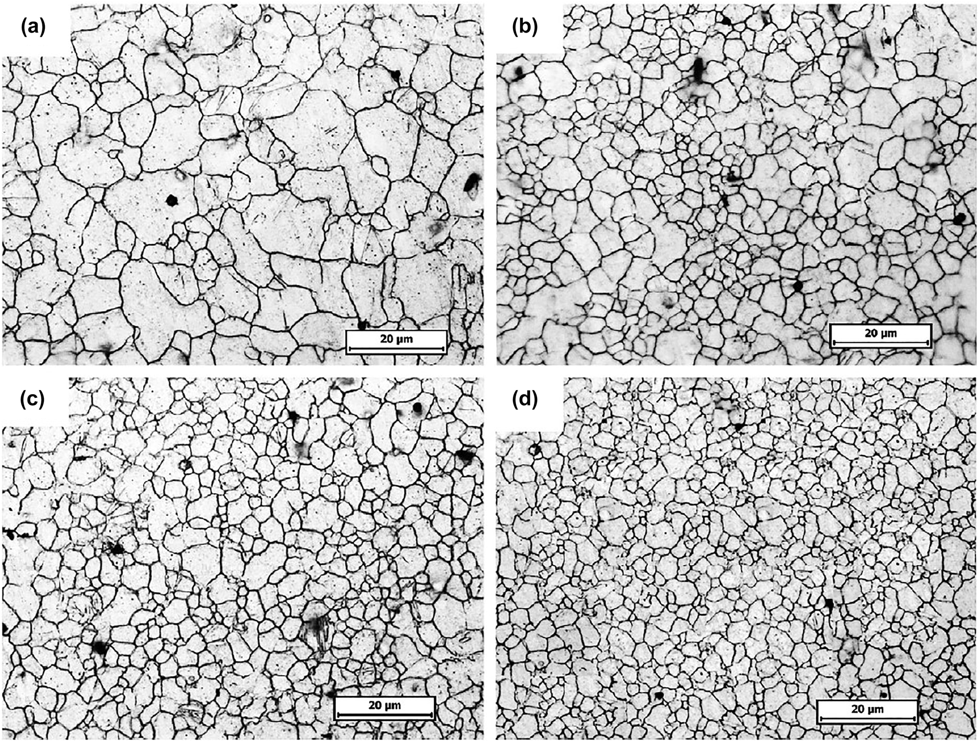
Surface microstructure of the AZ31B alloy (a) before LSP, after LSP with (b) single impact, (c) double impact, and (c) triple impact. Reprinted from (Zhang et al. 2010b) with permission from Elsevier.
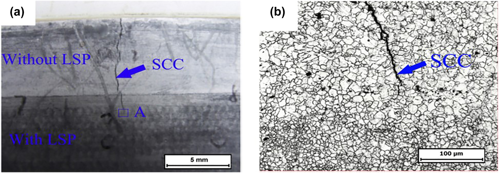
Comparison of SCC crack propagation in AZ31B Mg alloy (a) with and without LSP (visual observation), (b) microstructure of rectangular region A in Figure 12a. Reprinted from (Zhang et al. 2010b) with permission from Elsevier.
Equal channel angular pressing (ECAP) is one of the widely covered SPD processes which improves both the strength and ductility through the introduction of texture and grain refinement. Xu et al. (2013) reported the improvement in mechanical properties brought by ECAP in AZ31 alloy. With an increase in the number of passes up to 8, the grain refinement happened continuously which resulted in the increment of the microhardness value. The color-coded hardness maps of the samples pointed towards the homogenization of hardness throughout the sample with an increase in the number of passes. The micro-tensile testing done at ambient temperature and high temperature with a constant strain rate of 1 × 10⁻³ s−1 showed an improvement in ductility with an increase in the number of ECAP process. Apart from influencing mechanical properties, it also influences the corrosion response of the alloy due to the reduction in grain size. Birbilis et al. (2010) conducted a study to correlate the corrosion parameters with the variation of microstructure brought about by the ECAP process. With an increase in ECAP pass from 0 to 8, the average grain size of the pure Mg sample reduced to 2.6 µm from several hundred µm. The corrosion current value decreased from >11 × 10−6 to <6 × 10⁻⁶ A/cm2 in 0.1 M NaCl due to reduction in grain size brought about by the increase in the number of passes. The study hypothesized that the increase in the number density of high-angle grain boundaries enhances coherent oxide formation on the surface which acts as a passivating layer and retard corrosion kinetics. In a recent study reported by Peron et al. (2020), the effect of ECAP on the corrosion performance and SCC susceptibility of AZ31 alloy were investigated. After a single pass of ECAP, the corrosion current density in SBF decreases by three times in comparison to the as-received AZ31 sample which is due to faster formation of the oxide layer (passivation) as a result of grain refinement. The slow strain rate testing of the samples in air and SBF revealed that the susceptibility indices, IUTS &
3.2.2 Physical methods for surface modification
PVD is one of the prominent surface modification techniques to produce a thin layer of coating from a vapor phase. For this purpose, the substrate material is preheated and placed in a chamber containing vapors of the desired coating. Generally, in the case of Mg, the substrate temperature is maintained below 180 °C to avoid material instability, which alters the adhesiveness and corrosion behavior of the coatings (Poinern et al. 2012). Bakhsheshi-Rad et al. (2017) synthesized ZnO, ZnO/Ca3ZrSi2O9 coating on Mg–1Ca–1Zn as-cast Mg alloy by PVD. The effectiveness of the coatings is tested by immersing those in the SBF solution for 14 days. Figure 13(a–k) shows the effect of SBF on the surface morphology of all the samples. Energy dispersive X-ray spectroscopy (EDS) analysis of the red marked area was also presented to understand the surface chemistry. From this figure, it can be said that ZnO and ZnO/Ca3ZrSi2O9 coating showed a high amount of Ca and P, which indicates the formation of Ca-phosphate compounds found in bone. Especially, ZnO/Ca3ZrSi2O9 showed better bioactivity with the formation of HA all over the surface, which is the reason for obtaining high-intensity peaks of Ca-P in this case. The positive impact of ZnO/Ca3ZrSi2O9 coating on Mg ion release, pH, and corrosion rate was also remarkable, which can be seen from Figure 14(a), (b) and (c), respectively. The Mg ion release was decreased with an increase in soaking time for all the samples. The ion release was decreased from 423 to 350 ppm, 272 to 200 ppm, and 93 to 64 ppm for uncoated sample, ZnO coated sample, and ZnO/Ca3ZrSi2O9 coated sample, respectively, in 3–14 days of immersion in SBF. This result supports the obtained lower value of the corrosion rate for the ZnO/Ca3ZrSi2O9 coated sample, which can be seen in Figure 14(c). The pH value of the ZnO/Ca3ZrSi2O9 coated sample also remained lower at each point of time. As per Abela (2015), substrate surface preparation, optimization in chamber base pressure, and selection of proper vapor sources are the keys to producing high-quality PVD coatings.
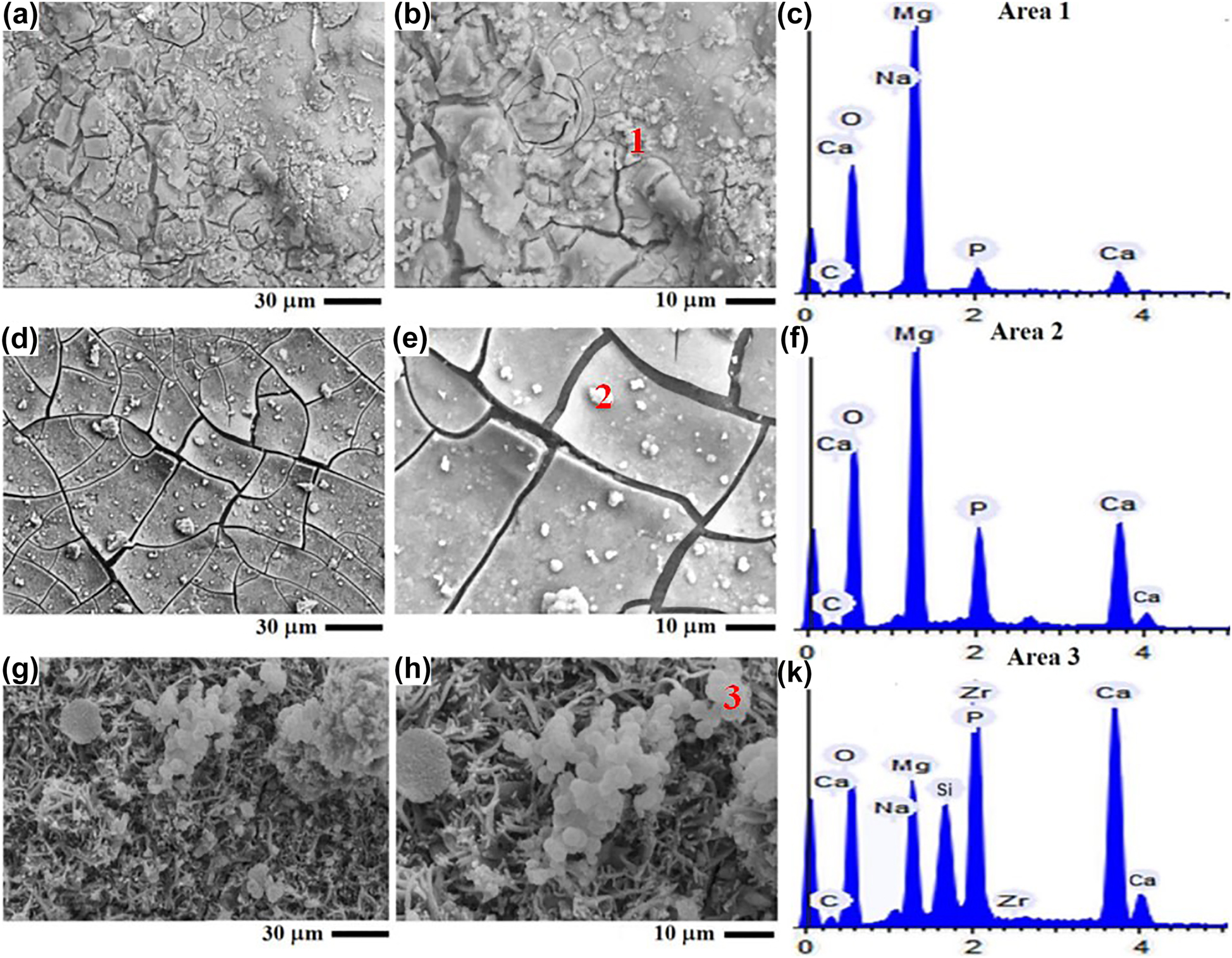
Surface micrographs of (a, b) uncoated sample, (d, e) ZnO coated sample, (g, h) ZnO/Ca3ZrSi2O9 coated sample, (c, f, k) EDS analysis of marked area after 14 days immersion in SBF. Reprinted from (Bakhsheshi-Rad et al. 2017) with permission from Elsevier.
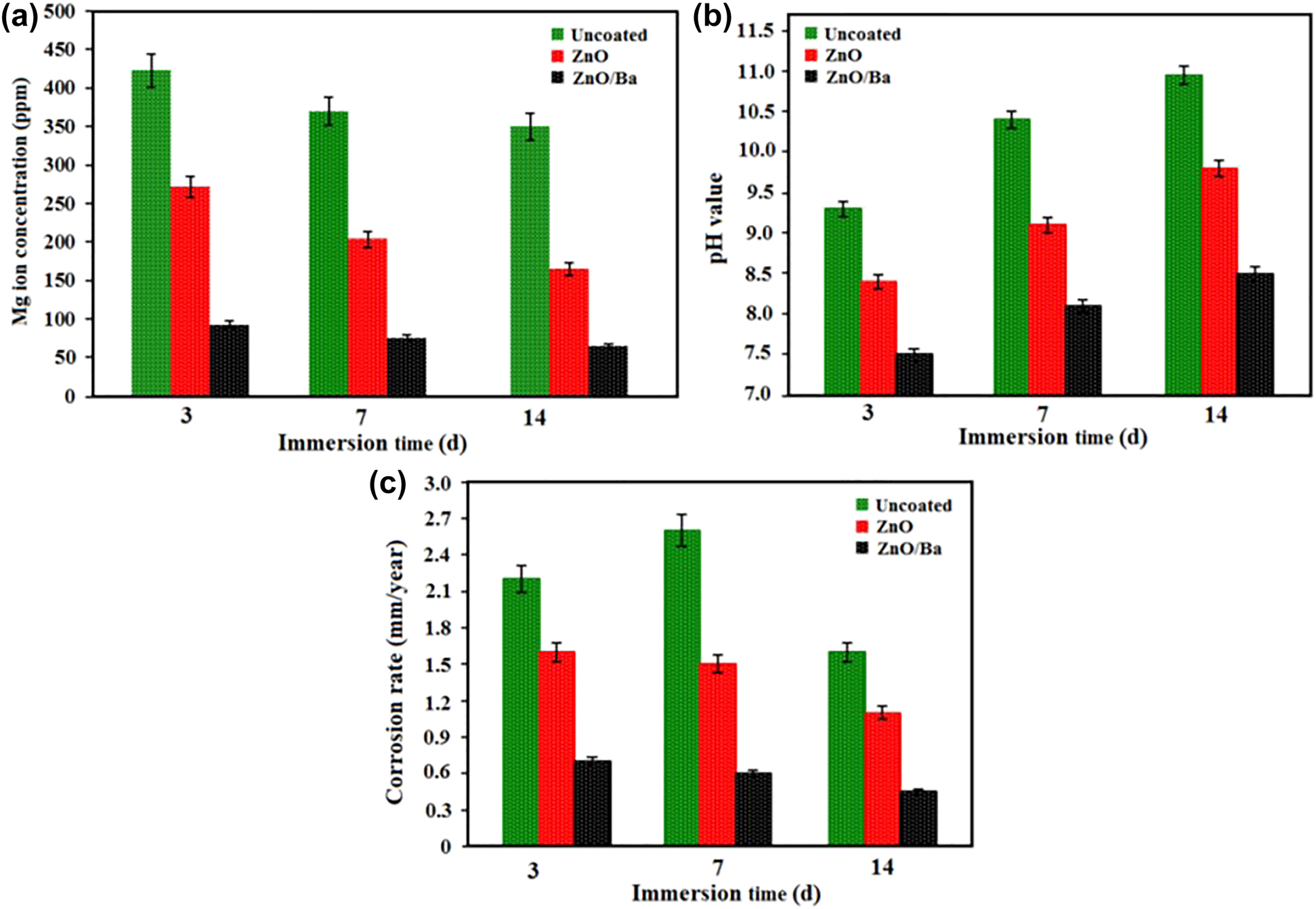
Variation of (a) Mg ion concentration, (b) pH value, (c) corrosion rate of uncoated sample, ZnO coated sample, and ZnO/Ca3ZrSi2O9 coated sample with immersion time in SBF. Reprinted from (Bakhsheshi-Rad et al. 2017) with permission from Elsevier.
Thermal spray coating is another important physical method for surface modification. The advantage of using thermal spray coating includes ease of thickness adjustment of the coating (Yang et al. 2011a), enhancing the substrate fatigue resistance (Dayani 2017), and biocorrosion resistance (Sun et al. 2001). Pardo et al. (2009) evaluated the surface morphology and corrosion performance of AZ31, AZ91D, and AZ80 following thermal spray (TS) Al coating. Additionally, the effect of cold-pressing (CP) post-treatment on TS samples is also evaluated. The microstructural investigation on Al-TS coating revealed the presence of interconnected porous structures. With CP post-treatment, the coatings became more homogenous due to reduced porosity and better interfacial bonding between coating and substrate surface. The potentiodynamic polarization curves for all samples, generated after 7 days of immersion in 3.5 wt% NaCl showed a relatively lower corrosion rate in the case of the AZ alloys which were provided the Al-TS-CP coating scheme. The corrosion current densities for Al-TS-CP coated samples were found to be 10−6 A/cm2 which is two orders lesser than the recorded corrosion density of Al-TS coated samples, i.e., 10−4 A/cm2. Bioactive coatings such as HA coatings can also be coated on Mg alloys by employing thermal spray methods (Sun et al. 2002). Similarly, hydrothermal coatings can also be used to develop corrosion resistant and antibacterial coatings over Mg alloys. Zou et al. (2019) prepared zinc loaded montmorillonite (MMT) coating and Fan et al. (2022) fabricated gentamicin-MMT coating over AZ31 alloy through hydrothermal method which improved the corrosion resistance, antibacterial activity and biocompatibility of base alloy.
3.2.3 Chemical methods for surface modification
3.2.3.1 Micro arc oxidation (MAO) coatings
MAO is widely used in industries to improve the surface properties of magnesium alloys. In this process, modification of the growing oxide film occurs when a potential higher than the dielectric breakdown potential is applied (Zhang 2010). Application of higher potential results in localized plasma reactions re-modifying the growing oxide film over the substrate. This method produces a relatively thick and dense oxide layer, which has grown both inwards and outwards of the substrate surface (Wan et al. 2016). As per Zhang and Zhang (2009), forming a coating via MAO is essentially a competition between the dissolution of base metal and new coating development. The AZ91 alloy showed a mass loss in the first 5 s of deposition, and then the mass of the sample was increased due to the formation of new coating. Additionally, with an increase in surface treatment time, the coating became thick and porous which can be seen from Figure 15(a–c). This type of observation is persistent with other studies related to MAO coating (Durdu and Usta 2012; Hwang et al. 2009; Kim et al. 2008; Wang et al. 2009b).
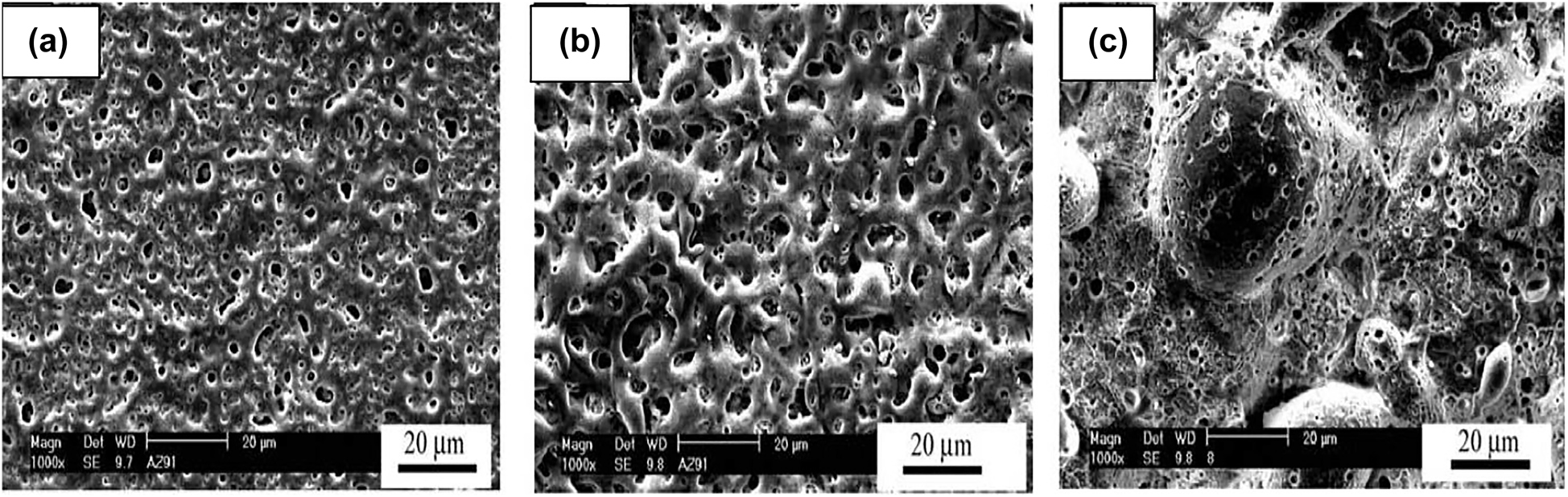
The morphology of MAO coating after (a) 1 min, (b) 5 min, and (c) 20 min. Reprinted from (Zhang and Zhang 2009) with permission from Elsevier.
As per Narayanan et al. (2014), the formation of micropores and cracks on the MAO coatings have both advantageous and harmful effects. The porous structure can release residual stress, improve mechanical interlocking effect, and uniform stress distribution. On the contrary, if the volume fraction of pore is high, more surface area will be available for the adsorption of harmful anions from the physiological environment. Due to this phenomenon, corrosion will proceed at a faster rate than usual and percolate to the underlying metal. Initially, the corrosion rate is less and the retention of mechanical properties is high (Ma et al. 2014; Ryu and Hong 2010). However, in the long run, the MAO coated alloys tend to dissolve faster than the uncoated alloys. One such result was published by Fischerauer et al. (2013) on micro-computer tomography (µ-CT) study in rats by using MAO coated ZX50. They reported a decrease in degradation rate from 1.7 mm/year to 0.25 mm/year with MAO surface treatment over ZX50 alloy in the initial phase. Additionally, the osteoconductive nature of the MAO coating is confirmed by this study. However, in due course of time, the degradation rate was found to increase ten folds, supporting the idea of rapid corrosion due to the porous morphology of MAO coating. The morphology of the MAO coating directly affects the corrosion performance of the implant, which can be tuned with varying electrolyte constituents & concentration (Chai et al. 2008), and input voltage (Zhang et al. 2008). Chai et al. (2008) evaluated the effect of oxysalt (sodium silicate, sodium aluminate, sodium phosphate, and sodium molybdate) addition in the electrolyte on the anticorrosion property of the coating. The coatings prepared from an electrolyte containing sodium silicate showed improved anticorrosion properties in comparison to other oxysalts. Chen et al. (2012) documented the effect of various electrochemical parameters such as frequency, current density, and duty cycle, etc., on coating performance. For this study, a dual electrolyte system (NaAlO2, Na3PO4) was used to carry out MAO on ZK60 alloy, and 3.5 wt% NaCl was used as the corrosion testing media. This study reported an increase in the number and size of pores with an increase in current density and a mild increase in corrosion rate with an increase in frequency. On the other hand, the corrosion rate remained constant up to a duty cycle of 40% beyond which it increases rapidly. In contrast, MAO coatings do not have a significant impact on mechanical properties, which was confirmed by Wu et al. (2007). From this study, one can conclude that selection of optimum electrochemical parameters and electrolyte constituents is the key to achieve better coating performance.
Besides controlling the above factors, providing a secondary layer of polymeric coating on the primary MAO coating is found to be beneficial in controlling the porosity of the surface (Bakhsheshi-Rad et al. 2016; Lu et al. 2011). Generally, polymeric coatings exhibit a swelling tendency in the aqueous environment while used as the main coating layer on an Mg alloy. This phenomenon results in a decrease in interfacial bond strength between the bone and the implant, which can lead to coating rupture (Narayanan et al. 2014). But, the incorporation of polymeric coating on MAO coating showed promising results by sealing the existing pores. One such study was reported by Zeng et al. (2014). In this study, the effect of introducing poly lactic acid (PLA) on to the MAO coating over Mg–1.2Li–1.12Ca–1.0Y alloy was discussed in detail. With the incorporation of PLA, the pore number reduced from 15,332 to 3489 over a fixed area. The average pore diameter also decreased from 8.61 to 2.26 µm. Due to the reduction in pore number and size, the corrosion current value also reduced from 6.31 × 10⁻⁶ A/cm2 to 1.70 × 10⁻⁶ A/cm2 in SBF corrosion media. The corrosion potential also shifted in a nobler direction, implying an improvement in anticorrosion behavior. Another noteworthy improvement is a steady hydrogen evolution rate over 30 h of immersion time in the case of MAO/PLA coating. In contrast, the hydrogen evolution rate for MAO coated sample increased up to five-fold just within 30 h of immersion time. Bakhsheshi-Rad et al. (2016) also reported a reduction in porosity and a decreased corrosion rate with MAO/PLA duplex coating on Mg–Ca alloy. Besides, the MAO/PLA duplex coating showed an improved hydrophobicity (contact angle 95.30°) in comparison to MAO coating alone (contact angle 22.90°) which may be the reason of its improved corrosion performance. Li et al. (2014a) conducted a similar study by dip coating the MAO treated Mg in polycaprolactone (PCL) which produces a MAO/PCL duplex coating. With increase in wt% of PCL solution from 0 to 7, the porosity level in the MAO coating reduced significantly and this brought about a decrease in the corrosion rate. The reduction in corrosion current density value was significant for MAO-7PCL coating, i.e., 0.81 × 10⁻⁶ A/cm2 for MAO-4PCL to 0.0045 × 10⁻⁶ A/cm2 for MAO-7PCL. The reduction of corrosion density value by two orders of magnitude can be attributed to the formation of thicker layer of PCL on the MAO coating using 7 wt% PCL solution for dip coating. This study also suggested that by tailoring the thickness of the polymer layer in a duplex coating, the lifetime of an Mg alloy bio implant can be enhanced.
In certain works, calcium phosphate nanoparticles were introduced in the MAO electrolyte to form bioactive coatings. The incorporation of any biocompatible nanoparticle significantly decreases the porosity level in the coating. Lin et al. (2014) used hydroxyapatite (HA) nanoparticles, which is present in our bone (Boushey et al. 2001), in phosphate electrolyte to form a bioactive coating over ZK60 alloy. Incorporation of 1 g/l HA in the electrolyte at a voltage of 420 V and an oxidation time of 5 min caused the sample to exhibit the lowest value of corrosion current density among all the samples. The corrosion current density was reduced by almost three orders of magnitude from 1.18 × 10⁻⁵A/cm2 for ZK60 to 8.5 × 10⁻⁸ A/cm2 for the bioactive coated ZK60. Additionally, cracks are evident after 30 days in MGO coating without HA in thirty days of immersion in Hank’s solution (Figure 16(a) and (c)). In contrast, no such cracking can be seen in the case of MAO coating containing HA particles (Figure 16(b) and (d)). The HA containing coating remained intact after 30 days of immersion and showed apatite formation over the coating. Hence, the incorporation of HA proved to be an efficient way to improve corrosion resistance, bioactivity, and coating integrity over time. The presence of protein in the physiological environment can also affect the performance of MAO coatings. Zheng et al. (2020) reported a study which outlined the synergistic effect of protein adsorption on MAO coating to restrict the corrosion kinetics. In this study, bovine serum albumin (BSA) was added to the corrosion media (PBS) to investigate the effect of protein on the degradation behavior of MAO coated alloy. Addition of BSA to PBS facilitated the formation of (RCH(NH2)COO)2Mg compounds which mitigated the dissolution of Mg(OH)2 thus reducing the corrosion rate. This study indicated that MAO coated Mg alloys may perform better in in-vivo conditions.
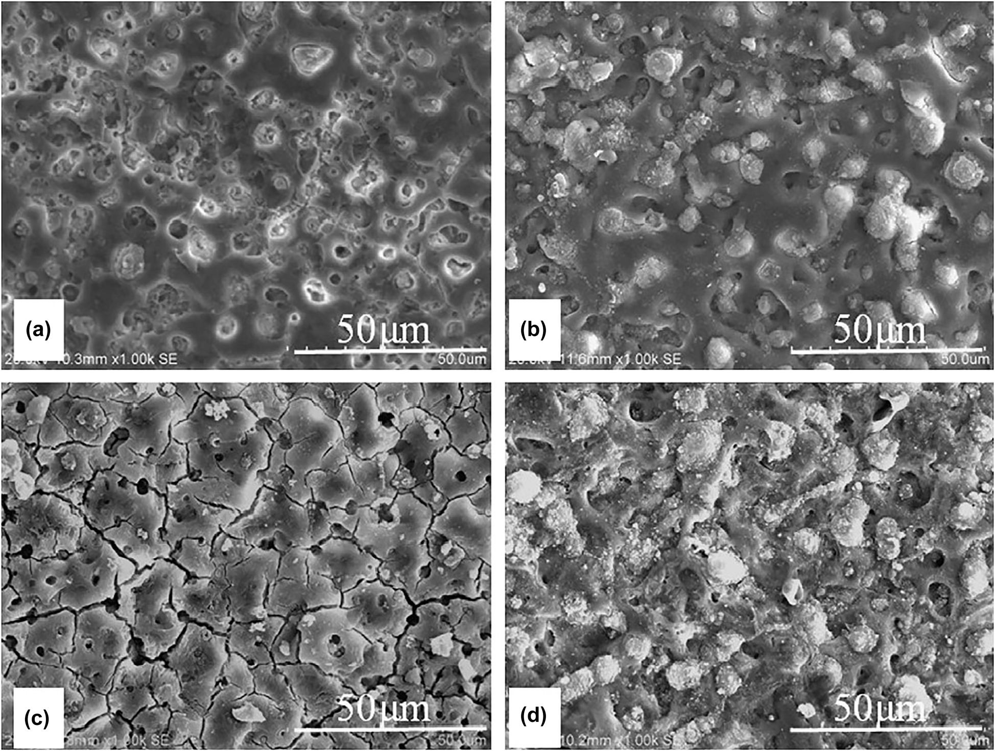
Surface morphology of (a) MAO coating, (b) MAO + HA coating before immersion and (c) MAO coating, (d) MAO + HA coating after 30 days of immersion in Hank’s solution. Reprinted from (Lin et al. 2014) with permission from Elsevier.
3.2.3.2 Chemical conversion coatings
These coatings are produced by chemical reactions of the substrate material in an aqueous solution resulting in the formation of the oxide layer and other complex compounds. The coating bath usually contains phosphate, carbonate, and fluoride, etc. (Hornberger et al. 2012). The benefits of using chemical conversion coating lie in its simplicity of operation and low cost. These coatings may be applied as a primary coating over the surface or as a secondary coating over MAO coated surface. Sometimes, chemical conversion coatings are used to form a base layer for electrophoretic coatings.
Phosphate conversion coatings have gained a lot of attention due to their bio-compatibility with bone. Additionally, it has been used as an alternative to chromate conversion coating due to its nontoxicity (Zhou et al. 2008). Jayaraj et al. (2019) prepared a lanthanum phosphate coating on the AZ31 alloy by employing a chemical conversion technique. This study concluded that the corrosion resistance of the coating would decrease with the presence of hydroxide compounds, which can lead to the formation of cracks in the coating. This type of observation and explanation is supported by Phuong and Moon (2014). Phuong and Moon (2014) compared the corrosion performance of zinc phosphate conversion coating (ZPCC) and magnesium phosphate coating (MPCC) on AZ31 alloy in 0.5 M NaCl solution. The corrosion performance of ZPCC was found to be poor in comparison to MPCC due to the presence of a high-volume fraction of pores on the coating. Ca-phosphate coating is one of the widely used phosphate coating owing to its excellent osteoconductivity, biocompatibility, and the ability to form biomimetic HA (Cui et al. 2013). Liu et al. (2014a) used the Ca-phosphate coating as a secondary surface treatment over an MAO coated pure Mg to improve the performance of the coating. The presence of HA and dicalcium phosphate dihydrate (DCPD) on the coating was confirmed from XRD analysis. However, with the increase in electrolyte temperature, the formation of DCPD was inhibited, resulting in enhanced HA formation. During the immersion test in Kokubo’s SBF, the sample showed the formation and growth of bone-like apatite with appreciable corrosion resistance. In a recent study done by Amaravathy and Kumar (2019), the advantageous effect of Sr doping on Zn–Ca–P (ZCP) conversion coating can be seen in terms of reduction in degradation rate and enhancement in bioactivity. AZ31 magnesium alloy was used as the substrate in this case. The strontium nitrate (source of Sr) was varied as 0, 0.5, 1, 1.5 wt% in the phosphate bath. As the doping level of Sr increased from 0 to 1.5 wt%, the hydrophobicity of the coating was increased from 89 ± 6° to 112 ± 8° which minimizes the interaction between the surface and the surrounding fluid. The in-vitro study in SBF showed a corrosion rate of 0.7 mg/cm2/h for 1.5 wt% Sr doped ZCP after 100 h of immersion which is much lesser than the corrosion rate of the uncoated sample (2.49 mg/cm2/h) after the same immersion time. With an increase in Sr content, the volume of evolved hydrogen and change in pH with immersion time was lowered. The Ca/P ratio for Sr doped ZCP coating after the in-vitro test was found to be 1.55 which is closer to that of HA. Sr doped ZCP also showed superior cell viability than the AZ31 and ZCP coated sample in cytotoxicity evaluation.
Similar to phosphate conversion coating, fluoride conversion coatings are also used to improve the functionality of coating (Drábiková et al. 2017; Fintová et al. 2019; Li et al. 2017; Pereda et al. 2011). Chiu et al. (2007) did a detailed microstructural and corrosion study on fluoride conversion coating on the Mg substrate. The corrosion resistance was increased by this coating, but the increase was minimal compared to phosphate coatings. On the other hand, Yan et al. (2014) reported a significant increase in polarization resistance from 500 Ω cm2 for the bare alloy to 429,640 Ω cm2 for fluoride coated alloy. However, the contrasting results from (Chiu et al. 2007) and (Yan et al. 2014) may be due to the use of different substrate material and corrosive medium. In the former study, the coating was performed on a pure Mg substrate, while in the latter study, it utilized an AZ31B alloy substrate. From Section 3.1, it was clear that the corrosion resistance of the AZ series alloy is higher than pure Mg. Another noticeable difference is that Chiu et al. (2007) used Hanks’s solution as the corrosive medium and potentiodynamic polarization test for the quantification of corrosion rate, while Yan et al. (2014) used simulated blood plasma as a test solution and electrochemical impedance spectroscopy (EIS) to quantify the corrosion resistance. Apart from the high polarization resistance of fluoride coated AZ31B alloy, the formation of the HA phase after 90 days of immersion in simulated blood plasma can be seen from Figure 17(1d), which indicates the bioactivity of the coating. The degradation of the surface with time can be seen from Figure 17. The traces of coating can be found up to 120 days, after which the substrate alloy started corroding. The positive impact of fluoride conversion coating on corrosion performance of other AZ alloys such as AZ61 is also confirmed by Fintová et al. (2019).
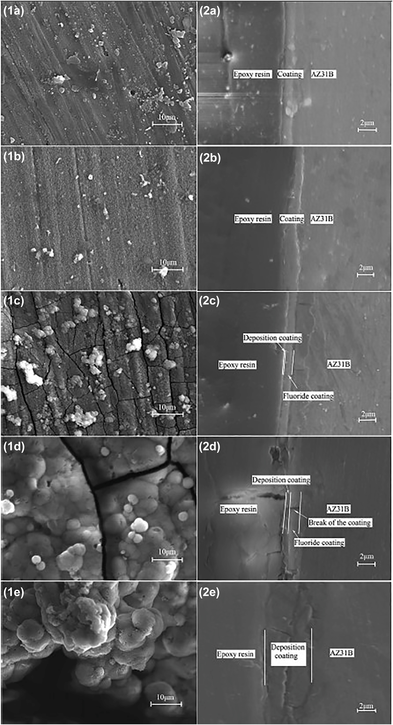
Surface morphologies (1) and cross-sectional morphologies (2) of fluoride conversion coating on AZ31 alloy after (a) 5 days, (b) 20 days, (c) 45 days, (d) 90 days, (e) 120 days of immersion in simulated blood plasma. Reprinted from (Yan et al. 2014) with permission from Elsevier.
3.2.3.3 Electrophoretic deposition (EPD)
EPD method is widely used to achieve porosity free uniform thickness coating. Complex shapes and contours can also be coated with ease by using this method. As per Van der Biest and Vandeperre (1999), EPD is a two-step deposition technique. The first steps involve the forced movement of the suspended colloidal particles in an aqueous medium towards an electrode under the application of an electric field. This process is known as electrophoresis. In the second step, the collected particles at the electrode form a coherent deposit. The advantage of using this method includes automation, throwing power adjustment, better adherence, and formation of a dense coating as compared to dip or spray coating (Pierce 1981). This deposition method is used to form nanocomposite functional coating on a bare substrate or MAO coated substrate. Various bioactive coating can be developed, such as bioactive glass (BG) (Rojaee et al. 2014a), nanostructured akermanite (Ca2MgSiO7) (Razavi et al. 2014a), HA (Kumar et al. 2016), chitosan, bredigite (Ca7Mg(SiO4)4) (Razavi et al. 2013) over Mg alloy to mitigate the corrosion rate. Rojaee et al. (2014b) compared the effectiveness of EPD coating over the sol-gel dip-coating method by preparing HA coating on the AZ91 alloy. Sol–gel coating is specifically used for producing thin inorganic coatings. In this method, the substrate is withdrawn from an aqueous solution followed by gravitational draining, solvent evaporation, and condensation reaction, which in turn results in the formation of a solid film (Brinker and Hurd 1994). By comparing these SEM images provided in (Rojaee et al. 2014b), it is quite clear that EPD coating is rougher in comparison to the sol–gel coating, which has proven to enhance the osteoblastic cell adhesion and growth (Seyfoori et al. 2013). Potentiodynamic polarization tests of the sample indicated that the value of corrosion current density for the EPD coated sample is 2.21 × 10⁻⁶ A/cm2, which is marginally lower than the value obtained for sol-gel coated sample, i.e., 2.83 × 10⁻⁶ A/cm2 in SBF solution. Similarly, Heise et al. (2017) deposited chitosan/BG45S5 composite coating on WE43 alloy by using the EPD technique. The coating showed excellent adhesion with the formation of bioactive hydroxycarbonateapatite after one day immersion in SBF which was confirmed by FTIR and EDX results. Surface pretreatment on substate alloy before the deposition of coating was found to be beneficial in terms of corrosion protection. Rojaee et al. (2014b) reported an animal in vivo study by using EPD of diopside (CaMgSi2O6) on MAO coated AZ91 alloy. From Figure 18(a) we can see that with an increase in culture time, the pH of the culture medium increases for all the samples. However, in comparison to bare AZ91 and MAO coated samples, diopside/MAO coated samples showed steady increase in pH value. Within 7 days, the pH value of medium increased from 8.8 to 9.5 for AZ91, 8.1 to 8.8 for MAO coated AZ91 alloy, and 7.9 to 8.3 diopside/MAO coating. A similar trend was also seen in the case of Mg ion concentration (Figure 18(b)). The Mg ion concentration was found to be least at each point of test time for diopside/MAO coating.
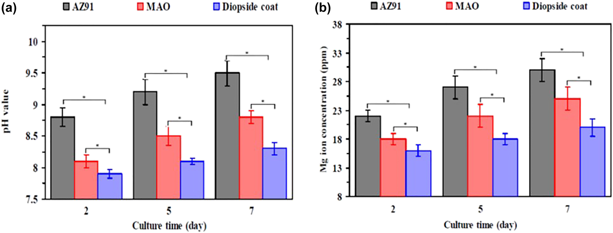
The variation of (a) pH value, and (b) Mg ion concentration (ppm) with culture time of 2, 5, 7 days in DMEM medium. Reprinted from (Razavi et al. 2014b) with permission from Elsevier.
4 Advancements in state of the art in Mg bio-implants to improve corrosion and stress corrosion cracking
4.1 Nanostructured Mg alloys
Nanostructuring is an effective means to enhance mechanical properties (Meyers et al. 2006) and corrosion resistance (Ralston and Birbilis 2010) while mitigating the property anisotropy and tension-compression asymmetry in magnesium (Fatemi et al. 2018). Basically, two approaches, namely the bottom-up and top-down approaches are employed to manufacture nanostructured Mg-based products. In the bottom-up approach, nano-powders of Mg are sintered into final products (Kumar and Pandey 2020) (Rai et al. 2020) whereas in the top-down approach severe plastic deformation (SPD) of the surface has been done to form nanostructured product (Kim and Kim 2004). The powder metallurgy method demands the production of nanocrystalline Mg powder which is itself challenging owing to the reactive nature of Mg. Then the powder needs to be sintered without causing any significant grain growth which is very difficult to achieve in practice. Due to this complexity of the bottom-up approach, the nanocrystalline Mg products are generally obtained by SPD processes (Estrin and Vinogradov 2010).
Recently, Parfenov et al. (2020) reported the effect of nanostructuring on the corrosion behavior of Mg–1Ca alloy. The as-cast coarse-grained (CG) Mg–1Ca alloy was subjected to room temperature high pressure torsion (HPT) treatment to produce nanostructured (nanocrystalline/NC) sample (grain size less than 100 nm). Then all the samples were tested in Ringer’s solution for 32 days at 37 ± 2 °C. The NC sample survived the 32 days of immersion test while the CG sample disintegrated just after four days of immersion. The SEM investigation revealed that the low corrosion resistance of the CG alloy attributed to the presence of coarser
4.2 Open porous Mg-based scaffolds
In general, bone in the body of a mammal is grouped into two categories, i.e., cortical bone and cancellous or trabecular bone. The cortical bone is denser than cancellous bone and comprises nearly 80% of the bone mass with a porosity level of 3–12% (Cooper et al. 2004). On the other hand, the cancellous bones have an open porous structure with a porosity range of 52–88% (Renders et al. 2007) which helps the passage of essential fluids through the bone. To mimic the porous property of bones, open porous scaffolds have been used to promote tissue ingrowth and provide better mechanical interlocking between the scaffold and surrounding tissue (Mour et al. 2010). Though a high corrosion rate remains an issue, other properties like enhanced osteoblast proliferation and differentiation make Mg-based materials a good choice for scaffold application (Yoshizawa et al. 2014). There are various conventional manufacturing techniques to produce open and interconnected pores such as the space holder method, melt infiltration in a preform, melt vacuum solidification foaming, etc. (Vahidgolpayegani et al. 2017). Still, very few studies reported in the literature on the in-vivo and in-vitro performance of open porous Mg-based scaffolds. One such study was reported by Witte et al. (2006a, 2007, where the open porous Mg alloy (AZ91) scaffold was produced using the space holder method. The scaffold was used as a subchondral bone replacement in rabbits to evaluate its effect on cartilage regeneration. The AZ91 scaffold degraded at a higher rate but showed enhanced osteoconductivity at the rim of the implant during the degradation process without negatively affecting the surrounding tissues. This study also addressed the need for any protective coatings like Ca-P or fluoride conversion coating to reduce the initial high rate of corrosion in the scaffold. A recent study done by Jia et al. (2018) investigated the relationship between the particles used in the space holder method (NaCl is used in their study), pore characteristics, and mechanical behavior of the scaffold. They have prepared two different templates utilizing spherical and irregular polyhedral NaCl particles, respectively, by hot press sintering process (Figure 19(a) and (b)). Subsequently, infiltration casting was used to produce the green compact of Mg and the NaCl template. Then NaCl was leached out to produce open porous Mg scaffolds. The scaffold corresponding to a template made out of spherical NaCl particle (S-scaffold) showed superior interconnectivity of pores and stable compressive deformability due to uniform spatial porous structure as comparison to template made out of irregular NaCl particle (I-scaffold) which consisted of irregular pore structure. The I-scaffold showed a higher value of surface area/object volume in comparison to S-scaffold which may alter the corrosion properties of the scaffolds. However, the investigation of corrosion properties is not included in this study.
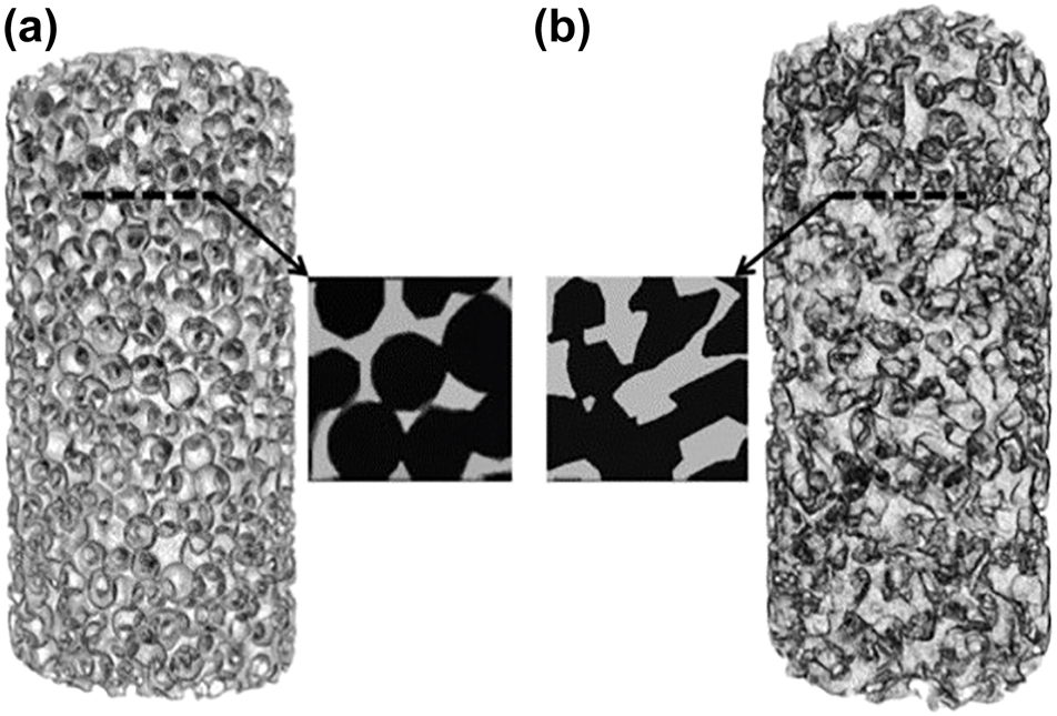
Morphologies of NaCl templates (a) made out of spherical particle and (b) made out of irregular polyhedral particle, respectively, reconstructed from micro-CT results. Reprinted from (Jia et al. 2018) with permission from Elsevier.
Another in-vivo and in-vitro study on open porous Mg-based scaffold prepared using Mg W4 (MgY4) short fibers through the sintering route was reported by Bobe et al. (2013). This study reported a corrosion rate of 0.16 mm/year after six weeks of in-vivo testing which is 24 times lesser than the corrosion rate determined from in-vitro testing (3.88 mm/year) in DMEM medium with 10% fetal calf serum (FCS) after 24 h. With the increase in in-vivo test duration up to twelve weeks, the corrosion rate was further reduced to 0.08 mm/year without any sign of inflammation or gas cavities at the implantation site. Additionally, new bone formation was observed after six weeks of implantation with the presence of both osteoblast and osteoclast. Liu et al. (2014b) also confirmed the synergetic properties of Mg scaffolds in terms of enhanced osteogenesis. Apart from bio-degradability and osteogenesis, the mechanical properties like elastic modulus and the compression strength of the open porous scaffold should match with that of the bone where it is being implanted. Towards this end, Seyedraoufi and Mirdamadi (2013) manufactured open porous Mg–Zn scaffolds using the powder metallurgy route. The porosity level and the mechanical properties of Mg–4 wt% Zn and Mg–6 wt% Zn scaffolds were compared against the pure Mg scaffolds. The Mg–Zn samples showed a porosity level of 19–36% with pore size in the range of 150–400 µm. Both the sintering temperature and the Zn content were found to affect the mechanical properties such as compressive strength and Young’s modulus. However, the effect of sintering temperature on mechanical properties did not follow any clear trend, but both the sample showed a reduction in compressive strength and Young’s modulus with the increase in the volume fraction of pores. The compressive strength and Young’s modulus values were found to be higher for Mg–6 wt% Zn scaffolds which were attributed to the mixed effect of grain refinement and dispersion strengthening brought by higher wt% of Zn. The compressive strength of Mg–Zn scaffold and porous magnesium scaffold were found to be 15–60 MPa and 15–31 MPa, respectively, which closer to the compressive strength of natural bone.
Better control over the porosity and interconnected pore network facilitates proper tuning of the corrosion and mechanical properties as per the requirement. But obtaining precise control over the level of porosity, pore size, distribution, and interconnectivity is difficult by using conventional methods. To overcome these challenges, advanced processes such as laser perforation, fiber deposition hot pressing, and solid freeform fabrication (SFF) techniques are used now. A study done by Geng et al. (2009) reported the application of laser perforation technique to produce a honeycomb-shaped open porous Mg scaffold in which the pore diameter was 0.5 mm. This technique is advantageous in terms of the production of holes with accurate positioning and high repeatability without leaving any working residue on the sample. Geng et al. (2009) also incorporated β-tricalcium phosphate (β-TCP) coatings on the porous scaffold to enhance its bioactivity and to retard the corrosion kinetics. This study optimized the porosity level (42–50%) in the porous scaffold for which the compressive strength and the elastic modulus value match with the compressive strength and the elastic modulus of cancellous bone. The in-vitro biodegradation test in MEM revealed that the pH variation in the β-TCP coated Mg scaffold was from 7.58 to 7.72 over a period of 12 days which is always lesser than that of the pH change in the uncoated porous Mg scaffold (8.11–8.12) for the same testing period. This observation supports the idea of reduction in corrosion kinetics by retarding the Mg2+ ion release due to the presence of β-TCP precipitation layer. Cytocompatibility tests on the β-TCP coated Mg scaffold involving UMR106 (the human osteosarcoma cells), cultured in MEM medium at 37 °C, 5% CO2 showed improved cell adhesion and proliferation rate in comparison to the pure Mg counterpart. Fiber deposition hot pressing (FDHP) is another effective technology to achieve a better control over the pore size and pore distribution. This technique was implemented by Zhang et al. (2014) to produce 3D porous Mg scaffold with an interconnected network structure. The scaffold was made by lining up the two different Mg fibers layer by layer with a layer spacing of 0.5 mm and alternative fiber layers were perpendicular to each other. In this manner, scaffolds were prepared with varying porosity levels in the range of 33–54%. The pore diameter in the axial direction and the lateral direction were found to be 270–300 µm and 110–175 µm, respectively. The compression testing in both the axial and lateral direction of the scaffold revealed that the compressive strength and Young’s modulus decreases with increase in porosity in both the direction. The compressive strength and Young’s modulus of the scaffolds varied between 11.1 and 30.3 MPa and 0.09–0.39 GPa which are in the range of cancellous bone mechanical properties. This study showed a higher value of compressive strength and a lower value of Young’s modulus for the porous Mg scaffold as a comparison to the porous Mg scaffold produced by Geng et al. (2009). Recently, Wang et al. (2020) published an article that combined the open porous scaffold technology with coating technology. An open porous Mg–Nd–Zn–Zr alloy (JDBM) scaffold was prepared using a previously patented technique (Yuan and Jia 2018). Then a brushite (CaH2PO4.2H2O) coating, termed as DCPD, was applied on the scaffold using the chemical deposition method. The JDBM-DCPD scaffolds performed better in terms of bone regeneration, growth and integration in comparison with JDBM with MgF2 coating which can be seen from Figure 20(a–d). Figure 20(a) and (b) depict the Van Gieson staining of undecalcified sections and quantitative analysis of newly formed bone, respectively. The red portions in Figure 20(a) correspond to the bone tissues and black correspond to residual scaffold material. From both Figure 20(a) and (b), it is clear that the new bone formation and growth are more in the case of JDBM-DCPD scaffold. Similarly, HE and Masson staining presented in Figure 20(c) and (d), respectively, are showing newly mineralized bone tissue (NB) in the samples. It is clear that the JDBM-DCPD sample depicted enhanced NB & vascular formation (black arrow in Figure 20(c)) and presence of more osteoid (black arrow in Figure 20(d)) in comparison with other samples. The enhanced bone repair capacity of the JDBM-DCPD sample was ascribed to an appreciable reduction in the release of Mg2+ ion which provides sufficient time for mesenchymal stem cell adhesion, proliferation and differentiation. Although this study evaluated the mechanical properties such as yield strength and elastic modulus of the DCPD coated scaffolds, fatigue strength determination under corrosive environment is missing which plays a significant role in in-vivo applications.
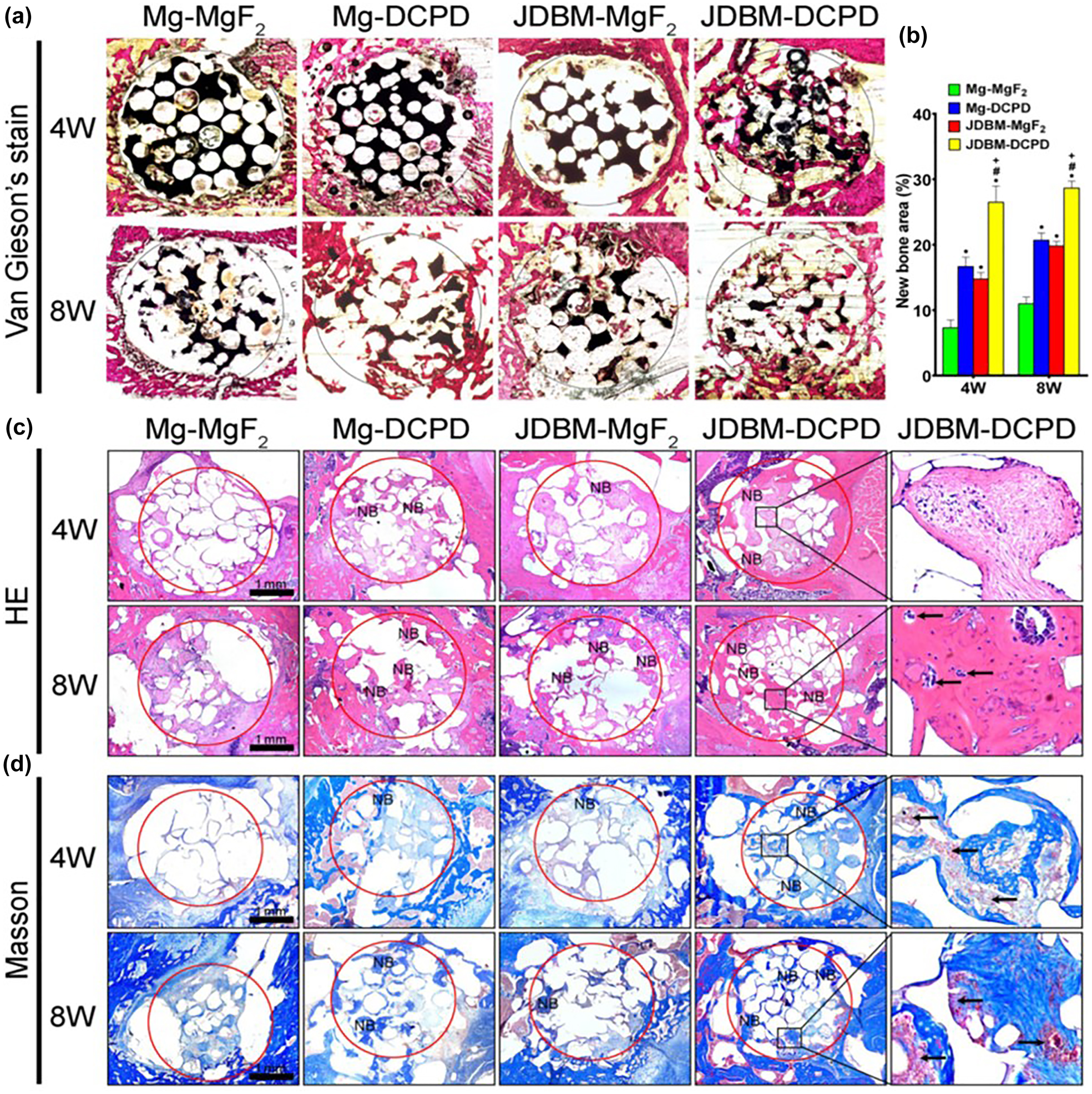
Comparison of bone regeneration, growth and integration in different scaffold-coating cases.
(a) Van Gieson staining of undecalcified sections (red portion – bone tissue, black portion – residual scaffold material), (b) quantitative analysis of newly formed bone, (c) HE staining (black arrow – vascular formation), (d) Masson staining (black arrow – osteoid) showing newly mineralized bone tissue (NB). (*, # and + represent P < 0.05 or statically significant when compared with Mg-MgF2, Mg-DCPD and JDBM-MgF2, respectively). Reprinted from (Wang et al. 2020) with permission from Elsevier.
Solid freeform fabrication (SFF), which is also known as rapid prototyping/additive manufacturing, is gaining importance in the production of open porous Mg-based scaffolds in recent time. It involves the layer-by-layer construction of complex 3D scaffolds with the help of computer aided design (CAD) files. This technique is a bottom-up approach and does not require any part-specific tooling or manual control. As working with Mg powders is not safe because of its reactive nature, an indirect SFF method has been introduced by Staiger et al. (2010) and Nguyen et al. (2011). In their work, a topologically ordered open cell porous Mg (TOPM) scaffold was prepared incorporating both 3D printing and low pressure assisted gravity casting. Their study pointed out that decreasing the pore size in the CAD model increases the percentage error in porosity in the end product. It is due to the difficulty associated with infiltrating the NaCl paste into the polymer template produced by 3D printing. Despite the better control over the porosity and interconnected network structure, the in-vitro and in-vivo testing of this TOPM for bone interfacial application is still remaining. Selective laser melting (SLM) is a rapid prototyping process in which a high-power density laser is used to selectively melt and fuse metallic powders layer by layer to fabricate near net shape process (Aboulkhair et al. 2019). Li et al. (2018b) used the SLM technique to fabricate a topologically ordered Mg-REE (WE43) scaffold based on a diamond unit cell. This study is quite important as WE43 Mg alloy is known to be better than pure Mg in terms of corrosion resistance. This study evaluated the hydrogen evolution rate of WE43 scaffold in revised SBF with 5% fetal bovine serum (FBS), degradation of mechanical properties through compression testing, and cytotoxicity in MG-63 (human osteoblast-like cell line) cultured in Dulbecco’s modified eagle medium (DMEM) with 10% FBS at 37 °C, 95% humidity and 5% CO2. The hydrogen evolution rate was found to be 0.17 ml/cm2 day after four weeks which is lower than the degradation rate of as-cast WE43 alloy (0.3 ml/cm2 day) (Hänzi et al. 2009). The compression testing showed an increase in Young’s modulus value from 690 to 1220 MPa in the immersion period of 7 days due to an increase in the amount of degradation product which act as ceramic particles in the scaffold. However, after seven days, the value was reduced near to its initial value due to structural change in the scaffold and no noticeable change was observed from 28 days immersion period. On the other hand, the yield strength of the scaffold was reduced gradually from 24 to 14 MPa over a 28 days immersion period. The cytotoxicity evaluation done up to 72 h reported level 0 toxicity (as per ISO 10993-5 and -12) except for 24 h data. This study also addressed the need for bioactive coating for enhanced cell adhesion and proliferation. A recent study by Li et al. (2019) examined the fatigue behavior of WE43 scaffold as the extension of their previous study (Li et al. 2018b). The fatigue tests were performed in both air and in r-SBF at varying stress levels from 0.2
As per our knowledge, the assessment of different alloy-surface modification combinations in the area of additively manufactured Mg-based open porous scaffold is very scarce in the literature to date. In addition, no study included the biodegradation behavior of these scaffolds under the applications of cyclic load (except (Li et al. 2019)) which is the major research gap in this area.
4.3 Use of bulk metallic glass (BMG) as an implant material
During the last two decades, BMGs have been explored as a potential biomaterial owing to their multiple favorable properties such as high corrosion resistance, wear resistance, and mechanical strength (Schroers et al. 2009; Telford 2004). The enhancement in above-mentioned properties essentially comes from its amorphous structure (absence of long-range order). Specifically, Mg-based BMGs gained attention in the scientific community. It possesses biocompatibility and higher corrosion resistance compared to the polycrystalline Mg alloys (Yuan and Jia 2018), which may address the issue of rapid degradation of Mg alloys in the physiological environment. There have been numerous studies involving the in-vivo and in-vitro performance of various Mg-based BMGs in the last decade. Among them, the Mg–Zn–Ca ternary system is one of the widely explored BMG. Ramya et al. (2015) reported a comparative study between two partially amorphous (BMG) and fully crystalline Mg–Zn–Ca systems having the same chemical compositions. Their study revealed that with the increase in Zn content in both amorphous and crystalline Mg–Zn–Ca system, the corrosion rate was reduced in SBF testing medium. Additionally, the amorphous systems showed a significant decrease in corrosion rate in comparison to the crystalline systems having the same chemical composition. Similarly, Gu et al. (2010a) compared the corrosion behavior and cellular response of two Mg–Zn–Ca BMGs (Mg66Zn30Ca4 & Mg70Zn25Ca5). The corrosion current density for Mg66Zn30Ca4 was found to be 3.53 µA/cm2 in SBF medium which is lesser than the corrosion current density value of Mg70Zn25Ca5 and as rolled pure Mg (11.2 µA/cm2 and 36.8 µA/cm2, respectively) due to the presence of Zn at a higher amount. Also, the cytocompatibility tests confirmed the enhanced cell proliferation and cell adherence to the BMG surface as comparison to the as-rolled pure Mg surface. Chen et al. (2017) further modified the surface of Mg–Zn–Ca BMG (Mg65.2Zn28.8Ca6) by employing a porous Si-containing coating over it through micro arc oxidation (MAO). A significant two order decrease in corrosion current density was observed after the incorporation of MAO coating over the Mg65.2Zn28.8Ca6 BMG tested in SBF medium. The corrosion current density value for uncoated BMG was found to be 7.5 × 10⁻⁶ A/cm2 whereas the MAO coated BMG showed a corrosion current density value of 7.23 × 10⁻⁸ A/cm2. The corrosion potential also shifted to nobler value, i.e., from −1.345 V versus SCE for uncoated BMG to −1.244 V versus SCE for MAO coated BMG. Also, hydroxyapatite formation over the MAO coated surface was observed after seven days of immersion in SBF which ensured its bioactivity. Other elements such as Sr, Yb, Y, Cu and Pd are also added to form different BMGs which are reported in Table 4. Although the Mg-based BMGs exhibit superior corrosion resistance than their crystalline counterpart, low ductility limits their use due to the possibility of catastrophic brittle failure. To address this issue, Zhang et al. (2012) prepared an in situ formed Mg75Cu13.33Y6.67Zn5 BMG composite which showed superior mechanical properties along with good biocorrosion resistance. The compressive strength of Mg75Cu13.33Y6.67Zn5 composite and Mg75Cu13.33Y6.67Zn5 BMG were found to be 1040 and 840 MPa, respectively. A significant increase in plastic strain from 3 to 19% was observed when the Mg75Cu13.33Y6.67Zn5 BMG was replaced by Mg75Cu13.33Y6.67Zn5 composite. The corrosion current density of both the BMG and composite in Hank’s solution was found to be within the same order. Similarly, Wong et al. (2017) investigated the corrosion behavior and mechanical strength of Mg60Zn35Ca5 BMG, Mg67Zn28Ca5 BMG, and Mg60Zn35Ca5 BMG composite with Ti particle, and Mg67Zn28Ca5 with Ti particle in SBF. Cytotoxicity test was conducted in MG63 human osteosarcoma cell line cultured in DMEM with 10% FBS at 37 °C, 5% CO2 atmosphere. After 12 weeks of immersion in SBF, the degradation rate of Mg60Zn35Ca5 BMG and Mg60Zn35Ca5 – Ti BMG composite was found to be 0.67 mm/year and 0.26 mm/year. On the other hand, Mg67Zn28Ca5 BMG and Mg67Zn28Ca5-Ti BMG composite were completely degraded to debris in 11 and 4 weeks, respectively. The Mg67Zn28Ca5 system performed unsatisfactorily in terms of biodegradability due to more amount of Mg and poor adherence of Ti particle to the BMG matrix. The compressive strength of Mg60Zn35Ca5 BMGC was decreased from 807 to 154 MPa after 12 weeks of immersion in SBF. In addition, all the samples were found to be slightly toxic as per ISO 10993-5 standard. The failure mode of the BMGC was reported to be of ductile nature due to the presence of ductile metallic particles in the BMG matrix. It is mentioned in (Wong et al. 2017) that the structure of samples changed from amorphous to crystalline after immersion in SBF within four weeks, which implies low crystallization resistance of the sample. Hence, a thorough study on the crystallization resistance and stability of these BMGC are needed to be carried out.
In-vitro corrosion behavior of recently developed Mg–Zn–Ca based BMGs.
| BMGs | Testing medium | Corrosion current density (A/cm2) | References |
|---|---|---|---|
| Mg20Ca20Zn20Sr20Yb20 | Hank’s solution | 9.16 × 10⁻⁶ | Li et al. (2013) |
| Mg70Zn23Ca5Pd2 | Hank’s solution | 2.1 × 10⁻³ | González et al. (2012) |
| Mg66Zn23Ca5Pd6 | 2.7 × 10⁻³ | ||
| Mg66Zn30Ca3Sr1 | Phosphate-buffered saline (PBS) | 6.5 × 10⁻⁵ | Li et al. (2015) |
| Mg75Cu13.33Y6.67Zn5 | Hank’s solution | 1.7 × 10⁻⁵ | Zhang et al. (2012) |
| Mg75Cu13.33Y6.67Zn5 (BMG composite) | Hank’s solution | 9.1 × 10⁻⁵ | Zhang et al. (2012) |
Apart from the corrosion and compression behavior evaluation, Mg–Zn–Ca BMG was also assessed in terms of cyclic loading performance by Wang et al. (2009a). As per their study, the fatigue limit for Ca65Mg15Zn20 BMG was found to be 140 MPa at 106 cycles which is lesser than the Zr-based BMGs. The fatigue data obtained was highly variable and scattered which indicates the need for further studies on this area. Li et al. (2016) evaluated the CF behavior of Mg66Zn30Ca3Sr1 BMG. The compression–compression fatigue test was conducted both in air and phosphate-buffered saline (PBS) solution. The endurance limit for the Mg66Zn30Ca3Sr1 BMG in air and PBS was recorded to be 370 and 150 MPa, respectively. The significant decrease in the endurance limit of the BMG in the PBS medium implies the severity of the physiological environment on the fatigue property of BMGs. The corrosion pits acted as initiation sites for peeling-off fracture that escalated with time and the BMG finally failed through fragmented fracture which can be seen from Figure 21(a–d). The reported fatigue strength of Mg66Zn30Ca3Sr1 BMG both in air and PBS were found to be significantly higher than the fatigue strength of extruded AZ61 (Němcová et al. 2014), AZ80-T5b (Bhuiyan et al. 2010), AZ31 (Nan et al. 2008), WE43, and AZ91D (Gu et al. 2010b). The fatigue limit of all the above-mentioned extruded samples in air, 3.5 wt% NaCl, 5 wt% NaCl environment, 3 wt% NaCl, and SBF was scattered between 20 and 80 MPa. It is important to note that the fatigue testing mentioned in (Bhuiyan et al. 2010; Nan et al. 2008; Němcová et al. 2014) was done in 3.5 wt% NaCl medium, whereas Gu et al. (2010b) was done in SBF. Hence, a systematic evaluation of the fatigue property of Mg66Zn30Ca3Sr1 BMG in 3.5 wt% NaCl and SBF are necessary to compare the results obtained from Li et al. (2016). Additionally, the fatigue property of the crystalline counterpart of Mg66Zn30Ca3Sr1 BMG needs to be evaluated to quantify the change in fatigue strength by using BMG. Recently, Mg-based BMG systems (Mg–Ca–Au and Mg–Ca–Au–Yb) were reported by Baulin et al. (2018) which showed appreciable resistance to crystallization and were found to be thermally stable. The Young’s modulus of the proposed system and pure crystalline Mg were found to be 55 ± 5 and 45 GPa which reduces the risk of osteolysis in patients. However, the biodegradation behavior, cytotoxicity test, and fatigue evaluation of this newly developed Mg-based BMG are still in progress and not reported in the literature.
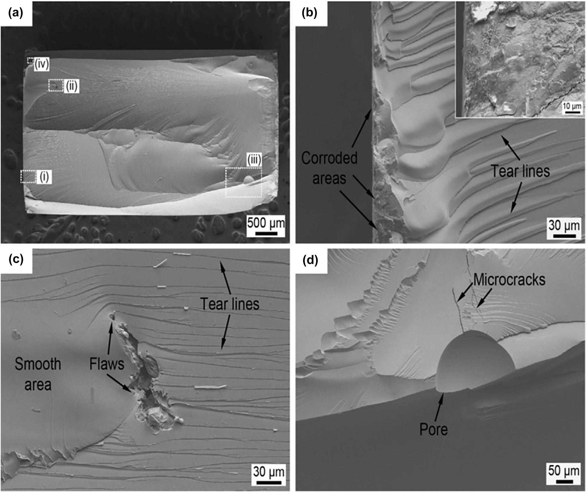
SEM micrographs showing fatigue fracture morphologies of the Mg66Zn30Ca3Sr1 BMG tested in PBS (maximum stress = 250 MPa): (a) overall view of the failed specimen, (b–d) and the inset of Figure 21b are the magnified images for the regions (i), (ii), (iii) and (iv) in Figure 21a, respectively. Reprinted from (Li et al. 2016) with permission from Elsevier.
5 Clinical outcomes of Mg-based implant
The clinical use of Mg-based implants was started at the end of the 18th century with the use of Mg in ligatures for suturing wounds (Witte 2015). Gradually, the clinical trials on pure Mg and Mg alloy implants have been documented up to 1948 which can be seen from Figure 22(a). However, clinical trials in this field paused for half a decade after 1950 due to some difficulties such as the risk of fatality, very high in-vivo degradation rate, gas cavity formation, and insufficient medical instrumentation to monitor the in-vivo degradation process. The development in medical imaging techniques such as computer tomography (CT) and micro-CT (µ-CT) enabled medical professionals to observe the degradation process of temporary implants along with the restoration of the defects in anatomy noninvasively. Hence, the clinical trials again escalated from 2005 as seen from Figure 22(b). Despite the rapid technological development in the medical sector, the risk of internal organ toxicity, implant failure, and the possibility of fatality still exist which limits the use of new Mg-based implants for human clinical trials.
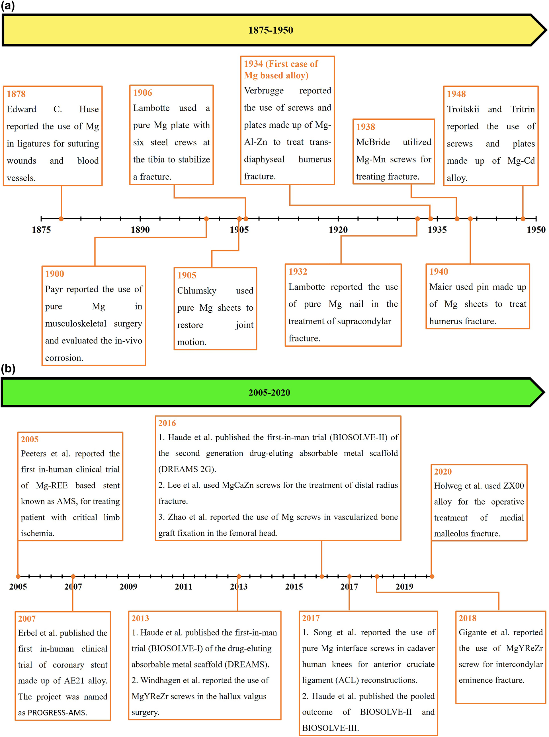
Timeline of human clinical trials using Mg-based implants.
(a) All the studies in between 1875 and 1950 were compiled from Witte (2015). (b) The case studies between 2005 and 2020 correspond to Erbel et al. (2007); Gigante et al. (2018); Haude et al. (2013, 2016b, 2017); Holweg et al. (2020a); Lee et al. (2016); Peeters et al. (2005); Song et al. (2017); Windhagen et al. (2013); Zhao et al. (2016). No clinical trials in between 1950 and 2005.
Figure 23 depicts the Mg implants which have already been used in a variety of surgeries in human beings. Mg-based coronary stents and compression screws are two widely used implants for coronary angioplasty surgery and hallux valgus corrective surgery, respectively. The first-in-human clinical trial of Mg-based absorbable metal stent (AMS) was reported by Peeters et al. (2005) and the trial was further expanded by Bosiers (2009). In this study (Peeters et al. 2005), twenty patients with symptomatic critical limb ischemia (CLI) received one or two AMS for suboptimal angioplasty. The three months follow up showed promising performance in terms of primary clinical patency and limb salvage rates in the treatment of below knee lesions in CLI patients. This study led to the use of Mg alloy stent in coronary angioplasty to remove the blockage in the coronary arteries. Erbel et al. (2007) and Waksman et al. (2009) reported the first nonrandomized multicenter human trial (PROGRESS-AMS) of bioabsorbable Mg stents on 63 patients. Although the AMS in the human coronary did not show any early or late adversative findings, the stent suffered from the problem of very rapid degradation (completely degraded within 4 months). This study reported the need for drug eluting stents (DES) with some protective coating to minimize both the neointima formation and degradation rate. Haude et al. (2013, 2016) reported the in-human trial of drug eluting AMS (DREAMS) which was basically Poly-lactide-co-glycolide (PLGA) coated Mg alloy stent loaded with paclitaxel drug. This trial is widely known as BIOSOLVE-I. The degradation rate of the stent used in BIOSOLVE-I was reduced significantly in comparison to AMS used in (Erbel et al. 2007). This trial showed excellent long-term outcomes with no scaffold thrombosis and a low rate of target lesion failure (TLF). Subsequently, Haude et al. (2016) conducted BIOSOLVE-II in-human trial with a second-generation DREAMS, namely DREAMS 2G Magmaris. It is a polylactic-l-acid (PLLA) coated Mg alloy stent loaded with sirolimus drug. It showed excellent corrosion and clinical performance in comparison to BIOSOLVE-I trial. The mean scaffold area loss after six months in BIOSOLVE-II trial was found to be 0.5% which was much lesser than the mean scaffold area loss in BIOSOLVE-I trial, i.e., 11.1%. Similarly, a lesser decrease in mean lumen area was recorded in the case of BIOSOLVE-II trial. After six months, the decrease in mean lumen area in the case of BIOSOLVE-II and BIOSOLVE-I trial was found to be 15.3 and 2.4%, respectively. Hence, the degradation and clinical performance of DREAMS 2G Magmaris is more balanced than the DREAMS. Furthermore, results from BIOSOLVE-III (pivotal trial) and short-term BIOSOLVE-IV were also reported in Haude et al. (2017, 2020 and Galli et al. (2019), respectively, which showed less target lesion failure. Other coatings over Mg alloy stents such as polydopamine (PDA), poly (ester urethane) urea (PEUU) etc. have been discussed in details in the review reported by Zhang et al. (2021).
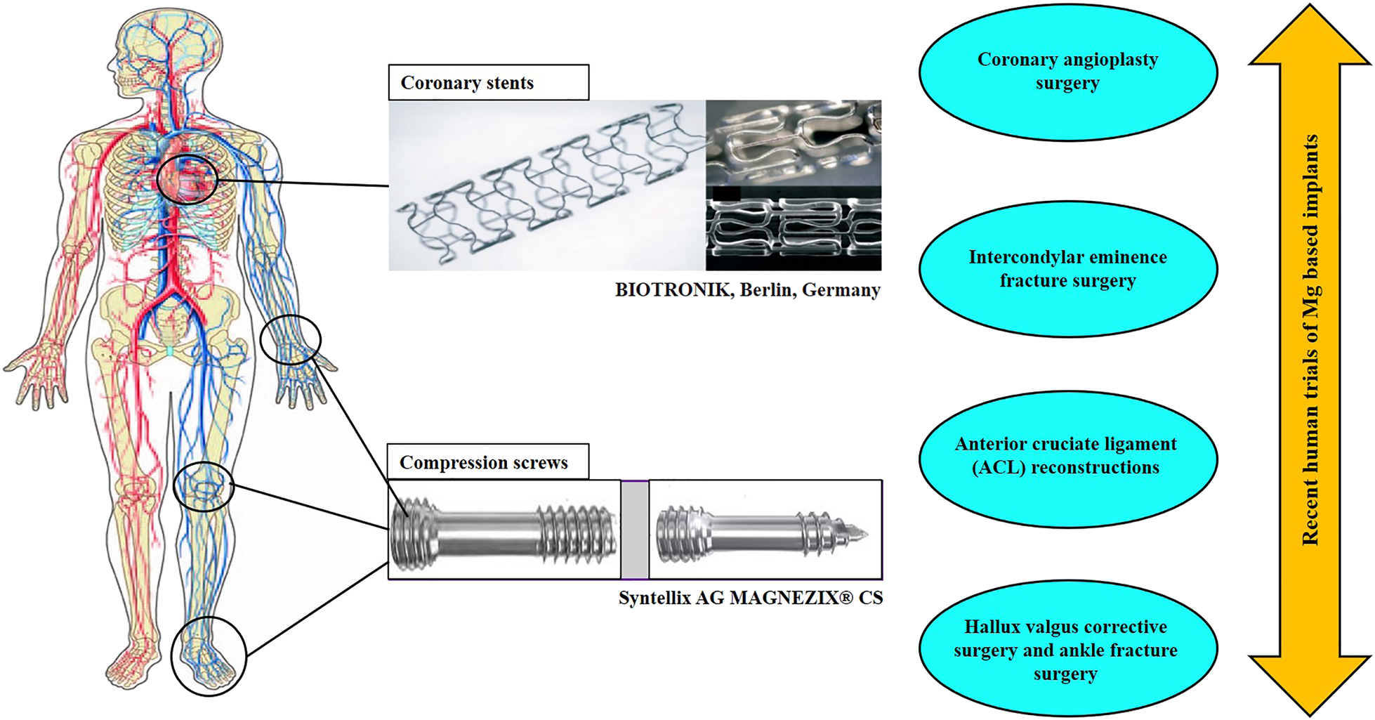
Representative of the Mg-based implants which have been used in human clinical trials recently.
The use of MgYREZr (MAGNEZIX) compression screws is found to be pertinent in hallux valgus corrective surgery (Atkinson et al. 2019; Choo et al. 2019; Klauser 2019) and Intercondylar eminence fracture surgery (Gigante et al. 2018). For mild hallux valgus, MAGNEZIX screws showed comparable clinical results with the conventional Ti screws. Additionally, the screw uniformly degraded in one-year post-operation without causing any adverse effect on wound healing. Similarly, ZX00 screws were used to treat an ankle fracture (Holweg et al. 2020a), which showed nearly 50% volume loss in 52 weeks (1 year) postoperatively. This study also showed complete consolidation of fracture within 12 weeks of the surgery without any complications. The lesser degradation rate in the case of ZX00 screws can be attributed to the presence of REEs in the Mg alloy as discussed in Section 3.1.2. Another Mg alloy screw (Mg–5wt%Ca–1wt%Zn) showed promising results in the treatment of distal radius fracture surgery reported by Lee et al. (2016). This study demonstrated successful long term (1 year) clinical study of the Mg–5wt%Ca–1wt%Zn alloy in 53 patients. The degradation behavior of the screw and the simultaneous bone healing process can be seen from Figure 24(a–c). Within one-year follow-up period, the Mg alloy screw completely degraded with the formation of biomimicking calcification matrix (CCP) at the corroding interface. At the end of one year, the newly formed bone substituted the degrading screw resulting in the complete healing of the fracture.
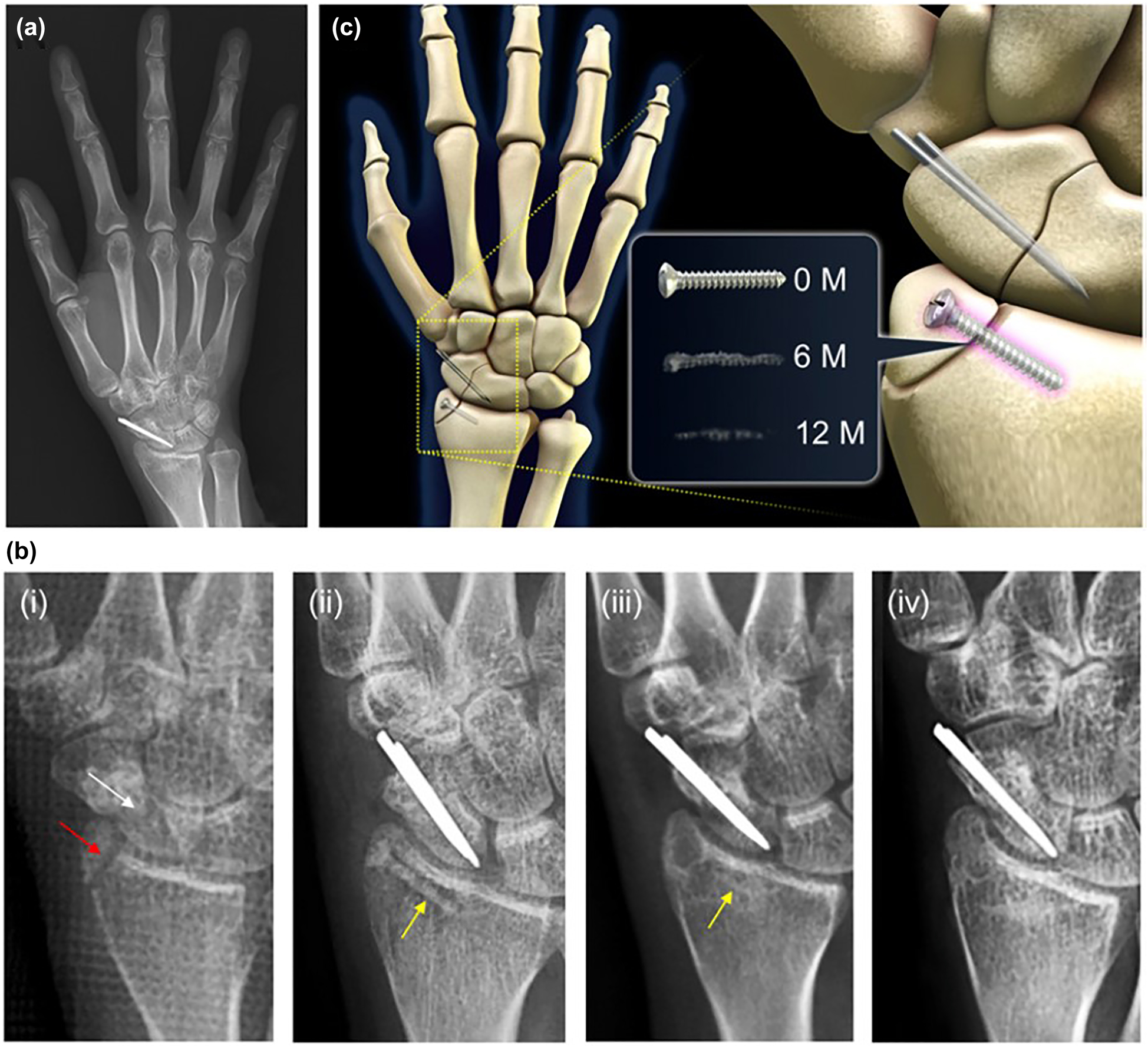
X-ray images of (a) 1-year post-surgery of the distal radius fracture site showing complete degradation of Mg alloy screw with bone healing. (b) X-ray images showing (i) the distal radius fracture (red arrow) and the scaphoid nonunion (white arrow) before the surgical procedure, (ii) Mg–5wt%Ca–1wt%Zn alloy screw (yellow arrow) to fix the distal radius fracture and conventional stainless steel (SS) implant for the scaphoid nonunion, (iii) 6-month post-surgery, (iv) 1-year post-surgery. (c) Schematic representation of the fracture site depicting the progression of degradation process in the Mg–5wt%Ca–1wt%Zn alloy screw with time. Figure reproduced from Lee et al. (2016) with permission from PNAS.
In addition to all the above-mentioned clinical trials, other studies including both human and animal subjects are precisely documented in Table 5. Although Mg alloy-based stents and screws are the only two implants used in human trials, many other implants have been tested in variety of animal models which can be seen from Table 5. Recently, Naujokat et al. (2017) reported the clinical use of WE43/MgYREZr alloy plate and screw in minipigs for osteosynthesis. The result was compared against the conventional Ti plate and screw. Although the bio-efficacy of the Ti group was found to be superior in terms of bone-to-implant contact ratio at a certain post-surgery time point and osteotomy healing duration, the Mg alloy-based implant performed satisfactorily without causing any allergic reaction or delay in wound healing. In this case, the use of a bioactive coating over the surface of the WE43 implant may reduce the lacunas formed adjacent to the bone which can address the issue of less bone-to-implant contact ratio and osteotomy healing.
Recent clinical applications of Mg based implants.
| Implant material | Study type | Implantation site/surgery name | Degradation in-vivo | Toxicity evaluation/complications | Biocompatibility/bio-efficacy evaluation | References |
|---|---|---|---|---|---|---|
| MgYREZr (MAGNEZIX) screw compared against conventional Ti screw | Clinical study: human trials | Hallux valgus corrective surgery | (a) Nearly complete absorption of the MAGNEZIX screw in 1 year follow up period; (b) no issues related to strength and peri-implant fracture in MAGNEZIX | Development of superficial cellulitis in very few patients regardless of screw material | (a) Complete healing of osteotomy in all patients; (b) comparable results between MAGNEZIX and Ti screws in terms of prolonged wound healing and deep infection | (Atkinson et al. 2019; Choo et al. 2019; Klauser 2019; Plaass et al. 2016; Windhagen et al. 2013) |
| ZX00 screws | Clinical study: human trial | Ankle fracture surgery | 50% loss of screw volume after 52 weeks | No postoperative complications | (a) The complete consolidation of ankle fracture was observed after 12 weeks; (b) REE free ZX00 showed unimpeded fracture healing in the early clinical study. | (Holweg et al. 2020a) |
| MgYREZr (MAGNEZIX) screw | Clinical study: human trials | Intercondylar eminence fracture surgery by ARIF (arthroscopically assisted reduction and internal fixation) technique | Complete resorption of fixation device at 6 months follow up | No toxicity | (a) Screws were homogeneously replaced by newly formed bone without any loosening of the fixation device; (b) potential alternative to conventional nonresorbable fixation device | Gigante et al. (2018) |
| Pure Mg interface screw compared against conventional polylactic acid (PLA) interface screw | Clinical study: human cadaver knees | Anterior cruciate ligament (ACL) reconstructions in proximal tibia fixation | Not assessed | Toxicity need to be checked | (a) Similar postoperative fixation effects and mechanical stability in both pure Mg screw and PLA screw; (b) however, degradation behavior needs to be addressed | Song et al. (2017) |
| PLGA coated Mg alloy stent loaded with paclitaxel | Clinical study: human trials (BIOSOLVE-I) | Coronary stent | 11.1% mean scaffold area loss in 6 months | No cardiac death or scaffold thrombosis | Appropriate clinical and angiographic performance with a good safety profile | (Haude et al. 2013, 2016a) |
| PLLA coated Mg alloy stent loaded with sirolimus | Clinical study: human trials (BIOSOLVE-II) | Coronary stent | 0.48% mean scaffold area loss in 6 months | No scaffold thrombosis | (a) Lesser degradation as compared to the stent used in BIOSOLVE I; (b) potential alternative to polymeric absorbable stents | (Garcia-Garcia et al. 2018; Haude et al. 2016b) |
| Mg–5Ca–1Zn alloy screw | Clinical study: human trial | Distal radius fracture surgery | Nearly complete degradation of screws in 12 months follow up period | Not addressed | (a) Biomimicking calcification matrix formation at the interface of implant and bone as the degradation of implant proceeds; (b) this implant can completely avoid the need of a second surgery | Lee et al. (2016) |
| Pure Mg screws | Clinical study: human trial | Fixation of vascularized bone flaps during osteonecrosis of femoral head (ONFH) surgery | The diameter of the Mg screw was reduced by 25.2 ± 1.8% within 12 months post-surgery | No serum level abnormalities in the kidney and liver after the implantation | This study showed better bone flap stabilization in comparison to the conventional approach without any bone flap fixation | Zhao et al. (2016) |
| LAE442 pins | Animal study: sheep, pig, rabbit | Intramedullary interlocked nailing system (sheep, pig), femur (rabbit) | (a) The volume of pin decreased by 10.14% in 24 weeks; (b) long term study on rabbit showed 41.1% volume loss in 9 months | (a) Accumulation of REE in inner organs; (b) slightly inferior clinical tolerance as compared to austenitic ss alloy | Good clinical compatibility with appropriate degradation rate | (Angrisani et al. 2016; Hampp et al. 2013; Krause et al. 2010; Rössig et al. 2015; Thomann et al. 2009; Witte et al. 2006b) |
| LAE442 and Mg–La2 open porous scaffold (β-tricalcium phosphate (TCP) used as control) | Animal study: rabbit | Femur of a rabbit | LAE442 scaffold showed a volume loss of 11.1% in 36 weeks whereas Mg–La2 scaffold showed a volume loss of 72.8% in 20 weeks | Slight swelling around the wound area after the surgery upto 5 days | The homogeneous degradation behavior and better osseointegration of LAE442 scaffold make it as a good choice for weight bearing bone defects | Kleer et al. (2019) |
| Mg–0.45Zn–0.45Ca (ZX00) screws | Animal study: sheep | Fixation of osteotomy in right tibia of a sheep | 9 and 10.1% decrease in implant volume in proximal and distal screws, respectively, after 12 weeks of implantation | Not addressed | (a) Fracture consolidation after 12 weeks with the formation of new bone on screw surfaces; (b) the screw retains its most of the volume and structure up to the bone consolidation period | (Grün et al. 2018; Holweg et al. 2020b) |
| Pure Mg pins compared against stainless steel pins. | Animal study: rat, rabbit | Medullary cavity in the rat distraction osteogenesis model, tibia of rabbit | A reduction of Mg pin volume to 60.57 ± 1.41% after nine-week post operation period | No internal organ disfunction | Pure Mg pins performed better than the ss pin in terms of accelerated bone regeneration and bone maturation | (Hamushan et al. 2020; Yu et al. 2018) |
| Mg–6Zn–0.5Sr interface screw | Animal study: rabbit | Tendon graft fixation to the femoral tunnel in a rabbit | Complete degradation of screws within 12 week post-operation | Not addressed | It outperformed conventionally used PLA in terms of peri-tunnel bone loss, load to failure in femur-tendon graft-tibia complexes (FTGTC) after ACL reconstruction surgery | (Wang et al. 2017, 2018) |
| Mg–xCa alloy (x = 0.5 or 5.0 wt%) | Animal study: rabbit | Femoral condyle of rabbit | (a) Complete degradation of Mg–5Ca alloy within two weeks post-surgery; (b) Mg–0.5Ca alloy retained 51% of its initial volume after 4 weeks of operation | Mg–5Ca implant suffered rapid degradation and caused prolonged inflammation of surgery area | Enhanced bone volume fraction over a selected volume of Mg–0.5Ca implant as compared to Mg–5Ca | Makkar et al. (2018) |
| MAGNEZIX | Animal study: minipig, rabbit | Plate and screw for osteosynthesis in minipigs, screw in left femora of rabbit | 22 mm3 volume of the plate (out of 340 mm3) showed a lower density than the bulk in 30 weeks during degradation | (a) Lower bone-to-implant contact ratio was observed in MAGNEZIX implant as compared to Ti implant (72 vs. 94% at week 30); (b) no clinical side effects such as inflammation, allergic reaction and delay in wound healing | (a) MAGNEZIX implant and Ti implant both showed 100% osteotomy healing by the end of 15 weeks; (b) however, formation of lacunas necessitates more study on plate and screw MAGNEZIX implant | (Naujokat et al. 2017; Waizy et al. 2014) |
| Rapidly solidified RS66 | Animal study: rabbit | Cylindrical implants of RS66 were placed into the femur condyle, into muscle and under the skin of adult rabbits | Volume remaining of RS66 implants after 8 weeks: in bone (condyle) = 28.65%; in muscle (intramuscular) 19.23%; under the skin (subcutaneous) 11.18% | No gas cavity formation, no enhanced bone formation | Highly biocompatible biomaterial with no signs of inflammation | Willbold et al. (2013) |
| MgNd2 | Animal study: minipig | Frontal sinus stent in minipigs | (a) Rapid stent degradation in first 45 days; (b) 61 ± 26.7 mm3 volume of the stent was retained out of 170 ± 4.8 mm3 (original volume) after 180 days | Swab test indicated increase in bacterial colonization in the frontal sinus | Potential material for frontal sinus stent application | Durisin et al. (2016) |
| MgF2 coated MgNd2 cylinders | Animal study: minipig | Frontal sinus of minipigs | The implant was not degraded completely upto 6 months owing to the presence of protective MgF2 coating | Evidence of locally confined foreign body reaction without any bacterial superinfection | Potential implant material for frontal sinus surgery application | Weber et al. (2015) |
| As extruded ZK60 (MAO coated and bare) compared against poly-l-lactic acid (PLLA) pins | Animal study: rat | Right distal femur of mice (along the trans-epicondylar axis from the medial condyle) | In vivo corrosion rate estimated from micro CT: (a) bare ZK60: 3.25 ± 0.41 (2 weeks) 0.67 ± 0.04 (26 weeks); (b) MAO coated ZK60: 2.61 ± 0.41 (2 weeks) 0.58 ± 0.04 (26 weeks) | No significant effect on internal organs | (a) Osteoconductivity and osteointegration of Mg alloy was better in comparison with PLLA; (b) issue of high rate of corrosion was not well addressed by MAO coating | Qi et al. (2014) |
| Anticatabolic drug zoledronic acid (ZA)-loaded polylactic acid/brushite bilayer coating on a Mg–Nd–Zn–Zr alloy (denoted as Mg/ZA/CaP) | Animal study: rat | Intramedullary nail for osteoporotic fracture surgery in rats | Mg/ZA/CaP retained 76.4% of its initial volume after 12 weeks whereas pure Mg retained only 56% | No sign of histological abnormalities in the internal organs | Mg/ZA/CaP nails outperformed both Mg/CaP (without ZA loading) and stainless-steel nails in terms of mechanical performance, fracture healing capacity, and accelerated bone regeneration | Li et al. (2018a) |
| TiO2/MgO nanolayer on ZK60 rod | Animal study: rat | Right/left femur of rat | TiO2/MgO coated ZK60 rod retained 94% of the initial volume whereas untreated ZK60 rods showed a retention of only 70% of implant volume after 8-week post-surgery | Not addressed | TiO2/MgO coated ZK60 rod seems promising than the uncoated ZK60 in terms of corrosion resistance, osteoconductivity, and restoration of mechanical properties of surrounding bone | Lin et al. (2019) |
| Mg-phenolic network deposition (tannic acid, TA) on Mg–Zn alloy | Animal study: rat | Cranial bone defect surgery in rat | Formation of more stable passivation layer which reduces the degradation rate | No complications | Mg-phenolic network deposited sample showed better osteocompatibility than the bare Mg–Zn alloy | Asgari et al. (2019) |
6 Conclusion and outlook
From the past two decades, the advancements that occurred in the area of Mg alloy bioimplants are remarkable. Most Mg alloys are tested in in-vitro environments to quantify their corrosion and SCC response in different solutions simulating the physiological environment. The growing interest in the field of Mg-based implants and scaffold is producing a myriad of research opportunities. Despite aggressive research in this area, some aspects remain unaddressed which are essential for further development. Addressing the following aspects can lead to production of highly corrosion and SCC resistant Mg based implant in future which will be appropriate for in-vivo application.
The discovery of new potential Mg alloys by high throughput research is a viable way to reach one step near to our ideal alloy for temporary implant application. It can significantly lessen the resources required for testing by screening out underperforming alloys.
Novel Mg based multi principal element alloys need to be explored to overcome the demerits associated with conventional Mg based alloys. For this purpose, density functional theory calculations and/or data driven machine learning approach may be taken.
Development of Mg-based new BMG systems can also efficiently handle the existing corrosion and SCC problems. The assessment of the crystallinity resistance of BMGs is vital in this context as the advantageous corrosion properties are directly linked with the amorphous structure of the BMGs.
The route of surface modification also showed some promising results in improving corrosion resistance, corrosion assisted cracking, and producing bio-inductive coatings. It is important to note that no single technique for surface modification can offer every desired functionality. So, combinations of proper techniques can provide the desired functionality of the coating. For instance, LSP can mitigate SCC and CF in an Mg alloy by increasing the fatigue strength but may not protect the alloy from the general mode of corrosion. Similarly, the MAO coating is promising in terms of mechanical interlocking effect, and HA coatings are bio-inductive. Hence, a combination of LSP with MAO or HA can improve the functionality of the coating.
Until now, most of the published studies have been done in in-vitro testing conditions without any application of mechanical stress. So, the tested implant material may not perform satisfactorily in the in-vivo environment due to the presence of different types of mechanical stresses on the implant. Hence, the understanding of the nature of mechanical stress involved in an implant location and mimicking the stress conditions in in-vitro tests will lead to a closer relationship between the in-vitro & in-vivo environment. This step demands the need for online mechanochemical testing devices.
The dimension and shape of the sample used in the in-vitro study should be appropriately designed to closely resemble the geometry of actual implants. This step will ensure proper stress distribution over the implant.
Additionally, all the materials and their corrosion by-products should be appropriately investigated for cell toxicity within the physiological condition before the in-vivo application.
About the authors

Priyabrata Das obtained his B. Tech. in Mechanical Engineering from Biju Patnaik University of Technology, Odisha and his M. Tech. degree in Metallurgical and Materials Engineering from the Indian Institute of Technology (Bhubaneswar) in 2017 and 2020, respectively. He is currently pursuing his doctoral research at Indian Institute of Technology Delhi in the area of biomaterials. Priyabrata is recipient of the institute silver medal for academic excellence in the M. Tech. program and has published three peer reviewed papers in international journals.

T. S. Sampath Kumar is head of the Medical Materials Laboratory, Department of Metallurgical and Materials Engineering, Indian Institute of Technology Madras. He received his PhD in Materials Engineering from Indian Institute of Science, Bangalore in 1986. His research interests are in nanostructured implants, multifunctional nanocarriers, injectable bone cements, electrospun 3D scaffolds and biomaterials from eggshell waste. He has published more than 125 papers in peer reviewed journals, 3 book chapters and has 3 patents to his credit.

Dr. Kisor K. Sahu obtained a Master’s degree in Metallurgy from Indian Institute of Science, Bangalore and a PhD from Graduate School of Energy Sciences, Kyoto University with a scholarship by Japan Government (MEXT). He is currently on the faculty at School of Minerals, Metallurgical and Materials Engineering, IIT Bhubaneswar. He is presently an editorial board member of Scientific Reports. He has published more than 40 peer reviewed papers in journals of international repute.

Dr. Srikant Gollapudi is currently on the faculty at the School of Minerals, Metallurgical and Materials Engineering, IIT Bhubaneswar. He obtained his Bachelor’s degree in Metallurgical Engineering from NIT Rourkela, his Master’s degree in Metallurgy from IISc, Bangalore and his PhD in Materials Science and Engineering from NC State University, USA. His research interests are in corrosion, titanium and magnesium. He has more than 30 publications in well-known peer reviewed journals and 7 patent filings (3 granted and 4 under review).
-
Author contributions: All the authors have accepted responsibility for the entire content of this submitted manuscript and approved submission.
-
Research funding: None declared.
-
Conflicts of interest: The authors declare no conflicts of interest regarding this article.
References
Abela, S. (2015). Physical vapour deposition on Mg alloys for biomedical applications. In: Surface Modification of Magnesium and Its Alloys for Biomedical Applications, II. Woodhead Publishing, Cambridge, UK, pp. 81–100.10.1016/B978-1-78242-078-1.00004-9Search in Google Scholar
Aboulkhair, N.T., Simonelli, M., Parry, L., Ashcroft, I., Tuck, C., and Hague, R. (2019). 3D printing of aluminium alloys: additive manufacturing of aluminium alloys using selective laser melting. Prog. Mater. Sci. 106: 100578. https://doi.org/10.1016/j.pmatsci.2019.100578.Search in Google Scholar
Agarwal, S., Curtin, J., Duffy, B., and Jaiswal, S. (2016). Biodegradable magnesium alloys for orthopaedic applications: a review on corrosion, biocompatibility and surface modifications. Mater. Sci. Eng. C 68: 948–963. https://doi.org/10.1016/j.msec.2016.06.020.Search in Google Scholar PubMed
Amaral, M., Lopes, M.A., Silva, R.F., and Santos, J.D. (2002). Densification route and mechanical properties of Si3N4-bioglass biocomposites. Biomaterials 23: 857–862. https://doi.org/10.1016/s0142-9612(01)00194-6.Search in Google Scholar PubMed
Amaravathy, P. and Kumar, T.S.S. (2019). Bioactivity enhancement by Sr doped Zn-Ca-P coatings on biomedical magnesium alloy. J. Magnes. Alloy. 4: 584–596. https://doi.org/10.1016/j.jma.2019.05.014.Search in Google Scholar
Angrisani, N., Reifenrath, J., Zimmermann, F., Eifler, R., Meyer-Lindenberg, A., Vano-Herrera, K., and Vogt, C. (2016). Biocompatibility and degradation of LAE442-based magnesium alloys after implantation of up to 3.5 years in a rabbit model. Acta Biomater. 44: 355–365. https://doi.org/10.1016/j.actbio.2016.08.002.Search in Google Scholar PubMed
Apps, P.J., Karimzadeh, H., King, J.F., and Lorimer, G.W. (2003). Precipitation reactions in magnesium-rare earth alloys containing yttrium, gadolinium or dysprosium. Scripta Mater. 48: 1023–1028. https://doi.org/10.1016/s1359-6462(02)00596-1.Search in Google Scholar
Asgari, M., Yang, Y., Yang, S., Yu, Z., Yarlagadda, P.K., Xiao, Y., and Li, Z. (2019). Mg–phenolic network strategy for enhancing corrosion resistance and osteocompatibility of degradable magnesium alloys. ACS Omega 4: 21931–21944. https://doi.org/10.1021/acsomega.9b02976.Search in Google Scholar PubMed PubMed Central
Atkinson, H.D., Khan, S., Lashgari, Y., and Ziegler, A. (2019). Hallux valgus correction utilising a modified short scarf osteotomy with a magnesium biodegradable or titanium compression screws - a comparative study of clinical outcomes. BMC Muscoskel. Disord. 20: 334. https://doi.org/10.1186/s12891-019-2717-7.Search in Google Scholar PubMed PubMed Central
Avedesian, M.M. and Baker, H. (1999). Handbook committee: magnesium and magnesium alloys. ASM International, Materials Park, Ohio, pp. 19–20.Search in Google Scholar
Bagherifard, S., Hickey, D.J., Fintová, S., Pastorek, F., Fernandez-Pariente, I., Bandini, M., Webster, T.J., and Guagliano, M. (2018). Effects of nanofeatures induced by severe shot peening (SSP) on mechanical, corrosion and cytocompatibility properties of magnesium alloy AZ31. Acta Biomater. 66: 93–108. https://doi.org/10.1016/j.actbio.2017.11.032.Search in Google Scholar PubMed
Bakhsheshi-Rad, H.R., Abdul-Kadir, M.R., Idris, M.H., and Farahany, S.J.C.S. (2012). Relationship between the corrosion behavior and the thermal characteristics and microstructure of Mg-0.5Ca-xZn alloys. Corrosion Sci. 64: 184–197. https://doi.org/10.1016/j.corsci.2012.07.015.Search in Google Scholar
Bakhsheshi-Rad, H.R., Hamzah, E., Ebrahimi-Kahrizsangi, R., Daroonparvar, M., and Medraj, M. (2016). Fabrication and characterization of hydrophobic microarc oxidation/poly-lactic acid duplex coating on biodegradable Mg-Ca alloy for corrosion protection. Vacuum 125: 185–188. https://doi.org/10.1016/j.vacuum.2015.12.022.Search in Google Scholar
Bakhsheshi-Rad, H.R., Hamzah, E., Ismail, A.F., Aziz, M., Kasiri-Asgarani, M., Akbari, E., Jabbarzare, S., Najafinezhad, A., and Hadisi, Z. (2017). Synthesis of a novel nanostructured zinc oxide/baghdadite coating on Mg alloy for biomedical application: in-vitro degradation behavior and antibacterial activities. Ceram. Int. 43: 14842–14850. https://doi.org/10.1016/j.ceramint.2017.07.233.Search in Google Scholar
Baltzer, N. and Copponnex, T. (Eds.) (2014). Precious metals for biomedical applications. Woodhead Publishing, Elsevier, Cambridge, UK.Search in Google Scholar
Baulin, O., Fabregue, D., Kato, H., Liens, A., Wada, T., and Pelletier, J.M. (2018). A new, toxic element-free Mg-based metallic glass for biomedical applications. J. Non-Cryst. Solids 481: 397–402. https://doi.org/10.1016/j.jnoncrysol.2017.11.024.Search in Google Scholar
Bekmurzayeva, A., Duncanson, W.J., Azevedo, H.S., and Kanayeva, D. (2018). Surface modification of stainless steel for biomedical applications: revisiting a century-old material. Mater. Sci. Eng. C 93: 1073–1089. https://doi.org/10.1016/j.msec.2018.08.049.Search in Google Scholar PubMed
Bhuiyan, M.S., Mutoh, Y., Murai, T., and Iwakami, S. (2010). Corrosion fatigue behavior of extruded magnesium alloy AZ80-T5 in a 5% NaCl environment. Eng. Fract. Mech. 77: 1567–1576.10.1016/j.engfracmech.2010.03.032Search in Google Scholar
Birbilis, N., Easton, M.A., Sudholz, A.D., Zhu, S.M., and Gibson, M.A. (2009). On the corrosion of binary magnesium-rare earth alloys. Corrosion Sci. 51: 683–689.10.1016/j.corsci.2008.12.012Search in Google Scholar
Birbilis, N., Ralston, K.D., Virtanen, S., Fraser, H.L., and Davies, C.H.J. (2010). Grain character influences on corrosion of ECAPed pure magnesium. Corrosion Eng. Sci. Technol. 45: 224–230. https://doi.org/10.1179/147842209x12559428167805.Search in Google Scholar
Bobe, K., Willbold, E., Morgenthal, I., Andersen, O., Studnitzky, T., Nellesen, J., Tillmann, W., Vogt, C., Vano, K., and Witte, F. (2013). In vitro and in vivo evaluation of biodegradable, open-porous scaffolds made of sintered magnesium W4 short fibres. Acta Biomater. 9: 8611–8623. https://doi.org/10.1016/j.actbio.2013.03.035.Search in Google Scholar PubMed
Bosiers, M. (2009). AMS INSIGHT-absorbable metal stent implantation for treatment of below-the-knee critical limb ischemia: 6-month analysis. Cardiovasc. Intervent. Radiol. 32: 424–435. https://doi.org/10.1007/s00270-008-9472-8.Search in Google Scholar PubMed PubMed Central
Boushey, C.J., Coulston, A.M., Rock, C.L., and Monsen, E. (Eds.) (2001). Nutrition in the prevention and treatment of disease. Academic press, California, USA.Search in Google Scholar
Brinker, C.J.J. and Hurd, A. (1994). Fundamentals of sol-gel dip-coating. J. Phys. III: 4. https://doi.org/10.1051/jp3:1994198.10.1051/jp3:1994198Search in Google Scholar
Cahoon, J.R. and Paxton, H.W. (1968). Metallurgical analyses of failed orthopedic implants. J. Biomed. Mater. Res. 2: 1–22. https://doi.org/10.1002/jbm.820020102.Search in Google Scholar PubMed
Chai, L., Yu, X., Yang, Z., Wang, Y., and Okido, M. (2008). Anodizing of magnesium alloy AZ31 in alkaline solutions with silicate under continuous sparking. Corrosion Sci. 50: 3274–3279. https://doi.org/10.1016/j.corsci.2008.08.038.Search in Google Scholar
Chakraborty Banerjee, P., Al-Saadi, S., Choudhary, L., Harandi, S.E., and Singh, R. (2019). Magnesium implants: prospects and challenges. Materials 12: 136. https://doi.org/10.3390/ma12010136.Search in Google Scholar PubMed PubMed Central
Chen, J., Wang, Z., and Lu, S. (2012). Effects of electric parameters on microstructure and properties of MAO coating fabricated on ZK60 Mg alloy in dual electrolyte. Rare Met. 31: 172–177. https://doi.org/10.1007/s12598-012-0486-7.Search in Google Scholar
Chen, S., Tu, J., Hu, Q., Xiong, X., Wu, J., Zou, J., and Zeng, X. (2017). Corrosion resistance and in vitro bioactivity of Si-containing coating prepared on a biodegradable Mg-Zn-Ca bulk metallic glass by micro-arc oxidation. J. Non-Cryst. Solids 456: 125–131. https://doi.org/10.1016/j.jnoncrysol.2016.11.011.Search in Google Scholar
Chiu, K.Y., Wong, M.H., Cheng, F.T., and Man, H.C. (2007). Characterization and corrosion studies of fluoride conversion coating on degradable Mg implants. Surf. Coating. Technol. 202: 590–598. https://doi.org/10.1016/j.surfcoat.2007.06.035.Search in Google Scholar
Choo, J.T., Lai, S.H.S., Tang, C.Q.Y., and Thevendran, G. (2019). Magnesium-based bioabsorbable screw fixation for hallux valgus surgery – a suitable alternative to metallic implants. Foot Ankle Surg. 25: 727–732. https://doi.org/10.1016/j.fas.2018.09.001.Search in Google Scholar PubMed
Choudhary, L., Raman, R.S., Hofstetter, J., and Uggowitzer, P.J. (2014). In-vitro characterization of stress corrosion cracking of aluminium-free magnesium alloys for temporary bio-implant applications. Mater. Sci. Eng. C 42: 629–636. https://doi.org/10.1016/j.msec.2014.06.018.Search in Google Scholar PubMed
Cooper, D.M.L., Matyas, J.R., Katzenberg, M.A., and Hallgrimsson, B. (2004). Comparison of microcomputed tomographic and microradiographic measurements of cortical bone porosity. Calcif. Tissue Int. 74: 437–447. https://doi.org/10.1007/s00223-003-0071-z.Search in Google Scholar PubMed
Crubzy, E., Murail, P., Girard, L., and Bernadou, J.P. (1998). False teeth of the Roman world. Nature 391: 29. https://doi.org/10.1038/34067.Search in Google Scholar PubMed
Cui, W., Beniash, E., Gawalt, E., Xu, Z., and Sfeir, C. (2013). Biomimetic coating of magnesium alloy for enhanced corrosion resistance and calcium phosphate deposition. Acta Biomater. 9: 8650–8659. https://doi.org/10.1016/j.actbio.2013.06.031.Search in Google Scholar PubMed
Davenport, A.J., Padovani, C., Connolly, B.J., Stevens, N.P., Beale, T.A., Groso, A., and Stampanoni, M. (2006). Synchrotron X-ray microtomography study of the role of y in corrosion of magnesium alloy WE43. Electrochem. Solid State Lett. 10: C5.10.1149/1.2400727Search in Google Scholar
Dayani, S.B. (2017). Improvement of fatigue and corrosion-fatigue resistance of AZ31B cast alloy by cold spray coating and top coating. Doctoral dissertion, University of Waterloo. Waterloo, Canada.Search in Google Scholar
Ding, K. and Ye, L. (2006). Laser shock peening : performance and process simulations. Woodhead Publishing, Cambridge, UK.10.1201/9781439823620Search in Google Scholar
Ding, Y., Wen, C., Hodgson, P., and Li, Y. (2014). Effects of alloying elements on the corrosion behavior and biocompatibility of biodegradable magnesium alloys: a review. J. Mater. Chem. B 2: 1912–1933. https://doi.org/10.1039/c3tb21746a.Search in Google Scholar PubMed
Drábiková, J., Fintová, S., Tkacz, J., Doležal, P., and Wasserbauer, J. (2017). Unconventional fluoride conversion coating preparation and characterization. Anti-corrosion Methods & Mater. 64: 613–619.10.1108/ACMM-02-2017-1757Search in Google Scholar
Durdu, S. and Usta, M. (2012). Characterization and mechanical properties of coatings on magnesium by micro arc oxidation. Appl. Surf. Sci. 261: 774–782. https://doi.org/10.1016/j.apsusc.2012.08.099.Search in Google Scholar
Durisin, M., Seitz, J.M., Reifenrath, J., Weber, C.M., Eifler, R., Maier, H.J., Lenarz, T., and Klose, C. (2016). A novel biodegradable frontal sinus stent (MgNd2): a long-term animal study. Eur. Arch. Oto-Rhino-Laryngol. 273: 1455–1467. https://doi.org/10.1007/s00405-015-3774-7.Search in Google Scholar PubMed
Eliades, T., Pratsinis, H., Kletsas, D., Eliades, G., and Makou, M. (2004). Characterization and cytotoxicity of ions released from stainless steel and nickel-titanium orthodontic alloys. Am. J. Orthod. Dentofacial Orthop. 125: 24–29. https://doi.org/10.1016/j.ajodo.2003.09.009.Search in Google Scholar PubMed
Erbel, R., Di Mario, C., Bartunek, J., Bonnier, J., de Bruyne, B., Eberli, F.R., Erne, P., Haude, M., Heublein, B., Horrigan, M., et al.. (2007). Temporary scaffolding of coronary arteries with bioabsorbable magnesium stents: a prospective, non-randomised multicentre trial. Lancet 369: 1869–1875. https://doi.org/10.1016/s0140-6736(07)60853-8.Search in Google Scholar
Estrin, Y. and Vinogradov, A. (2010). Fatigue behaviour of light alloys with ultrafine grain structure produced by severe plastic deformation: an overview. Int. J. Fatig. 32: 898–907. https://doi.org/10.1016/j.ijfatigue.2009.06.022.Search in Google Scholar
Fan, L., Sun, W., Zou, Y., Xu, Q.Q., Zeng, R.C., and Tian, J. (2022). Enhanced corrosion resistance, antibacterial activity and biocompatibility of gentamicin-montmorillonite coating on Mg alloy-in vitro and in vivo studies. J. Mater. Sci. Technol. 111: 167–180. https://doi.org/10.1016/j.jmst.2021.08.089.Search in Google Scholar
Fatemi, S.M., Zarei-Hanzaki, A., Eskandari, M., and Haghshenas, M. (2018). Alleviation of mechanical anisotropy in ultrafine/nano-grained AZ31 magnesium alloy. J. Mater. Eng. Perform. 27: 4270–4279. https://doi.org/10.1007/s11665-018-3485-5.Search in Google Scholar
Feng, H., Liu, S., Du, Y., Lei, T., Zeng, R., and Yuan, T. (2017). Effect of the second phases on corrosion behavior of the Mg-Al-Zn alloys. J. Alloys Compd. 695: 2330–2338. https://doi.org/10.1016/j.jallcom.2016.11.100.Search in Google Scholar
Fintová, S., Drábiková, J., Pastorek, F., Tkacz, J., Kuběna, I., Trško, L., Hadzima, B., Minda, J., Doležal, P., Wasserbauer, J., et al.. (2019). Improvement of electrochemical corrosion characteristics of AZ61 magnesium alloy with unconventional fluoride conversion coatings. Surf. Coating. Technol. 357: 638–650.10.1016/j.surfcoat.2018.10.038Search in Google Scholar
Fischerauer, S.F., Kraus, T., Wu, X., Tangl, S., Sorantin, E., Hänzi, A.C., Löffler, J.F., Uggowitzer, P.J., and Weinberg, A.M. (2013). In vivo degradation performance of micro-Arc-oxidized magnesium implants: a micro-CT study in rats. Acta Biomater. 9: 5411–5420. https://doi.org/10.1016/j.actbio.2012.09.017.Search in Google Scholar PubMed
Galli, S., Verheye, S., Wlodarczak, A., Bennett, J., Torzewski, J., Wiemer, M., Haude, M., Nuruddin, A.A., Starmer, G., Buck, T., et al.. (2019). Safety and performance of the resorbable magnesium scaffold, Magmaris in a real-world setting - 12-month follow-up of first 600 subjects in biosolve-IV registry. JACC Cardiovasc. Interv. 12: S39. https://doi.org/10.1016/j.jcin.2019.01.140.Search in Google Scholar
Ganesh, P., Sundar, R., Kumar, H., Kaul, R., Ranganathan, K., Hedaoo, P., Raghavendra, G., Kumar, S.A., Tiwari, P., Nagpure, D.C., et al.. (2014). Studies on fatigue life enhancement of pre-fatigued spring steel specimens using laser shock peening. Mater. Des. 54: 734–741. https://doi.org/10.1016/j.matdes.2013.08.104.Search in Google Scholar
Garcia-Garcia, H.M., Haude, M., Kuku, K., Hideo-Kajita, A., Ince, H., Abizaid, A., Tölg, R., Lemos, P.A., von Birgelen, C., Christiansen, E.H., et al.. (2018). In vivo serial invasive imaging of the second-generation drug-eluting absorbable metal scaffold (Magmaris — DREAMS 2G) in de novo coronary lesions: insights from the BIOSOLVE-II First-In-Man Trial. Int. J. Cardiol. 255: 22–28. https://doi.org/10.1016/j.ijcard.2017.12.053.Search in Google Scholar PubMed
Geng, F., Tan, L., Zhang, B., Wu, C., He, Y., Yang, J., and Yang, K. (2009). Study on β-TCP coated porous Mg as a bone tissue engineering scaffold material. J. Mater. Sci. Technol. 25: 123–129.Search in Google Scholar
Gigante, A., Setaro, N., Rotini, M., Finzi, S.S., and Marinelli, M. (2018). Intercondylar eminence fracture treated by resorbable magnesium screws osteosynthesis: a case series. Injury 49: S48–S53. https://doi.org/10.1016/j.injury.2018.09.055.Search in Google Scholar PubMed
Gollapudi, S. (2012). Grain size distribution effects on the corrosion behaviour of materials. Corrosion Sci. 62: 90–94. https://doi.org/10.1016/j.corsci.2012.04.040.Search in Google Scholar
González, S., Pellicer, E., Fornell, J., Blanquer, A., Barrios, L., Ibánez, E., Solsona, P., Surinach, S., Baró, M.D., Nogués, C., et al.. (2012). Improved mechanical performance and delayed corrosion phenomena in biodegradable Mg-Zn-Ca alloys through Pd-alloying. J. Mech. Behav. Biomed. Mater. 6: 53–62. https://doi.org/10.1016/j.jmbbm.2011.09.014.Search in Google Scholar PubMed
Grün, N.G., Holweg, P., Tangl, S., Eichler, J., Berger, L., Van den Beucken, J.J., Löffler, J.F., Klestil, T., and Weinberg, A.M. (2018). Comparison of a resorbable magnesium implant in small and large growing-animal models. Acta Biomater. 78: 378–386. https://doi.org/10.1016/j.actbio.2018.07.044.Search in Google Scholar PubMed
Gu, X., Zheng, Y., Zhong, S., Xi, T., Wang, J., and Wang, W. (2010a). Corrosion of, and cellular responses to Mg-Zn-Ca bulk metallic glasses. Biomaterials 31: 1093–1103. https://doi.org/10.1016/j.biomaterials.2009.11.015.Search in Google Scholar PubMed
Gu, X.N., Zhou, W.R., Zheng, Y.F., Cheng, Y., Wei, S.C., Zhong, S.P., Xi, T.F., and Chen, L.J. (2010b). Corrosion fatigue behaviors of two biomedical Mg alloys - AZ91D and WE43 - in simulated body fluid. Acta Biomater. 6: 4605–4613. https://doi.org/10.1016/j.actbio.2010.07.026.Search in Google Scholar PubMed
Gu, X.N., Li, N., Zheng, Y.F., and Ruan, L. (2011). In vitro degradation performance and biological response of a Mg-Zn-Zr alloy. Mater. Sci. Eng. B 176: 1778–1784. https://doi.org/10.1016/j.mseb.2011.05.032.Search in Google Scholar
Guo, Y., Sealy, M.P., and Guo, C. (2012). Significant improvement of corrosion resistance of biodegradable metallic implants processed by laser shock peening. CIRP Ann. - Manuf. Technol. 61: 583–586. https://doi.org/10.1016/j.cirp.2012.03.125.Search in Google Scholar
Gupta, A., Cecen, A., Goyal, S., Singh, A.K., and Kalidindi, S.R. (2015). Structure-property linkages using a data science approach: application to a non-metallic inclusion/steel composite system. Acta Mater. 91: 239–254. https://doi.org/10.1016/j.actamat.2015.02.045.Search in Google Scholar
Gupta, V.B., Anitha, S., Hegde, M.L., Zecca, L., Garruto, R.M., Ravid, R., Shankar, S.K., Stein, R., Shanmugavelu, P., and Rao, K.J. (2005). Aluminium in Alzheimer’s disease: are we still at a crossroad? Cell. Mol. Life Sci. 62: 143–158. https://doi.org/10.1007/s00018-004-4317-3.Search in Google Scholar PubMed PubMed Central
Hakimi, O., Aghion, E., and Goldman, J. (2015). Improved stress corrosion cracking resistance of a novel biodegradable EW62 magnesium alloy by rapid solidification, in simulated electrolytes. Mater. Sci. Eng. C 51: 226–232. https://doi.org/10.1016/j.msec.2015.03.001.Search in Google Scholar PubMed
Hampp, C., Angrisani, N., Reifenrath, J., Bormann, D., Seitz, J.M., and Meyer-Lindenberg, A. (2013). Evaluation of the biocompatibility of two magnesium alloys as degradable implant materials in comparison to titanium as non-resorbable material in the rabbit. Mater. Sci. Eng. C 33: 317–326. https://doi.org/10.1016/j.msec.2012.08.046.Search in Google Scholar PubMed
Hamushan, M., Cai, W., Zhang, Y., Lou, T., Zhang, S., Zhang, X., Cheng, P., Zhao, C., and Han, P. (2020). High-purity magnesium pin enhances bone consolidation in distraction osteogenesis model through activation of the VHL/HIF-1α/VEGF signaling. J. Biomater. Appl. 35: 224–236. https://doi.org/10.1177/0885328220928550.Search in Google Scholar PubMed
Hänzi, A.C., Gunde, P., Schinhammer, M., and Uggowitzer, P.J. (2009). On the biodegradation performance of an Mg-Y-RE alloy with various surface conditions in simulated body fluid. Acta Biomater. 5: 162–171. https://doi.org/10.1016/j.actbio.2008.07.034.Search in Google Scholar PubMed
Hänzi, A.C., Gerber, I., Schinhammer, M., Löffler, J.F., and Uggowitzer, P.J. (2010). On the in vitro and in vivo degradation performance and biological response of new biodegradable Mg-Y-Zn alloys. Acta Biomater. 6: 1824–1833.10.1016/j.actbio.2009.10.008Search in Google Scholar PubMed
Harandi, S.E. and Singh Raman, R.K. (2015). Appropriate mechanochemical conditions for corrosion-fatigue testing of magnesium alloys for temporary bioimplant applications. JOM 67: 1137–1142.10.1007/s11837-015-1387-7Search in Google Scholar
Harandi, S.E. and Singh Raman, R.K. (2017). Corrosion fatigue of a magnesium alloy under appropriate human physiological conditions for bio-implant applications. Eng. Fract. Mech. 186: 134–142. https://doi.org/10.1016/j.engfracmech.2017.09.031.Search in Google Scholar
Hatamleh, O., Lyons, J., and Forman, R. (2007). Laser and shot peening effects on fatigue crack growth in friction stir welded 7075-T7351 aluminum alloy joints. Int. J. Fatig. 29: 421–434. https://doi.org/10.1016/j.ijfatigue.2006.05.007.Search in Google Scholar
Haude, M., Erbel, R., Erne, P., Verheye, S., Degen, H., Böse, D., Vermeersch, P., Wijnbergen, I., Weissman, N., Prati, F., et al.. (2013). Safety and performance of the drug-eluting absorbable metal scaffold (DREAMS) in patients with de-novo coronary lesions: 12 month results of the prospective, multicentre, first-in-man BIOSOLVE-I trial. Lancet 381: 836–844. https://doi.org/10.1016/s0140-6736(12)61765-6.Search in Google Scholar
Haude, M., Erbel, R., Erne, P., Verheye, S., Degen, H., Vermeersch, P., Weissman, N., Prati, F., Bruining, N., Waksman, R., et al.. (2016a). Safety and performance of the DRug-Eluting Absorbable Metal Scaffold (DREAMS) in patients with de novo coronary lesions: 3-year results of the prospective, multicentre, first-in-man BIOSOLVE-I trial. EuroIntervention 12: e160–e166. https://doi.org/10.4244/eij-d-15-00371.Search in Google Scholar PubMed
Haude, M., Ince, H., Abizaid, A., Toelg, R., Lemos, P.A., von Birgelen, C., Christiansen, E.H., Wijns, W., Neumann, F.J., Kaiser, C., et al.. (2016b). Safety and performance of the second-generation drug-eluting absorbable metal scaffold in patients with de-novo coronary artery lesions (BIOSOLVE-II): 6 month results of a prospective, multicentre, non-randomised, first-in-man trial. Lancet 387: 31–39. https://doi.org/10.1016/s0140-6736(15)00447-x.Search in Google Scholar
Haude, M., Ince, H., Kische, S., Abizaid, A., Tölg, R., Alves, L.P., Van Mieghem, N., Verheye, S., Von Birgelen, C., and Christiansen, E. (2017). Sustained safety and clinical performance of a drug-eluting absorbable metal scaffold up to 24 months: pooled outcomes of BIOSOLVE-II and BIOSOLVE-III behalf of the BIOSOLVE-II and BIOSOLVE-III investigators BIOSOLVE-II and III. EuroIntervention 13: 432–439. https://doi.org/10.4244/EIJ-D-17-00254.Search in Google Scholar PubMed
Haude, M., Ince, H., Kische, S., Toelg, R., Van Mieghem, N.M., Verheye, S., von Birgelen, C., Christiansen, E.H., Barbato, E., Garcia-Garcia, H.M., et al.. (2020). Sustained safety and performance of the second-generation sirolimus-eluting absorbable metal scaffold: pooled outcomes of the BIOSOLVE-II and -III trials at 3 years. Cardiovasc. Revascularization Med. 21: 1150–1154. https://doi.org/10.1016/j.carrev.2020.04.006.Search in Google Scholar PubMed
He, S.M., Zeng, X.Q., Peng, L.M., Gao, X., Nie, J.F., and Ding, W.J. (2007). Microstructure and strengthening mechanism of high strength Mg-10Gd-2Y-0.5Zr alloy. J. Alloys Compd. 427: 316–323. https://doi.org/10.1016/j.jallcom.2006.03.015.Search in Google Scholar
Heise, S., Höhlinger, M., Hernández, Y.T., Palacio, J.J.P., Ortiz, J.A.R., Wagener, V., Virtanen, S., and Boccaccini, A.R. (2017). Electrophoretic deposition and characterization of chitosan/bioactive glass composite coatings on Mg alloy substrates. Electrochim. Acta 232: 456–464. https://doi.org/10.1016/j.electacta.2017.02.081.Search in Google Scholar
Hermawan, H., Ramdan, D., and Djuansjah, J.R.P. (2011). Metals for biomedical applications. In: Biomedical engineering: from theory to applications. I. InTech, Rijeka, Croatia, pp. 411–430.10.5772/19033Search in Google Scholar
Hoffheinz, S. and Dimitroff, N. (1928). Experimentelle Studie über die Resorption parenteral zugeführten metallischen Magnesiums. Deutsche Zeitschrift für Chirurgie 208: 346–353. https://doi.org/10.1007/bf02827373.Search in Google Scholar
Holweg, P., Herber, V., Ornig, M., Hohenberger, G., Donohue, N., Puchwein, P., Leithner, A., and Seibert, F. (2020a). A lean bioabsorbable magnesium-zinc-calcium alloy ZX00 used for operative treatment of medial malleolus fractures. Bone Jt. Res. 9: 477–483. https://doi.org/10.1302/2046-3758.98.bjr-2020-0017.r2.Search in Google Scholar PubMed PubMed Central
Holweg, P., Berger, L., Cihova, M., Donohue, N., Clement, B., Schwarze, U., Sommer, N.G., Hohenberger, G., van den Beucken, J.J., Seibert, F., et al.. (2020b). A lean magnesium–zinc–calcium alloy ZX00 used for bone fracture stabilization in a large growing-animal model. Acta Biomater. 113: 646–659. https://doi.org/10.1016/j.actbio.2020.06.013.Search in Google Scholar PubMed
Hornberger, H., Virtanen, S., and Boccaccini, A.R. (2012). Biomedical coatings on magnesium alloys - a review. Acta Biomater. 8: 2442–2455. https://doi.org/10.1016/j.actbio.2012.04.012.Search in Google Scholar PubMed
Hort, N., Huang, Y.D., Fechner, D., Störmer, M., Blawert, C., Witte, F., Vogt, C., Drücker, H., Willumeit, R., Kainer, K.U., et al.. (2010). Magnesium alloys as implant materials - principles of property design for Mg-RE alloys. Acta Biomater. 6: 1714–1725. https://doi.org/10.1016/j.actbio.2009.09.010.Search in Google Scholar PubMed
Hwang, D.Y., Kim, Y.M., Park, D.Y., Yoo, B., and Shin, D.H. (2009). Corrosion resistance of oxide layers formed on AZ91 Mg alloy in KMnO4 electrolyte by plasma electrolytic oxidation. Electrochim. Acta 54: 5479–5485. https://doi.org/10.1016/j.electacta.2009.04.047.Search in Google Scholar
Inoue, K., Suenaga, N., Oizumi, N., Yamaguchi, H., Miyoshi, N., Taniguchi, N., Morita, S., Munemoto, M., Kurata, S., and Tanaka, Y. (2020). Humeral bone resorption after reverse shoulder arthroplasty using uncemented stem. J. Shoulder Elbow Surg. Int. 4: 138–143. https://doi.org/10.1016/j.jses.2019.11.007.Search in Google Scholar PubMed PubMed Central
Jacobs, J.J., Hallab, N.J., Skipor, A.K., and Urban, R.M. (2003). Metal degradation products: a cause for concern in metal-metal bearings? Clin. Orthop. Relat. Res. 417: 139–147, Lippincott Williams and Wilkins. https://doi.org/10.1097/01.blo.0000096810.78689.62.Search in Google Scholar PubMed
Jafari, S., Harandi, S.E., and Singh Raman, R.K. (2015). A review of stress-corrosion cracking and corrosion fatigue of magnesium alloys for biodegradable implant applications. JOM 67: 1143–1153. https://doi.org/10.1007/s11837-015-1366-z.Search in Google Scholar
Jawahir, I.S., Puleo, D.A., and Schoop, J. (2016). Cryogenic machining of biomedical implant materials for improved functional performance, Life and Sustainability. Procedia CIRP 46: 7–14. https://doi.org/10.1016/j.procir.2016.04.133.Search in Google Scholar
Jayaraj, J., Rajesh, K.R., Raj, S.A., Srinivasan, A., Ananthakumar, S., Dhaipule, N.G.K., Kalpathy, S.K., Pillai, U.T.S., and Mudali, U.K. (2019). Investigation on the corrosion behavior of lanthanum phosphate coatings on AZ31 Mg alloy obtained through chemical conversion technique. J. Alloys Compd. 784: 1162–1174.10.1016/j.jallcom.2019.01.121Search in Google Scholar
Jia, G., Hou, Y., Chen, C., Niu, J., Zhang, H., Huang, H., Xiong, M., and Yuan, G. (2018). Precise fabrication of open porous Mg scaffolds using NaCl templates: relationship between space holder particles, pore characteristics and mechanical behavior. Mater. Des. 140: 106–113. https://doi.org/10.1016/j.matdes.2017.11.064.Search in Google Scholar
Kannan, M.B. and Raman, R.K.S. (2008). In vitro degradation and mechanical integrity of calcium-containing magnesium alloys in modified-simulated body fluid. Biomaterials 29: 2306–2314. https://doi.org/10.1016/j.biomaterials.2008.02.003.Search in Google Scholar PubMed
Kannan, M.B., Dietzel, W., Blawert, C., Atrens, A., and Lyon, P. (2008). Stress corrosion cracking of rare-earth containing magnesium alloys ZE41, QE22 and Elektron 21 (EV31A) compared with AZ80. Mater. Sci. Eng. A 480: 529–539. https://doi.org/10.1016/j.msea.2007.07.070.Search in Google Scholar
Kawamura, N., Nakao, Y., Ishikawa, R., Tsuchida, D., and Iijima, M. (2020). Degradation and biocompatibility of AZ31 magnesium alloy implants in vitro and in vivo: a micro-computed tomography study in rats. Materials 13: 473. https://doi.org/10.3390/ma13020473.Search in Google Scholar PubMed PubMed Central
Kelly, R.G. (2003). Electrochemical thermodynamics and corrosion. In: Schweitzer, P.A. (Ed.), Electrochemical thermodynamics and kinetics of relevance to corrosion. New York: Marcel Dekker, pp. 9–54.10.1201/9780203909133.ch2Search in Google Scholar
Kim, H.K. and Kim, W.J. (2004). Microstructural instability and strength of an AZ31 Mg alloy after severe plastic deformation. Mater. Sci. Eng. A 385: 300–308. https://doi.org/10.1016/s0921-5093(04)00882-2.Search in Google Scholar
Kim, Y.K., Lee, M.H., Prasad, M.N., Park, I.S., Lee, M.H., Seol, K.W., and Bae, T.S. (2008). Surface characteristics of magnesium alloys treated by anodic oxidation using pulse power. Adv. Mater. Res. 47: 1290–1293. https://doi.org/10.4028/www.scientific.net/amr.47-50.1290.Search in Google Scholar
Kirkland, N.T., Lespagnol, J., Birbilis, N., and Staiger, M.P. (2010). A survey of bio-corrosion rates of magnesium alloys. Corrosion Sci. 52: 287–291. https://doi.org/10.1016/j.corsci.2009.09.033.Search in Google Scholar
Klauser, H. (2019). Internal fixation of three-dimensional distal metatarsal I osteotomies in the treatment of hallux valgus deformities using biodegradable magnesium screws in comparison to titanium screws. Foot Ankle Surg. 25: 398–405. https://doi.org/10.1016/j.fas.2018.02.005.Search in Google Scholar PubMed
Kleer, N., Julmi, S., Gartzke, A.K., Augustin, J., Feichtner, F., Waselau, A.C., Klose, C., Maier, H.J., Wriggers, P., and Meyer-Lindenberg, A. (2019). Comparison of degradation behaviour and osseointegration of the two magnesium scaffolds, LAE442 and La2, in vivo. Materialia 8: 100436.10.1016/j.mtla.2019.100436Search in Google Scholar
Kokubo, T., Kim, H.M., and Kawashita, M. (2003). Novel bioactive materials with different mechanical properties. Biomaterials 24: 2161–2175. https://doi.org/10.1016/s0142-9612(03)00044-9.Search in Google Scholar PubMed
Koo, Y., Jang, Y., and Yun, Y. (2017). A study of long-term static load on degradation and mechanical integrity of Mg alloys-based biodegradable metals. Mater. Sci. Eng. B 219: 45–54. https://doi.org/10.1016/j.mseb.2017.02.009.Search in Google Scholar PubMed PubMed Central
Kraus, T., Fischerauer, S.F., Hänzi, A.C., Uggowitzer, P.J., Löffler, J.F., and Weinberg, A.M. (2012). Magnesium alloys for temporary implants in osteosynthesis: in vivo studies of their degradation and interaction with bone. Acta Biomater. 8: 1230–1238. https://doi.org/10.1016/j.actbio.2011.11.008.Search in Google Scholar PubMed
Krause, A., Von der Höh, N., Bormann, D., Krause, C., Bach, F.W., Windhagen, H., and Meyer-Lindenberg, A. (2010). Degradation behaviour and mechanical properties of magnesium implants in rabbit tibiae. J. Mater. Sci. 45: 624–632. https://doi.org/10.1007/s10853-009-3936-3.Search in Google Scholar
Kulkarni, R.K., Moore, E.G., Hegyeli, A.F., and Leonard, F. (1971). Biodegradable poly(lactic acid) polymers. J. Biomed. Mater. Res. 5: 169–181. https://doi.org/10.1002/jbm.820050305.Search in Google Scholar PubMed
Kumar, A. and Pandey, P.M. (2020). Development of Mg based biomaterial with improved mechanical and degradation properties using powder metallurgy. J. Magnes. Alloy. 8: 883–898. https://doi.org/10.1016/j.jma.2020.02.011.Search in Google Scholar
Kumar, R.M., Kuntal, K.K., Singh, S., Gupta, P., Bhushan, B., Gopinath, P., and Lahiri, D. (2016). Electrophoretic deposition of hydroxyapatite coating on Mg-3Zn alloy for orthopaedic application. Surf. Coating. Technol. 287: 82–92.10.1016/j.surfcoat.2015.12.086Search in Google Scholar
Lee, J.W., Han, H.S., Han, K.J., Park, J., Jeon, H., Ok, M.R., Seok, H.K., Ahn, J.P., Lee, K.E., Lee, D.H., et al.. (2016). Long-term clinical study and multiscale analysis of in vivo biodegradation mechanism of Mg alloy. Proc. Natl. Acad. Sci. U.S.A. 113: 716–721. https://doi.org/10.1073/pnas.1518238113.Search in Google Scholar PubMed PubMed Central
Lei, T., Tang, W., Cai, S.H., Feng, F.F., and Li, N.F. (2012). On the corrosion behaviour of newly developed biodegradable Mg-based metal matrix composites produced by in situ reaction. Corrosion Sci. 54: 270–277. https://doi.org/10.1016/j.corsci.2011.09.027.Search in Google Scholar
Li, H., Pang, S., Liu, Y., Sun, L., Liaw, P.K., and Zhang, T. (2015). Biodegradable Mg-Zn-Ca-Sr bulk metallic glasses with enhanced corrosion performance for biomedical applications. Mater. Des. 67: 9–19. https://doi.org/10.1016/j.matdes.2014.10.085.Search in Google Scholar
Li, H., Liu, Y., Pang, S., Liaw, P.K., and Zhang, T. (2016). Corrosion fatigue behavior of a Mg-based bulk metallic glass in a simulated physiological environment. Intermetallics 73: 31–39. https://doi.org/10.1016/j.intermet.2016.03.003.Search in Google Scholar
Li, H.F., Xie, X.H., Zhao, K., Wang, Y.B., Zheng, Y.F., Wang, W.H., and Qin, L. (2013). In vitro and in vivo studies on biodegradable CaMgZnSrYb high-entropy bulk metallic glass. Acta Biomater. 9: 8561–8573. https://doi.org/10.1016/j.actbio.2013.01.029.Search in Google Scholar PubMed
Li, L.H., Narayanan, T.S., Kim, Y.K., Kong, Y.M., Park, I.S., Bae, T.S., and Lee, M.H. (2014a). Deposition of microarc oxidation-polycaprolactone duplex coating to improve the corrosion resistance of magnesium for biodegradable implants. Thin Solid Films 562: 561–567. https://doi.org/10.1016/j.tsf.2014.04.004.Search in Google Scholar
Li, N., Li, Y.D., Li, Y.X., Wu, Y.H., Zheng, Y.F., and Han, Y. (2014b). Effect of surface mechanical attrition treatment on biodegradable Mg-1Ca alloy. Mater. Sci. Eng. C 35: 314–321. https://doi.org/10.1016/j.msec.2013.11.010.Search in Google Scholar PubMed
Li, Q., Zhu, P., Chen, S., Zhang, B., and Yang, K. (2017). In vitro study on degradation of AZ31B magnesium alloy with fluoride conversion coating. Mater. Technol. 32: 409–414. https://doi.org/10.1080/10667857.2016.1262311.Search in Google Scholar
Li, G., Zhang, L., Wang, L., Yuan, G., Dai, K., Pei, J., and Hao, Y. (2018a). Dual modulation of bone formation and resorption with zoledronic acid-loaded biodegradable magnesium alloy implants improves osteoporotic fracture healing: an in vitro and in vivo study. Acta Biomater. 65: 486–500. https://doi.org/10.1016/j.actbio.2017.10.033.Search in Google Scholar PubMed
Li, Y., Zhou, J., Pavanram, P., Leeflang, M.A., Fockaert, L.I., Pouran, B., Tümer, N., Schröder, K.U., Mol, J.M.C., Weinans, H., et al.. (2018b). Additively manufactured biodegradable porous magnesium. Acta Biomater. 67: 378–392. https://doi.org/10.1016/j.actbio.2017.12.008.Search in Google Scholar PubMed
Li, Y., Jahr, H., Zhang, X.Y., Leeflang, M.A., Li, W., Pouran, B., Tichelaar, F.D., Weinans, H., Zhou, J., and Zadpoor, A.A. (2019). Biodegradation-affected fatigue behavior of additively manufactured porous magnesium. Addit. Manuf. 28: 299–311. https://doi.org/10.1016/j.addma.2019.05.013.Search in Google Scholar
Li, Z., Gu, X., Lou, S., and Zheng, Y. (2008). The development of binary Mg-Ca alloys for use as biodegradable materials within bone. Biomaterials 29: 1329–1344. https://doi.org/10.1016/j.biomaterials.2007.12.021.Search in Google Scholar PubMed
Lin, X., Wang, X., Tan, L., Wan, P., Yu, X., Li, Q., and Yang, K. (2014). Effect of preparation parameters on the properties of hydroxyapatite containing micro-arc oxidation coating on biodegradable ZK60 magnesium alloy. Ceram. Int. 40: 10043–10051. https://doi.org/10.1016/j.ceramint.2014.02.104.Search in Google Scholar
Lin, Z., Wu, S., Liu, X., Qian, S., Chu, P.K., Zheng, Y., Cheung, K.M., Zhao, Y., and Yeung, K.W. (2019). A surface-engineered multifunctional TiO2 based nano-layer simultaneously elevates the corrosion resistance, osteoconductivity and antimicrobial property of a magnesium alloy. Acta Biomater. 99: 495–513. https://doi.org/10.1016/j.actbio.2019.09.008.Search in Google Scholar PubMed
Liu, C., Zheng, H., Gu, X., Jiang, B., and Liang, J. (2019). Effect of severe shot peening on corrosion behavior of AZ31 and AZ91 magnesium alloys. J. Alloys Compd. 770: 500–506. https://doi.org/10.1016/j.jallcom.2018.08.141.Search in Google Scholar
Liu, M., Schmutz, P., Uggowitzer, P.J., Song, G., and Atrens, A. (2010). The influence of yttrium (Y) on the corrosion of Mg-Y binary alloys. Corrosion Sci. 52: 3687–3701. https://doi.org/10.1016/j.corsci.2010.07.019.Search in Google Scholar
Liu, G., Tang, S., Li, D., and Hu, J. (2014a). Self-adjustment of calcium phosphate coating on micro-arc oxidized magnesium and its influence on the corrosion behaviour in simulated body fluids. Corrosion Sci. 79: 206–214. https://doi.org/10.1016/j.corsci.2013.11.011.Search in Google Scholar
Liu, Y.J., Yang, Z.Y., Tan, L.L., Li, H., and Zhang, Y.Z. (2014b). An animal experimental study of porous magnesium scaffold degradation and osteogenesis. Braz. J. Med. Biol. Res. 47: 715–720. https://doi.org/10.1590/1414-431x20144009.Search in Google Scholar PubMed PubMed Central
Logan, H.L. (1958). Mechanism of stress-corrosion cracking in the AZ31B magnesium alloy. J. Res. Natl. Bur. Stand. 61: 503–508. https://doi.org/10.6028/jres.061.043.Search in Google Scholar
Lu, P., Cao, L., Liu, Y., Xu, X., and Wu, X. (2011). Evaluation of magnesium ions release, biocorrosion, and hemocompatibility of MAO/PLLA-modified magnesium alloy WE42. J. Biomed. Mater. Res. B 96B: 101–109. https://doi.org/10.1002/jbm.b.31744.Search in Google Scholar PubMed
Lumley, R. (Ed.) (2018). Fundamentals of aluminium metallurgy: production, processing and applications. Woodhead Publishing, Elsevier, Cambridge, UK.Search in Google Scholar
Lunder, O., Nisancioglu, K. and Hansen, R.S. (1993). Corrosion of die cast magnesium-aluminum alloys. In: SAE Technical Paper 930755.10.4271/930755Search in Google Scholar
Ma, X., Zhu, S., Wang, L., Ji, C., Ren, C., and Guan, S. (2014). Synthesis and properties of a bio-composite coating formed on magnesium alloy by one-step method of micro-arc oxidation. J. Alloys Compd. 590: 247–253. https://doi.org/10.1016/j.jallcom.2013.12.145.Search in Google Scholar
Mahajan, A. and Sidhu, S.S. (2018). Surface modification of metallic biomaterials for enhanced functionality: a review. Mater. Technol. 33: 93–105.10.1080/10667857.2017.1377971Search in Google Scholar
Makkar, P., Sarkar, S.K., Padalhin, A.R., Moon, B.G., Lee, Y.S., and Lee, B.T. (2018). In vitro and in vivo assessment of biomedical Mg–Ca alloys for bone implant applications. J. Appl. Biomater. Funct. Mater. 16: 126–136. https://doi.org/10.1177/2280800017750359.Search in Google Scholar PubMed
Mcbride, E.D. (1938). Absorbable metal in bone surgery: a further report on the use of magnesium alloys. J. Am. Med. Assoc. 111: 2464–2467. https://doi.org/10.1001/jama.1938.02790530018007.Search in Google Scholar
Meyers, M.A., Mishra, A., and Benson, D.J. (2006). Mechanical properties of nanocrystalline materials. Prog. Mater. Sci. 51: 427–556. https://doi.org/10.1016/j.pmatsci.2005.08.003.Search in Google Scholar
Mishra, R.S. and Ma, Z.Y. (2005). Friction stir welding and processing. Mater. Sci. Eng. R Rep. 50: 1–78. https://doi.org/10.1016/j.mser.2005.07.001.Search in Google Scholar
Mour, M., Das, D., Winkler, T., Hoenig, E., Mielke, G., Morlock, M.M., and Schilling, A.F. (2010). Advances in porous biomaterials for dental and orthopaedic applications. Materials 3: 2947–2974.10.3390/ma3052947Search in Google Scholar
Nagels, J., Stokdijk, M., and Rozing, P.M. (2003). Stress shielding and bone resorption in shoulder arthroplasty. J. Shoulder Elbow Surg. 12: 35–39. https://doi.org/10.1067/mse.2003.22.Search in Google Scholar PubMed
Nan, Z.Y., Ishihara, S., and Goshima, T. (2008). Corrosion fatigue behavior of extruded magnesium alloy AZ31 in sodium chloride solution. Int. J. Fatig. 30: 1181–1188. https://doi.org/10.1016/j.ijfatigue.2007.09.005.Search in Google Scholar
Narayanan, T.S.N.S., Park, I.S., and Lee, M.H. (2014). Strategies to improve the corrosion resistance of microarc oxidation (MAO) coated magnesium alloys for degradable implants: prospects and challenges. Prog. Mater. Sci. 60: 1–71. https://doi.org/10.1016/j.pmatsci.2013.08.002.Search in Google Scholar
Narayanan, T.S.N.S., Park, I.S., and Lee, M.H. (Eds.) (2015). Surface modification of magnesium and its alloys for biomedical applications: modification and coating techniques. Woodhead Publishing, Elsevier, Cambridge, UK.10.1016/B978-1-78242-077-4.00002-4Search in Google Scholar
Naujokat, H., Seitz, J.M., Açil, Y., Damm, T., Möller, I., Gülses, A., and Wiltfang, J. (2017). Osteosynthesis of a cranio-osteoplasty with a biodegradable magnesium plate system in miniature pigs. Acta Biomater. 62: 434–445. https://doi.org/10.1016/j.actbio.2017.08.031.Search in Google Scholar PubMed
Němcová, A., Skeldon, P., Thompson, G.E., Morse, S., Čížek, J., and Pacal, B. (2014). Influence of plasma electrolytic oxidation on fatigue performance of AZ61 magnesium alloy. Corrosion Sci. 82: 58–66.10.1016/j.corsci.2013.12.019Search in Google Scholar
Nguyen, T.L., Staiger, M.P., Dias, G.J., and Woodfield, T.B. (2011). A novel manufacturing route for fabrication of topologically-ordered porous magnesium scaffolds. Adv. Eng. Mater. 13: 872–881. https://doi.org/10.1002/adem.201100029.Search in Google Scholar
Nielsen, K. (1987). Corrosion of metallic implants. Br. Corrosion J. 22: 272–278. https://doi.org/10.1179/000705987798271352.Search in Google Scholar
Niinomi, M. (1998). Mechanical properties of biomedical titanium alloys. Mater. Sci. Eng. A 243: 231–236. https://doi.org/10.1016/s0921-5093(97)00806-x.Search in Google Scholar
Niinomi, M. (2002). Recent metallic materials for biomedical applications. Metall. Mater. Trans. 33: 477–486. https://doi.org/10.1007/s11661-002-0109-2.Search in Google Scholar
Niinomi, M. (2003). Fatigue performance and cyto-toxicity of low rigidity titanium alloy, Ti–29Nb–13Ta–4.6 Zr. Biomaterials 24: 2673–2683. https://doi.org/10.1016/s0142-9612(03)00069-3.Search in Google Scholar PubMed
Niinomi, M. (2008). Metallic biomaterials. J. Artif. Organs 11: 105–110. https://doi.org/10.1007/s10047-008-0422-7.Search in Google Scholar PubMed
Nisancioglu, K. (1990). Corrosion mechanism of AZ91 magnesium alloy. In: Proc. of 47th Annual World Magnesium Conf. 1990, pp. 43–50.Search in Google Scholar
Pardo, A., Casajús, P., Mohedano, M., Coy, A.E., Viejo, F., Torres, B., and Matykina, E. (2009). Corrosion protection of Mg/Al alloys by thermal sprayed aluminium coatings. Appl. Surf. Sci. 255: 6968–6977. https://doi.org/10.1016/j.apsusc.2009.03.022.Search in Google Scholar
Parfenov, E.V., Kulyasova, O.B., Mukaeva, V.R., Mingo, B., Farrakhov, R.G., Cherneikina, Y.V., Yerokhin, A., Zheng, Y.F., and Valiev, R.Z. (2020). Influence of ultra-fine grain structure on corrosion behaviour of biodegradable Mg-1Ca alloy. Corrosion Sci. 163: 108303. https://doi.org/10.1016/j.corsci.2019.108303.Search in Google Scholar
Peeters, P., Bosiers, M., Verbist, J., Deloose, K., and Heublein, B. (2005). Preliminary results after application of absorbable metal stents in patients with critical limb ischemia. J. Endovasc. Ther. 12: 1–5. https://doi.org/10.1583/04-1349r.1.Search in Google Scholar PubMed
Pereda, M.D., Alonso, C., Gamero, M., Del Valle, J.A., and De Mele, M.F.L. (2011). Comparative study of fluoride conversion coatings formed on biodegradable powder metallurgy Mg: the effect of chlorides at physiological level. Mater. Sci. Eng. C 31: 858–865. https://doi.org/10.1016/j.msec.2011.01.010.Search in Google Scholar
Peron, M., Skaret, P.C., Fabrizi, A., Varone, A., Montanari, R., Roven, H.J., Ferro, P., Berto, F., and Torgersen, J. (2020). The effect of equal channel angular pressing on the stress corrosion cracking susceptibility of AZ31 alloy in simulated body fluid. J. Mech. Behav. Biomed. Mater. 106: 103724. https://doi.org/10.1016/j.jmbbm.2020.103724.Search in Google Scholar PubMed
Phakatkar, A.H., Shirdar, M.R., Qi, M.L., Taheri, M.M., Narayanan, S., Foroozan, T., Sharifi-Asl, S., Huang, Z., Agrawal, M., Lu, Y.P., et al.. (2020). Novel PMMA bone cement nanocomposites containing magnesium phosphate nanosheets and hydroxyapatite nanofibers. Mater. Sci. Eng. C 109: 110497. https://doi.org/10.1016/j.msec.2019.110497.Search in Google Scholar PubMed
Pierce, P. (1981). The physical chemistry of the cathodic electrodeposition process. J. Coat. Technol. Res. 53: 52–67.Search in Google Scholar
Plaass, C., Ettinger, S., Sonnow, L., Koenneker, S., Noll, Y., Weizbauer, A., Reifenrath, J., Claassen, L., Daniilidis, K., Stukenborg-Colsman, C., et al.. (2016). Early results using a biodegradable magnesium screw for modified chevron osteotomies. J. Orthop. Res. 34: 2207–2214. https://doi.org/10.1002/jor.23241.Search in Google Scholar PubMed
Poinern, G.E.J., Brundavanam, S., and Fawcett, D. (2012). Biomedical magnesium alloys: a review of material properties, surface modifications and potential as a biodegradable orthopaedic implant. Am. J. Biomed. Eng. 2: 218–240.10.5923/j.ajbe.20120206.02Search in Google Scholar
Pu, Z., Dillon, O.W.Jr, Jawahir, I.S., and Puleo, D.A. (2010). Microstructural changes of AZ31 magnesium alloys induced by cryogenic machining and its influence on corrosion resistance in simulated body fluid for biomedical applications. In: ASME 2010 International Manufacturing Science and Engineering Conference, pp. 271–277.10.1115/MSEC2010-34234Search in Google Scholar
Pu, Z., Song, G.L., Yang, S., Dillon, O.W., Puleo, D.A., and Jawahir, I.S. (2011). Cryogenic burnishing of AZ31B Mg alloy for enhanced corrosion resistance. Magnes. Technol. 2011: 513–518. https://doi.org/10.1002/9781118062029.ch95.Search in Google Scholar
Qi, Z.R., Zhang, Q., Tan, L.L., Lin, X., Yin, Y., Wang, X.L., Yang, K., and Wang, Y. (2014). Comparison of degradation behavior and the associated bone response of ZK60 and PLLA in vivo. J. Biomed. Mater. Res. 102: 1255–1263. https://doi.org/10.1002/jbm.a.34795.Search in Google Scholar PubMed
Rai, N., Samantaray, B.K., Rajulapati, K.V., Ravi, R., Bakshi, S.R., Koundinya, N.T.B.N., Kottada, R.S., and Gollapudi, S. (2020). Theoretical and experimental studies on thermal stability of nanocrystalline Mg–Mo alloy. Materialia 14: 100933, https://doi.org/10.1016/j.mtla.2020.100933.Search in Google Scholar
Ralston, K.D. and Birbilis, N. (2010). Effect of grain size on corrosion: a review. Corrosion 66: 0750051–07500513. https://doi.org/10.5006/1.3462912.Search in Google Scholar
Ramya, M., Sarwat, S.G., Udhayabanu, V., Subramanian, S., Raj, B., and Ravi, K.R. (2015). Role of partially amorphous structure and alloying elements on the corrosion behavior of Mg-Zn-Ca bulk metallic glass for biomedical applications. Mater. Des. 86: 829–835. https://doi.org/10.1016/j.matdes.2015.07.154.Search in Google Scholar
Razavi, M., Fathi, M., Savabi, O., Razavi, S.M., Beni, B.H., Vashaee, D., and Tayebi, L. (2013). Surface modification of magnesium alloy implants by nanostructured bredigite coating. Mater. Lett. 113: 174–178. https://doi.org/10.1016/j.matlet.2013.09.068.Search in Google Scholar
Razavi, M., Fathi, M., Savabi, O., Razavi, S.M., Beni, B.H., Vashaee, D., and Tayebi, L. (2014a). Controlling the degradation rate of bioactive magnesium implants by electrophoretic deposition of akermanite coating. Ceram. Int. 40: 3865–3872.10.1016/j.apsusc.2014.05.130Search in Google Scholar
Razavi, M., Fathi, M., Savabi, O., Razavi, S.M., Heidari, F., Manshaei, M., Vashaee, D., and Tayebi, L. (2014b). In vivo study of nanostructured diopside (CaMgSi2O6) coating on magnesium alloy as biodegradable orthopedic implants. Appl. Surf. Sci. 313: 60–66. https://doi.org/10.1016/j.apsusc.2014.05.130.Search in Google Scholar
Renders, G.A.P., Mulder, L., Van Ruijven, L.J., and Van Eijden, T.M.G.J. (2007). Porosity of human mandibular condylar bone. J. Anat. 210: 239–248. https://doi.org/10.1111/j.1469-7580.2007.00693.x.Search in Google Scholar PubMed PubMed Central
Rojaee, R., Fathi, M., Raeissi, K., and Taherian, M. (2014a). Electrophoretic deposition of bioactive glass nanopowders on magnesium based alloy for biomedical applications. Ceram. Int. 40: 7879–7888. https://doi.org/10.1016/j.ceramint.2013.12.135.Search in Google Scholar
Rojaee, R., Fathi, M., and Raeissi, K. (2014b). Comparing nanostructured hydroxyapatite coating on AZ91 alloy samples via sol-gel and electrophoretic deposition for biomedical applications. IEEE Trans. NanoBioscience 13: 409–414. https://doi.org/10.1109/TNB.2014.2338931.Search in Google Scholar PubMed
Rokhlin, L.L. (2003). Magnesium alloys containing rare earth metals: structure and properties. Taylor & Francis, London.10.1007/s10856-009-3956-1Search in Google Scholar PubMed
Rosalbino, F., De Negri, S., Saccone, A., Angelini, E., and Delfino, S. (2010). Bio-corrosion characterization of Mg-Zn-X (X = Ca, Mn, Si) alloys for biomedical applications. J. Mater. Sci. Mater. Med. 21: 1091–1098. https://doi.org/10.1007/s10856-009-3956-1.Search in Google Scholar
Rössig, C., Angrisani, N., Helmecke, P., Besdo, S., Seitz, J.M., Welke, B., Fedchenko, N., Kock, H., and Reifenrath, J. (2015). In vivo evaluation of a magnesium-based degradable intramedullary nailing system in a sheep model. Acta Biomater. 25: 369–383. https://doi.org/10.1016/j.actbio.2015.07.025.Search in Google Scholar PubMed
Ryu, H.S. and Hong, S.H. (2010). Corrosion resistance and antibacterial properties of Ag-containing MAO coatings on AZ31 magnesium alloy formed by microarc oxidation. J. Electrochem. Soc. 157: C131. https://doi.org/10.1149/1.3301829.Search in Google Scholar
Saal, J.E., Kirklin, S., Aykol, M., Meredig, B., and Wolverton, C. (2013). Materials design and discovery with high-throughput density functional theory: the open quantum materials database (OQMD). JOM 65: 1501–1509. https://doi.org/10.1007/s11837-013-0755-4.Search in Google Scholar
Scheideler, L., Füger, C., Schille, C., Rupp, F., Wendel, H.P., Hort, N., Reichel, H.P., and Geis-Gerstorfer, J. (2013). Comparison of different in vitro tests for biocompatibility screening of Mg alloys. Acta Biomater. 9: 8740–8745. https://doi.org/10.1016/j.actbio.2013.02.020.Search in Google Scholar PubMed
Schroers, J., Kumar, G., Hodges, T.M., Chan, S., and Kyriakides, T.R. (2009). Bulk metallic glasses for biomedical applications. JOM 61: 21–29. https://doi.org/10.1007/s11837-009-0128-1.Search in Google Scholar
Sealy, M.P., Guo, Y.B., Caslaru, R.C., Sharkins, J., and Feldman, D. (2016). Fatigue performance of biodegradable magnesium-calcium alloy processed by laser shock peening for orthopedic implants. Int. J. Fatig. 82: 428–436. https://doi.org/10.1016/j.ijfatigue.2015.08.024.Search in Google Scholar
Seitz, J.M., Eifler, R., Bach, F.W., and Maier, H.J. (2014). Magnesium degradation products: effects on tissue and human metabolism. J. Biomed. Mater. Res. 102: 3744–3753. https://doi.org/10.1002/jbm.a.35023.Search in Google Scholar
Seyedraoufi, Z.S. and Mirdamadi, S. (2013). Synthesis, microstructure and mechanical properties of porous Mg-Zn scaffolds. J. Mech. Behav. Biomed. Mater. 21: 1–8. https://doi.org/10.1016/j.jmbbm.2013.01.023.Search in Google Scholar
Seyfoori, A., Mirdamadi, S., Mehrjoo, M., and Khavandi, A. (2013). In-vitro assessments of micro arc oxidized ceramic films on AZ31 magnesium implant: degradation and cell-surface response. Prog. Nat. Sci.: Mater. Int. 23: 425–433. https://doi.org/10.1016/j.pnsc.2013.06.008.Search in Google Scholar
Sikora, E. (2012). A quest for the holy grail of bioimplants - a review of recent developments in biodegradable metals for medical applications. Recent Pat. Mater. Sci. 2: 81–97.10.1016/j.engfracmech.2014.08.009Search in Google Scholar
Singh Raman, R.K., Jafari, S., and Harandi, S.E. (2015). Corrosion fatigue fracture of magnesium alloys in bioimplant applications: a review. Eng. Fract. Mech. 137: 97–108.10.1016/j.jot.2016.09.001Search in Google Scholar
Song, B., Li, W., Chen, Z., Fu, G., Li, C., Liu, W., Li, Y., Qin, L., and Ding, Y. (2017). Biomechanical comparison of pure magnesium interference screw and polylactic acid polymer interference screw in anterior cruciate ligament reconstruction - a cadaveric experimental study. J. Orthop. Translat. 8: 32–39. https://doi.org/10.1016/j.jot.2016.09.001.Search in Google Scholar
Song, G. (2007). Control of biodegradation of biocompatable magnesium alloys. Corrosion Sci. 49: 1696–1701.10.1002/(SICI)1527-2648(199909)1:1<11::AID-ADEM11>3.0.CO;2-NSearch in Google Scholar
Song, G.L. and Atrens, A. (1999). Corrosion mechanisms of magnesium alloys. Adv. Eng. Mater. 1: 11–33. https://doi.org/10.1002/(sici)1527-2648(199909)1:1<11::aid-adem11>3.0.co;2-n.10.5006/1.3674295Search in Google Scholar
Song, S., Song, G.L., Shen, W., and Liu, M. (2012). Corrosion and electrochemical evaluation of coated magnesium alloys. J. Sci. Eng. 68: 015005-1–015005-12, doi:https://doi.org/10.5006/1.3674295.Search in Google Scholar
Staiger, M.P., Pietak, A.M., Huadmai, J., and Dias, G. (2006). Magnesium and its alloys as orthopedic biomaterials: a review. Biomaterials 27: 1728–1734. https://doi.org/10.1016/j.biomaterials.2005.10.003.Search in Google Scholar PubMed
Staiger, M.P., Kolbeinsson, I., Kirkland, N.T., Nguyen, T., Dias, G., and Woodfield, T.B. (2010). Synthesis of topologically-ordered open-cell porous magnesium. Mater. Lett. 64: 2572–2574. https://doi.org/10.1016/j.matlet.2010.08.049.Search in Google Scholar
Stroganov, G.B., Savitsky, E.M., Tikhova, N.M., Terekhova, V.F., Volkov, M.V., Sivash, K.M., and Borodkin, V.S. (1972). Magnesium-base alloy for use in bone surgery, United States patent US 3,687,135.10.1016/j.jbiomech.2014.12.021Search in Google Scholar PubMed
Sumner, D.R. (2015). Long-term implant fixation and stress-shielding in total hip replacement. J. Biomech. 48: 797–800. https://doi.org/10.1016/j.jbiomech.2014.12.021.Search in Google Scholar
Sun, L., Berndt, C.C., Gross, K.A., and Kucuk, A. (2001). Material fundamentals and clinical performance of plasma-sprayed hydroxyapatite coatings: a review. J. Biomed. Mater. Res. 58: 570–592. https://doi.org/10.1002/jbm.1056.Search in Google Scholar PubMed
Sun, L., Berndt, C.C., Khor, K.A., Cheang, H.N., and Gross, K.A. (2002). Surface characteristics and dissolution behavior of plasma-sprayed hydroxyapatite coating. J. Biomed. Mater. Res. 62: 228–236. https://doi.org/10.1002/jbm.10315.Search in Google Scholar PubMed
Sun, Y., Zhang, B., Wang, Y., Geng, L., and Jiao, X. (2012). Preparation and characterization of a new biomedical Mg-Zn-Ca alloy. Mater. Des. 34: 58–64. https://doi.org/10.1016/j.matdes.2011.07.058.Search in Google Scholar
Sunil, B.R., Kumar, T.S., Chakkingal, U., Nandakumar, V., and Doble, M. (2014). Friction stir processing of magnesium-nanohydroxyapatite composites with controlled in vitro degradation behavior. Mater. Sci. Eng. C 39: 315–324. https://doi.org/10.1016/j.msec.2014.03.004.Search in Google Scholar PubMed
Taïr, K., Kharoubi, O., Taïr, O.A., Hellal, N., Benyettou, I., and Aoues, A. (2016). Aluminium-induced acute neurotoxicity in rats: treatment with aqueous extract of Arthrophytum (Hammada scoparia). J. Acute Dis. 5: 470–482.10.1016/S1369-7021(04)00124-5Search in Google Scholar
Telford, M. (2004). The case for bulk metallic glass. Mater. Today 7: 36–43. https://doi.org/10.1016/s1369-7021(04)00124-5.Search in Google Scholar
Thomann, M., Krause, C., Bormann, D., Von der Höh, N., Windhagen, H., and Meyer-Lindenberg, A. (2009). Comparison of the resorbable magnesium . alloys LAE442 und MgCa0.8 concerning their mechanical properties, their progress of degradation and the bone-implant-contact after 12 months implantation duration in a rabbit model. Mater. Werkst. 40: 82–87. https://doi.org/10.1002/mawe.200800412.Search in Google Scholar
Tsai, M.H., Chen, M.S., Lin, L.H., Lin, M.H., Wu, C.Z., Ou, K.L., and Yu, C.H. (2011). Effect of heat treatment on the microstructures and damping properties of biomedical Mg-Zr alloy. J. Alloys Compd. 509: 813–819. https://doi.org/10.1016/j.jallcom.2010.09.098.Search in Google Scholar
Underwood, E.J. (1971). Trace elements in human and animal nutrition, 3. New York, USA: Academic Press.10.1016/B978-0-08-101289-5.00002-0Search in Google Scholar
Vahidgolpayegani, A., Wen, C., Hodgson, P., and Li, Y. (2017). Production methods and characterization of porous Mg and Mg alloys for biomedical applications. In: Metallic foam bone. Woodhead Publishing, Cambridge, USA, pp. 25–82.10.1088/2051-672X/ab269cSearch in Google Scholar
Vaira Vignesh, R., Padmanaban, R., and Govindaraju, M. (2019). Investigations on the surface topography, corrosion behavior, and biocompatibility of friction stir processed magnesium alloy AZ91D. Surf. Topogr. Metrol. Prop. 7: 025020. https://doi.org/10.1088/2051-672x/ab269c.Search in Google Scholar
Van der Biest, O.O. and Vandeperre, L.J. (1999). Electrophoretic deposition of materials. Annu. Rev. Mater. Sci. 29: 327–352. https://doi.org/10.1146/annurev.matsci.29.1.327.Search in Google Scholar
Van Phuong, N. and Moon, S. (2014). Comparative corrosion study of zinc phosphate and magnesium phosphate conversion coatings on AZ31 Mg alloy. Mater. Lett. 122: 341–344. https://doi.org/10.1016/j.matlet.2014.02.065.Search in Google Scholar
Waizy, H., Diekmann, J., Weizbauer, A., Reifenrath, J., Bartsch, I., Neubert, V., Schavan, R., and Windhagen, H. (2014). In vivo study of a biodegradable orthopedic screw (MgYREZr-alloy) in a rabbit model for up to 12 months. J. Biomater. Appl. 28: 667–675. https://doi.org/10.1177/0885328212472215.Search in Google Scholar PubMed
Waksman, R., Erbel, R., Di Mario, C., Bartunek, J., de Bruyne, B., Eberli, F.R., Erne, P., Haude, M., Horrigan, M., Ilsley, C., et al.. (2009). Early- and long-term intravascular ultrasound and angiographic findings after bioabsorbable magnesium stent implantation in human coronary arteries. JACC Cardiovasc. Interv. 2: 312–320. https://doi.org/10.1016/j.jcin.2008.09.015.Search in Google Scholar PubMed
Wan, P., Tan, L., and Yang, K. (2016). Surface modification on biodegradable magnesium alloys as orthopedic implant materials to improve the bio-adaptability: a review. J. Mater. Sci. Technol. 32: 827–834. https://doi.org/10.1016/j.jmst.2016.05.003.Search in Google Scholar
Wang, J., Xu, J., Song, B., Chow, D.H., Yung, P.S.H., and Qin, L. (2017). Magnesium (Mg) based interference screws developed for promoting tendon graft incorporation in bone tunnel in rabbits. Acta Biomater. 63: 393–410. https://doi.org/10.1016/j.actbio.2017.09.018.Search in Google Scholar PubMed
Wang, J., Wu, Y., Li, H., Liu, Y., Bai, X., Chau, W., Zheng, Y., and Qin, L. (2018). Magnesium alloy based interference screw developed for ACL reconstruction attenuates peri-tunnel bone loss in rabbits. Biomaterials 157: 86–97. https://doi.org/10.1016/j.biomaterials.2017.12.007.Search in Google Scholar PubMed
Wang, J.Y., Wicklund, B.H., Gustilo, R.B., and Tsukayama, D.T. (1996). Titanium, chromium and cobalt ions modulate the release of bone-associated cytokines by human monocytes/macrophages in vitro. Biomaterials 17: 2233–2240. https://doi.org/10.1016/0142-9612(96)00072-5.Search in Google Scholar PubMed
Wang, K. (1996). The use of titanium for medical applications in the USA. Mater. Sci. Eng. A 213: 134–137. https://doi.org/10.1016/0921-5093(96)10243-4.Search in Google Scholar
Wang, L., Shinohara, T., and Zhang, B.P. (2010). Influence of chloride, sulfate and bicarbonate anions on the corrosion behavior of AZ31 magnesium alloy. J. Alloys Compd. 496: 500–507. https://doi.org/10.1016/j.jallcom.2010.02.088.Search in Google Scholar
Wang, W., Jia, G., Wang, Q., Huang, H., Li, X., Zeng, H., Ding, W., Witte, F., Zhang, C., Jia, W., et al.. (2020). The in vitro and in vivo biological effects and osteogenic activity of novel biodegradable porous Mg alloy scaffolds. Mater. Des. 189: 108514. https://doi.org/10.1016/j.matdes.2020.108514.Search in Google Scholar
Wang, G., Liaw, P.K., Senkov, O.N., Miracle, D.B., and Morrison, M.L. (2009a). Mechanical and fatigue behavior of Ca65Mg15Zn 20 bulk-metallic glass. Adv. Eng. Mater. 11: 27–34. https://doi.org/10.1002/adem.200800313.Search in Google Scholar
Wang, Y.M., Wang, F.H., Xu, M.J., Zhao, B., Guo, L.X., and Ouyang, J.H. (2009b). Microstructure and corrosion behavior of coated AZ91 alloy by microarc oxidation for biomedical application. Appl. Surf. Sci. 255: 9124–9131. https://doi.org/10.1016/j.apsusc.2009.06.116.Search in Google Scholar
Weber, C.M., Eifler, R., Seitz, J.M., Maier, H.J., Reifenrath, J., Lenarz, T., and Durisin, M. (2015). Biocompatibility of MgF2-coated MgNd2 specimens in contact with mucosa of the nasal sinus-A long term study. Acta Biomater. 18: 249–261. https://doi.org/10.1016/j.actbio.2015.03.003.Search in Google Scholar PubMed
Wen, Z., Wu, C., Dai, C., and Yang, F. (2009). Corrosion behaviors of Mg and its alloys with different Al contents in a modified simulated body fluid. J. Alloys Compd. 488: 392–399. https://doi.org/10.1016/j.jallcom.2009.08.147.Search in Google Scholar
Willbold, E., Kalla, K., Bartsch, I., Bobe, K., Brauneis, M., Remennik, S., Shechtman, D., Nellesen, J., Tillmann, W., Vogt, C., et al.. (2013). Biocompatibility of rapidly solidified magnesium alloy RS66 as a temporary biodegradable metal. Acta Biomater. 9: 8509–8517. https://doi.org/10.1016/j.actbio.2013.02.015.Search in Google Scholar PubMed
Willbold, E., Gu, X., Albert, D., Kalla, K., Bobe, K., Brauneis, M., Janning, C., Nellesen, J., Czayka, W., Tillmann, W., et al.. (2015). Effect of the addition of low rare earth elements (lanthanum, neodymium, cerium) on the biodegradation and biocompatibility of magnesium. Acta Biomater. 11: 554–562. https://doi.org/10.1016/j.actbio.2014.09.041.Search in Google Scholar PubMed
Williams, D.F. (1976). Corrosion of implant materials. Annu. Rev. Mater. Sci. 6: 237–266. https://doi.org/10.1146/annurev.ms.06.080176.001321.Search in Google Scholar
Windhagen, H., Radtke, K., Weizbauer, A., Diekmann, J., Noll, Y., Kreimeyer, U., Schavan, R., Stukenborg-Colsman, C., and Waizy, H. (2013). Biodegradable magnesium-based screw clinically equivalent to titanium screw in hallux valgus surgery: short term results of the first prospective, randomized, controlled clinical pilot study. Biomed. Eng. Online 12: 1–10. https://doi.org/10.1186/1475-925X-12-62.Search in Google Scholar PubMed PubMed Central
Witte, F. (2015). Reprint of: The history of biodegradable magnesium implants: a review. Acta Biomater. 23: S28–S40. https://doi.org/10.1016/j.actbio.2015.07.017.Search in Google Scholar PubMed
Witte, F., Kaese, V., Haferkamp, H., Switzer, E., Meyer-Lindenberg, A., Wirth, C.J., and Windhagen, H. (2005). In vivo corrosion of four magnesium alloys and the associated bone response. Biomaterials 26: 3557–3563. https://doi.org/10.1016/j.biomaterials.2004.09.049.Search in Google Scholar PubMed
Witte, F., Reifenrath, J., Müller, P.P., Crostack, H.A., Nellesen, J., Bach, F.W., Bormann, D., and Rudert, M. (2006a). Cartilage repair on magnesium scaffolds used as a subchondral bone replacement. Mater. Werkst. 37: 504–508. https://doi.org/10.1002/mawe.200600027.Search in Google Scholar
Witte, F., Fischer, J., Nellesen, J., Crostack, H.A., Kaese, V., Pisch, A., Beckmann, F., and Windhagen, H. (2006b). In vitro and in vivo corrosion measurements of magnesium alloys. Biomaterials 27: 1013–1018. https://doi.org/10.1016/j.biomaterials.2005.07.037.Search in Google Scholar PubMed
Witte, F., Ulrich, H., Palm, C., and Willbold, E. (2007). Biodegradable magnesium scaffolds: Part II: peri-implant bone remodeling. J. Biomed. Mater. Res. 81: 757–765. https://doi.org/10.1002/jbm.a.31293.Search in Google Scholar PubMed
Wong, P.C., Tsai, P.H., Li, T.H., Cheng, C.K., Jang, J.S.C., and Huang, J.C. (2017). Degradation behavior and mechanical strength of Mg-Zn-Ca bulk metallic glass composites with Ti particles as biodegradable materials. J. Alloys Compd. 699: 914–920. https://doi.org/10.1016/j.jallcom.2017.01.010.Search in Google Scholar
Wu, G., Fan, Y., Gao, H., Zhai, C., and Zhu, Y.P. (2005). The effect of Ca and rare earth elements on the microstructure, mechanical properties and corrosion behavior of AZ91D. Mater. Sci. Eng. A 408: 255–263. https://doi.org/10.1016/j.msea.2005.08.011.Search in Google Scholar
Wu, K., Wang, Y.Q., and Zheng, M.Y. (2007). Effects of microarc oxidation surface treatment on the mechanical properties of Mg alloy and Mg matrix composites. Mater. Sci. Eng. A 447: 227–232. https://doi.org/10.1016/j.msea.2006.10.133.Search in Google Scholar
Xin, Y., Huo, K., Hu, T., Tang, G., and Chu, P.K. (2009). Corrosion products on biomedical magnesium alloy soaked in simulated body fluids. J. Mater. Res. 24: 2711–2719. https://doi.org/10.1557/jmr.2009.0323.Search in Google Scholar
Xu, J., Shirooyeh, M., Wongsa-Ngam, J., Shan, D., Guo, B., and Langdon, T.G. (2013). Hardness homogeneity and micro-tensile behavior in a magnesium AZ31 alloy processed by equal-channel angular pressing. Mater. Sci. Eng. A 586: 108–114. https://doi.org/10.1016/j.msea.2013.07.096.Search in Google Scholar
Yan, T., Tan, L., Zhang, B., and Yang, K. (2014). Fluoride conversion coating on biodegradable AZ31B magnesium alloy. J. Mater. Sci. Technol. 30: 666–674. https://doi.org/10.1016/j.jmst.2013.12.015.Search in Google Scholar
Yang, J., Cui, F., and Lee, I.S. (2011a). Surface modifications of magnesium alloys for biomedical applications. Ann. Biomed. Eng. 39: 1857–1871. https://doi.org/10.1007/s10439-011-0300-y.Search in Google Scholar PubMed
Yang, L., Huang, Y., Peng, Q., Feyerabend, F., Kainer, K.U., Willumeit, R., and Hort, N. (2011b). Mechanical and corrosion properties of binary Mg-Dy alloys for medical applications. Mater. Sci. Eng. B 176: 1827–1834. https://doi.org/10.1016/j.mseb.2011.02.025.Search in Google Scholar
Yoshizawa, S., Brown, A., Barchowsky, A., and Sfeir, C. (2014). Magnesium ion stimulation of bone marrow stromal cells enhances osteogenic activity, simulating the effect of magnesium alloy degradation. Acta Biomater. 10: 2834–2842. https://doi.org/10.1016/j.actbio.2014.02.002.Search in Google Scholar PubMed
Yu, Y., Lu, H., and Sun, J. (2018). Long-term in vivo evolution of high-purity Mg screw degradation — local and systemic effects of Mg degradation products. Acta Biomater. 71: 215–224. https://doi.org/10.1016/j.actbio.2018.02.023.Search in Google Scholar PubMed
Yuan, G. and Jia, G. (2018). Preparation method and application of three-dimensional interconnected porous magnesium-based metal. United States patent US2018/0037976A1.10.1007/s11706-014-0264-6Search in Google Scholar
Zeng, R.C., Qi, W.C., Song, Y.W., He, Q.K., Cui, H.Z., and Han, E.H. (2014). In vitro degradation of MAO/PLA coating on Mg-1.21Li-1.12Ca-1.0Y alloy. Front. Mater. Sci. 8: 343–353. https://doi.org/10.1007/s11706-014-0264-6.Search in Google Scholar
Zhang, R., Zhou, X., Gao, H., Mankoci, S., Liu, Y., Sang, X., Qin, H., Hou, X., Ren, Z., Doll, G.L., et al.. (2018). The effects of laser shock peening on the mechanical properties and biomedical behavior of AZ31B magnesium alloy. Surf. Coating. Technol. 339: 48–56. https://doi.org/10.1016/j.surfcoat.2018.02.009.Search in Google Scholar
Zhang, R.F. (2010). Film formation in the second step of micro-arc oxidation on magnesium alloys. Corrosion Sci. 52: 1285–1290. https://doi.org/10.1016/j.corsci.2009.12.025.Search in Google Scholar
Zhang, R.F., Shan, D.Y., Chen, R.S., and Han, E.H. (2008). Effects of electric parameters on properties of anodic coatings formed on magnesium alloys. Mater. Chem. Phys. 107: 356–363. https://doi.org/10.1016/j.matchemphys.2007.07.027.Search in Google Scholar
Zhang, R.F. and Zhang, S.F. (2009). Formation of micro-arc oxidation coatings on AZ91HP magnesium alloys. Corrosion Sci. 51: 2820–2825. https://doi.org/10.1016/j.corsci.2009.08.009.Search in Google Scholar
Zhang, X., Li, X.W., Li, J.G., and Sun, X.D. (2014). Preparation and mechanical property of a novel 3D porous magnesium scaffold for bone tissue engineering. Mater. Sci. Eng. C 42: 362–367. https://doi.org/10.1016/j.msec.2014.05.044.Search in Google Scholar PubMed
Zhang, L.N., Hou, Z.T., Ye, X., Xu, Z.B., Bai, X.L., and Shang, P. (2013a). The effect of selected alloying element additions on properties of Mg-based alloy as bioimplants: a literature review. Front. Mater. Sci. 7: 227–236. https://doi.org/10.1007/s11706-013-0210-z.Search in Google Scholar
Zhang, X.B., He, X.C., Xue, Y.J., Wang, Z.Z., and Wang, Q. (2013b). Effects of Sr on microstructure and corrosion resistance in simulated body fluid of as cast Mg-Nd-Zr magnesium alloys. Corrosion Eng. Sci. Technol. 49: 345–351. https://doi.org/10.1179/1743278213y.0000000143.Search in Google Scholar
Zhang, X.L., Chen, G., and Bauer, T. (2012). Mg-based bulk metallic glass composite with high bio-corrosion resistance and excellent mechanical properties. Intermetallics 29: 56–60. https://doi.org/10.1016/j.intermet.2012.04.018.Search in Google Scholar
Zhang, E., Chen, H., and Shen, F. (2010a). Biocorrosion properties and blood and cell compatibility of pure iron as a biodegradable biomaterial. J. Mater. Sci. Mater. Med. 21: 2151–2163. https://doi.org/10.1007/s10856-010-4070-0.Search in Google Scholar PubMed
Zhang, Y., You, J., Lu, J., Cui, C., Jiang, Y., and Ren, X. (2010b). Effects of laser shock processing on stress corrosion cracking susceptibility of AZ31B magnesium alloy. Surf. Coating. Technol. 204: 3947–3953. https://doi.org/10.1016/j.surfcoat.2010.03.015.Search in Google Scholar
Zhang, Z.Q., Yang, Y.X., Li, J.A., Zeng, R.C., and Guan, S.K. (2021). Advances in coatings on magnesium alloys for cardiovascular stents – a review. Bioact. Mater. 6: 4729–4757. https://doi.org/10.1016/j.bioactmat.2021.04.044.Search in Google Scholar PubMed PubMed Central
Zhao, D., Huang, S., Lu, F., Wang, B., Yang, L., Qin, L., Yang, K., Li, Y., Li, W., Wang, W., et al.. (2016). Vascularized bone grafting fixed by biodegradable magnesium screw for treating osteonecrosis of the femoral head. Biomaterials 81: 84–92. https://doi.org/10.1016/j.biomaterials.2015.11.038.Search in Google Scholar PubMed
Zheng, Y.F., Gu, X.N., and Witte, F. (2014). Biodegradable metals. Mater. Sci. Eng. R Rep. 77: 1–34. https://doi.org/10.1016/j.mser.2014.01.001.Search in Google Scholar
Zheng, Z.Q., Wang, L., Zeng, M.Q., Zeng, R.C., Kannan, M.B., Lin, C.G., and Zheng, Y.F. (2020). Biodegradation behavior of micro-arc oxidation coating on magnesium alloy-from a protein perspective. Bioact. Mater. 5: 398–409. https://doi.org/10.1016/j.bioactmat.2020.03.005.Search in Google Scholar PubMed PubMed Central
Zhou, W., Shan, D., Han, E.H., and Ke, W. (2008). Structure and formation mechanism of phosphate conversion coating on die-cast AZ91D magnesium alloy. Corrosion Sci. 50: 329–337. https://doi.org/10.1016/j.corsci.2007.08.007.Search in Google Scholar
Zhu, L., Li, Q., Zhang, Q., and Chen, X. (2018). Dynamic precipitation in Mg–8.08Gd–2.41Sm–0.30Zr alloy during hot compression. Materials 11: 2147. https://doi.org/10.3390/ma11112147.Search in Google Scholar PubMed PubMed Central
Zou, Y.H., Wang, J., Cui, L.Y., Zeng, R.C., Wang, Q.Z., Han, Q.X., Qiu, J., Chen, X.B., Chen, D.C., Guan, S.K., et al.. (2019). Corrosion resistance and antibacterial activity of zinc-loaded montmorillonite coatings on biodegradable magnesium alloy AZ31. Acta Biomater. 98: 196–214. https://doi.org/10.1016/j.actbio.2019.05.069.Search in Google Scholar PubMed
Supplementary Material
The online version of this article offers supplementary material (https://doi.org/10.1515/corrrev-2021-0088).
© 2022 Walter de Gruyter GmbH, Berlin/Boston
Articles in the same Issue
- Frontmatter
- Reviews
- Corrosion, stress corrosion cracking and corrosion fatigue behavior of magnesium alloy bioimplants
- Recent reviews on bio-waste materials for corrosion protection of metals
- Original Articles
- Phase field modeling of corrosion damage
- Corrosion sensitivity analysis of pipelines in CO2 and H2S coexisting environment in the South China Sea
- Experimental study on corrosion characteristics of ATOMET 4601 + TiC alloy steels
- Neural network method for the modeling of SS 316L elbow corrosion based on electric field mapping