Corrosion of copper intrauterine devices: review and recent developments
-
David M. Bastidas
David M. Bastidas is an associate professor of corrosion engineering at The University of Akron. He received his PhD in materials science and engineering and BSc in chemistry from the University of Barcelona (Spain). His research focuses on the corrosion of steel in concrete, inhibition, and Cu-IUD corrosion. He is a member of research and infrastructure committees of NACE and the vice chairman of corrosion of steel in concrete WP of EFC.

 ,
Benjamin Valdez
,
Benjamin Valdez
Benjamin Valdez is a professor at the Institute of Engineering, UABC and was the Institute’s director from 2006 to 2013. He has PhD and MSc in chemistry and BSc in chemical engineering. He was appointed a member of the Mexican Academy of Science and the National System of Researchers in Mexico. He was a guest editor of
Corrosion Reviews . His activities include corrosion research, consultancy, and control in industrial plants and environments.
,
Michael Schorr
Michael Schorr is a professor (Dr. honoris causa) at the Institute of Engineering, UABC. He received his PhD in materials engineering and BSc in chemistry from the Technion-Israel Institute of Technology. He was an editor of
Corrosion Reviews (1986–2004). His research focuses on volatile corrosion inhibitors in industrial environments. He is a corrosion consultant and a professor in Israel, United States, Latin America, and Europe.
and
Jose M. Bastidas
Jose M. Bastidas is a full professor at the CENIM, CSIC. He received his PhD in chemistry from Complutense University of Madrid, Spain. He was a postdoctoral Ramsey fellow at the University of Manchester (UK). His research focuses on corrosion and inhibition mechanisms, protection methods and numerical modeling in biomedical materials, chemical industry, heritage science, construction, and transportation.

Abstract
A systematic review of the literature about the corrosion of copper in intrauterine devices (IUDs) was conducted, an important topic of copper application that apparently may not be well known to a broad corrosion audience. Copper IUDs (Cu-IUDs) are one of the most widely used contraceptive methods around the world, particularly in China, India, and Latin America. The contraceptive method is based on the release of copper ions from a Cu-IUD. Copper ions enhance the inflammatory response in the uterine cavity and reach concentrations in the luminal fluids of the genital tract, which are toxic for spermatozoa and embryos. A description is made of the different types of Cu-IUD used, the traditional T-shaped device, copper nanoparticles inside a polymeric matrix, and other shapes. This review aims to discuss the main parameters affecting the efficiency of a Cu-IUD, the contraceptive mechanism, and the shape of the device. The high copper corrosion rate immediately after insertion in the uterus (“burst release”) is discussed, which presents values of the order of up to 296 μg/day, causing side effects such as bleeding and pain, with an exponential decay defining a steady-state plateau after 1–2 months of insertion with values of 40 μg/day for a 200 mm2 Cu-IUD. This plateau is maintained over the life span of a Cu-IUD, in which the copper dissolution rate is as low as 2 μg/day for a Cu-IUD with indomethacin keeping up the contraceptive action mechanism, the concentration of copper that needs to be higher than 10−6 mol/l.
1 Introduction
Nowadays, intrauterine devices (IUDs) are considered as the second most widely used contraceptive method (Sitruk-Ware, 2006; Winner et al., 2012; Wright et al., 2012; Tatum & Connell, 2013). The distribution of IUD users for women ages 15–49 years is 83% in Asia (64% of them are in China), 8% in Europe, 4% in Latin America, 4% in Africa, 1% in the United States, and 0.03% in Oceania (Buhling et al., 2014; Norman et al., 2015; Kavanaugh & Jerman, 2018).
Copper-bearing IUDs (Cu-IUD; TCu380A), which are recommended by the World Health Organization, consist of a T-framed plastic body to which copper is attached, the copper sleeves are located on the horizontal arms, and the vertical stem is wound around the copper wire (Figure 1, right; Bastidas et al., 2010). The number of Cu-IUD indicates the copper surface area, for instance, 380 mm2 for a TCu380A and 300 mm2 for a CuT300. A direct relationship has been reported between the size of a Cu-IUD (higher contact surface with the endometrium) and the quantity of synthesized prostaglandin E2 (PGE2) (Timonen, 1976; Roldán Rodríguez-Marín et al., 1993). According to the ASTM B170–99 (2015) standard and the unified numbering system (UNS), the copper wire is referred to as grade 1 (also designed as C10100) of about 0.40±0.01 mm in diameter, has a purity of at least 99.99%, and is the highest purity oxygen-free electronic copper with a maximum limit of 0.0005%. The copper sleeves are of the same UNS C10100 grade as the wire.
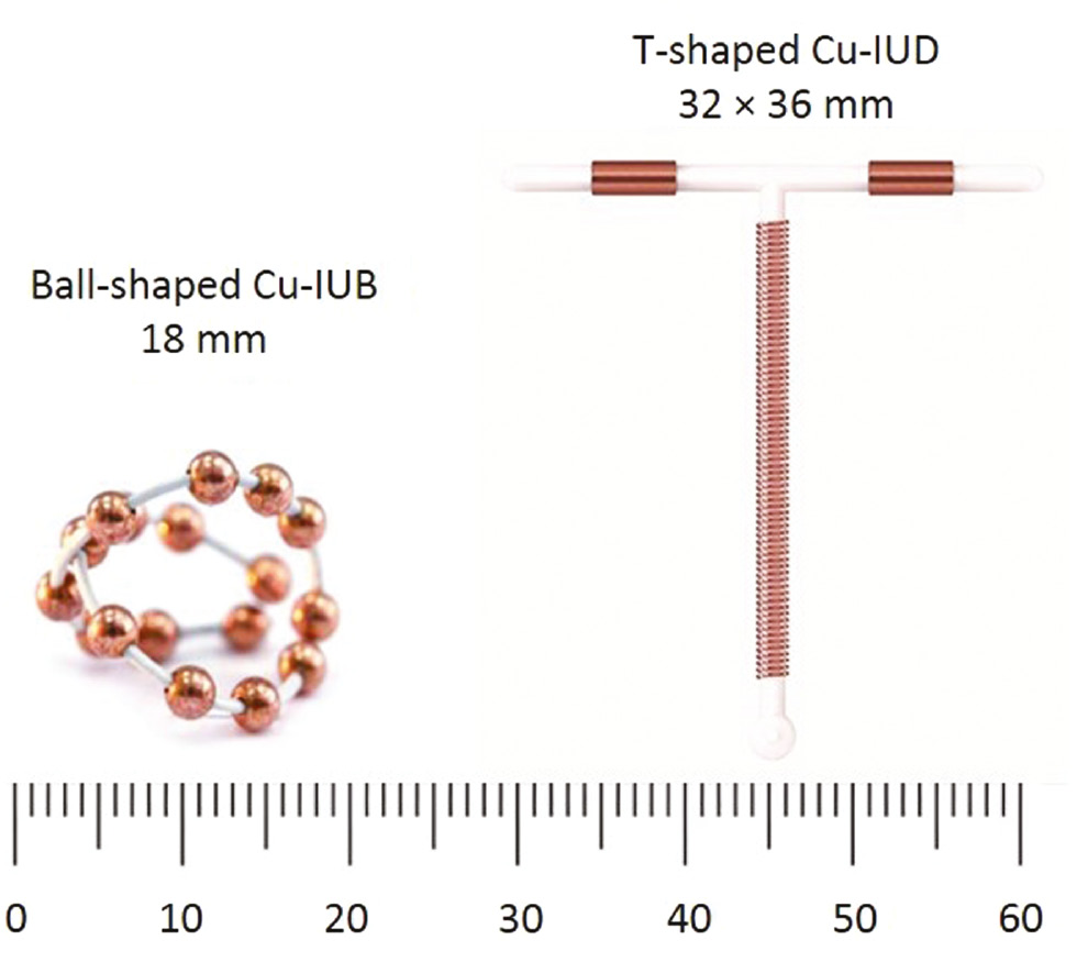
Cu-IUD: (left) IUB-shaped Cu-IUB and (right) T-shaped Cu-IUD.
Several shapes of Cu-IUDs are frequently used. Most countries used at least one T-shaped Cu-IUD. For instance, Mona Lisa in France and Belgium, Mirena in Argentina, Australia, Brazil, Canada, Colombia, Germany, Mexico, New Zealand, Peru, Spain, Sweden, The Netherlands, United Kingdom, and Para Gard and Skyla in the United States and China use stainless-steel rings despite their higher failure and expulsion rates (up to 10% per year; Bernstein et al., 1972; Orlans, 1974; Buhling et al., 2014).
Throughout the world, about 100 million Cu-IUDs are used as follows: China (more than 80 million), Vietnam (~4 million), Indonesia (3.5 million), Egypt (3 million), Mexico (2.5 million), Turkey (2.2 million), Colombia (0.7 million), Cuba (0.6 million), and Peru (0.5 million; Arancibia et al., 2003). Alvarez et al. (2012) indicated that approximately 162 million women worldwide used Cu-IUDs.
2 History and woman’s uterus
In all the treatises on IUDs, it is mentioned that Bedouin camel drivers had the habit of introducing in the uterus of the camels a rounded and smooth stone, the size of an almond, so that they would not be pregnant in the crossings of the desert (Margulies, 1975).
Towards 1930, the devices began to be used. The first consisted of a piece of silkworm gut of about 5 cm in length with a silver thread. Later, it was used in the form of a ring surrounded by silver. To introduce the device, a small fork was used, with which it forced to deform the ring in a guaranteed way; when removing the fork, it recovered the primitive round form.
Oppenheimer in Israel (1959) used polyethylene (PE) plastic material that has good “memory” and designed the IUD Margulies spiral-shaped, with a tail also of plastic, which was outside the uterus to allow extraction (Oppenheimer, 1959). Lippes designed an S-shaped IUD and the applicator was a thin plastic tube into which the S was inserted that was straight; when the applicator was removed, the device recovered the S-shape inside the uterus (Lippes, 1965). With these three basic ideas, the plastic, the applicator, and the tractor wire, different models were designed, so that almost every country has its own.
Anatomical and functional considerations of the uterus led Tatum (1974) to devise an IUD in the form of a T. IUDs with metals (copper, silver, and zinc) began to be used with good results. Van Os in Holland (Rhemrev et al., 1985) designed a model that does not require an applicator, similar in shape to the Dalkon shield, endowed with lateral spicules that hinder its expulsion and also with copper wire in the central stem (Zipper et al., 1976). Although Zipper et al. (1969) described the contraceptive effect of Cu-IUDs and zinc-bearing IUDs on rabbits, and different models of Cu-IUDs were proposed (Tatum, 1974), copper was known as a potent spermicide for more than 130 years (Roblero et al., 1996).
The woman’s uterus (womb) is hollow and characterized by a pear shape (typical dimensions of about 7.6 cm long, 4.5 cm broad, and 3.0 cm thick; weights about 60 g) in the lower abdomen between the bladder and the rectum. The measurement of the uterine cavity is an important but not definitive means to decrease the rates of unwanted effects (Kurz, 1985). The broader, upper part of the uterus is the corpus (with two layers of tissue; the outer layer is the myometrium), and the narrow lower part is the cervix. In women of childbearing age, the inner layer of the uterus (endometrium) goes through a series of monthly changes, because of menstrual cycle (Diez-Febrer et al., 1993).
3 Cu-IUDs
Cu-IUDs are classified as nonhormonal IUDs. These devices deliver cupric ions that serve as antifertility agents. A minor percentage of copper is on the surface of the shield and dissociation studies indicated that less than 5% of the total copper is released (Jones et al., 1973). Existing Cu-IUDs, which comprise a PE body and copper wire wound around the stem, are one of the most widely used form of birth control worldwide due to its advantages of safety and reversibility, high efficiency, economy, and long lasting (3–5 years). Nowadays, there are about 150 million IUD users all over the world (Bahamondes et al., 2003).
On the IUD shape, the T-shaped Cu-IUD is the most popular (Figure 1, right). Anchor-, U-, and 7-shaped Cu-IUDs are currently used. Pelvic inflammatory disease (PID) is the great workhorse to Cu-IUD, especially for American authors, from the misfortune that caused the Dalkon shield IUD in the early 1970s (De Castro, 1993). For these devices, shape is one of the main drawbacks, as they become difficult to extract and in some cases they can break a part while inside the uterus. IUD containing a gold or platinum core electrolytically coated with copper has also been used to minimize the wire fragmentation risk (Gal-Or et al., 1982).
3.1 Copper nanoparticles mixed with low-density PE
Although the antifertility efficiency of Cu-IUDs can be comparable to those of female or male sterilization, some side effects of Cu-IUDs are PID, menorrhagia, intermenstrual bleeding, and uterine perforation and so on. Nevertheless, the reaction of cupric ions on the cellular membranes with spermicidal and bactericidal effects is breaking the idea of the Cu-IUD-PID association. To overcome the limitations of conventional Cu-IUDs, copper nanoparticles mixed with low-density PE were proposed as contraceptive devices. Polymer matrix composites were used due to their superiority of controlled release of drug and continuous matrix phase (Ramakrishnan et al., 2015). The composites are filled with copper nanoparticles considering that the size effect of copper nanoparticles can increase the Cu(II) ion transformation ratio in a simulated uterine solution (SUS; Cai et al., 2005b; Xia et al., 2006a; Hu et al., 2013). Ultrafine-grained bulk copper fabricated by severe plastic deformation with excellent mechanical properties (Lugo, 2008) possesses excellent corrosion resistance in SUS (Xu et al., 2004, 2012; Shaamash et al., 2005; Wen et al., 2006; Xia et al., 2006b; Li et al., 2007; Liang et al., 2008).
3.2 Copper intrauterine ball (IUB)
Recently, in the literature, there has been a new IUD, named as IUB, consisting of copper specimens that take a three-dimensional spherical form, and 17 pure copper spheres are threaded over a shape memory alloy NiTiNol wire, a Ti-based alloy with highly pure Ni, and a commercial pure Ti (Baram et al., 2014; Wiebe, 2014; Wiebe & Trussell, 2016; Figure 1, left). NiTiNol presents the ability of deforming force, allowing flexions while always returning to its preset shape. Goldstuck and Wildemeersch (2017) have reported that the IUB has a more physiological fit in both nulliparous and multiparous users because of its extreme flexibility and small dimensions (~12 mm diameter). IUB is 14%–25% smaller than each arm of the common CuT380 IUD, and probably, this new design may reduce the high rate of expulsion in the first year of use (27%; Matsubayashi et al., 2007; Madden et al., 2014; Wiebe & Trussell, 2016). On the contrary, it has been reported that the NiTiNol shape memory alloy, in spite of its high Ni content (49% Ni and 51% Ti), is biocompatible and presents a high corrosion resistance in artificial saliva, and the Ni release is very low (~0.1 ppm; El Medawar et al., 2002).
4 Bactericidal effect of copper
Copper inhibits the growth of some bacteria, such as Neisseria gonorrhoeae and meningococci, preventing the transmission of sexual diseases such as syphilis, gonococci, or soft chancre (Valdéz et al., 2008). Enterobacter are commonly found in vaginal culture. The incidence of the following bacteria is as follows: Staphylococcus aureus 16%, Staphylococcus epidermis 18%, Pseudomonas aeruginosa 5%, Escherichia coli 25%, Candida albicans 20%, N. gonorrhoeae 2%, and Candida dublinieses 12% (Carrillo-Beltrán, 2009). Neisseria gonorrhoeae and Neisseria meningitidis (Meningococcus) have also been reported (De Castro, 1993). Figure 2 shows a scanning electron microscopy (SEM) micrograph of copper surface colonized by Actinomyces israelii biofilm (Valdéz et al., 2008).
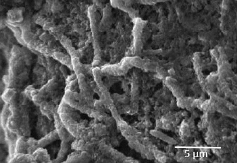
SEM micrograph of A. israelii biofilm on the copper surface.
Cu-IUDs inhibit spermatozoa motility, where Cu2+ ions significantly affect sperm incubation (Ullmann & Hammerstein, 1972; Barwin & Tuttle, 1978; Roblero et al., 1996). Copper changes the endometrium and human blastocysts are found to be sensitive to copper as well. It has been tested that the presence of copper is toxic to the embryo, thus playing a contraceptive effect (Brinster & Cross, 1972).
Actinomyces israelii biofilms developed in IUD used for a long time showed a branched growth adhered to the copper surface by its own extracellular polymers as demonstrated by Carrillo et al. (2010). In addition, they found in microbial growths in synthetic medium that A. israelii is capable of surviving copper toxicity due the porous structure of the biofilm.
5 Copper corrosion
A lack of information exits in the literature on the corrosion of Cu-IUDs using native (natural) uterine fluid, the small amount of fluid in the endometrial cavity, 5–35 μl in the midluteal phase and about 80–180 μl in midcycle (Casslén, 1986), and the difficulty associated with the extraction of representative fluid induces the use of SUS (Xue et al., 1998; Zhu et al., 1999; Bastidas et al., 2000).
5.1 Corrosion of copper in acidic and alkaline solutions
Given that, depending on the menstrual cycle and the user, the pH of the uterus cavity has been reported from 5.9 to 8.0 (Sedlis et al., 1967; Johnson Jr et al., 1976), it is of interest to discuss copper corrosion in acid and alkaline media.
The corrosion of copper in hydrochloric acid solution has been widely studied in the literature by different researchers (Crundwell, 1992; Diard et al., 1998; Polo et al., 2003). There is unanimity among different researchers in that chloride, complexes (Cu-CuCl2−), and insoluble products are the parameters that control the corrosion process; the diffusion of different soluble species, mainly CuCl2−, controls the kinetics of the corrosion process. In general, copper oxidizes forming chlorinated complexes of copper (I) at a rate that depends on the existing chloride concentration and is independent of pH and the mass transfer process. Copper does not corrode in nonoxidizing acid environments (Finsgar & Milosev, 2010). In the anodic dissolution of copper in hydrochloric acid solution, it is common to find a passivation of the material. The first stage is an electronic transfer process in which the chloride ion participates in the formation of the CuCl insoluble species adsorbed on the copper surface following the Langmuir isotherm (kc=θ/(1–θ)), where k is the equilibrium constant of the adsorption reaction, c is the concentration of the adsorbed species, and θ the degree of coating of the copper surface; the value of θ is assumed to be very small; θ<<1; Crundwell, 1992; Barcia, 1993; Diard et al., 1998). The second stage is a chemical reaction in which the CuCl species participates in the formation of the complex CuCl2− (Polo et al., 2003).
Copper has also been studied using sulfuric acid solution (Clerc & Alkire, 1991; Tromans & Ahmed, 1998). At low sulfuric acid concentrations, the copper solution is the main anodic process. However, at higher acid concentrations, metal passivation occurs (Bastidas et al., 2003). The passivation process has been related to the formation of a layer of cuprous oxide (Cu2O, cuprite) and another layer of cupric oxide (CuO, tenorite). A more general mechanism has also been proposed consisting of a dissolution-precipitation process. The cuprite and tenorite formed during the anodic polarization in contact with H2SO4 can undergo transformations to Cu, Cu2SO4, and CuSO4 (Tromans & Ahmed, 1998). It has also been reported that passivation occurs through the formation and growth of a CuSO4 layer instead of the corresponding cuprite (Clerc & Alkire, 1991), and copper oxides are stable only in the pH range of 8–12 (Finsgar & Milosev, 2010).
In the chemical treatments of cleaning and pickling of copper and its alloys, the use of organic acids is frequent. Figure 3 shows a Nyquist plot for copper in 5.0% citric acid after 96 h experimentation. A well-defined semicircle and a small tail (diffusion process) at high and low frequencies, respectively, can be observed. Figure 3 (inset) includes the equivalent circuit used to fit impedance data. The solution resistance (Rs) was 36 Ω cm2. The polarization resistance (Rp) was 4780 Ω cm2, which allows the monitoring of the corrosion rate. Using Faraday’s law, the loss of copper was determined to be 0.7 mg/cm2. The CPE parameter (characterized, as a first approximation, by the electrochemical double-layer (Cdl) and the dimensionless n exponent, n=1.0) was 24 μF/cm2. The diffusion process was simulated by the Warburg impedance (Zw; Zw=σ(i – j)ω−½), where σ is the Warburg diffusion coefficient, which was 150 Ω cm2 s−½. The strong acidity of the mineral acids allows a fast and also economic cleaning of the copper-based materials. However, organic acids have the advantage that, being less aggressive than mineral acids, they allow the formation of metal complexes and are not dangerous and are easy to handle and ecologically acceptable (Bastidas & Otero, 1996; 2002; Otero, 1996a; Polo et al., 2002; Cano et al., 2003). Finally, in the cleaning treatment of copper, the use of alkaline solutions is common. The aqueous alkaline solutions containing D(+)glucose and formaldehyde are an alternative to the conventional cleaning solution (Otero & Bastidas, 1996b; Cano et al., 2001b, 2005).
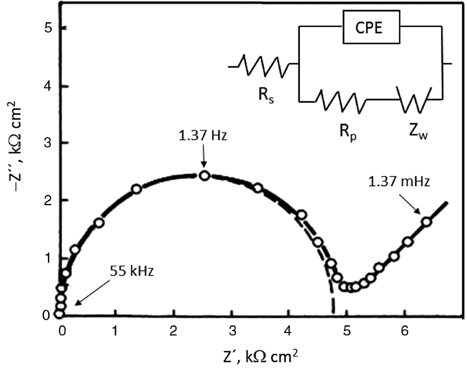
Nyquist plot for copper in 5.0% citric acid after 96 h experimentation at room temperature.
Figure 4 shows a schematic representation of copper corrosion mechanism on a Cu-IUD, which is characterized by a small central zone with restricted access of oxygen (anodic zone) surrounded by a big zone with free access of oxygen (cathodic zone). The presence of corrosion products deposited on the copper surface and the precipitation process of calcite (CaCO3), generated by the presence of sodium bicarbonate and calcium chloride as the components of SUS used (see Table 1), produce aeration differential cells causing pitting corrosion (Oster, 1972; Rosenfeld et al., 1981; Bastidas et al., 2001, 2010; Burstein & Liu, 2007; Alonso et al., 2016):
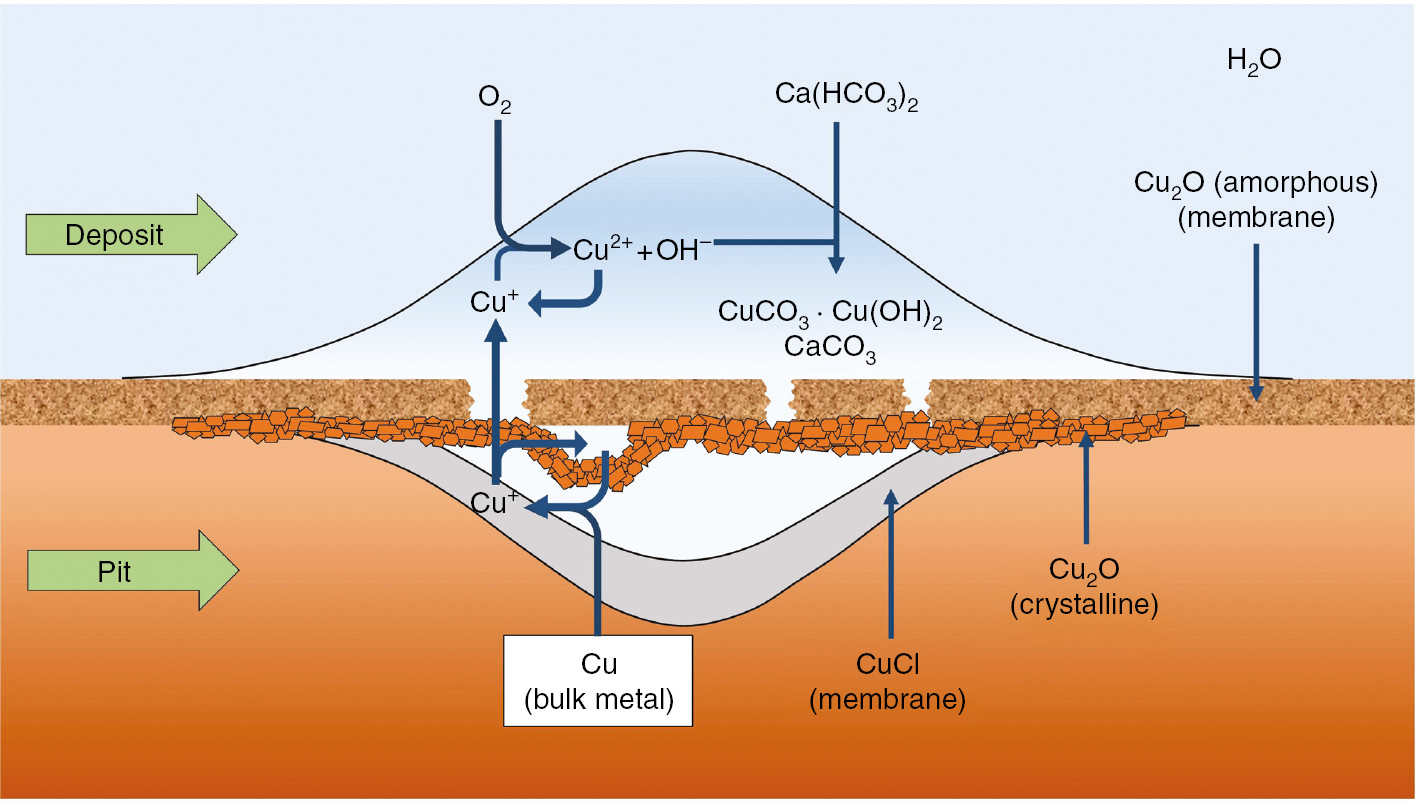
Copper pitting corrosion mechanism.
Chemical composition of SUS used (Zhang et al., 1996; Mora et al., 2002).
| Reference | Concentration (g/l) |
||||||||||||||
|---|---|---|---|---|---|---|---|---|---|---|---|---|---|---|---|
| NaHCO3 | NaH2PO4·2H2O | Glucose | Urea | Albumin | CaCl2 | KCl | NaCl | ||||||||
| Zhang et al., 1996 | 0.25 | 0.072 | 0.50 | – | – | 0.167 | 0.224 | 4.97 | |||||||
| Mora et al., 2002 | 0.25 | 0.072 | 0.50 | 0.48 | – | 0.167 | 0.224 | 4.97 | |||||||
| Mora et al., 2002 | 0.25 | 0.072 | 0.50 | – | 35 | 0.167 | 0.224 | 4.97 | |||||||
| Mora et al., 2002 | 0.25 | 0.072 | 0.50 | 0.48 | 35 | 0.167 | 0.224 | 4.97 | |||||||
Reaction inside a precipitated deposit:
Cathodic reaction taking place outside of the amorphous membrane of cuprite (Cu2O), a native oxide spontaneously generated on the copper surface with a porous structure (Cano et al., 2001a,b; Bastidas et al., 2005):
Anodic reaction taking place inside of the amorphous membrane of cuprite:
Reaction between Cu2+, Eq. (3), and the copper from the wall of the pit (Cu(s)):
5.2 Corrosion of copper in SUS
Table 1 includes the chemical composition of SUS used in the literature (Zhang et al., 1996; Mora et al., 2002).
Figure 5 shows copper dissolution rate against albumin content, using SUS (Table 1), for different times of experimentation, albumin content, pH 6.3 and 8.0, and 0.2 atm of oxygen pressure. It can be observed that the behavior is similar for both pH 6.3 and 8.0 with a high dissolution rate up to 500–700 μg/day for 1 day and 110–250 μg/day for 30 days of experimentation. Figure 6 shows an SEM micrograph for the copper specimen of Figure 5 after 1 day of experimentation. It can be observed that there is a broken and nonadherent layer of corrosion products, which allows the diffusion of copper ions for the contraceptive effect. Pitting corrosion was detected on the copper surface after the removal of the corrosion products.
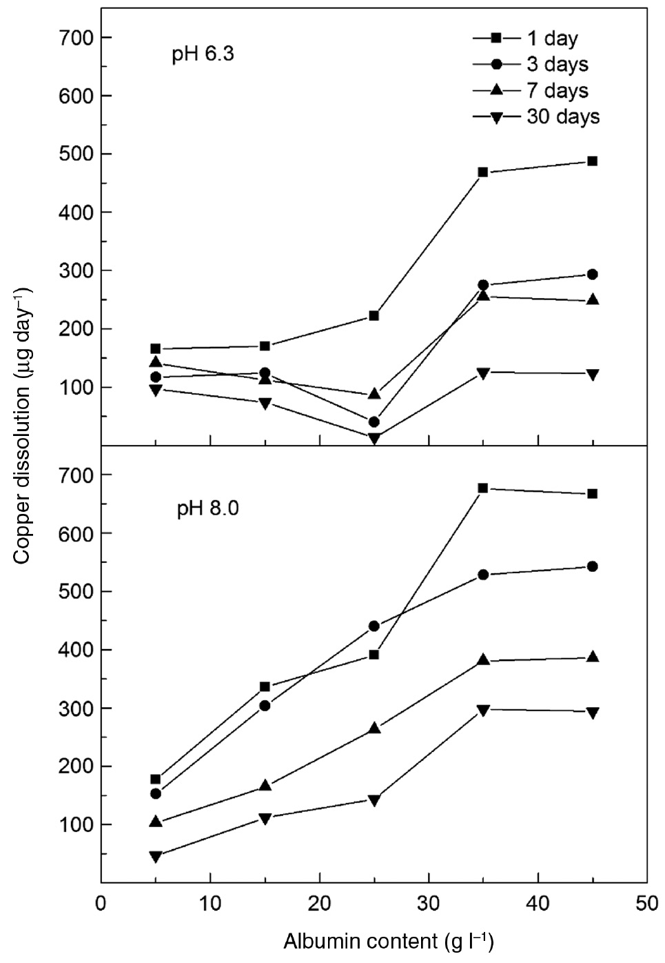
Copper dissolution rate vs. albumin content for copper surface area of 200 mm2 in SUS (Table 1) without albumin (Mora et al., 2002).
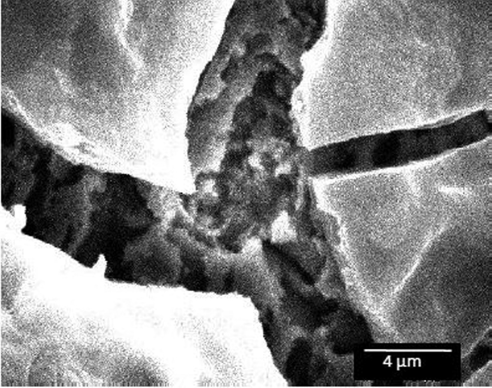
SEM micrograph for copper immersed in SUS (Table 1) with urea and albumin, pH 8.0, and 0.2 atm of oxygen pressure (Mora et al., 2002).
It has been reported that there is a much higher release of copper in women during the first two cycles of use and after this a relative constant release for 1 year (Hagenfeldt, 1972, 1987). Previous studies have shown that Cu-IUD devices exhibit fast copper dissolution rates during the early stages of exposure to the uterine fluid, commonly referred to as “burst release” causing bleeding and pain (Zhang et al., 1996; Bastidas et al., 2000; De la Cruz et al., 2005; Gao et al., 2007; Pereda et al., 2008; Zhang et al., 2015). Anomalous bleeding occurs during the first few months after Cu-IUD insertion. This could lead to persistent menorrhagia, where Cu-IUD device removal is recommended. Typical blood loss within the menstrual period is about 35 ml (Barwin & Tuttle, 1978). The accumulation of cupric ions in solution results with time in the deposition of corrosion products in the IUD surface (Bank et al., 1975). This partially protective film decreases the Cu dissolution rate until steady-state Cu release kinetics is observed (Bastidas et al., 2001; Valdéz et al., 2003a,b).
Figure 7 shows the copper dissolution rate vs. time for different pH values using SUS (Table 1; Mora et al., 2002) without albumin and in the absence of additional pressure of oxygen at pH 5.0, 6.3, and 8.0. It can be observed that the behavior is similar for the three pH values with an exponential decay of copper corrosion with time in the first 10 days of experimentation, and after that, a steady state is defined with 0.5–1.5 μg/day copper dissolution. This process is defined as the burst release phenomenon. At the beginning of the experiment, the pits generated increase the copper surface area exposed to SUS originating a high corrosion rate (Figure 8). Copper release in utero reported in the literature varies in a wide range from 44 to 296 μg/day after the first month of insertion (Akinla et al., 1975; Kjaer et al., 1993; Patai et al., 2003) and decreasing to a steady-state value of 7 μg/day after 12 months of insertion (Thiery et al., 1982). In vitro experiments performed by Zhou et al. (2010) reported a copper release rate of as low as 2 μg/day in the steady state and the Cu-IUD with indomethacin was effective for contraceptive use.
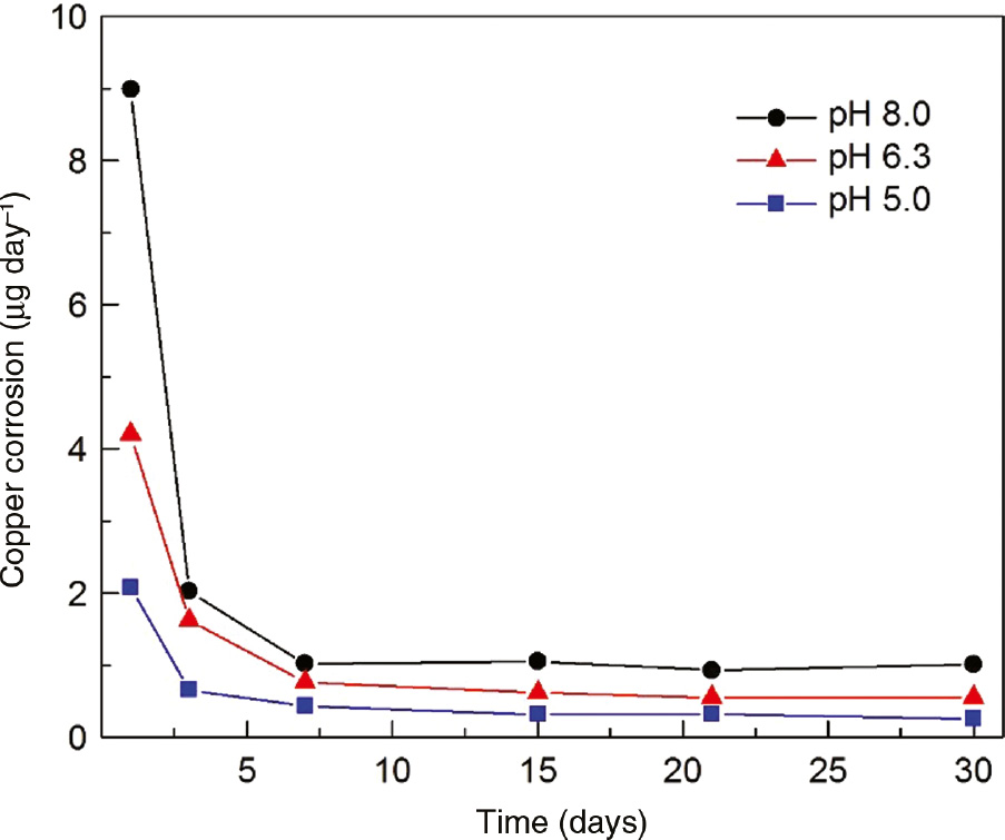
Copper corrosion rate vs. time. Copper surface area of 200 mm2 in SUS (Table 1) without albumin (Mora et al., 2002).
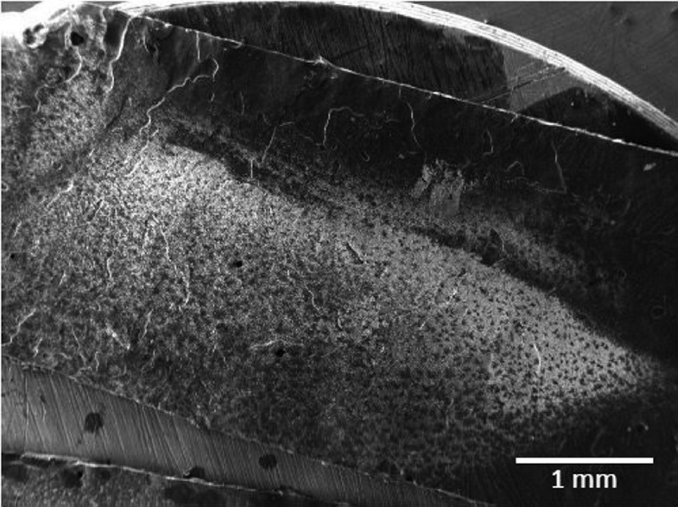
Field emission-SEM morphology of a copper specimen exposed to SUS (Table 1) without albumin (Mora et al., 2002). Pits can be observed over the entire surface.
The quantification of in vitro burst release process can be done using different techniques, such as inductively coupled plasma (ICP)-optical emission spectroscopy (OES) with a lower limit of parts per billon, ICP-mass spectrometry (MS), flame atomic absorption spectroscopy (FAAS), atomic absorption spectroscopy (AAS), or diethyl ammonium salt of diethyldithiocarbamic acid (DAD) in spectrophotometric measurements because the sensibility is enough to study the process (Bastidas et al., 2019a,b). Anodic stripping voltammetry has also been used to quantify the amount of copper (Arancibia et al., 2003).
Copper release rate depends on different issues such as copper grade, effective surface area, and pH. An interesting alternative to reduce the undesirable burst release phenomenon is the use of organic inhibitors such as thiourea and benzotriazole. However, research is required to introduce a corrosion inhibitor in the uterus without risks avoiding secondary effects, among others (Thiery et al., 1982; Bastidas et al., 2000; Scendo, 2008; Turok, 2011; Wildemeersch et al., 2014; Zhang et al., 2015). Promising results have been reported by Alvarez et al. (2012) for purine (C5H4N4) with inhibitory efficiency higher than 98%. This heterocyclic compound is present in the structure of RNA and DNA, and probably, it is nontoxic for the body.
A characteristic coloring is one of the most distinguishing features of cupric ion chelates. Visual absorption spectra can be used to quantify the cupric ion content (Mora et al., 2002). The diethyl ammonium salt of DAD acid forms a yellow chelate with the cupric ion. Absorbance measurements performed at a wavelength of 446 nm using an HACH Model DR/3000 spectrophotometer (Figure 9), which shows a calibration of absorbance vs. cupric ion concentration, showed that the absorbance is proportional to cupric ion in the range from 10 to 80 μmol/l.
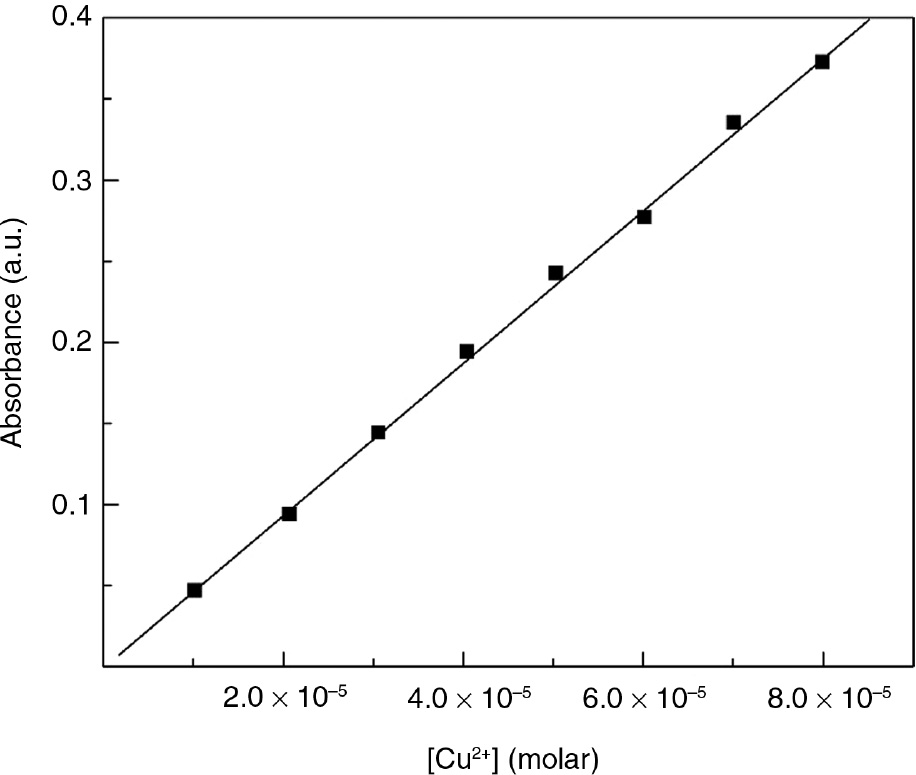
Calibration plot for copper dissolution, absorbance vs. molar concentration of copper.
The morphology of the corrosion products and deposits is a nonuniform layer, showing some paths through which copper ions can be released, favoring copper to act as a contraceptive device (Patai et al., 1998, 2004; Bastidas et al., 2003; Berthou et al., 2003). Thus, a nonadherent and broken layer is formed, and a compact layer of cuprite is produced underneath, where sulfur and chloride pits were generated. Pitting corrosion is generally observed on the copper surface after the removal of the corrosion product layers. Copper has a contraceptive effect due to cupric ion release into the uterine environment (Ortiz et al., 1996; Cai et al., 2005a).
Pereda et al. (2009) has demonstrated that the early fragmentation of Cu-IUDs cannot be attributed to stress corrosion cracking (SCC). A number of wire samples were metallographically mounted, sectioned, and observed under the optical microscope. No cracks were found. Copper wires are twisted onto an inert plastic rod during the manufacturing process of the Cu-IUD, so that residual stresses are present in the copper wires. This fact, together with the existence of a chloride and organic compounds containing medium like the uterine fluid environment, may lead to SCC. It is known that the presence of Cu2+ ions at high concentrations of the order of 1 m can originate from the SCC of copper (Arancibia et al., 2003; Farina et al., 2005). The shorter fracture times observed in some cases may be attributed to a high uniform corrosion rate that reduces the wire sections and leads to rupture by the overloading of the remaining smaller section. The addition of cupric ions to the solutions at concentrations similar to those measured in uterine fluid (20 mg/l=3×10−3m) did not lead to SCC in copper (Farina et al., 2005).
6 Cu-IUD mechanism
The accurate mechanism of action of the multifunctional effect of a Cu-IUD in the antifertility process is still not known. Pregnancy is prevented by a combination of a metal or plastic frame; a foreign body effect is the most important phenomenon in the antifertility process; the chemical and biochemical reactions are related to the type of material of which the foreign body is composed, causing sterile inflammation that is toxic to sperm; and the specific action of copper is mainly through the release of copper (Zipper et al., 1969; Ortiz et al., 1996; Ortiz & Croxatto, 2007; Chen et al., 2006). For instance, to ensure the antifertility effect of a Cu-IUD, the concentration of the released cupric (Cu2+) ions in uterine solution needs to be higher than 10−6 mol/l (Figure 10; Araya et al., 2003). Copper ions delivered from Cu-IUDs enhance the inflammatory response in the uterine cavity and reach concentrations in the luminal fluids of the genital tract that are toxic for spermatozoa and embryos (Jones et al., 1973; Middleton & Kennedy, 1975; Holland & White, 1998; Linder & Hazegh-Azam, 1996; Ortiz et al., 1996; Bastidas & Simancas, 1997; Bastidas et al., 2001; Sivin, 1997; Mishell, 1998; van Os & Edelman, 1998; Homouda, 2002; Araya et al., 2003; Caliskan et al., 2003). It is believed that the presence of Cu-IUD in the endometrial cavity increases the concentration of Cu2+, accompanied by the synthesis of lipids such as PG that are involved in inflammation and motility effects (Hagenfeldt, 1987), which gains the tubal contractility (higher-frequency intensity), and white blood cells (macrophages, within the uterine and tubal fluids; Kosonen, 1978, 1980; Koch & Vogel, 1980; De Castro et al., 1986; De Castro & González-Gancedo, 1987, De Castro 1993; Grillo et al., 2010). These compounds prevent the encounter of healthy gametes, the formation of viable embryos, and their ability to reach the uterus (motility effect). In other words, they prevent the fertilization of the ovum by interference with sperm function and transport within the uterus and tubes (Carrascosa et al., 2018). In addition, the presence of copper can result in the oxidation of sulfhydryl (-SH) groups of acid fibroblast growth factor causing the loss of the myogenic activity of the protein (Oster, 1972; Oster & Salgo 1975; Beltrán-García et al., 1998, 2000; Lozano et al., 2000; Cunha et al., 2001; Stanford & Mikolajczyk, 2002).
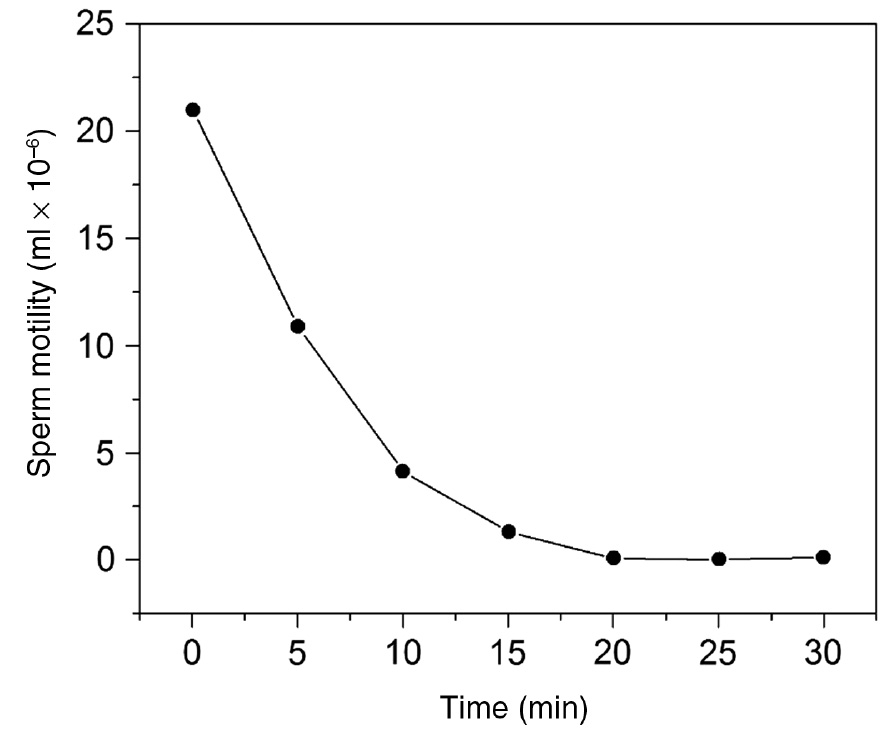
Sperm motility vs. time for a cupric ion concentration of 8 μmol/l CuCl2 in Ringer’s solution (0.72 NaCl, 0.03 KCl, 0.29 MgSO4, and 0.26 Na3PO4, concentration as g/100 ml), pH 7.2, and temperature of 37°C.
The endocrinologic mechanisms of reproductive processes may be summarized as follows (Figure 11). The ovary during the menstrual cycle produces two hormones (H; estrogen and progesterone) carrying information to a specific protein structure, receptor (R), located on the cytoplasmic membrane. Copper ions (Cu2+) from a Cu-IUD may alter the relationship with oligoelements (Cu, Zn, Mo, Nb, and Mn) having a negative impact on the motility (Figure 11, bottom; De Castro, 1993). The H reaches the endometrial territory through the arteries that irrigate the uterus. Once there, it comes in contact with its specific R and causes changes in the protein structure of the uterus that induce their way through the cytoplasm to reach the nucleus. During this first phase, the R is called the hormone-cytoplasmic receptor (HRc) complex. This inactivated complex starts its way to the cell nucleus through a movement called translocation (Ortega et al., 1991; Kadanali et al., 2001; Kleszczewski et al., 2003; Savaris & Chies, 2003; Karasahin et al., 2011). While approaching the nucleus, the complex undergoes a transmutation called transformation to bind to the nuclear chromatin and modify in an active complex, the so-called nuclear hormone-receptor complex (HRn; Figure 11; De Castro, 1993). Once the active complex is located inside the cell nucleus, an action is produced on the DNA, which entails the appearance of an mRNA to carry the information of the specialized work of the cytoplasmic ribosomes and to regulate the intracellular protein synthesis. Once the function of the active complex has been completed, it passes back to the cytoplasm, deactivating and undergoing a recycling process to start the whole process again.
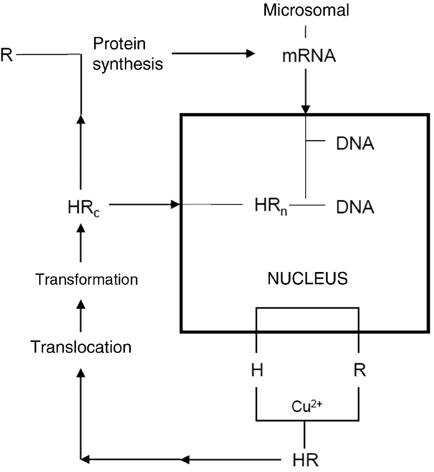
Recycling reproductive process mechanism. H, hormone; HR, hormone-receptor complex; HRc, hormone-cytoplasmic receptor complex; HRn, nuclear hormone-receptor complex; mRNA, RNA (carrier of genetic codes); R, receptor.
7 Conclusion and recommendations
Chemical analyses (ICP-OES, ICP-MS, FAAS, and AAS), absorbance measurements, and electrochemical methods are adequate for the quantification of the copper release process of a Cu-IUD in an SUS environment. The SUS described in the literature are suitable to perform in in vitro experiments on the copper release of a Cu-IUD.
Burst release phenomenon taking place in the first 3 weeks of Cu-IUD insertion in the uterus cavity (copper release of 5–10 μg/day followed by a low steady-state dissolution process with ~0.5 μg/day), producing bleeding and pain, needs to be avoided or minimized, so the phenomenon needs a in-depth investigation. Results reported in the literature using corrosion inhibitors as pretreatment of SUS are hopeful and need to be studied in detail.
Microbial biofilms can coexist on the Cu-IUD surfaces due to the porous structure formed with the extracellular polymeric matrix. The interaction between these microbial growths and the copper ions released does not affect the efficacy of the contraceptive function of the device.
Studies are required to understand the mechanism of action of a Cu-IUD. For instance, (1) the copper dissolution rate of 2 μg/day for a Cu-IUD with indomethacin after a long time of insertion in the uterus and (2) the released cupric ions in the uterine solution that need to be higher than 10−6 mol/l are important findings that require to be studied in depth. Another example is that the minimum copper release rate necessary for a Cu-IUD to be efficient is not known.
About the authors

David M. Bastidas is an associate professor of corrosion engineering at The University of Akron. He received his PhD in materials science and engineering and BSc in chemistry from the University of Barcelona (Spain). His research focuses on the corrosion of steel in concrete, inhibition, and Cu-IUD corrosion. He is a member of research and infrastructure committees of NACE and the vice chairman of corrosion of steel in concrete WP of EFC.

Benjamin Valdez is a professor at the Institute of Engineering, UABC and was the Institute’s director from 2006 to 2013. He has PhD and MSc in chemistry and BSc in chemical engineering. He was appointed a member of the Mexican Academy of Science and the National System of Researchers in Mexico. He was a guest editor of Corrosion Reviews. His activities include corrosion research, consultancy, and control in industrial plants and environments.

Michael Schorr is a professor (Dr. honoris causa) at the Institute of Engineering, UABC. He received his PhD in materials engineering and BSc in chemistry from the Technion-Israel Institute of Technology. He was an editor of Corrosion Reviews (1986–2004). His research focuses on volatile corrosion inhibitors in industrial environments. He is a corrosion consultant and a professor in Israel, United States, Latin America, and Europe.

Jose M. Bastidas is a full professor at the CENIM, CSIC. He received his PhD in chemistry from Complutense University of Madrid, Spain. He was a postdoctoral Ramsey fellow at the University of Manchester (UK). His research focuses on corrosion and inhibition mechanisms, protection methods and numerical modeling in biomedical materials, chemical industry, heritage science, construction, and transportation.
-
Funding: D.M. Bastidas acknowledges funding from The University of Akron.
References
Akinla O, Luukkainen T, Timonen H. Important factors in the use-effectiveness of the copper-T-200 IUD. Contraception 1975; 12: 696–707.10.1016/S0010-7824(75)80052-7Search in Google Scholar
Alonso C, Casero E, Roman E, Campos SF-P, de Mele MFL. Effective inhibition of the early copper ion burst release by purine adsorption in simulated uterine fluids. Electrochim Acta 2016; 189: 54–63.10.1016/j.electacta.2015.12.093Search in Google Scholar
Alvarez F, Schilardi PL, de Mele MFL. Reduction of the burst release of copper ions from copper-based intrauterine devices by organic inhibitors. Contraception 2012; 85: 91–98.10.1016/j.contraception.2011.05.011Search in Google Scholar PubMed
Arancibia V, Peña C, Allen HE, Lagos G. Characterization of copper in uterine fluids of patients who use the copper T-380A intrauterine device. Clin Chim Acta 2003; 332: 69–78.10.1016/S0009-8981(03)00124-4Search in Google Scholar PubMed
Araya R, Gómez-Mora H, Vera R, Bastidas JM. Human spermatozoa motility analysis in a Ringer’s solution containing cupric ions. Contraception 2003; 67: 161–163.10.1016/S0010-7824(02)00477-8Search in Google Scholar
ASTM B170-99 (2015). Standard specification for oxygen-free electrolytic copper-refinery shapes. West Conshohocken, PA: ASTM International, 2015.Search in Google Scholar
Bahamondes L, Faundes A, Sobreira-Lima B, Lui-Filho JF, Pecci P, Matera S. TCu 380A IUD: a reversible permanent contraceptive method in women over 35 years of age. Contraception 2003; 72: 337–341.10.1016/j.contraception.2004.12.026Search in Google Scholar PubMed
Bank HL, Williamson HO, Manning K. Scanning electron microscopy of copper-containing intrauterine devices: long-term changes in utero. Fertil Steril 1975; 26: 503–503.10.1016/S0015-0282(16)41170-2Search in Google Scholar
Baram I, Weinstein A, Trussell J. The IUB, a new invented IUD: a brief report. Contraception 2014; 89: 139–141.10.1016/j.contraception.2013.10.017Search in Google Scholar PubMed PubMed Central
Barcia OE. Mattos OR, Pébére N, Tribollet B. Mass-transport study for the electrodissolution of copper in 1 M hydrochloric acid solution by impedance. J Electrochem Soc 1993; 140: 2825–2832.10.1149/1.2220917Search in Google Scholar
Barwin BN, Tuttle S. Jolly EE. The intrauterine contraceptive device. Can Med Assoc J 1978; 118: 53–58.Search in Google Scholar
Bastidas JM, Otero E. A comparative study of benzotriazole and 2-amino-5-mercapto-1,3,4-thiadiazole as copper corrosion inhibition in acid media. Mater Corros 1996; 47: 333–337.10.1002/maco.19960470606Search in Google Scholar
Bastidas JM, Simancas J. Characterization of corrosion products on a copper containing intrauterine device during storage at room temperature. Biomaterials 1997; 18: 247–250.10.1016/S0142-9612(96)00136-6Search in Google Scholar
Bastidas JM, Cano E, Mora N. Copper corrosion-simulated uterine solutions. Contraception 2000; 61: 395–399.10.1016/S0010-7824(00)00124-4Search in Google Scholar PubMed
Bastidas JM, Mora N, Cano E, Polo JL. Characterization of copper corrosion products originated in simulated uterine fluids and on packaged intrauterine devices. J Mater Sci Mater M 2001; 12: 391–397.10.1023/A:1011288701499Search in Google Scholar
Bastidas JM, Pinilla P, Polo JL, Cano E. Adsorption of benzotriazole on copper electrode surface in citric acid media. Corrosion 2002; 58: 922–931.10.5006/1.3280782Search in Google Scholar
Bastidas JM, Pinilla P, Cano E, Polo JL, Miguel S. Copper corrosion inhibition by triphenylmethane derivatives in sulfuric acid media. Corros Sci 2003; 45: 427–449.10.1016/S0010-938X(02)00123-3Search in Google Scholar
Bastidas DM, Cano E, Mora EM. Influence of oxygen, albumin and pH on copper dissolution in a simulated uterine fluid. Eur J Contracept Reprod Health Care 2005; 10: 123–130.10.1080/13625180500131642Search in Google Scholar PubMed
Bastidas DM, Criado M, Fajardo S, La Iglesia MV, Cano E, Bastidas JM. Copper deterioration: causes, diagnosis and risk minimization. Int Mater Rev 2010; 55: 99–127.10.1179/095066009X12506721665257Search in Google Scholar
Bastidas DM, Daniëls F, Velerius T, Bastidas JM. In vitro comparative study on Cu-IUD burst release in simulated uterine solution using two quantification techniques. Contraception 2019a (in press).Search in Google Scholar
Bastidas DM, Valdéz-Salas B, Beltrán-Partida E, Bastidas JM. Copper nano and microparticle formation on Cu-IUDs during in-vitro testing. Contraception 2019b (in press).Search in Google Scholar
Beltrán-García MJ, Espinosa A, Pérez-Zapata AJ, Ogura T. In vitro cell toxicity and catalase destruction by metallic copper of the intrauterine device. Pathophysiology 1998; 5: 82.10.1016/S0928-4680(98)80599-3Search in Google Scholar
Beltrán-García MJ, Espinosa A, Herrera N, Pérez-Zapata AJ, Beltrán-García C, Ogura T. Formation of copper oxychloride and reactive species as causes of uterine injury during copper oxidation of Cu-IUD. Contraception 2000; 61: 99–103.10.1016/S0010-7824(00)00085-8Search in Google Scholar
Bernstein GS, Israel R, Seword P, Mishell Jr DR. Clinical experience with the Cu-7 intrauterine device. Contraception 1972; 6: 99–107.10.1016/0010-7824(72)90051-0Search in Google Scholar PubMed
Berthou J, Chretien FC, Driguez PA. Degradation of copper IUDs in utero. The process of metallic corrosion. A scanning electron microscope study. Gynecol Obstet Fertil 2003; 31: 29–42.10.1016/S1297-9589(02)00013-9Search in Google Scholar
Brinster RL, Cross PC. Effect of copper on the preimplantation mouse embryo. Nature 1972; 238: 398–399.10.1038/238398a0Search in Google Scholar PubMed
Buhling KJ, Zite NB, Lotke P, Black K. Worldwide use of intrauterine contraception: a review. Contraception 2014; 89: 162–173.10.1016/j.contraception.2013.11.011Search in Google Scholar PubMed
Burstein GT, Liu C. Nucleation of corrosion pits in Ringer’s solution containing bovine serum. Corros Sci 2007; 49: 4296–4306.10.1016/j.corsci.2007.05.018Search in Google Scholar
Cai S, Xia X, Xie C. Research on Cu2+ transformations of Cu and its oxides particles with different sizes in simulated uterine solution. Corros Sci 2005a; 47: 1039–1047.10.1016/j.corsci.2004.06.015Search in Google Scholar
Cai S, Xia X, Xie C. Corrosion behavior if copper/LDPE nanocomposites in simulated uterine solution. Biomaterials 2005b; 26: 2671–2676.10.1016/j.biomaterials.2004.08.003Search in Google Scholar PubMed
Caliskan E, Öztürk N, Dilbaz BÖ, Dilbaz S. Analysis of risk factors associated with uterine perforation by intrauterine devices. Eur J Contracept Reprod H C 2003; 8: 150–155.10.1080/ejc.8.3.150.155Search in Google Scholar
Cano E, Bastidas JM, Polo JL, Mora N. Study of the effect of acetic acid vapor on copper corrosion at 40 and 80% relative humidity. J Electrochem Soc 2001a; 148: B431–B437.10.1149/1.1404968Search in Google Scholar
Cano E, López MF, Simancas S, Bastidas JM. X-ray photoelectron spectroscopy study on the chemical composition of copper tarnish products formed at low humidities. J Electrochem Soc 2001b; 148: E26–E30.10.1149/1.1344547Search in Google Scholar
Cano E, Pinilla P, Polo JL, Bastidas JM. Copper corrosion inhibition by fast green, fuchsia acid and basic compounds in citric acid solution. Mater Corros 2003; 54: 222–228.10.1002/maco.200390050Search in Google Scholar
Cano E, Polo JL, La Iglesia A, Bastidas JM. Rate control for copper tarnishing. Corros Sci 2005; 47: 977–987.10.1016/j.corsci.2004.06.026Search in Google Scholar
Carrascosa JP, Cotan D, Jurado I, Oropesa-Avila M, Sánchez-Martin P, Savaris RF, Tan J, Sánchez-Alcazar JA, Tan SL, Horcajadas JA. The effect of copper on endometrial receptivity and induction of apoptosis on decidualized human endometrial stronal cells. Reprod Sci 2018; 25: 985–999.10.1177/1933719117732165Search in Google Scholar PubMed
Carrillo-Beltrán M. In: Corrosion induced by microorganism in copper intrauterine devices [in Spanish]. Mexicali, Mexico: Autonomous University of Baja California. 2009: 12: 25.Search in Google Scholar
Carrillo M, Valdéz B, Schorr M, Vargas L, Álvarez L, Zlatev R, Stoytcheva M. In-vitro Actinomyces israelii’s biofilm development on IUD copper surfaces. Contraception 2010; 81: 261–264.10.1016/j.contraception.2009.09.008Search in Google Scholar PubMed
Casslén B. Uterine fluid volume. Cyclic variations and possible extrauterine contributions. J Reprod Med 1986; 31: 506–510.Search in Google Scholar
Chen Z, Meng H, Xing G, Chen C, Zhao Y, Jia G, Wang T, Yuan H, Ye C, Zhao F, Chai Z, Zhu C, Fang X, Ma B, Wan L. Acute toxicological effects of copper nanoparticles in vivo. Toxicol Lett 2006; 163: 109–120.10.1016/j.toxlet.2005.10.003Search in Google Scholar PubMed
Clerc C, Alkire R. Effect of benzotriazole on surface processes during copper electrodissolution in sulfuric acid. J Electrochem Soc 1991; 138: 25–33.10.1149/1.2085552Search in Google Scholar
Crundwell FK. The anodic dissolution of copper in hydrochloric acid solutions. Electrochim Acta 1992; 37: 2707–2714.10.1016/0013-4686(92)85197-SSearch in Google Scholar
Cunha ACRD, Dorea JG, Cantuaria AA. Intrauterine device and maternal copper metabolism during lactation. Contraception 2001; 63: 37–39.10.1016/S0010-7824(00)00191-8Search in Google Scholar PubMed
De Castro A. Mechanism of action of intrauterine devices. In: Fernández-Bolaños J, García-Triguero A, editors. The contraception by intrauterine device [in Spanish]. ELA edition. Madrid, Spain, 1993: 71–83.Search in Google Scholar
De Castro A, González-Gancedo P. Mechanism of action of high-load copper IUDs. Adv Contracept 1987; 4: 185–190.10.1007/BF01849436Search in Google Scholar PubMed
De Castro A, González-Gancedo P, Contreras F, Lapeña G. The effect of copper ions in vivo on specific hormonal endometrial receptors. Adv Contracept 1986; 2: 399–404.10.1007/BF02340058Search in Google Scholar PubMed
De la Cruz D, Cruz A, Arteaga M, Castillo L, Tovalin H. Blood copper levels in Mexican users of the T380A IUD. Contraception 2005; 72: 122–125.10.1016/j.contraception.2005.02.009Search in Google Scholar PubMed
Diard JP, Le Canut JM, Le Gorrec B, Montella C. Copper electrodissolution in 1 M HCl at low current densities. II. Electrochemical impedance spectroscopy study. Electrochim Acta 1998; 43: 2485–2501.10.1016/S0013-4686(97)10156-6Search in Google Scholar
Diez-Febrer E, Fuster-Cortina V, González-Santana A, Plana-Royo A. Late complications. II. Metrorrhagia. In: Fernández-Bolaños J, Garcia-Triguero A, editors. The contraception by intrauterine device [in Spanish]. ELA edition. Madrid, Spain, 1993: 137–157.Search in Google Scholar
El Medawar L, Rocher P, Hornez JC, Traisnel M, Breme J, Hildebrand HF. Electrochemical and cytocompatibility assessment of NiTiNol memory shape alloy for orthodontic use. Biomol Eng 2002; 19: 153–160.10.1016/S1389-0344(02)00041-2Search in Google Scholar
Farina SB, Duffó GS, Galvele JR. Stress corrosion cracking of copper and silver, specific effect of the metal cations. Corros Sci 2005; 47: 239–245.10.1016/j.corsci.2004.05.019Search in Google Scholar
Finsgar M, Milosev I. Inhibition of copper corrosion by 1,2,3-benzotriazole: a review. Corros Sci 2010; 52: 2737–2749.10.1016/j.corsci.2010.05.002Search in Google Scholar
Gal-Or L, Gonen R, Zilberman A, Scharf M. Corrosion of a new copper-gold or copper-platinum intrauterine device. J Biomed Mater Res 1982; 16: 785–798.10.1002/jbm.820160605Search in Google Scholar PubMed
Gao J, Li Y, Liu L, Gu X. Releasing of cupric ion of three types of copper-bearing intrauterine contraceptive device in simulated uterine fluid. J Reprod Contracept 2007; 18: 33–39.10.1016/S1001-7844(07)60005-4Search in Google Scholar
Goldstuck ND, Wildemeersch D. Prevention of intrauterine contraceptive device expulsion and intolerance: determination of the anchor mechanism. Clin Obstet Gynecol Reprod Med 2017; 3: 1–8.10.15761/COGRM.1000173Search in Google Scholar
Grillo CA, Reigosa MA, de Mele MAFL. Does over-exposure to copper ions released from metallic copper induce cytotoxic and genotoxic effects on mammalian cells? Contraception 2010; 81: 343–349.10.1016/j.contraception.2009.12.003Search in Google Scholar PubMed
Hagenfeldt W. Studies on mode of action of copper-T device. Acta Endocrinol 1972; 71: 169–169.10.1530/acta.0.071S009Search in Google Scholar
Hagenfeldt K. The role of prostaglandins and allied substances in uterine haemostasis. Contraception 1987; 36: 23–35.10.1016/0010-7824(87)90059-XSearch in Google Scholar PubMed
Holland MK, White IG. Heavy metals and human spermatozoa. III. The toxicity of copper ions for spermatozoa. Contraception 1998; 38: 685–695.10.1016/0010-7824(88)90050-9Search in Google Scholar PubMed
Homouda K. The effect on intrauterine device position and performance of a modified TCu380A insertion technique. Eur J Contracept Reprod H C 2002; 7: 31–35.10.1080/ejc.7.1.31.35Search in Google Scholar
Hu LX, He J, Hou L, Wang H, Li J, Xie C, Duan Z, Sun LK, Wang X, Zhu C. Biological evaluation of the copper/low-density polyethylene nanocomposite intrauterine device. PLoS One 2013; 8: e74128.10.1371/journal.pone.0074128Search in Google Scholar PubMed PubMed Central
Johnson Jr AB, Maness RF, Wheeler RG. Calcareous deposits formed on IUDs in human exposures. Contraception 1976; 14: 507–517.10.1016/0010-7824(76)90002-0Search in Google Scholar PubMed
Jones RW, Gregson NM, Elstein M. Effect of copper-containing intrauterine contraceptive devices on human cells in culture. Br Med J 1973; 2: 520–523.10.1136/bmj.2.5865.520Search in Google Scholar PubMed PubMed Central
Kadanali S, Varoglu E, Komec D, Oslu H. Evaluation of active and passive transport mechanisms in genital tracts of IUD-bearing women with radionuclide hysterosalpingoscintigraphy. Contraception 2001; 63: 41–45.10.1016/S0010-7824(00)00190-6Search in Google Scholar PubMed
Karasahin KE, Keskin U, Turok DK, Cleland K, Raymond E, Trussell J, Cheng L, Zhu H, Kozinszky Z, Lieng M. Copper IUD and emergency contraception. Contraception 2011; 84: 205–207.10.1016/j.contraception.2010.12.005Search in Google Scholar PubMed
Kavanaugh ML, Jerman J. Contraceptive method used in the United States: trends and characteristics between 2008, 2012 and 2014. Contraception 2018; 97: 14–21.10.1016/j.contraception.2017.10.003Search in Google Scholar PubMed PubMed Central
Kjaer A, Laursen K, Thormann L, Borggaard O, Lebech PE. Copper release from copper intrauterine devices removed after up to 8 years of use. Contraception 1993; 47: 349–358.10.1016/0010-7824(93)90032-3Search in Google Scholar PubMed
Kleszczewski T, Modzelewska B, Bal W, Sipowicz M, Kostrzewska A. Cu(II) complexation potentiates arginine vasopressin action on nonpregnant human myometrium in vivo. Contraception 2003; 67: 477–483.10.1016/S0010-7824(03)00041-6Search in Google Scholar PubMed
Koch UJ, Vogel M. Effects of the MLCu250 IUD on the endometrium and sperm migration together with clinical results. Contracept Deliv Syst 1980; 1: 37–48.Search in Google Scholar
Kosonen A. Corrosion of copper in utero. Fertil Steril 1978; 30: 59–65.10.1016/S0015-0282(16)43397-2Search in Google Scholar
Kosonen A. Corrosion of copper in utero. In: Hafez ESE, van Os WAA, editors. Medical intrauterine devices physiological and clinical aspects. London: Martinus Nijhoff, 1980: 22–29.10.1007/978-94-009-8872-9_3Search in Google Scholar
Kurz KH. Cavimeter uterine measurement and IUD clinical correlation. In: Zatuchni GI, Goldsmith A, Sciarra JJ, editors. Intrauterine contraception: advances and future prospects. Philadelphia: Harper and Row Pub., 1985: 142–162.Search in Google Scholar
Li J, Suo J, Huang X, Ye C, Wu X. Release behavior of copper ion in a novel contraceptive composite. Contraception 2007; 76: 233–237.10.1016/j.contraception.2007.04.010Search in Google Scholar PubMed
Liang J, Li Y, Gu X, Gao Y, Liu J. Investigation of the release behavior of cupric ion for three types of Cu-IUDs and indomethacin for medicated Cu-IUD in simulated uterine fluid. Contraception 2008; 77: 299–302.10.1016/j.contraception.2007.12.005Search in Google Scholar PubMed
Linder MC, Hazegh-Azam M. Copper biochemistry and molecular biology. Am J Clin Nutr 1996; 63: 797S–811S.10.1093/ajcn/63.5.797Search in Google Scholar PubMed
Lippes J. Contraception with intrauterine plastic loops. Am J Obstet Gynecol 1965; 93: 1024–1029.10.1016/0002-9378(65)90166-3Search in Google Scholar PubMed
Lozano RM, Pineda-Lucena A, González C, Jiménez MA, Cuevas P, Redondo-Horcajo M, Sanz JM, Rico M, Giménez-Gallego G. H NMR structural characterization of a nonmitogenic, vasodilatory, ischemia-protector and neuromodulatory acid fibroblast growth factor. Biochemistry 2000; 39: 4982–4993.10.1021/bi992544nSearch in Google Scholar PubMed
Lugo N. Llorca N, Cabrera JM, Horita Z. Microstructure and mechanical properties of pure copper deformed severely by equal-channel angular pressing and high pressure torsion. Mater Sci Eng A 2008; 477: 366–371.10.1016/j.msea.2007.05.083Search in Google Scholar
Madden T, Mcnicholas C, Zhao Q, Secura GM, Eisenberg DL, Peipert JF. Association of age and parity with intrauterine device expulsion. Obstet Gynecol 2014; 124: 718–726.10.1097/AOG.0000000000000475Search in Google Scholar PubMed PubMed Central
Margulies L. History of intrauterine devices. Bull N Y Acad Med 1975; 51: 662–667.Search in Google Scholar
Matsubayashi H, Kitaya K, Yamaguchi K, Nishiyama R, Takaya Y, Ishikawa T. Is the serum copper concentration a risk factor for implantation failure? BMC Res Notes 2007; 10: 37.10.1186/s13104-017-2708-4Search in Google Scholar PubMed PubMed Central
Middleton JC, Kennedy M. The biological actions of endouterine copper. Contraception 1975; 11: 209–225.10.1016/0010-7824(75)90031-1Search in Google Scholar PubMed
Mishell Jr DR. Intrauterine devices: mechanism of action, safety, and efficacy. Contraception 1998; 58: S45–S53.10.1016/S0010-7824(98)00082-1Search in Google Scholar PubMed
Mora N, Cano E, Mora EM, Bastidas JM. Influence of pH and oxygen on copper corrosion in simulated uterine fluid. Biomaterials 2002; 23: 667–671.10.1016/S0142-9612(01)00154-5Search in Google Scholar
Norman WV, Soon JA, Panagiotoglou D, Albert A, Zed PJ. The acceptability of contraception task-sharing among pharmacists in Canada, the ACT-Pharm study. Contraception 2015; 92: 55–61.10.1016/j.contraception.2015.03.013Search in Google Scholar PubMed
Oppenheimer W. Prevention of pregnancy by a Graefenberg ring method — a revaluation after 28 years experience. Am J Obstet Gynecol 1959; 78: 446–454.10.1016/0002-9378(59)90203-0Search in Google Scholar PubMed
Orlans FB. Copper IUDs: a review of the literature. Contraception 1974; 10: 543–559.10.1016/0010-7824(74)90121-8Search in Google Scholar PubMed
Ortega S, Schaeffer MT, Soderman D, Disalvo J, Linemeyer DL, Giménez-Gallego G, Thomas KA. Conversion of cysteine to serine residues alters the activity, stability, and heparin dependence of acidic fibroblast growth factor. J Biol Chem 1991; 266: 5842–5846.10.1016/S0021-9258(19)67674-XSearch in Google Scholar
Ortiz ME, Croxatto HB. Copper-T intrauterine device and levonorgestrel intrauterine system: biological bases of their mechanism of action. Contraception 2007; 75: S16–S30.10.1016/j.contraception.2007.01.020Search in Google Scholar PubMed
Ortiz ME, Croxatto HB, Bardin CW. Mechanisms of action of intrauterine devices. Obstet Gynecol Surv 1996; 51: S42–S51.10.1097/00006254-199612000-00014Search in Google Scholar PubMed
Oster GK. Chemical reactions of the copper intrauterine device. Fertil Steril 1972; 23: 18–23.10.1016/S0015-0282(16)38703-9Search in Google Scholar
Oster GK, Salgo MP. The copper intrauterine device and its mode of action. N Engl J Med 1975; 28: 432–438.10.1056/NEJM197508282930906Search in Google Scholar PubMed
Otero E. Bastidas JM. Cleaning of two hundred year-old copper works of art using citric acid with and without benzotriazole and 2-amino-5-mercapto-1,3,4-thiadiazole. Mater Corros 1996a; 47: 133–138.10.1002/maco.19960470303Search in Google Scholar
Otero E, Bastidas JM. Study of two new aqueous alkaline copper cleaning solutions and comparison with a conventional bath. Mater Corros 1996b; 47: 511–515.10.1002/maco.19960470906Search in Google Scholar
Patai K, Berenyi M, Sipos M, Noszal B. Characterization of calcified deposits on contraceptive intrauterine devices. Contraception 1998; 58: 305–308.10.1016/S0010-7824(98)00114-0Search in Google Scholar PubMed
Patai K, Szilagyi G, Noszal B, Szentmariay I. Local tissue effects of copper-containing intrauterine devices. Fertil Steril 2003; 80: 1281–1283.10.1016/S0015-0282(03)01171-3Search in Google Scholar
Patai K, Devenyi L, Zelko R. Comparison of surface morphology and composition of intrauterine devices in relation to patient complaints. Contraception 2004; 70: 149–152.10.1016/j.contraception.2004.03.006Search in Google Scholar PubMed
Pereda MD, Reigosa M, Fernandez-Lorenzo de Mele M. Relationship between radial diffusion of copper ions released from a metal disk and cytotoxic effects. Comparison with results obtained using extracts. Bioelectrochemistry 2008; 72: 94–101.10.1016/j.bioelechem.2007.11.008Search in Google Scholar PubMed
Pereda MD, Farina SB, Fernandez-Lorenzo M. Is the early fragmentation of intrauterine devices caused by stress corrosion cracking? Acta Biomater 2009; 5: 3240–3246.10.1016/j.actbio.2009.04.033Search in Google Scholar PubMed
Polo JL, Cano E, Pinilla P, Bastidas JM. Adsorption of fuchsin basic on copper in citric acid. Adsorpt Sci Technol 2002; 20: 1023–1036.10.1260/026361702321705294Search in Google Scholar
Polo JL, Pinilla P, Cano E, Bastidas JM. Trifenylmethane compounds as copper corrosion inhibitors in hydrochloric acid solution. Corrosion 2003; 59: 414–423.10.5006/1.3277573Search in Google Scholar
Ramakrishnan R, Bharaniraja B, Aprem AS. Controlled release of copper from an intrauterine device using a biodegradable polymer. Contraception 2015; 92: 585–588.10.1016/j.contraception.2015.08.014Search in Google Scholar PubMed
Rhemrev PE, Van Os WAA, Kleinhout J, Ne Nooyer CC. Comparison of three multiload IUDs: MLCu 250, MLCu375 and MLAgCu250. Adv Contracept 1985; 1: 31–36.10.1007/BF01849141Search in Google Scholar PubMed
Roblero L, Guadarrama A, Lopez T, ZegersHochschild F. Effect of copper ion on the motility, viability, acrosome and fertilizing capacity of human spermatozoa in vitro. Reprod Fertil Dev 1996; 8: 871–874.10.1071/RD9960871Search in Google Scholar PubMed
Roldán Rodríguez-Marín A, García-Triguero A, López-Durán I. Late complications. I. Pain. In: Fernandez-Bolaños J, García-Triguero A, editors. In: The contraception by intrauterine device [in Spanish]. ELA edition. Madrid, Spain, 1993: 125–136.Search in Google Scholar
Rosenfeld J, Williams WS, Sharma NK. SEM analysis of copper-containing intrauterine implants. J Biomed Mater Res 1981; 15: 605–610.10.1002/jbm.820150414Search in Google Scholar PubMed
Savaris R, Chies JAB. Copper ions dynamically regulate β3 integrin subunit expression in Ishikawa cells. Contraception 2003; 67: 247–249.10.1016/S0010-7824(02)00511-5Search in Google Scholar PubMed
Scendo M. Inhibition of copper corrosion in sodium nitrate solutions with nontoxic inhibitors. Corros Sci 2008; 50: 1584–1592.10.1016/j.corsci.2008.02.015Search in Google Scholar
Sedlis A, Kandemir E, Sone ML. Intrauterine pH of women using stainless steel contraceptive device. Obstet Gynecol 1967; 30: 114–117.Search in Google Scholar
Shaamash AH, Sayed GH, Hussien MM, Shaaban MM. A comparative study of the levonorgestrel-releasing intrauterine system Mirena versus copper T380A intrauterine device during lactation: breast-feeding performance, infant growth and infant development. Contraception 2005; 72: 346–351.10.1016/j.contraception.2005.04.004Search in Google Scholar PubMed
Sitruk-Ware R. Contraception: an international perspective. Contraception 2006; 73: 215–222.10.1016/j.contraception.2005.08.019Search in Google Scholar PubMed
Sivin I. Mechanism of action of intrauterine contraception devices. Am J Obstet Gynecol 1997; 177: 718–719.10.1016/S0002-9378(97)70175-3Search in Google Scholar PubMed
Stanford JB, Mikolajczyk RT. Mechanisms of action of intrauterine devices: update and estimation of postfertilization effects. Am J Obstet Gynecol 2002; 187: 1699–1708.10.1067/mob.2002.128091Search in Google Scholar PubMed
Tatum HJ. Copper-bearing intrauterine devices. Clin Obstet Gynecol 1974; 17: 93–119.10.1097/00003081-197403000-00011Search in Google Scholar PubMed
Tatum HJ, Connell EB. Intrauterine contraceptive devices. In: Filshie M, Guilleband J, editors. Contraception: science and practice. London, UK: Butterworths, 2013: 144–173.10.1016/B978-0-407-01720-7.50013-8Search in Google Scholar
Thiery M, Schmidt F, Tatum HJ. Copper loss from the copper T model TCu220C. Contraception 1982; 26: 295–302.10.1016/0010-7824(82)90077-4Search in Google Scholar PubMed
Timonen H. Copper release from copper-T intrauterine devices. Contraception 1976; 14: 25–38.10.1016/S0010-7824(76)80005-4Search in Google Scholar
Tromans D, Ahmed T. Active/passive behavior of copper in strong sulfuric acid. J Electrochem Soc 1998; 145: 601–608.10.1149/1.1838310Search in Google Scholar
Turok DK. The copper IUD and emergency contraception. Contraception 2011; 84: 205–206.10.1016/j.contraception.2010.12.004Search in Google Scholar
Ullmann G, Hammerstein J. Inhibition of sperm motility in vitro by copper wire. Contraception 1972; 6: 71–76.10.1016/S0010-7824(72)80007-6Search in Google Scholar
Valdéz B, Rosas N, Carrillo M, Pérez T, Ogura T, Beltrán GC, Beltrán JMG. Corrosion characteristics of copper-based IUDs. Anti-Corros Method Mater 2003a; 50: 129–135.10.1108/00035590310463993Search in Google Scholar
Valdéz B, Sampedro J, Carrillo M, Schorr M, Pérez T, Hernández-Duque G. Corrosion of copper in contraceptive intrauterine implants. Corros Rev 2003b; 21: 215–230.10.1515/CORRREV.2003.21.2-3.215Search in Google Scholar
Valdéz B, Carrillo M, Zlatev R, Stoytcheva M, Schorr M, Cobo J, Pérez T, Bastidas JM. Influence of Actinomyces israelii biofilm on the corrosion behavior of copper IUD. Anti-Corros Method Mater 2008; 55: 55–59.10.1108/00035590810859412Search in Google Scholar
Van Os WAA, Edelman DA. New directions in IUCD development. Adv Contracept 1998; 14: 41–44.10.1023/A:1006523626646Search in Google Scholar
Wen F, Xie C, Cai S, Gui Y. Electrochemical behaviour of copper/LDPE composites in the simulated uterine solution. Electrochim Acta 2006; 51: 5606–5611.10.1016/j.electacta.2006.02.034Search in Google Scholar
Wiebe E. A comparison of early complication rates in three IUDs-ING-IUS, copper T and frameless copper. Contraception 2014; 90: 312–312.10.1016/j.contraception.2014.05.069Search in Google Scholar
Wiebe E, Trussell J. Discontinuation rates and acceptability during 1 year of using the intrauterine ball (the SCu380A). Contraception 2016; 93: 364–366.10.1016/j.contraception.2015.12.002Search in Google Scholar PubMed PubMed Central
Wildemeersch D, Sabbe PJ, Dowsett MG, Flexer V, Thompson P, Walker D, Thomas PA, Adriaens A. Assessment of copper corrosion from frameless copper IUDs after long-term in utero residence. Contraception 2014; 90: 454–459.10.1016/j.contraception.2014.05.009Search in Google Scholar PubMed
Winner B, Peipert JF, Zhao QH, Buckel C, Madden T, Allsworth JE, Secura GM. Effectiveness of long-acting reversible contraception. N Engl J Med 2012; 366: 1998–2007.10.1056/NEJMoa1110855Search in Google Scholar PubMed
Wright R, Frost C, Turok D. Advanced practice clinician perceptions of and experience with the copper IUD for emergency contraception: a qualitative study. Contraception 2012; 86: 321–321.10.1016/j.contraception.2012.05.132Search in Google Scholar
Xu T, Lei H, Cai SZ, Xia XP, Xie CS. The release of cupric ion in simulated uterine: new material nano-Cu/low-density polyethylene used for intrauterine devices. Contraception 2004; 70: 153–157.10.1016/j.contraception.2004.02.018Search in Google Scholar PubMed
Xia X, Cai S, Xie C. Preparation, structure and thermal stability of Cu/LDPE nanocomposites. Mater Chem Phys 2006a; 95: 122–129.10.1016/j.matchemphys.2005.05.010Search in Google Scholar
Xia X, Xie C, Cai S, Wen F, Zhu C, Yang X. Effect of the loading and size of copper particles on the mechanical properties of novel Cu/LDPE composites for use in intrauterine devices. Mater Sci Eng A 2006b; 429: 329–333.10.1016/j.msea.2006.05.045Search in Google Scholar
Xu XX, Nie FL, Wang YB, Zhang JX, Zheng W, Li L, Zheng YF. Effective inhibition of the early copper ion burst release with ultra-fine grained copper and single crystal copper for intrauterine device application. Acta Biomater 2012; 8: 886–896.10.1016/j.actbio.2011.10.018Search in Google Scholar PubMed
Xue H, Xu N, Zhang C. Corrosion behavior of copper in a copper bearing intrauterine device in the presence of indomethacin. Contraception 1998; 57: 49–53.10.1016/S0010-7824(97)00207-2Search in Google Scholar PubMed
Zhang A, Xu N, Yang B. The corrosion behavior of copper in simulated uterine fluid. Corros Sci 1996; 38: 635–641.10.1016/0010-938X(95)00154-CSearch in Google Scholar
Zhang S, Li Y, Yu P, Chen T, Zhou W, Liu J. In vitro release of cupric ion from intrauterine devices: influence of frame, shape, copper surface area and indomethacin. Biomed Microdevices 2015; 17: 19.10.1007/s10544-014-9924-7Search in Google Scholar PubMed
Zhou X, Li Y, Jiang X, Qiu L, Liu J. Release of copper and indomethacin from intrauterine devices immersed in simulated uterine fluid. Eur J Contracept Reprod Health Care 2010; 15: 205–212.10.3109/13625181003782860Search in Google Scholar PubMed
Zhu J, Xu N, Zhang C. Characteristics of copper corrosion in simulated uterine fluid in the presence of protein. Adv Contracept 1999; 15: 179–190.10.1023/A:1006793231274Search in Google Scholar PubMed
Zipper ZA, Tatum HJ, Pastene L, Medel M, Rivera M. Metallic copper as an intrauterine contraceptive adjunct to the ‘T’ device. Am J Obstet Gynecol 1969; 105: 1274–1278.10.1016/0002-9378(69)90302-0Search in Google Scholar PubMed
Zipper J, Medel M, Osorio A. Goldschmith A. Long-term use effectiveness of the Cu-7-200 IUD. Int J Obstet Gynecol 1976; 14: 142–144.10.1002/j.1879-3479.1976.tb00583.xSearch in Google Scholar PubMed
© 2019 Walter de Gruyter GmbH, Berlin/Boston
Articles in the same Issue
- Frontmatter
- In this issue
- Reviews
- Corrosion performance of cold deformed austenitic stainless steels for biomedical applications
- Corrosion of copper intrauterine devices: review and recent developments
- Chromate-free chemical conversion coatings for aluminum alloys
- A review on graphene-based polymer composite coatings for the corrosion protection of metals