Analytical approaches and preparation of biological, food and environmental samples for analyses of zearalenone and its metabolites
-
Renata Gadzała-Kopciuch
 ,
Anna Kuźniewska
,
Anna Kuźniewska

Abstract
Zearalenone (ZEN) is a mycotoxin that occurs in all stages of plant growth and development and exerts harmful effects on humans and animals. Zearalenone is easily absorbed in the digestive tract, and it is metabolized in the intestinal wall and the liver. Zearalenone has several derivatives: zearalenone, zearalanone (ZAN), α-zearalenol (α-ZEL), β-zearalenol (β-ZEL), α-zearalanol (α-ZAL) and β-zearalanol (β-ZAL). These substances have a high affinity for estrogen receptors, and they can gradually affect the endocrine system. Excess ZEN and its metabolites are excreted with urine and bile. This paper analyzes ZEN metabolism and investigates the presence of ZEN and its metabolites in urine. Since the isolation of ZEN and its metabolites from different matrices still poses a significant problem, the paper also presents various sample preparation methods (including liquid-solid extraction, liquid-liquid extraction and other techniques) as well as sensitive and specific chromatographic techniques, including liquid chromatography (LC) with fluorescence and mass spectrometry detection, gas chromatography (GC) and thin-layer chromatography (TLC).
1 Introduction
Mycotoxins are secondary metabolites produced by numerous fungi of the genera Aspergillus, Fusarium, and Penicillium. Mycotoxins may occur in food and feed as a result of fungal infections in crops. Due to their specific metabolism, the ingested mycotoxins are accumulated in different organs or tissues [1,2]. In 1960, the outbreak of the turkey X disease, which killed 100,000 ducks, turkeys and pheasants in the United Kingdom, led to a breakthrough in our understanding of mycotoxin-induced diseases. The affected animals were fed the same peanut mixture contaminated with a toxin produced mainly by Aspergillus flavus bacteria [2,3].
Zearalenone (ZEN) is a lactone of 6-[10-hydroxy-6-oxy-trans-1-undecenyl]-B-resorcylic acid with a molecular weight of 318.364 g mL-1 [4, 5, 6]. Due to the presence of a double bond between C11 and C12, two stereoisomeric forms of ZEN are possible: trans-ZEN and cis-ZEN. The structure of ZEN is similar to that of natural estrogen, which enables the mycotoxin to gradually influence the hormonal system by binding to estrogen receptors [7]. As regards estrogen receptors, cis-ZEN is more similar to natural estrogen than trans-ZEN (Figure 1) [8,9]. The term “zearalenone” is a portmanteau combining zea for maize, ral for resorcylic acid lactone, en for the olefin double bond, and one for the ketone group. Zearalenone and its metabolites form α and β derivatives [10].
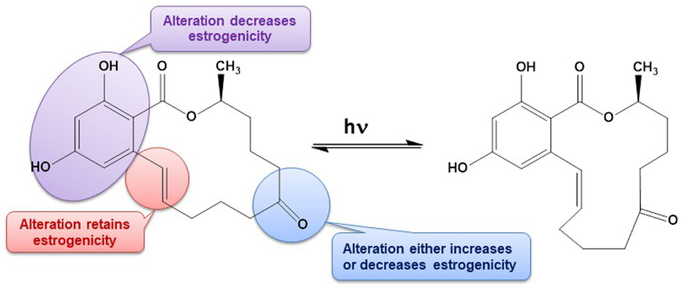
Structure of cis (a) and trans (b) zearalenone (ZEN).
Zearalenone has genotoxic, teratogenic, hemotoxic, immunotoxic, carcinogenic and hepatotoxic properties [11]. The maximum dose of ZEN is 50 μg kg-1 in cereals and maize-based snacks, 20 μg kg-1 in infant formulas and child products, and 200 μg kg-1 in unprocessed maize [12]. The maximum limit for direct human consumption is 100 μg kg-1 [13].
Zearalenone metabolites are formed via two major biotransformation pathways. The first pathway during which α- and β-zearalenol (ZEL) are formed involves hydroxylation catalyzed by 3α- and 3β-hydroxy-steroid dehydrogenases (HSDs). In the second pathway, ZEN and its metabolites are conjugated with glucuronic acid and catalyzed by uridine diphosphate glucuronyl transferases (UDPGT) [5,14]. Zearalenone and its metabolites are endocrine disruptors due to their structure and high affinity for estrogen receptors. Zearalenone undergoes metabolic changes, including reduction of the main α- and β-metabolites of zearalenol and conjugation of ZEN and its metabolites with glucuronic or sulfuric acid (Figure 2). The above processes increase the polarity of ZEN molecules and influence the elimination of toxic substances from the body [15]. Excess ZEN and its metabolites are excreted from the body with urine and bile. Various methods for enriching and isolating toxic compounds from biological matrices such as blood, tissues or urine have been developed [16,17].
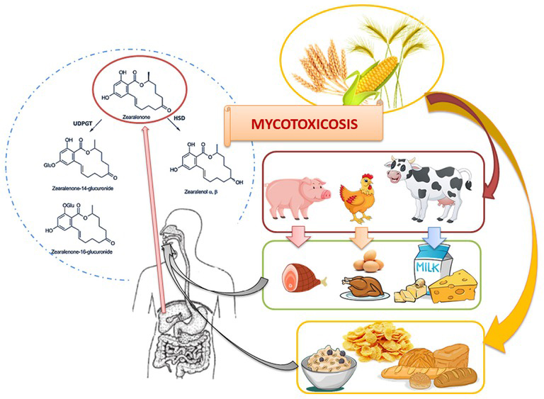
The infection pathway and zearalenone metabolism.
The isolation of analytes from different matrices, mainly biological and environmental samples, and the purification and enrichment of extracts pose considerable problems in analytical practice. The most common methods of isolating ZEN and its metabolites include solid-phase extraction with sorbents such as octadecyl [18,19] as well as immunosorbents [20] and molecularly imprinted polymers [21]. Pressurized liquid extraction (PLE) [22] and accelerated solvent extraction (ASE) [4] are also excellent methods for isolating the analyte. Various chemical and biochemical methods are used to quantify ZEN and its metabolites. Zearalenone concentrations in biological and environmental samples are generally very low.
Different quantitative indicators are used to determine the concentrations of ZEN and its metabolites. Instrumental analyses should be characterized by high sensitivity and a low limit of detection and quantification. Trace amounts of ZEN in extracts are determined by liquid chromatography with different detection methods and gas chromatography.
The aim of this study was to describe different analytical approaches in preparation of biological, food and environmental samples for analyses of zearalenone and its metabolites. Special attention was also paid to the assessment of the exposure of living organisms to the influence of ZEN and the appearance of its metabolites in urine.
2 Urine as a biological sample for assessing the effects of exposure to ZEN
There is a general scarcity of research on the toxic and harmful effects of ZEN compounds in humans. Most studies have been conducted on animal samples which are also important for evaluating the health risks associated with ZEN exposure in humans. In both humans and animals, ZEN and its metabolites are excreted mainly with urine. The research related to animals’ use has been complied with all the relevant national regulations and institutional policies for the care and use of animals. Similarly, in the case of research to human use has been complied with all the relevant national regulations, institutional policies and in accordance the tenets of the Helsinki Declaration, and has been approved by the authors’ institutional review board or equivalent committee.
The biological recovery of ZEN from porcine urine depends on the route of administration. Urine recovery ranges from 14% to 45.38% of the applied dose after oral administration, and from 45.4% to 77% after intravenous injection [23,24]. In a study by Dänicke et al. [25,26], piglets were fed a diet with increasing inclusion levels of wheat contaminated with Fusarium toxin over a period of 35 days. The toxin was identified by hydrolysis with β-glucuronidase/sulfatase, immunoaffinity clean-up and high-performance liquid chromatography (HPLC) with fluorescence detection. A linear dose relationship was observed with an increase in ZEN, α-ZEL and β-ZEL concentration in urine. The average urine recovery for this mycotoxin was 94.75% and 85%, respectively.
In the work of Gambacorta et al. [24], piglets were administered a single dietary bolus of mixtures of deoxynivalenol (DON), aflatoxin B1 (AFB1), fumonisin B1 (FB1), ochratoxin A (OTA) and ZEN. Urine samples were collected three times over a 24-hour period. The samples were hydrolyzed with β-glucuronidase/sulfatase, purified on an immunoaffinity column (IAC; Myco6in1, Vicam) and Oasis® HLB solid phase extraction. Mycotoxin biomarkers were detected in each urine sample, and β-ZEL was identified only at the highest dose of ZEN. The percentage of the mycotoxin excreted in 24 h was calculated based on the volume and concentration of urine in the analyte. The mean percentage of ZEN excreted in urine during 24 h was 36.8% (28.4% and 8.3% in a 3:1 ratio of ZEN/α-ZEL).
Warth et al. [27] monitored a 27-year-old male patient for the content of ZEN excreted in urine. The patient was administered a naturally contaminated diet for four days. After enzymatic hydrolysis, total ZEN concentration was measured in 24-hour urine samples. The total ZEN content (mainly in the form of glucuronide) ranged from 0.30 to 0.59 ng mL-1, which corresponded to 7.0-13.2% of the administered daily dose. It should also be noted that α-and β-ZEL were not identified in excreted urine
A study performed on 30 men and 30 women in Germany [28] revealed the presence of ZEN, α-ZEL and β-ZEL in all urine samples. The mycotoxin and its metabolites were identified by LC-MS/MS after enzymatic hydrolysis and were purified on the immunoaffinity column. The concentration of ZEN ranged from 0.04 to 0.28 ng mL-1, the content of α-ZEL – from 0.06 to 0.45 ng mL-1, and β-ZEL – from 0.01 to 0.20 ng mL-1. Significant gender-related differences were not reported in the composition and concentrations of the analytes in urine. The values noted in the majority of the exposed individuals were below the tolerable daily intake level of 0.25 μg kg-1 BW set by EFSA, but higher values were observed in some subjects.
The urinary excretion of ZEN appears to be a reliable indicator of human exposure to this mycotoxin. The concentrations of ZEN and its metabolites, including α-and β-ZEL, provide valuable information about exposure to modified ZEN mycotoxins which co-occur in food and contribute to human exposure. The health risks associated with the exposure to ZEN have been researched extensively in animals (mainly pigs), but far less frequently in humans. Research has revealed that the formation and distribution of ZEN metabolites are similar in humans and animals. Despite the fact that pigs were administered a higher dose than humans, the proportions of reduced forms of ZEN in urine were comparable. These findings considerably expand our understanding of ZEN’s toxicity.
3 Mycotoxin isolation methods
The sample preparation process exerts the greatest influence on the results of quantitative analyses. Different sample preparation procedures (Figure 3) are used to optimize the efficiency of the analytical procedure.
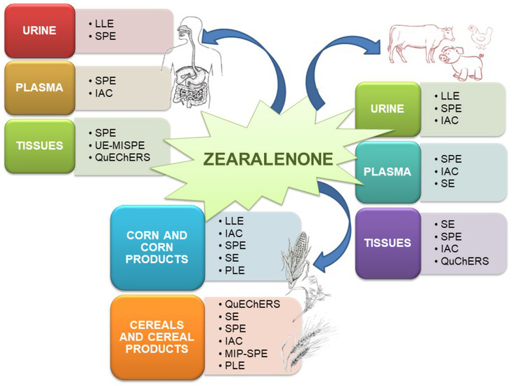
Various extraction techniques for isolation of zearalenone and its metabolites.
3.1 Solid-phase extraction
During solid-phase extraction, a solution of the analyte is passed through an extraction column containing the adsorbent. The analytes are absorbed by the particle bed, and the column is washed to remove the impurities. In the next step, the absorbed analyte is washed out with a suitable solvent. Numerous examples of solid-phase extraction with different sorbents (Table 1), including silica gel modified by the octadecyl hydrocarbon chain (C18) [19,20,34], Florisil [19] and Oasis® HLB polymers [24], as well as extract purification tools such as immunosorbents (IAC) [20,35,36] can be found in the literature.
Solid-phase extraction with different sorbents and a comparison of recovery values
| Mycotoxin | Matrix | Sample preparation | Recovery | References |
|---|---|---|---|---|
| ZEN | Endometrial cancer | UE-MISPE | 82.5-97.9% | [16] |
| α-ZEL | ||||
| ZEN | ||||
| α-ZAL | ||||
| β-ZAL | Human, bovine and swine | C18/Florisil | 87-108% | [19] |
| α-ZEL | urine | |||
| β-ZEL | ||||
| ZAN | ||||
| ZEN | ||||
| α-ZEL | Urine | IACs (Myco6in1, VICAM) | nd*) | [23] |
| β-ZEL | ||||
| Serum | 66-88% | |||
| ZEN | Feedstuffs | 74-82% | ||
| α-ZEL | Bile | IACs (Easi-ExtractTM ZON, R-Biopharm) | 81-104% | [25,26] |
| β-ZEL | Urine | 75-94% | ||
| Liver | 66-88% | |||
| ZEN | ||||
| α-ZEL | Urine | IACs (ZearalaTest™,VICAM) | nd*) | [28] |
| β-ZEL | ||||
| ZEN | ||||
| α-ZAL | Bovine feed | SPE (C18) | 79% | [29] |
| β-ZAL | Urine | 82% | ||
| ZEN | Rainbow trout organs | IACs (ZearalaTest™WB, VICAM) | 85% | [30] |
| ZEN | Functional food | QuEChERS (DSPE) | 83-91.5% | [31] |
| ZEN | Corn and corn products | IACs | 92-114% | [33] |
| ZEN | Urine | SPE RP-C18 (Phenomenex) | 86-102% | [34] |
| Animal tissue | ||||
| ZEN | Blood | IACs (ZearalaTest™,VICAM) | nd*) | [35] |
*) nd - no data
Jodlbauer et al. [34] used the octadecyl sorbent to isolate ZEN and its derivatives from urine and animal tissue samples. The recovery of ZEN and its metabolites from urine and tissues ranged from 86% to 102% in the evaluated method. Andres et al. [19] used C18 and Florisil as sorbents in the process of ZEN isolation and purification. Both sorbents were tested on urine samples, and Florisil was found to be less selective. The octadecyl sorbent was selected for further analyses, where ZEN recovery ranged from 92% to 108%.
In other studies, ZEN and its metabolites were isolated from urine, plasma and equine feces, and were purified by solid-phase extraction (SPE) and immunoaffinity chromatography (IAC) [20]. An ammonium acetate buffer with pH 4.8 and D2-ZAN was initially added to the urine sample. The solution was incubated with β-glucuronidase/ arylsulfhatase solution and brought to pH 4. The solution was applied to a sorbent filled with ISOLUTE® C18, which was previously conditioned with methanol and H2O. The sorbent was washed with 30% MeOH (2 mL) and dried under vacuum. The analytes were eluted with methanol (1.25 mL), and the eluate was diluted with a phosphate buffer solution at pH 7.4. The extract was purified with an immunosorbent (Easi-Extract®Zearalenone) which was previously conditioned with phosphate buffered saline solution (PBS). The cited study demonstrated that effective sample preparation and sensitivity of the method are determined by the complexity of the analyzed matrix. Single-stage purification before IAC was sufficient for plasma samples, whereas urine samples had to be purified by both SPE and IAC. Urine recovery ranged from 56% to 100%.
Zearalenone and its metabolites can also be isolated with molecularly imprinted polymers (MIP). In the work of Mausia et al. [21], the zearalenone template was synthesized by precipitation polymerisation and applied as a sorbent in molecularly imprinted solid-phase extraction (MISPE). The sorbent was characterized by high stability and affinity for the ZEN molecule. The sorbent was previously conditioned with acetonitrile; the sample was applied in an acetonitrile/ water mixture (60/40% v/v) and washed with an ACN/ H2O mixture (30/70% v/v). The analytes were eluted with a 95/5% (v/v) mixture of methanol and acetic acid. The obtained recovery for ZEN ranged from 90.8% to 99.6%.
Comments: Since the introduction of solid phase extraction as a sample preparation method (especially for liquid samples), this technique has been widely used to isolate and enrich various compounds and has often been used to purify extracts from interfering compounds. The rapid development of SPE is due to progress aimed at the synthesis of new sorbents that are characterized by specific interactions with an isolated compound in order to achieve high recovery. Future research should focus on reducing the amount of sorbent used for extraction, thus minimizing or eliminating the use of toxic solvents. An alternative to traditional sorbents can be molecularly imprinted polymers (MIPs), which enable selective and specific interaction with the analyte. This ensures not only selective retention of the isolated compound, but also purification of the extract from interfering compounds. Undoubtedly, an important issue is the repeatability and reproducibility of the obtained sorbents.
3.2 QuEChERS
The quick, easy, cheap, effective, rugged and safe (QuEChERS) method is suitable for preparing biological samples in line with green chemistry rules. In the QuEChERS approach, the isolated compound is extracted with organic solvents with the addition of salt, and analyte and matrix components are split between the organic phase and water. A purified extract is obtained by dispersion and solid phase extraction. The QuEChERS method is simple, it relies on non-chlorinated solvents, involves fewer sample preparation steps, and can be used to analyze a larger number of samples. Pajewska at el. [17] and Woźniak at el. [37] used the QuEChERS technique to isolate ZEN and its metabolites from muscle tissues, and obtained a recovery rate of 56-115%. This technique was also used by Cunha and Fernandes [38] to analyze breakfast cereals and contaminated flour. The procedure was modified to produce a recovery rate of 67-101% for breakfast cereals and 52-103% for flour.
3.3 Novel methods of sample preparation for the isolation of ZEN and its metabolites
Zearalenone levels should be monitored in food and feed to protect humans and animals from consuming products contaminated with this mycotoxin. Therefore, innovative methods are being sought to minimize the health problems associated with toxins.
Kim et al. [39] developed an innovative system for removing and monitoring ZEN levels in feed, which offers an alternative to IAC. The proposed approach relies on the use of monoclonal antibodies (mAbs) and magnetic nanoparticles (MNP). In the cited study, ground and enriched samples were shaken and extracted in 70% methanol. The collected samples were passed through paper filters, and MNP-mAb conjugates were used to remove the mycotoxin. Zearalenone was removed magnetically from the supernatant, and its concentration was measured. The mycotoxin was also extracted by IAC for comparative purposes. The recovery rate was significantly higher in the proposed approach (92-100%) than in IAC (81-88%).
Amoli-Diva et al. [40] used dispersive liquid-liquid micro-extraction (DLLME) combined with microsolid phase extraction (μ-SPE) to determine ZEN levels in wheat samples. This simple and low-cost extraction method was characterized by satisfactory repeatability and reproducibility as well as high recovery in the range of 91.6% to 99.1%. Antep and Merdivan [41] investigated the effectiveness of the DLLME method for quantitative determination of ZEN in beer, with the aim of developing a simple, fast and sensitive extraction method. The obtained recoveries ranged from 71% to 108%. The simplicity, accuracy and low cost of the DLLME-μ-SPE method were confirmed by Hashemi et al. [42] who extracted ZEN from an acetonitrile mixture with the use of hydrophobic magnetic nanoparticles. The magnetic absorber was collected with an external magnet, and water from the supernatant was decanted. The recovery rate for maize samples was high in the range of 93.2% to 102.1%. The study demonstrated that ZEN levels in samples could be effectively determined with the use of the two-stage micro-extraction procedure based on DLLME and μ-SPE with hydrophobic magnetic nanoparticles.
Porto-Figueira et al. [43] combined μ-QuEChERS with ultrasound-assisted extraction (USAE) to improve the original QuEChERS method. The described approach involved a reduced amount of separating salts, smaller samples and fewer solvents. The cited authors demonstrated the potential of the proposed method.
Magnetic molecularly imprinted polymers (mMIP) were used for selective extraction of ZEN from various cereal flours [44]. Quercetin was the inert template, and it was also used to synthesize mMIP for ZEN. Flour extraction mixtures were prepared by centrifugation and sonication with acetonitrile and water (80:20, v/v) and 0.2% HCOOH. The extracts were centrifuged, separated and placed in centrifuge tubes, and ultra-pure water and 100 mg of mMIP were added. The solution was decanted, and the supernatant was removed with an external magnet. The analyte with mMIP was transferred to a water test tube, and the supernatant was centrifuged and removed. Zearalenone was eluted three times with methanol and twice with acetonitrile. The sample was centrifuged each time, and mMIP was magnetically decanted. The ZEN recovery rate was above 95%, which indicates that this method is useful for isolating trace amounts of the mycotoxin in food analyses.
Another unconventional method for ZEN detection was presented by Fang et al. [45]. The authors synthesized a new molecularly imprinted optosensing material (MIOM) by anchoring the MIP layer on ionic liquid-modified CdSe/ ZnS quantum dots. The MIOM was used to detect ZEN in maize, rice and wheat flours. The recovery rates ranged from 84% to 107%.
Comments: The novel approaches to sample preparation differ from conventional methods. The separation of the extracts from other co-extracted compounds in the matrix is also an important issue during sample preparation. Effective sample preparation and high sensitivity of the method are determined by the complexity of the matrix, therefore each step is an inherent element to obtaining the best possible recovery results. It is preferable to replace the expensive sorbents (e.g., antibodies) by magnetic particle imprinted polymers with structural analogs of the analytes. The developed methodologies for isolating ZEN and its metabolites with the use of mMIP allow obtaining high and not always repeatable recoveries, but they allow high efficiency of cleaning the samples from compounds that coexist in them as contaminants. Future research should focus on the search for a template for the molecular imprint and the development of a repeatable synthesis of the prepared polymers. In addition, new applications such as integrated artificial intelligence systems to predict appropriate extraction conditions are expected to ensure the future viability of this technique.
3.4 Solvent extraction
Liquid extraction is a popular method of extracting analytes from the matrix, including samples of food and infected animal tissues, in analyses of ZEN and its metabolites. Additional ultrasonic (UE) or microwave (MEA) energy can be used in this extraction technique, which can successfully increase the efficiency of extraction and shorten the time of its operation.
Pallaroni et al. [22] described the pressurized liquid extraction (PLE) method that relies on environmentally acceptable and less harmful organic solvents. Zearalenone was extracted from maize with a 50/50% (v/v) mixture of isopropanol and an aqueous solution of triethanolamine (1%) at 80°C. The obtained recovery was 85%.
Uracca et al. [4] determined the content of ZEN and α-zearalenone in cereal samples (corn, wheat, rice, barley, rye) and swine feed by ASE. Optimal extraction conditions were obtained by using a solvent mixture MeOH/ACN (50:50 v/v) at the temperature of 50°C, a pressure of 1500 psi, a static time of 5 min and flush volume 60%. The obtained recovery exceeded 96% for ZEN (RSD < 4.0%) and reached 98% for α-ZAL (RSD < 4.6%) [4].
Comments: One of the advantages of ASE method is that the process is automated. The solvent thoroughly penetrates the sample, which reduces extraction time and decreases cost associated with slow sample preparation.
4 Analytical methods
Various chemical and biochemical methods are used to quantify ZEN and its metabolites and minimize the health risks associated with exposure to mycotoxins. Biological and environmental samples contain trace amounts of these mycotoxins, which is why selective and sensitive identification techniques with low limits of detection (LOD) and determination (LOQ) are required. Various analytical techniques and the relevant LOD values for ZEN and its metabolites in different matrices are presented in Table 2.
Various analytical techniques for detecting ZEN and its metabolites in different matrices
| Mycotoxin | Matrix | Sample preparation | Technique | LOD | References |
|---|---|---|---|---|---|
| ZEN | |||||
| α-ZAL | |||||
| β-ZAL | broiler meat | SE, IACs | HPLC-DAD | 30-140 ng mL-1 | [52] |
| α-ZEL | |||||
| β-ZEL ZAN | |||||
| ZEN | maize and cereal-based feeds | SPE C18 | HPLC-FLD | 15 ng mL-1 | [53] |
| ZEN | plasma | IACs | HPLC-FLD | 5.94-14.30 ng mL-1 | [35] |
| ZEN | |||||
| ZAN | grain | SPE C18, IACs | HPLC-MS/MS | 0.5 ng g-1 | [18] |
| ZEN | |||||
| α-ZEL | cereal-based foods | SPE, IACs | HPLC-MS/MS | 1-10 ng g-1 | [54] |
| β-ZEL | |||||
| ZEN | |||||
| α-ZAL | |||||
| β-ZAL | sediments | PLE, SPE | GC-MS | 0.01-10.28 ng g-1 | [55] |
| α-ZEL | |||||
| β-ZEL ZAN | |||||
| ZEN | |||||
| α-ZEL | biological samples | SE, SPE | GC-MS | 0.06-0.6 ng mL-1 | [56] |
| β-ZEL | |||||
| ZEN | maize | SE, SPE C18 | HPLC, TLC | 20 ng g-1 | [57] |
4.1 High-performance liquid chromatography
The presence of ZEN and its metabolites can be determined by high-performance liquid chromatography (HPLC) with various detectors. Zearalenone is a naturally fluorescent compound, and it is identified by fluorimetric detection.
Ok et al. [36] isolated ZEN by HPLC with fluorimetric detection, where the SynergiTM Hydro-RP 80Å column (Phenomenex, 4.6 × 250 mm; 4 μm) was maintained at 40°C. The excitation and emission wavelengths were 275 nm and 450 nm, respectively. A H2O/ACN/MeOH (35/10/55% v/v/v) mixture was the mobile phase, and the flow rate was 1.0 mL min-1. The LOD value was 4.0 μg kg-1, and LOQ was determined at 10.0 μg kg-1.
Marczuk et al. [35] analyzed clinical cases of dairy cattle infected by Fusarium fungi, including ZEN. Blood samples were collected and subjected to IAC extraction with a Zearala-Test® column to determine the plasma concentration of ZEN. The extracts were analyzed by HPLC with fluorescent detection. An ODS Hypersil chromatographic column (4 × 250 mm; 5 μm) was used. The mobile phase was an ACN/MeOH/H2O mixture (46/8/46 or 8/10/82% v/v/v) with a flow rate of 1.8 mL min-1 or 1.5 mL min-1 at 30°C. The excitation and emission wavelengths were 218 and 438 nm, respectively.
Hewitt et al. [33] identified ZEN in corn and maize products with the use of an H2O/MeOH/ACN mixture as the mobile phase at an excitation wavelength of 274 nm and an emission wavelength of 440 nm. Pajewska et al. [17] relied on the QuEChERS technique to isolate ZEN and its metabolites from human tissues. The compounds were separated by HPLC with fluorimetric detection at the excitation and emission wavelengths of 270 and 440 nm, respectively. Zearalenone and α-ZEL were detected as compounds with the highest fluorescent intensity. High-performance liquid chromatography with fluorescence detection is characterized by high selectivity and sensitivity, and it is one of the most popular methods of ZEN detection.
Liquid chromatography coupled with mass spectrometry is becoming an increasingly popular detection technique despite its high cost. Liquid chromatography with one mass analyzer (LC-MS) and tandem mass spectrometry (LC-MS/MS) ensure unambiguous identification of the analyte. This method requires an ion source to determine the mass-to-charge ratio (m/z). Electrospray ionization (ESI) and atmospheric pressure chemical ionization (APCI) are the most widely used ionization techniques [46,47].
Jodlbauer et al. [34] relied on the LC-MS/MS technique with an APCI type ionization source to determine the concentrations of ZEN and its metabolites in urine and tissue samples collected from cows and pigs. The analysis was performed on the Supersphere®RP-18 Merck column (125 × 3.0 mm; 4 μm) at 35°C. The mobile phase was an ACN/MeOH/ H2O (10/45/45% v/v/v) mixture with 15 mM ammonium acetate at a flow rate of 0.5 mL min-1. The LOD values ranged from 0.1 to 0.5 μg L-1, and LOQ – from 0.5 to 1 μg L-1.
Soleimanes et al. [47] determined ZEN levels in cereal matrices by the UPLC-MS/MS technique with ESI in positive and negative ion mode. The LOD values in the tested matrices were determined at 0.1-0.4 ng g-1, and LOQ values – at 0.2-1 ng g-1. Liquid chromatography coupled with mass spectrometry is increasingly used in analyses, but other analytical methods are often selected due to the high cost of mass spectrometry.
4.2 Gas chromatography
Gas chromatography-mass spectrometry (GC-MS) is also used to analyze ZEN and its metabolites. A GC-MS method for detecting ZEN and its metabolites in samples of bovine urine, bile, meat and kidneys was described by the Standard Operating Procedure ARO/458 [48]. The GC-MS/MS method was also used by Rodríguez-Carrasco et al. [49] to detect ZEN in grain-based products. Dudziak [50] relied on GC-MS to detect ZEN in aqueous environments. Zearalenone concentrations were determined at 0.3 to 0.5 ng/L in water samples, and recovery rates ranged from 62% to 80%.
4.3 Thin-layer chromatography
Classical thin-layer chromatography (TLC) is a less accurate technique than GS-MS. Quantitative results are compared with the standard, and the purity, quality and concentration of the standards affect the accuracy of the analysis. This method has been used to detect ZEN in maize. The fluorescence of ZEN was compared against the applied standards. Samples that tested positive for ZEN in TLC were extracted again to validate the results [51].
Schaafsma et al. [57] relied on TLC to analyze Fusarium toxins in maize and wheat. A comparison of the costs associated with different analytical methods revealed that TLC was the cheapest technique. However, toxin concentrations were less accurately determined by TLC than HPLC.
5 Conclusions
The adverse effects of ZEN can contribute to many hormone-dependent diseases. Zearalenone is ubiquitous in food products, which spurs the search for effective methods of neutralizing this toxin. Research into ZEN metabolism provides crucial information for evaluating this mycotoxin’s effects in humans and animals. Quantitative analyses of ZEN in food and feed also play a very important role in such assessments. Effective analytical procedures are thus needed to minimize the negative impact of mycotoxins on living organisms and the environment. An attempt was made in this review article to present various methods for the isolation and quantification of ZEN and its metabolites. The optimal analytical method should be characterized by a simple sample preparation procedure because numerous analytical steps can lead to uncertainty in the accurate determination of the analytes. Several sample preparation techniques for different matrices were presented, and the applicability of various ZEN identification methods, mostly chromatography with different detection methods, was discussed.
List of Abbreviations
- ASE
accelerated solvent extraction
- DLLME
dispersive liquid-liquid microextraction
- DSPE
dispersive solid phase extraction
- GC-FID
gas chromatography–flame ionization detection
- HPLC
high-performance liquid chromatography
- IACs
immunoaffinity columns
- ILs
ionic liquids
- LOD
limit of detection
- LOQ
limit of quantification
- MAE
microwave-assisted extraction
- MSPE
magnetic solid-phase extraction
- PLE
pressurized liquid extraction
- SPE
solid-phase extraction
- RSD
relative standard deviation
- ZAN
zearalanone
- α-ZAL
α-zearalanol
- β-ZAL
β-zearalanol
- α-ZEL
α-zearalenol
- β-ZEL
β-zearalenol
- ZEN
zearalenone
Conflict of interest: Authors state no conflict of interest.
References
[1] Marin S, Ramos AJ, Cano-Sancho G, Sanchis V. Mycotoxins: occurrence, toxicology, and exposure assessment. Food Chem Toxicol. 2013;60:218-37.10.1016/j.fct.2013.07.047Search in Google Scholar PubMed
[2] Buszewska-Forajta M. Mycotoxins, invisible danger of feedstuff with toxic effect on animals. Toxicon. 2020;182:34-53.Schelstraete W, Devreese M, Croubels S. Comparative toxicokinetics of Fusarium mycotoxins in pigs and humans. Food Chem Toxicol. 2020;137:111140.10.1016/j.toxicon.2020.04.101Search in Google Scholar PubMed
[3] Urraca JL, Marazuela MD, Moreno-Bondi MC. Analysis for zearalenone and α-zearalenol in cereals and swine feed using accelerated solvent extraction and liquid chromatography with fluorescence detection. Anal Chim Acta. 2004;524:175-83.10.1016/j.aca.2004.03.093Search in Google Scholar
[4] Zinedine A, Soriano JM, Moltó JC, Mañes J. Review on the toxicity, occurrence, metabolism, detoxification, regulations and intake of zearalenone: an oestrogenic mycotoxin. Food Chem Toxicol. 2007;45(1):1-18.10.1016/j.fct.2006.07.030Search in Google Scholar PubMed
[5] Rogowska A, Pomastowski P, Sagandykova G, Buszewski B. Zearalenone and its metabolites: effect on human health, metabolism and neutralisation methods. Toxicon. 2019;162(March):46-56.10.1016/j.toxicon.2019.03.004Search in Google Scholar PubMed
[6] Binder SB., Schwartz-Zimmermann HE, Varga E, Bichl G, Michlmayr H, et al. Metabolism of zearalenone and its major modified forms in pigs. Toxins. 2017;9(2):1-15.10.3390/toxins9020056Search in Google Scholar PubMed PubMed Central
[7] Drzymala SS, Binder J, Brodehl A, Penkert M, Rosowski M, Garbe LA, et al. Estrogenicity of novel phase I and phase II metabolites of zearalenone and cis-zearalenone. Toxicon. 2015;105:10-2.10.1016/j.toxicon.2015.08.027Search in Google Scholar PubMed
[8] Gadzała-Kopciuch R, Kwaśniewska K, Ludwiczak A, Skrzyniarz P, Jakubowski R, Nowak W, et al. Towards a new approach for the description of cyclo−2,4-dihydroxybenzoate, a substance which effectively mimics zearalenone in imprinted polymers designed for analyzing selected mycotoxins in urine. Int J Mol Sci. 2019;20:1588; doi:10.3390/ijms20071588.10.3390/ijms20071588Search in Google Scholar PubMed PubMed Central
[9] Metzler M. Proposal for a uniform designation of zearalenone and its metabolites. Mycotoxin Res. 2011;27(1):1-3.10.1007/s12550-010-0075-2Search in Google Scholar PubMed
[10] Bai X, Sun Ch, Xu J, Liu D, Han Y, Wu S, et al. Detoxification of zearalenone from corn oil by adsorption of functionalized GO systems. Appl Surf Sci. 2018;430:198-207.10.1016/j.apsusc.2017.06.055Search in Google Scholar
[11] EFSA CONTAM Panel (EFSA Panel on Contaminants in the Food Chain). Scientific opinion on the risks to public health related to the presence of nickel in food and drinking water. EFSA J. 2015;13(2):1-124.10.2903/j.efsa.2015.4002Search in Google Scholar
[12] Han Z, Jiang K, Fan Z, Di Mavungu JD, Dong M, Guo W, et al. Multi-walled carbon nanotubes-based magnetic solid-phase extraction for the determination of zearalenone and its derivatives in maize by ultra-high performance liquid chromatography-tandem mass spectrometry. Food Control. 2017;79:177-84.10.1016/j.foodcont.2017.03.044Search in Google Scholar
[13] Olsen M, Pettersson H, Kiessling KH. Reduction of zearalenone to zearalenol in female rat liver by 3α‐ hydroxysteroid dehydrogenase. Acta Pharmacol Toxicol (Copenh). 1981;48(2):157-61.10.1111/j.1600-0773.1981.tb01602.xSearch in Google Scholar
[14] Ueberschär KH, Brezina U, Dänicke S. Zearalenone (ZEN) and ZEN metabolites in feed, urine and bile of sows: analysis, determination of the metabolic profile and evaluation of the binding forms. Landbauforsch Appl Agric Forestry Res. 2016;66(1):21-8.Search in Google Scholar
[15] Gadzała-Kopciuch R, Cendrowski K, Cesarz A, Kiełbasa P, Buszewski B. Determination of zearalenone and its metabolites in endometrial cancer by coupled separation techniques. Anal Bioanal Chem. 2011;401(7):2069-78.10.1007/s00216-011-5206-xSearch in Google Scholar
[16] Pajewska M, Łojko M, Cendrowski K, Sawicki W, Kowalkowski T, Buszewski B, et al. The determination of zearalenone and its major metabolites in endometrial cancer tissues. Anal Bioanal Chem. 2018;410(5):1571-82.10.1007/s00216-017-0807-7Search in Google Scholar
[17] Zöllner P, Jodlbauer J, Lindner W. Determination of zearalenone in grains by high-performance liquid chromatography-tandem mass spectrometry after solid-phase extraction with RP-18 columns or immunoaffinity columns. J Chromatogr A. 1999;858(2):167-74.10.1016/S0021-9673(99)00821-3Search in Google Scholar
[18] de Andrés F, Zougagh M, Castañeda G, Ríos A. Determination of zearalenone and its metabolites in urine samples by liquid chromatography with electrochemical detection using a carbon nanotube-modified electrode. J Chromatogr A. 2008;1212(1–2):54-60.10.1016/j.chroma.2008.09.112Search in Google Scholar PubMed
[19] Songsermsakul P, Sontag G, Cichna-Markl M, Zentek J, Razzazi-Fazeli E. Determination of zearalenone and its metabolites in urine, plasma and faeces of horses by HPLC-APCI-MS. J Chromatogr B Analyt Technol Biomed Life Sci. 2006;843(2):252-61.10.1016/j.jchromb.2006.06.012Search in Google Scholar PubMed
[20] Mausia T, de Smet D, Guorun Q, van Peteghem C, Zhang D, Wu A, et al. Molecularly imprinted polymers as specific adsorbents for zearalenone produced by precipitation polymerization and applied to mycotoxin production. Anal Lett. 2011;44(16):2633-43.10.1080/00032719.2011.553009Search in Google Scholar
[21] Pallaroni L, Von Holst C. Development of an extraction method for the determination of zearalenone in corn using less organic solvents. J Chromatogr A. 2004; 1055(1–2):247-9.10.1016/j.chroma.2004.08.147Search in Google Scholar PubMed
[22] Biehl ML, Prelusky DB, Koritz GD, Hartin KE, Buck WB, Locksley Trenholm H. Biliary excretion and enterohepatic cycling of zearalenone in immature pigs. Toxicol Appl Pharmacol. 1993;121(1):152-9.10.1006/taap.1993.1140Search in Google Scholar PubMed
[23] Gambacorta L, Solfrizzo M, Visconti A, Powers S, Cossalter AM, Pinton P, et al. Validation study on urinary biomarkers of exposure for aflatoxin B1, ochratoxin A, fumonisin B1, deoxynivalenol and zearalenone in piglets. World Mycotoxin J. 2013;6(3):299-308.10.3920/WMJ2013.1549Search in Google Scholar
[24] Dänicke S, Swiech E, Buraczewska L, Ueberschär KH. Kinetics and metabolism of zearalenone in young female pigs. J Anim Physiol Anim Nutr (Berl). 2005;89:268-76.10.1111/j.1439-0396.2005.00516.xSearch in Google Scholar PubMed
[25] Dänicke S, Brüssow KP, Valenta H, Ueberschär KH, Tiemann U, Schollenberger M. On the effects of graded levels of fusarium toxin contaminated wheat in diets for gilts on feed intake, growth performance and metabolism of deoxynivalenol and zearalenone. Mol Nutr Food Res. 2005;49:932-43.10.1002/mnfr.200500050Search in Google Scholar PubMed
[26] Warth B, Sulyok M, Berthiller F, Schuhmacher R, Krska R. New insights into the human metabolism of the Fusarium mycotoxins deoxynivalenol and zearalenone. Toxicol Lett. 2013;220(1):88-94.10.1016/j.toxlet.2013.04.012Search in Google Scholar PubMed
[27] Ali N, Gisela HD. Urinary biomarkers of exposure to the mycoestrogen zearalenone and its modified forms in German Adults. Arch Toxicol. 2018;92(8):2691-700.10.1007/s00204-018-2261-5Search in Google Scholar PubMed
[28] Juan C, Rittieni A, Yusa V, Manes J. Determination of zearalenone and its metabolites in bovine feeding stuff and assessment residues in bovine urine. International Conference on Food Innovation (Food Innova’2010), 25-29 October 2010, Valencia, Spain. https://images.engormix.com/s_news/ZEARALENONE_ZERANOL(4).pdfSearch in Google Scholar
[29] Woźny M, Obremski K, Jakimiuk E, Gusiatin M, Brzuzan P. Zearalenone contamination in rainbow trout farms in northeastern Poland. Aquaculture. 2013;416–417:209-11.10.1016/j.aquaculture.2013.09.030Search in Google Scholar
[30] Wu J, Zhao R, Chen B, Yang M. Determination of zearalenone in barley by high-performance liquid chromatography coupled with evaporative light scattering detection and natural occurrence of zearalenone in functional food. Food Chem. 2011;126(3):1508-11.10.1016/j.foodchem.2010.11.159Search in Google Scholar
[31] De Baere S, Osselaere A, Devreese M, Vanhaecke L, De Backer P, Croubels S. Development of a liquid-chromatography tandem mass spectrometry and ultra-high-performance liquid chromatography high-resolution mass spectrometry method for the quantitative determination of zearalenone and its major metabolites in chicken and pig plasma. Anal Chim Acta. 2012;756:37-48.10.1016/j.aca.2012.10.027Search in Google Scholar PubMed
[32] Hewitt TC, Flack CL, Kolodziejczyk JK, Chacon AM, D’Ovidio KL. Occurrence of zearalenone in fresh corn and corn products collected from local Hispanic markets in San Diego County, CA. Food Control. 2012;26(2):300-4.10.1016/j.foodcont.2012.01.035Search in Google Scholar
[33] Jodlbauer J, Zöllner P, Lindner W. Determination of Zearalenone and its metabolites in urine and tissue samples of cow and pig by LC-MS/MS. Mycotoxin Res. 2000;16(2 SUPPL):174-8.10.1007/BF02940030Search in Google Scholar PubMed
[34] Marczuk J, Obremski K, Lutnicki K, Gajecka M, Gajecki M. Zearalenone and deoxynivalenol mycotoxicosis in dairy cattle herds. Pol J Vet Sci. 2012;15(2):365-72.10.2478/v10181-012-0055-xSearch in Google Scholar PubMed
[35] Ok HE, Jung H, Lee SE, Peak O, Chun HS. Three liquid chromatographic methods for the analysis of aflatoxins in for different corn Zea Mays matrices. J Food Compos Anal. 2016;54:20-6.10.1016/j.jfca.2016.09.010Search in Google Scholar
[36] Wozniak B, Matraszek-Zuchowska I, Zmudzki J. Determination of stilbenes and resorcylic acid lactones in bovine, porcine and poultry muscle tissue by liquid chromatography-negative ion electrospray mass spectrometry and QuEChERS for sample preparation. J Chromatogr B Analyt Technol Biomed Life Sci. 2013;940:15-23.10.1016/j.jchromb.2013.09.018Search in Google Scholar PubMed
[37] Cunha SC, Fernandes JO. Development and validation of a method based on a QuEChERS procedure and heart-cutting GC–MS for determination of five mycotoxins in cereal products. J Sep Sci. 2010;33(4-5):600-9.10.1002/jssc.200900695Search in Google Scholar PubMed
[38] Kim HJ, Kim SH, Lee JK, Choi CU, Lee HS, Kang HG, et al. A novel mycotoxin purification system using magnetic nanoparticles for the recovery of aflatoxin Bl and zearalenone from feed. J Vet Sci. 2012;13(4):363-9.10.4142/jvs.2012.13.4.363Search in Google Scholar PubMed PubMed Central
[39] Amoli-Diva M, Taherimaslak Z, Allahyari M, Pourghazid K. Dispersive liquid-liquid microextraction coupled with magnetic nanoparticles for extraction of zearalenone in wheat samples. Nanochem Res. 2017;2(1):60-70.Search in Google Scholar
[40] Antep H. Mine, Merdivan M. Development for new dispersive liquid-liquid microextraction technique for the identification of zearalenone in beer. Anal Methods. 2012;4(12):4129-34.10.1039/c2ay25665gSearch in Google Scholar
[41] Hashemi M, Taherimaslak Z, Parvizi S, Torkejokar M. Spectro-fluorimetric Determination of zearalenone using dispersive liquid-liquid microextraction coupled to micro-solid phase extraction onto magnetic nanoparticles. RSC Advances. 2014;4(85):45065-73.10.1039/C4RA07684BSearch in Google Scholar
[42] Porto-Figueira P, Camacho I, Câmara JS. Exploring the potentialities of an improved ultrasound-assisted quick, easy, cheap, effective, rugged, and safe-based extraction technique combined with ultrahigh pressure liquid chromatography-fluorescence detection for determination of zearalenone in cereals. J Chromatogr A. 2015;1408:187-96.10.1016/j.chroma.2015.07.031Search in Google Scholar PubMed
[43] Cavaliere C, Antonelli M, Cerrato A, La Barbera G, Laganà A, Laus M, et al. A novel magnetic molecular imprinted polymer for selective extraction of zearalenone from cereal flours before liquid chromatography-tandem mass spectrometry determination. Toxins (Basel). 2019;11(9):1-12.10.3390/toxins11090493Search in Google Scholar PubMed PubMed Central
[44] Fang G, Fan C, Liu H, Pan M, Zhu H, Wang S. A novel molecularly imprinted polymer on CdSe/ZnS quantum dots for highly selective optosensing of mycotoxin zearalenone in cereal samples. RSC Advances. 2014;4(6):2764-71.10.1039/C3RA45172KSearch in Google Scholar
[45] Sforza S, Dall’asta C, Marchelli R. Recent advances in mycotoxin determination in food and feed by hyphenated chromatographic techniques/mass spectrometry. Mass Spectrom Rev. 2006;25(1):54-76.10.1002/mas.20052Search in Google Scholar PubMed
[46] Soleimany F, Jinap S, Faridah A, Khatib AA. UPLC-MS/MS for simultaneous determination of aflatoxins, ochratoxin A, zearalenone, DON, fumonisins, T-2 toxin and HT-2 toxin, in cereals. Food Control. 2012;25(2):647-53.10.1016/j.foodcont.2011.11.012Search in Google Scholar
[47] National Institute for Public Health and the Environment. Analysis of zeranol and metabolites in urine, bile, meat, liver and kidney by GC-MS. Standard Operating Procedure: ARO/458;1-9; https://www.rivm.nl/bibliotheek/digitaaldepot/ARO458EN.pdfSearch in Google Scholar
[48] Rodríguez-Carrasco Y, Moltó JC, Berrada H, Mañes J. A survey of trichothecenes, zearalenone and patulin in milled grain-based products using GC-MS/MS. Food Chem. 2014;146:212-9.10.1016/j.foodchem.2013.09.053Search in Google Scholar PubMed
[49] Dudziak M. Analysis of zearalenone in aqueous environment using GC-MS. Pol J Environ Stud. 2011;20(1):231-5.Search in Google Scholar
[50] Shotwell OL, Hesseltine CE, Goulden ML, Vandegraft EE. Survey of corn for aflatoxin, zearalenone, and ochratoxin. Cereal Chem. 1970;4(6):700-7.Search in Google Scholar
[51] Duca RC, Bravin F, Delaforge M, Vladescu L, Badea IA, Criste RD. Development of a new HPLC method used for determination of zearalenone and its metabolites in broiler samples. Influence of zearalenone on the nutritional properties of broiler meat. J Agric Food Chem. 2009;57(22):10497-504.10.1021/jf9014608Search in Google Scholar PubMed
[52] Llorent-Martínez EJ, Fernández-Poyatos MP, Ruiz-Medina A. Automated fluorimetric sensor for the determination of zearalenone mycotoxin in maize and cereals feedstuff. Talanta. 2019;191:89-93.10.1016/j.talanta.2018.08.049Search in Google Scholar PubMed
[53] Vendl O, Berthiller F, Crews C, Krska R. Simultaneous determination of deoxynivalenol, zearalenone, and their major masked metabolites in cereal-based food by LC-MS-MS. Anal Bioanal Chem. 2009;395(5):1347-54.10.1007/s00216-009-2873-ySearch in Google Scholar PubMed
[54] Kinani S, Bouchonnet S, Bourcier S, Porcher JM, Aït-Aïssa S. Study of the chemical derivatization of zearalenone and its metabolites for gas chromatography-mass spectrometry analysis of environmental samples. J Chromatogr A. 2008;1190(1–2):307-15.10.1016/j.chroma.2008.02.115Search in Google Scholar PubMed
[55] Blokland MH, Sterk SS, Stephany RW, Launay FM, Kennedy DG, van Ginkel LA. Determination of resorcylic acid lactones in biological samples by GC-MS. Discrimination between illegal use and contamination with fusarium toxins. Anal Bioanal Chem. 2006;384(5):1221-7.10.1007/s00216-005-0274-4Search in Google Scholar PubMed
[56] Schaafsma AW, Nicol RW, Savard ME, Sinha RC, Reid LM, Rottinghaus G. Analysis of Fusarium toxins in maize and wheat using thin layer chromatography. Mycopathologia. 1998;142(2):107-13.10.1023/A:1006937720711Search in Google Scholar
© 2020 Renata Gadzała-Kopciuch et al., published by De Gruyter
This work is licensed under the Creative Commons Attribution 4.0 International License.
Articles in the same Issue
- Review Articles
- Liquid-phase microextraction of polycyclic aromatic hydrocarbons: A review
- Colorimetric hand-held sensors and biosensors with a small digital camera as signal recorder, a review
- Mini-Review of Analytical Methods used in Quantification of Ellagic Acid
- Highly Sensitive and Robust Capillary Electrophoresis-Electrospray Ionization-Mass Spectrometry: Interfaces, Preconcentration Techniques and Applications
- Spectroscopic Determination of Two Beta-Blockers – Atenolol and Propanolol by Oxidative Derivatization Using Potassium Permanganate in Alkaline Medium
- Review on analytical methods for quantification of ADHD drugs in human biological samples
- Benzene, toluene, ethylbenzene, and xylene: Current analytical techniques and approaches for biological monitoring
- Advances in neurochemical measurements: A review of biomarkers and devices for the development of closed-loop deep brain stimulation systems
- Review on stationary phases and coating methods of MEMs gas chromatography columns
- Research Article
- Xanthene based resonance Rayleigh scattering and spectrofluorimetric probes for the determination of cyclobenzaprine: Application to content uniformity test
- Special Issue: SPECIAL ISSUE ON 25TH INTERNATIONAL SYMPOSIUM ON SEPARATION SCIENCES
- Chromatographic analysis of bio-oil formed in fast pyrolysis of lignocellulosic biomass
- Urinary carboxylic acids (UCAs) in subjects with autism spectrum disorder and their association with bacterial overgrowth
- Separation procedures in the identification of the hydrogenation products of biomass-derived hydroxymethylfurfural
- The effect of selenium, zinc and copper on the excretion of urinary modified nucleobases in rats treated with prostate cancer cells
- Analytical approaches and preparation of biological, food and environmental samples for analyses of zearalenone and its metabolites
- Nanosized zinc, epigenetic changes and its relationship with DMBA induced breast cancer in rats
- Special Issue: Bioanalytical Methods and Their Applications
- Single-molecule force spectroscopy: A facile technique for studying the interactions between biomolecules and materials interfaces
- Biological nanoscale fluorescent probes: From structure and performance to bioimaging
- Detection of metal ions in biological systems: A review
- Recent advances in animal origin identification of gelatin-based products using liquid chromatography-mass spectrometry methods: A mini review