Orbital Fat Prolapse
-
Leonid Skorin

A 79-year-old man presented with an asymptomatic yellowish mass on his temporal bulbar conjunctiva of the left eye. The patient's visual acuity was unaffected, and he had no history of ocular trauma, surgery, or thyroid dysfunction. Ocular examination revealed a soft, yellow, nontender mobile mass. Subconjunctival herniated orbital fat (HOF), also known as intraconal fat prolapse (image A, arrow), was diagnosed. The patient also had bilateral inferior and superior medial eyelid festoons, also known as extraconal fat prolapse (image B, arrows). The subconjunctival HOF was surgically removed. The patient declined blepharoplasty to remove the extraconal fat protrusion.
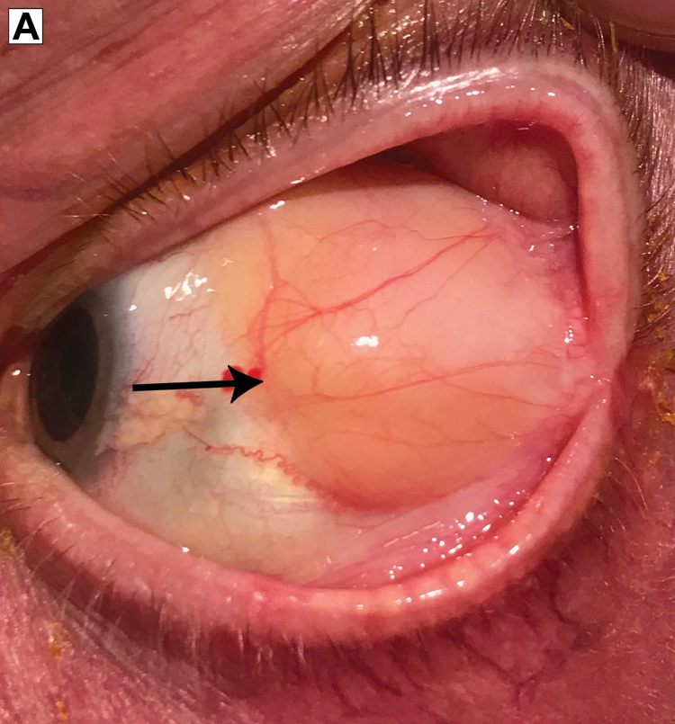
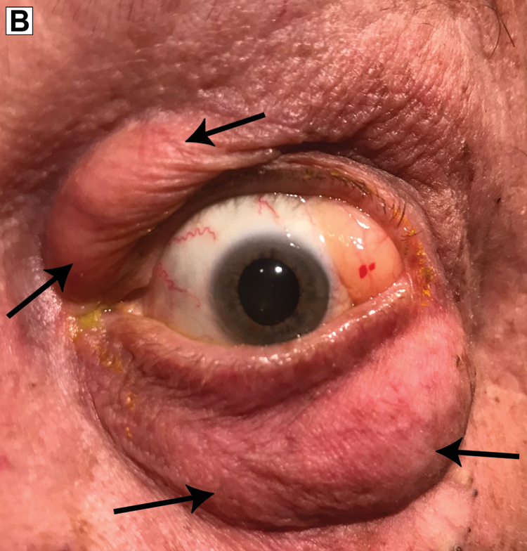
Extraconal fat prolapse is common, but subconjunctival HOF, which is benign, is rare.1,2 Patients may present with symptoms of dry eyes, discomfort, or cosmetic complaints.1,3 Clinically, subconjunctival HOF is typically located in the superotemporal quadrant and appears as a soft, yellow mass that is easily displaced using a cotton-tip applicator.3,4 These characteristics help distinguish it from conjunctival dermolipoma, conjunctival lymphoma, epidermoidal cyst, and prolapse of the lacrimal gland.1-4 Orbital computed tomography or magnetic resonance imaging identifies subconjunctival HOF as a low-density mass continuous with the intraconal fat.1,2,4,5 Histopathologic analysis reveals normal adipose tissue.5 Subconjunctival HOF is managed using transconjunctival excision.1,5 Extraconal fat protrusion can be corrected with blepharoplasty.2
References
1. Skorin L Jr, RinkC. Diagnosis and excision of subconjunctival herniation of orbital fat. Optom Vis Sci.2014;91(9):e236-e240.10.1097/OPX.0000000000000342Search in Google Scholar PubMed
2. Monner J , BenitoJR, ZayuelasJ, PalomaV, CastroV, SerraJM. Transconjunctival herniation of orbital fat.Ann Plast Surg1998;41(6):658-661.10.1097/00000637-199812000-00013Search in Google Scholar PubMed
3. Chatzistefanou KI , SamaraC, AsproudisI, et al. Subconjunctival orbital fat prolapse and thyroid associated orbitopathy: a clinical association. Clin Interv Aging. 2017;12:359-366.10.2147/CIA.S118955Search in Google Scholar PubMed PubMed Central
4. Wang X , YanJ. Subconjunctival orbital fat prolapse: an unsuspecting rare lesion.J Craniofac Surg.2015;26(2):e92-e94.10.1097/SCS.0000000000001066Search in Google Scholar PubMed
5. Lin CC , LiaoSL, LiouSW, ChenCC, WuYY, WoungLC. Subconjunctival herniated orbital fat mimicking adipocytic neoplasm.Optom Vis Sci.2015;92(10):1021-1026.10.1097/OPX.0000000000000693Search in Google Scholar PubMed
© 2018 American Osteopathic Association
This work is licensed under the Creative Commons Attribution-NonCommercial-NoDerivatives 4.0 International License.
Articles in the same Issue
- SURF
- Sleep and Lifestyle Habits of Osteopathic Emergency Medicine Residents During Training
- Addressing the Increased Incidence of Common Sexually Transmitted Infections
- ABSTRACTS
- Educating Leaders 2018: Abstracts From AACOM's Annual Conference
- OMT MINUTE
- Doming the Diaphragm in a Patient With Multiple Sclerosis
- EDITORIAL
- Resident Duty Hour Restrictions: The Implications Behind the New Data Ahead of the Single Accreditation System
- A Perspective on the Sleep and Lifestyle Habits of Emergency Medicine Residents
- LETTERS TO THE EDITOR
- Toxic Injury to the Gastointestinal Tract After Ipilimumab Therapy
- ORIGINAL CONTRIBUTION
- Using a Multitheory Model to Predict Initiation and Sustenance of Fruit and Vegetable Consumption Among College Students
- Predictors of Responsible Drinking or Abstinence Among College Students Who Binge Drink: A Multitheory Model Approach
- BRIEF REPORT
- Osteopathic Manipulative Therapy and Multiple Sclerosis: A Proof-of-Concept Study
- Diabetes Fellowship in Primary Care: A Survey of Graduates
- JAOA/AACOM MEDICAL EDUCATION
- Teaching Medical Students About Health Systems Science and Osteopathic Principles and Practice Using a Virtual World: The Envision Community Health Center
- CASE REPORT
- Hemiplegic Syndrome After Chopstick Penetration Injury in the Lateral Soft Palate of a Young Child
- CLINICAL IMAGES
- Orbital Fat Prolapse
- Unusual Cause of “Constipation”