Air in the eyelids! Pneumomediastinum and periorbital emphysema in a patient of dermatomyositis
-
Wei Wang


A 44-year-old man with a history of dermatomyositis presented to the clinic complaining of “swelling of the neck after a forced cough”. One month ago, he developed periorbital heliotrope rash and proximal muscle weakness. His serum creatine kinase level increased slightly (250 U/L), and his electroneuromyography showed myopathic abnormalities. The pathology of his right deltoid muscle biopsy revealed inflammatory myopathy with mild lymphocyte infiltration. A computerized tomography (CT) scan of the chest showed interstitial lung disease. A diagnosis of dermatomyositis was made, and the patient was given glucocorticoid and immunosuppressive therapy. Symptoms improved till several days ago when heavy cough developed and he felt a swelling neck. Physical examination revealed sub-cutaneous crepitation in the neck and chest wall. Chest X ray (Panel A) and CT scan (Panel B) confirmed pneumomediastinum (arrow) and subcutaneous emphysema (arrowhead). One day later, swelling and crepitation were also felt in his right eye lid. CT scan showed air in the periorbital and tempus region (Panel C, arrow). The patient was treated with methylprednisolone combined with cyclosporin and cyclophosphamide. There was complete resolution of the pneumomediastinum and subcutaneous emphysema 8 weeks later.
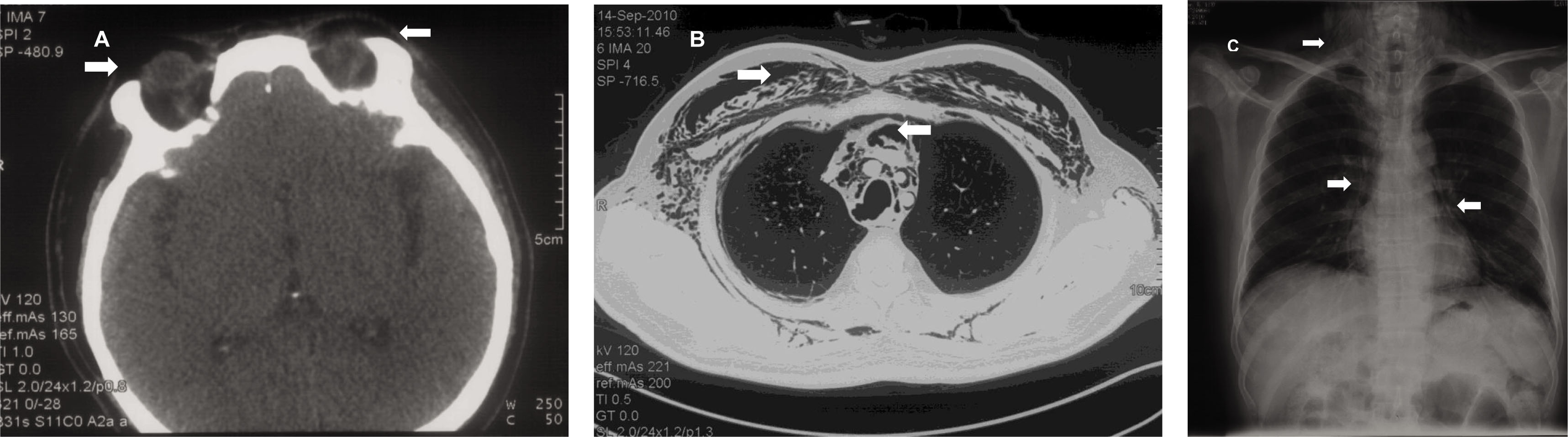
Panel A: A chest X ray demonstrated signs of pneumomediastinum (arrow) and subcutaneous emphysema (arrowhead) for this patient with dermatomyositis. Panel B: A Chest CT scan demonstrated signs of pneumomediastinum (arrow) and subcutaneous emphysema (arrowhead) for the patient with dermatomyositis. The bilateral lung fields were otherwise clear. Panel C: On a head CT scan, air was identified in the periorbital and tempus region (arrows).
Conflict of Interest
None declared.
© 2020 Wei Wang et al., published by Sciendo
This work is licensed under the Creative Commons Attribution-NonCommercial-NoDerivatives 4.0 International License.
Artikel in diesem Heft
- Editorial
- Biomarkers for treatment response in rheumatoid arthritis: Where are they?
- Criteria and Recommendation
- 2020 Chinese guidelines for the diagnosis and treatment of systemic lupus erythematosus
- The dawn of a new era of therapies in systemic lupus erythematosus
- Characteristics and long-term outcomes of patients with lupus-related protein-losing enteropathy: A retrospective study
- Vision loss, multiple cerebral infarction, ischemia of extremities: Systemic vasculitis or cardiac myxoma?
- Review
- Interleukin-17 inhibitors for the treatment of ankylosing spondylitis
- Communication
- Risk of malignancy and biologic therapy in rheumatic inflammatory diseases: A single-center experience
- Images
- Air in the eyelids! Pneumomediastinum and periorbital emphysema in a patient of dermatomyositis
- News
- The history of development of rheumatology in China: From a single center to the whole country