Preparation, characterization, and stability of lipid nanoparticles including unsaturated lipids
-
Yeseul Park
Yeseul Park is research fellow at Seoul National University of Science and Technology. She received her B.S. and M.S. degree from Seoul National University of Science and Technology, Korea, in 2021 and 2023, respectively. Her research interests are in biomimetic membranes and their applications.
Jin-Won Park is a professor at Seoul National University of Science and Technology. He received his B.S. degree from Korea University, Korea, in 1998 and his M.S. and Ph.D. degrees from Purdue University, USA, in 2003 and 2005, respectively. From 2007 to 2010, he was an assistant professor at Gachon University, Korea. Since 2010, he has been a professor at Seoul National University of Science and Technology. His research interests are in biomimetic membranes and their applications.


Abstract
Among the SARS-CoV-2 vaccines developed to date, the mRNA vaccines developed by the Pfizer/BioNTech and Moderna companies have been formulated using saturated lipids, specifically 1,2-distearoyl-sn-glycero-3-phosphocholine (DSPC), along with cholesterol. DSPC and cholesterol have the disadvantage of causing sclerosis. Therefore, in this study, lipid nanoparticles (LNPs) were prepared and characterized by replacing DSPC with 1,2-dioleoyl-sn-glycero-3-phosphocholine (DOPC) and excluding cholesterol. The results showed that the DOPC-based LNPs had a smaller diameter (75.67 nm) compared to the previous study and the polydispersity index (PI) indicated a good dispersion homogeneity, suggesting size uniformity. Additionally, the LNPs maintained their size between 57 nm and 152 nm and showed stable PI values (0.330–0.393) throughout the 25 days.
1 Introduction
Since the COVID-19 pandemic, mRNA vaccines have emerged as promising candidates among the various types of vaccines. The mRNA vaccines have several advantages over other vaccines [1]. Compared to DNA, which needs to enter the nucleus, mRNA only needs to reach the cytoplasm and has little risk of genomic integration. In comparison to both proteins and viral systems, mRNA manufacturing offers numerous advantages, including being cell-free and faster, and resulting in proteins with native glycosylation and conformational properties [2]. The use of mRNA has been limited due to the enzymatic degradation and low efficiency of cellular uptake of naked mRNA [3].
Lipid nanoparticles (LNPs) have been recognized as promising materials for nucleic acid delivery due to numerous advantages such as long-term stability, high loading capacity, and biocompatibility [4]. LNPs act as a protective capsule for mRNA, preventing it from enzymatic degradation until it reaches the cytosol of the target cell [5, 6]. The majority of LNPs are internalized by cells through a process called endocytosis. Once inside the cell, the LNPs are encapsulated within endosomes. The mRNA payload is released from the endosomes and enters the cytoplasm via endosomal escape [7, 8].
The mRNA vaccines introduced by Moderna (i.e., mRNA-1273) [9] and Pfizer-BioNTech (i.e., BNT162b) [10] used LNPs, produced by a nanoprecipitation process [11]. 1,2-Distearoyl-sn-glycero-3-phosphocholine (DSPC), which is known to be more suitable for nanoprecipitation than other lipid components, was selected as the component of the LNPs [12]. The use of saturated phosphatidylcholines with high melting temperatures, i.e. DSPC, can lead to the production of LNPs with remarkable stability. The potential drawback of highly stable LNPs formulated with these lipids is their limited ability to promote endosomal escape, which may negatively impact the delivery efficiency of encapsulated nucleic acids. To address this concern, phosphatidylcholines are typically mixed with cholesterols to produce highly stable LNPs, that retain their structural integrity and capability to facilitate endosomal escape [13].
Although LNPs composed of DSPC have shown high stability [7, 8], DSPC has the disadvantage of causing more sclerosis compared to an unsaturated lipid [14]. Due to the low melting temperatures of unsaturated lipids, LNPs composed of these lipids can become fluidized and are highly susceptible to opsonization by serum proteins [15]. From the previous studies, 1,2-dioleoyl-sn-glycero-3-phosphocholine (DOPC) is known to be the most effective and dominant unsaturated lipid component [16], [17], [18], [19], [20], [21], [22].
Cholesterol, which is widely used in LNP formulation, reduces the risk of drug leakage from the LNP core by filling in gaps between membranes and enhancing membrane rigidity [23, 24]. However, like DSPC, cholesterol is also a risk factor for atherosclerosis [25]. Therefore, despite its advantages, it is necessary to exclude cholesterol from the LNP components.
In this study, we aim to investigate the particle size and stability of LNPs prepared by replacing DSPC with DOPC and excluding cholesterol using dynamic light scattering (DLS) over 25 days. These characteristics may provide a platform for the DOPC-based LNPs of vaccines or therapies.
2 Experimental procedures
2.1 Materials
For LNPs preparation, the ionizable cationic lipid, 1,2-di-O-octadecenyl-3-trimethylammonium propane (DOTMA), was obtained from MedChemExpress. The 1,2-dioleoyl-sn-glycero-3-phosphocholine (DOPC) and 1,2-dioleoyl-sn-glycero-3-phosphoethanolamine-N-[methoxy(polyethylene glycol)-3000] (DOPE-PEG3000) were obtained from Avanti Polar Lipids. CleanCap EPO mRNA (858 nucleotides) was purchased from TriLink Biotechnologies.
2.2 LNP preparation
LNPs were prepared by the previously described ethanol injection method [26]. Briefly, lipids were dissolved in ethanol (99.5 %) and mixed in a desired molar ratio (cationic ionizable lipid:DOPC:DOPE-PEG ratios of 81:16:3). mRNA was dissolved in RNase-free sodium citrate buffer (50 mM, pH 3). After passing the solutions through a 200 nm filter, the aqueous and ethanolic solutions were mixed in a volume ratio of 3:1 via the injection method used for LNP production. The sterilized condition was used for each step of the procedure. After a further 30 min stirring, the collected LNPs were extruded several times using a 200 nm filter.
2.3 LNP characterization and stability
The size and stability of LNPs were determined by DLS measurements (nanoSAQLA, Otsuka). For this purpose, a 25 µL sample was injected into 975 µL 10 mM phosphate buffer (pH 7.4) and LNPs were stored for 25 days in a refrigerator at (2–5 °C). The sample size and stability were determined by DLS on the 4th, 11th, 18th, and 25th days. The number distributions were calculated using a solvent refractive index of 1.33.
3 Results
3.1 LNP size
The size distribution of the LNPs was determined by dynamic light scattering (DLS). As all conditions were maintained with the exception of cholesterol, a smaller diameter than the LNP diameter (180.06 nm ± 7.89 nm) from the previous publication was expected [1].
As expected, the results of the DLS measurement showed that the diameter of the LNPs varied between 57 nm and 152 nm (Figure 1a). The polydispersity index (PI) showed a high value (0.393) compared to the previous publication (0.104 ± 0.089). This difference in PI can be attributed to the calculation method, where PI is the square of the standard deviation divided by the square of the mean. In this study, the average size of the sample size (denominator) was smaller compared to that determined in the previous publications, resulting in high PI values. Therefore, the high PI value in this study does not mean that the variance is not homogeneous, and the prepared LNPs were generally small in size and showed good dispersion homogeneity.
σ = standard deviation, d = mean diameter.
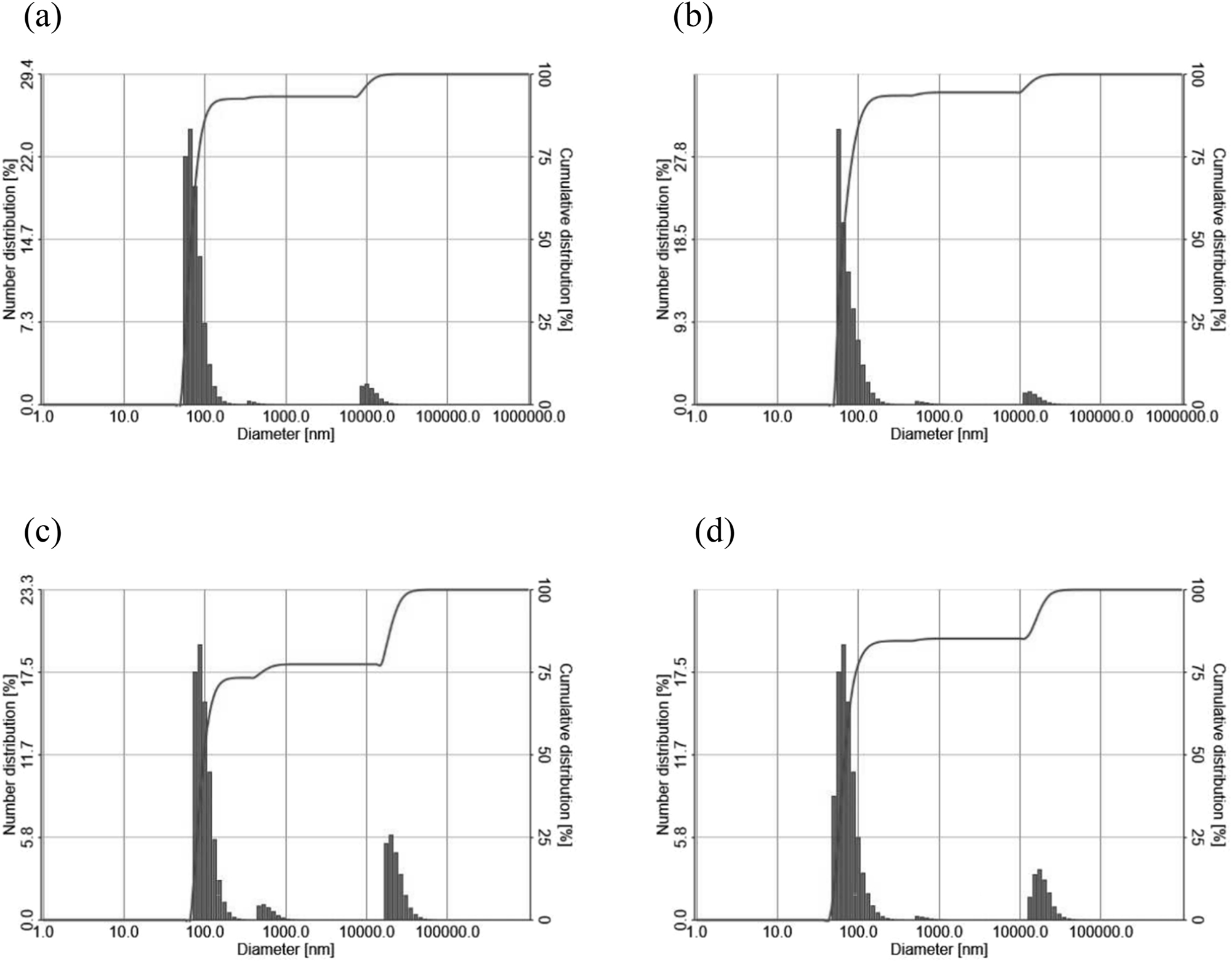
LNP diameter distributions were determined by DLS on the (a) 4th, (b) 11th, (c) 18th, and (d) 25th day.
3.2 LNP stability
To investigate the physical stability of LNPs, LNPs stored in a refrigerator (2–5 °C) were monitored by DLS for 25 days. In terms of LNP diameter, most LNPs maintained a size between 57 nm and 152 nm (Figure 1), and the average size of LNPs ranged from 74 nm to 100 nm (Table 1) over 25 days. There were few significant changes in the size of the LNPs compared to the first result (4th day). In addition, these results were similar to the results of the previous study (50–130 nm), where cholesterol was used and LNPs were prepared by microfluidics mixing, while the other experimental conditions were kept identical. The PI varied between 0.330 and 0.393 over 25 days.
LNP diameter distributions were determined by DLS on the 4th (a), 11th (b), 18th (c), and 25th (d) day; PI = polydispersity index.
| Observation period (days) | Average size (nm) | PI |
|---|---|---|
| 4 | 75.67 | 0.393 |
| 11 | 76.05 | 0.373 |
| 18 | 99.93 | 0.330 |
| 25 | 74.72 | 0.382 |
Cationic lipids, including DOTMA, are a key component in LNP formulations due to their ability to facilitate cellular uptake and endosomal release of mRNA. The stability of LNPs is heavily reliant on the presence and characteristics of the cationic lipid, as it significantly influences the successful delivery and performance of the formulation [27]. In previous study, when maintaining the DOTMA:lipid:cholesterol:cholesterol-PEG ratio at 45:18:35:2 and varying only the lipid component (EggPC, linolenic acid, oleic acid, linoleic acid), DOTMA based LNPs were relatively stable by showing a gradual increase in the particle size over the 4 weeks but the increase was not significant [28].
In this study, LNP formulations prepared by the ethanol injection method exhibited comparable diameter values to LNPs prepared by microfluidic mixing, and few significant changes in the size were observed over 25 days. Thus, it was concluded that the LNP formulation prepared by replacing saturated lipids (DSPC) with unsaturated lipids (DOPC) and excluding cholesterol, remained stable throughout the 25 day observation period. The structures of the components – DSPC, DOPC, DOPE, and cholesterol – are shown in Figure 2.
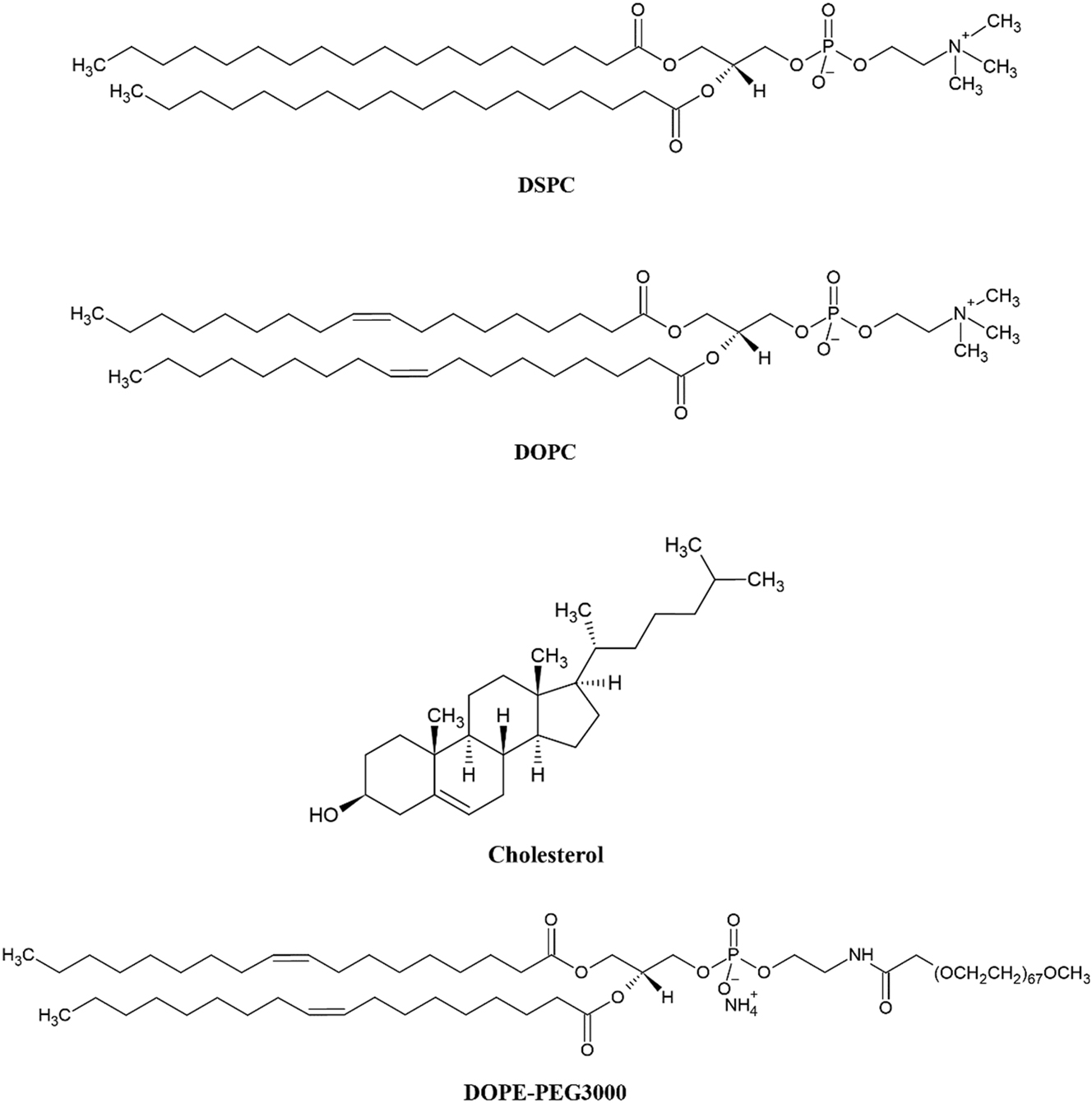
The chemical structure of helper lipids and other materials utilized in LNPs.
4 Conclusions
In this study, the particle size and stability of DOPC-based LNPs were investigated using dynamic light scattering (DLS) measurements. The results demonstrated that the LNPs prepared with a molar ratio of cationic ionizable lipid:DOPC:DOPE-PEG of 81:16:3 exhibited a smaller diameter of 75.67 nm, compared to previously reported LNPs, and the polydispersity index (PI) indicated a good dispersion homogeneity, suggesting that the LNPs were well-dispersed and had an uniform in size.
In addition, the physical stability of the LNPs was evaluated by monitoring the particle size and PI over 25 days. The LNPs maintained sizes ranging from 57 nm to 152 nm, and the PI varied between 0.330 and 0.393 throughout the observation period. The results for LNP size were similar to the previous study (50–130 nm) and it was concluded that the formulation of LNPs replacing saturated lipid (DSPC) with unsaturated lipid (DOPC) and excluding cholesterol, remained stable throughout the 25 day observation period.
The results of this study highlight the potential of DOPC-based LNPs as a platform for vaccine or therapeutic delivery systems. The smaller size and good dispersion homogeneity of these LNPs suggest the potentials for improved cellular uptake and efficient delivery of encapsulated nucleic acids. Moreover, the observed stability of DOPC-based LNPs over time indicates their suitability for storage and transport, which is crucial for practical applications.
About the authors
Yeseul Park is research fellow at Seoul National University of Science and Technology. She received her B.S. and M.S. degree from Seoul National University of Science and Technology, Korea, in 2021 and 2023, respectively. Her research interests are in biomimetic membranes and their applications.
Jin-Won Park is a professor at Seoul National University of Science and Technology. He received his B.S. degree from Korea University, Korea, in 1998 and his M.S. and Ph.D. degrees from Purdue University, USA, in 2003 and 2005, respectively. From 2007 to 2010, he was an assistant professor at Gachon University, Korea. Since 2010, he has been a professor at Seoul National University of Science and Technology. His research interests are in biomimetic membranes and their applications.
Acknowledgments
This study was supported by the Research Program funded by the SeoulTech (Seoul National University of Science and Technology).
-
Research ethics: Not applicable.
-
Author contributions: Yeseul Park conceptualized the study, carried out all experiments, analyzed all data, and wrote the first draft of the manuscript. Jin-Won Park conceptualized the study, supervised the study, reviewed, and edited the manuscript. All authors contributed to and approved the final draft of the manuscript.
-
Competing interests: The authors state no conflict of interest.
-
Research funding: None declared.
-
Data availability: Not applicable.
References
1. Wilson, B., Geetha, K. M. Lipid nanoparticles in the development of mRNA vaccines for COVID-19. J. Drug Delivery Sci. Technol. 2022, 74, 103553; https://doi.org/10.1016/j.jddst.2022.103553.Search in Google Scholar PubMed PubMed Central
2. Buschmann, M. D., Carrasco, M. J., Alishetty, S., Paige, M., Alameh, M. G., Weissman, D. Nanomaterial delivery systems for mRNA vaccines. Vaccines 2021, 9, 65; https://doi.org/10.3390/vaccines9010065.Search in Google Scholar PubMed PubMed Central
3. Wayment-Steele, H. K., Kim, D. S., Choe, C. A., Nicol, J. J., Wellington-Oguri, R., Watkins, A. M., Parra Sperberg, R. A., Huang, P., Participants, E., Das, R. Theoretical basis for stabilizing messenger RNA through secondary; structure design. Nucleic Acids Res. 2021, 49, 10604–10617; https://doi.org/10.1101/2020.08.22.262931.Search in Google Scholar PubMed PubMed Central
4. Pardi, N., Tuyishime, S., Muramatsu, H., Kariko, K., Mui, B., Tam, Y. K., Madden, T. D., Hope, M. J., Weissman, D. Expression kinetics of nucleoside-modified mRNA delivered in lipid nanoparticles to mice by various routes. J. Controlled Release 2015, 217, 345–351; https://doi.org/10.1016/j.jconrel.2015.08.007.Search in Google Scholar PubMed PubMed Central
5. Lindsay, K. E., Bhosle, S. M., Zurla, C., Beyersdorf, J., Rogers, K. A., Vanover, D., Xiao, P., Araínga, M., Shirreff, L. M., Pitard, B., Baumhof, P., Villinger, F., Santangelo, P. J. Visualization of early events in mRNA vaccine delivery in non-human primates via PET–CT and near-infrared imaging. Nat. Biomed. Eng. 2019, 3, 371–380; https://doi.org/10.1038/s41551-019-0378-3.Search in Google Scholar PubMed
6. Liang, F., Lindgren, G., Lin, A., Thompson, E. A., Ols, S., Röhss, J., John, S., Hassett, K., Yuzhakov, O., Bahl, K., Brito, L. A., Salter, H., Ciaramella, G., Loré, K. Efficient targeting and activation of antigen-presenting cells in vivo after modified mRNA vaccine administration in rhesus macaques. Mol. Ther. 2017, 25, 2635–2647; https://doi.org/10.1016/j.ymthe.2017.08.006.Search in Google Scholar PubMed PubMed Central
7. Degors, I. M. S., Wang, C., Rehman, Z. U., Zuhorn, I. S. Carriers break barriers in drug delivery: endocytosis and endosomal escape of gene delivery vectors. Acc. Chem. Res. 2019, 52, 1750–1760; https://doi.org/10.1021/acs.accounts.9b00177.Search in Google Scholar PubMed PubMed Central
8. Patel, S., Kim, J., Herrera, M., Mukherjee, A., Kabanov, A. V., Sahay, G. Brief update on endocytosis of nanomedicines. Adv. Drug Delivery Rev. 2019, 144, 90–111; https://doi.org/10.1016/j.addr.2019.08.004.Search in Google Scholar PubMed PubMed Central
9. Baden, L. R., El Sahly, H. M., Essink, B., Kotloff, K., Frey, S., Novak, R., Diemert, D., Spector, S. A., Rouphael, N., Creech, C. B., McGettigan, J., Khetan, S., Segall, N., Solis, J., Brosz, A., Fierro, C., Schwartz, H., Neuzil, K., Corey, L., Gilbert, P., Janes, H., Follmann, D., Marovich, M., Mascola, J., Polakowski, L., Ledgerwood, J., Graham, B. S., Bennett, H., Pajon, R., Knightly, C., Leav, B., Deng, W., Zhou, H., Han, S., Ivarsson, M., Miller, J., Zaks, T. Efficacy and safety of the mRNA-1273 SARS-CoV-2 vaccine. N. Engl. J. Med. 2021, 384, 403–416; https://doi.org/10.1056/nejmoa2035389.Search in Google Scholar
10. Polack, F., Thomas, S. J., Kitchin, N., Absalon, J., Gurtman, A., Lockhart, S., Perez, J. L., Pérez Marc, G., Moreira, E. D., Zerbini, C., Bailey, R., Swanson, K. A., Roychoudhury, S., Koury, K., Li, P., Kalina, W. V., Cooper, D., Frenck, R. W., Hammitt, L. L., Türeci, Ö., Nell, H., Schaefer, A., Ünal, S., Tresnan, D. B., Mather, S., Dormitzer, P. R., Şahin, U., Jansen, K. U., Gruber, W. C. Safety and efficacy of the BNT162b2 mRNA Covid-19 vaccine. N. Engl. J. Med. 2020, 383, 2603–2615; https://doi.org/10.1056/nejmoa2034577.Search in Google Scholar PubMed PubMed Central
11. Schoenmaker, L., Witzigmann, D., Kulkarni, J., Verbeke, R., Kersten, G., Jiskoot, W., Crommelin, D. J. A., mRNA-lipid nanoparticle COVID-19 vaccines: structure and stability, Int. J. Pharm. 2021, 601, 120586; https://doi.org/10.1016/j.ijpharm.2021.120586.Search in Google Scholar PubMed PubMed Central
12. Turnbull, I. C., Eltoukhy, A. A., Anderson, D. G., Costa, K. D. Lipidoid mRNA nanoparticles for myocardial delivery in rodents. In Cardiac Gene Therapy: Methods in Molecular Biology; Ishikawa K., Ed. Humana Press: New York, NY, Vol. 1521, 2017; pp. 153–166.10.1007/978-1-4939-6588-5_10Search in Google Scholar PubMed PubMed Central
13. Cheng, X., Lee, R. J. The role of helper lipids in lipid nanoparticles (LNPs) designed for oligonucleotide delivery. Adv. Drug Delivery Rev. 2016, 99, 129–137; https://doi.org/10.1016/j.addr.2016.01.022.Search in Google Scholar PubMed
14. Gennis, R. B. Biomembranes: Molecular Structure and Function; Springer Science & Business Media: New York, 2013.Search in Google Scholar
15. Yan, X., Scherphof, G. L., Kamps, J. A. A. M. Liposome opsonization. J. Liposome Res. 2005, 15, 109–139; https://doi.org/10.1081/lpr-64971.Search in Google Scholar PubMed
16. Kulkarni, J. A., Myhre, J. L., Chen, S., Tam, Y. Y. C., Danescu, A., Richman, J. M., Cullis, P. R. Design of lipid nanoparticles for in vitro and in vivo delivery of plasmid DNA. Nanomedicine 2017, 13, 1377–1387; https://doi.org/10.1016/j.nano.2016.12.014.Search in Google Scholar PubMed
17. Szlasa, W., Zendran, I., Zalesinska, A., Tarek, M., Kulbacka, J. Lipid composition of the cancer cell membrane. J. Bioenerg. Biomembr. 2020, 52, 321–342; https://doi.org/10.1007/s10863-020-09846-4.Search in Google Scholar PubMed PubMed Central
18. Zhi, D., Zhang, S., Wang, B., Zhao, Y., Yang, B., Yu, S. Transfection efficiency of cationic lipids with different hydrophobic domains in gene delivery. Bioconjugate Chem. 2010, 21, 563–577; https://doi.org/10.1021/bc900393r.Search in Google Scholar PubMed
19. Qiu, M., Glass, Z., Chen, J., Haas, M., Jin, X., Zhao, X., Rui, X., Ye, Z., Li, Y., Zhang, F., Xu, Q. Lipid nanoparticle-mediated codelivery of Cas9 mRNA and single-guide RNA achieves liver-specific in vivo genome editing of Angptl3. Proc. Natl. Acad. Sci. 2021, 118, e2020401118; https://doi.org/10.1073/pnas.2020401118.Search in Google Scholar PubMed PubMed Central
20. Tam, A., Kulkarni, J., An, K., Li, L., Dorscheid, D. R., Singhera, G. K., Bernatchez, P., Reid, G. S. D., Chan, K. Y. T., Witzigmann, D., Cullis, P., Lim, C. Lipid nanoparticle formulations for optimal RNA-based topical delivery to murine airways. Eur. J. Pharm. Sci. 2022, 176, 106234; https://doi.org/10.1016/j.ejps.2022.106234.Search in Google Scholar PubMed
21. van Meer, G., Voelker, D. R., Feigenson, G. W. Membrane lipids: where they are and how they behave. Nat. Rev. Mol. Cell Biol. 2008, 9, 112–124; https://doi.org/10.1038/nrm2330.Search in Google Scholar PubMed PubMed Central
22. Martin, D. D., Robbins, M. E., Spector, A. A., Wen, B. C., Hussey, D. H. The fatty acid composition of human gliomas differs from that found in nonmalignant brain tissue. Lipids 1996, 31, 1283–1288; https://doi.org/10.1007/bf02587914.Search in Google Scholar PubMed
23. Albertsen, C. H., Kulkarni, J. A., Witzigmann, D., Lind, M., Petersson, K., Simonsen, J. B. The role of lipid components in lipid nanoparticles for vaccines and gene therapy. Adv. Drug Delivery Rev. 2022, 188, 114416; https://doi.org/10.1016/j.addr.2022.114416.Search in Google Scholar PubMed PubMed Central
24. Sakurai, F., Nishioka, T., Yamashita, F., Takakura, Y., Hashida, M. Effects of erythrocytes and serum proteins on lung accumulation of lipoplexes containing cholesterol or DOPE as a helper lipid in the single-pass rat lung perfusion system. Eur. J. Pharm. Biopharm. 2001, 52, 165–172; https://doi.org/10.1016/s0939-6411(01)00165-5.Search in Google Scholar PubMed
25. von Eckardstein, A., Nofer, J. R., Assmann, G. High density lipoproteins and arteriosclerosis. Arterioscler., Thromb., Vasc. Biol. 2001, 21, 13–27; https://doi.org/10.1161/01.ATV.21.1.13.Search in Google Scholar PubMed
26. Sohi, N. A., Kiani, J., Arefian, E., Khosrojerdi, A., Fekrirad, Z., Ghaemi, S., Zim, M. K., Jalili, A., Bostanshirin, N., Soleimani, M. Development of an mRNA-LNP vaccine against SARS-CoV-2: evaluation of immune response in mouse and rhesus macaque. Vaccines 2021, 9, 1007; https://doi.org/10.3390/vaccines9091007.Search in Google Scholar PubMed PubMed Central
27. Maugeri, M., Nawaz, M., Papadimitriou, A., Angerfors, A., Camponeschi, A., Na, M., Hölttä, M., Skantze, P., Johansson, S., Sundqvist, M., Lindquist, J., Kjellman, T., Mårtensson, I.-L., Jin, T., Sunnerhagen, P., Östman, S., Lindfors, L., Valadi, H. Linkage between endosomal escape of LNP-mRNA and loading into EVs for transport to other cells. Nat. Commun. 2019, 10, 4333; https://doi.org/10.1038/s41467-019-12275-6.Search in Google Scholar PubMed PubMed Central
28. Wang, X., Yu, B., Ren, W., Mo, X., Zhou, C., He, H., Jia, H., Wang, L., Jacob, S. T., Lee, R. J., Ghoshal, K., Lee, L. J. Enhanced hepatic delivery of siRNA and microRNA using oleic acid based lipid nanoparticle formulations. J. Controlled Release 2013, 172, 690–698; https://doi.org/10.1016/j.jconrel.2013.09.027.Search in Google Scholar PubMed PubMed Central
© 2023 the author(s), published by De Gruyter, Berlin/Boston
This work is licensed under the Creative Commons Attribution 4.0 International License.
Articles in the same Issue
- Frontmatter
- Physical Chemistry
- Transition from a foam-like to an onion-like nanostructure in water-rich L3 phases
- Simulation of adsorption at rotating disk
- Wetting behaviour of ionic surfactants on the aluminium foil
- The interaction of ester functionalized amide gemini surfactants with polymers
- Biosurfactants
- The use of polyoxyethylene (20) cetyl ether in assessing the hydrophobicity of compounds of biomedical importance and in the process of drug release from microemulsions
- Novel Surfactants
- Synthesis, characterization, and evaluation of ethoxylated lauryl myristyl alcohol as non-ionic surfactants (surfethoxymers)
- Synthesis
- Synthesis and surface properties of branched-chain tertiary fatty alcohol sulfate surfactants
- Application
- Influence of surfactant concentration on selected quality parameters of fruit washing agents
- Preparation, characterization, and stability of lipid nanoparticles including unsaturated lipids
- Drug Delivery System
- Design, characterization, and evaluation of novel mucoadhesive nasal inserts for the treatment of hypertension
- Review
- An overview on eco-friendly polyglycerol esters of fatty acid, synthesis and applications