Synthesis, properties and application prospects in biomedical areas of unsaturated polyester resin modified with iron(II) clathrochelate
-
Przemysław Pączkowski

 ,
Sergey V. Shulga
,
Sergey V. Shulga

Abstract
The paper presents a number of properties of unsaturated polyester resins based on the post-consumer PET recyclate modified with iron(II) clathrochelate, such as thermal, mechanical, optical and visual. The iron compound was used as an accelerator instead of toxic low-molecular cobalt salts, commonly applied for curing of unsaturated polyester resins being also responsible for giving the resin materials an orange-red colour depending on the used amount. Moreover, the iron clathrochelate proved to be an ideal agent added to the polyester resin, giving it antimicrobial properties. Cytotoxicity tests showed that the tested materials were safe and did not cause any undesirable effects in contact with skin fibroblasts. Due to the use of the compound that gives polymers such broad properties, new perspectives are opening up in the preparation and application of polyester materials.
Introduction
The unsaturated polyester resin is a liquid polymer which cured (cross-linked with styrene, organic peroxides, named hardeners), maintains the solid shape characterized by exceptional strength and durability as well as resistance characteristics, or aesthetic qualities in relation to the imitation of materials. 1 , 2 Unsaturated polyester resins are mostly used in combination with reinforcing micro- and nanomaterials. 3 , 4 It is a well-known thermosetting polymer that is widely used for various applications. Thus, the polyester resins represent one of the absolute compounds applied in the most important wide ranges of industries: wood paints, chemical anchoring, composite materials, automotive and bathroom fixtures, corrugated panels, ribbed panels and others. 2 , 5 , 6 , 7 , 8 , 9 Currently, numerous examples in the literature detail the production and use of unsaturated polyester resins derived from post-consumer PET recyclate. 3 , 4 , 10
Clathrochelates of d-metals are coordination compounds of a new type in which the metal ion is in a three-dimensional ligand being isolated from external factors. 11 , 12 Symmetrical macrobicyclic clathrochelate metal complexes (Fe2+) are capable of forming bonds with polymer chain atoms through functional centers on the surface. In addition, iron(II) clathrochelates are reactive macrobicyclic precursors. 11 , 13 , 14
Encapsulation of a transition metal ion leads to the formation of complexes that demonstrate unprecedented chemical and photochemical stability as well as unusual properties. 11 , 15 The chelate compounds can be used as molecular scaffolds for electrocatalysts for hydrogen production as well as a distinct metallo-macrocyclic framework and a heterometallic coordination polymer. 16 , 17
The purpose of using clathrochelate metal complexes (Fe2+) in the polymeric structures is to establish the influence of macrobicyclic compounds which having an encapsulated metal ion in the rigid macrobicyclic environment, causes low toxicity, chemical and photochemical resistance of these compounds, as well as their propensity to form orderly molecular structures. 18
Iron (Fe2+) clathrochelate complexes were used to change the properties of synthetic polymers, such as epoxy resin. 19 The use of such a dye allows to expand the application of modified composites in various areas of modern industry. The interphase interactions of the filler with the polymeric structure change the physical and chemical properties and functional parameters, which is a current trend of modern materials science.
In the literature, there are several iron and vanadium compounds used as curing accelerators for unsaturated polyester resins. 20 It is believed that the addition of iron in the clathrochelate form can change the characteristics of the resin and will allow to get a new type of polymeric systems with styrene copolymerization without the use of toxic cobalt(II) compounds. 21 , 22 Iron(II) clathrochelate can be effective for arranging the polymer structure by the interraction polymer chains through the functional centers.
In the presented work, polymeric matrices based on unsaturated polyester cross-linked with different contents of iron(II) clathrochelate and without this additive were used for studies. Moreover, the antimicrobial properties of the obtained functional materials were determined. The potential application of polyesters in biomedical areas were identified.
Materials
Chemicals
The unsaturated polyester resin (UPR) from recycled PET (poly(ethylene terephthalate)), Estromal 14PB-06 NZ (acidic value 13.4 mg KOH g−1, viscosity at 23 °C 356 mPas, non-volatile content 61.2 wt%, and reactivity factor 1.53) was provided by LERG (Pustków, Poland). The blue-green syrupy liquid owes its colour to waste water bottles. Methyl ethyl ketone peroxide (MEKP, Luperox DHD-9) was purchased from Sigma-Aldrich (St. Louis, MO, USA) while N,N-dimethyl-p-toluidine (DMPT) from Merck.
Monochloromonodimethylamino clathrochelate FeBd2(Cl(Me2N)Gm)(BF)2 was prepared by the nucleophilic substitution reaction of dichloride macrobicyclic precursor FeBd2(Cl2Gm)(BF)2 (where Bd2− and Cl2Gm2− are α-benzyldioxime and dichloroglyoxime dianions, respectively) with dimethylamine, according to the procedure (Fig. 1). 23
![Fig. 1:
Structure of 1,8-bis(2-fluorobora)-2,7,9,14,15,20-hexaoxa-3,6,10,13,16,19-hexaaza-4,5,11,12-tetraphenyl-17-chloro-18-dimethylaminobicyclo[6.6.6]eicosa-3,5,10,12,16,18-hexa-ene(2−)iron(2+), FeBd2(Cl(Me2N)Gm)(BF)2 (monochloromonodimethylamino clathrochelate).](/document/doi/10.1515/pac-2025-0512/asset/graphic/j_pac-2025-0512_fig_001.jpg)
Structure of 1,8-bis(2-fluorobora)-2,7,9,14,15,20-hexaoxa-3,6,10,13,16,19-hexaaza-4,5,11,12-tetraphenyl-17-chloro-18-dimethylaminobicyclo[6.6.6]eicosa-3,5,10,12,16,18-hexa-ene(2−)iron(2+), FeBd2(Cl(Me2N)Gm)(BF)2 (monochloromonodimethylamino clathrochelate).
Resin curing
Polymeric materials based on the unsaturated polyester resin (UPR) with various contents of red-orange iron(II) clathrochelate powder (0.005, 0.01; 0.03, and 0.05 wt%) were prepared. A 5 mL solution of clathrochelate in dichloromethane was added to 100 g of the resin and stirred to obtain a homogeneous mixture. Then contents were heated at 55 °C for 2 h to evaporate methylene chloride.
For curing the previously prepared resin, 1 wt% MEKP as an initiator and 0.2 mL of N,N′-dimethyl-p-toluidine as an amine co-accelerator were used. The prepared solutions were degassed in the vacuum chamber from Vacuum Chambers (Białystok, Poland) and then poured into the cuboid-shaped molds. Gelation occurred after 15 min for the resin with 0.05 % clathrochelate, and after 40 min with 0.03 %. For the resins with 0.01 and 0.005 % of clathrochelate gelation took place after 2 and 6 h, respectively.
Further polymerization of polyesters took place at room temperature for 72 h, and then post-curing processes occured at an elevated temperature of 65–70 °C for 18 h.
The resin cured by 1 wt% of Luperox DHD-9 and 0.25 wt% of 4 % polymeric cobalt solution was used as the reference material.
Specimen preparation
In order to investigate the properties of the UPR-based materials, specimens in the form of squares or bars of appropriate dimensions were cut from the molds on a CNC milling machine MFG 8037P from Ergwind (Gdańsk, Poland).
Methods
Chemical characterization – STEM-EDS elemental mapping
The samples were ground in an agate mill to obtain a fine powder. The powder was flooded with 99.8 % ethanol to form a slurry and placed in an ultrasonic homogenizer for 10 s. Then, the slurry containing the sample was pipetted onto copper meshes (200 mesh inch−1 – 200 mesh) coated with lacey formvar stabilized with carbon and left on the paper to evaporate ethanol. Subsequently, the samples were transferred to the electron microscope. A high-resolution scanning transmission electron microscope Titan G2 60–300 kV from FEI company (Hillsboro, OR, USA) equipped with a field emission gun (FEG) was used to image the samples. Microscopic examination of the samples was made at the electron beam accelerating voltage of 300 kV. TEM imaging of the microstructure of the samples was performed in the bright field mode using the CCD camera as a detector.
Mapping of the samples determining the distribution of elements in the samples was carried out in the STEM mode, collecting the EDS spectrum from each place corresponding to the pixels of the map, point by point. The collected maps were presented in the form of a matrix of pixels, with the colour significant for the mapped element and the intensity corresponding to the percentage content of a given element.
Morphology of microstructure
To determine the morphology of iron(II) clathrochelate, micrographs of the micro-crystals sample were taken using the QUANTA 3D FEG high-resolution scanning electron microscope from FEI Company (Hillsboro, OR, USA) at an acceleration voltage of 5 kV. The sample was covered with a thin layer of Pd/Au to avoid electrostatic charging during the examination.
XRD analysis
The crystallography structure of the tested samples was examined by an Empyrean powder X-ray diffraction instrument (XRD) from PANalytical (Almelo, the Netherlands) using Cu-Kα nickel-filtered radiation of the wavelength 1.5405 Å in the Bragg-Brentano configuration. The data was recorded in the range from 4.7 to 70.6° (2Θ) with a step of 0.0263°.
Thermal and mechanical properties
Thermal properties
Thermal stabilities of the UPR with iron(II) clathrochelate were evaluated by thermogravimetry (TG) and differential thermogravimentry (DTG). The TG/DTG data were collected by means of a Netzsch STA 449F5 Jupiter (Selb, Germany). The test procedure was from 30 to 1000 °C at the heating rate of 10 °C min−1 in the oxidative atmosphere in accordance with the EN ISO 11358-1 standard. 24
Thermo-mechanical properties
The effect of temperature on the samples mechanical properties was investigated by the Vicat softening temperature (VST) tests using the Ceast HV3 apparatus by Instron (Turin, Italy) according to EN ISO 306. 25 The B120 measurement method was applied, with a force of 50 N and a heating rate of 120 °C h−1.
Mechanical properties
The characterization of the mechanical properties of the specimens was based on hardness, three-point bending and impact strength measurements.
The effect of the clathrochelate concentration on the polyester hardness was assessed using the 7206/H04 Shore durometer from Zwick (Ulm, Germany). The test was carried out in compliance with EN ISO 868, at the standard temperature of 23 °C ± 2 °C. 26 The final result was the arithmetic mean of five individual measurements (each obtained after 15 s).
Mechanical properties of the UPR-based samples were determined using the ZwickRoell Z010 universal testing machine from Zwick GmbH & Co. (Ulm, Germany). Mechanical properties determination was based on the three-point bending flexural test, where the samples of 65 mm × 10 mm × 4 mm diameter were used with a span of 50 mm be-tween the supports. The test speed was 5 mm min−1. The measurements were made according to the EN ISO 178 standard. 27 Finally, the arithmetic averaging of five measurements was taken for UPR. The flexural modulus (E f ) and strength (σ f ) as well as strain at break (ε f ), were determined.
The Charpy impact test was conducted in accordance with EN ISO 179-2, using a 639F-type impact hammer (Cometech Testing Machines, Taizhong, Taiwan) with a maximum pendulum energy of 5 J. 28 Resin specimens measuring 80 mm × 10 mm × 4 mm were used for the test.
Dynamic mechanical properties
Dynamic mechanical analysis (DMA) of the UPR-clathrochelate materials was carried out using a DMA-Q800 device (TA Instruments, New Castle, DE, USA) equipped with a dual cantilever clamp. Specimens with dimensions of 65 mm × 10 mm × 4 mm were subjected to a sinusoidal deformation at an amplitude of 0.01 mm and a constant frequency of 1.0 Hz over a temperature range from −150 to 200 °C. The heating rate was maintained at 5 °C min−1. DMA measurements were performed in accordance with EN ISO 6721-1:2019. 29 The viscoelastic properties of the cured materials were evaluated based on the temperature-dependent changes in storage modulus (E ′), loss modulus (E ″), and the loss or damping factor (tanδ). The glass transition temperature (T g ) was determined as the peak maximum of the tanδ. Additionally, the full width at half maximum (FWHM) of the tanδ peak was determined from the loss factor curves.
The crosslinking density (v e ) of the cured unsaturated polyester resin was estimated using a calculation method applicable to thermosetting polymers (Equation (1)): 30
v e – crosslinking density, mol m−3;
R – the universal gas constant, 8.314 m3 Pa mol−1 K−1;
T – the absolute temperature at T g + 50 °C, temperature in order to ensure the plateau zone, K.
Optical properties and visual characterization
UV–Vis spectroscopy
The UV-visible spectra of the specimens were analyzed based on absorption spectroscopy using a UV spectrophotometer, UV-2550 from Shimadzu (Kyoto, Japan) in the range from 200 to 900 nm at a scanning rate 200 nm min−1.
Visual characteristics – colorimetric parameters measurements
The sample colour was measured in accordance with ASTM E308, for which the Ci4200 spectrophotometer X-Rite (Grand Rapids, MI, USA) was used. 31 The colour is described in the CIELAB colour space defined by the Commission Internationale de l’Éclairage (International Commission on Illumination), commonly referred to as the CIE, in the L*, a*, b* area. The CIELAB variables form three-dimensional orthogonal coordinates. The parameter L* axis correlates with lightness, luminance–brightness ranged from 0 to 100, representing the grey scale from black to white (0 is black and 100 is white). The parameter a* axis describes the colour from greenness (negative values) to redness (positive values) whereas the parameter b* axis, from blueness (negative values) to yellowness (positive values) (Fig. 13).
The difference between the two colours-two points in the 3D colour space is described by the relationship between a sample colour (
where:
A standard observer can see a difference in colour when the
0
1 <
2 <
3.5 <
5 <

CIELAB colour space grading system (Graphical interpretation of the colour difference and colour space different from the pattern of a value less than
Contact angle measurements
The surface free energy (SFE) was determined using the Owens-Wendt method, based on direct measurements of the contact angle. Distilled water and diiodomethane were used as standard test liquids, with known values of the polar and dispersive components of the surface free energy. The SFE of CH2I2 and H2O was 53.2 and 72.8 mJ m−2, respectively. Dispersive and polar components for diiodomethane were equal to 50.8 and 2.4 mJ m−2, whereas for distilled water 21.8 and 51.0 mJ m−2. For the research, the video-based optical contact angle measuring system OCA 15EC from DataPhysics Instruments GmbH (Filderstadt, Germany) equipped with DSA100S drop shape analyser from KRÜSS GmbH (Hamburg, Germany) was employed. The result was captured using a CCD camera and measured 5 s after the 2 μL liquid droplet was formed. Contact angle measurements were made at a temperature of 21 ± 1 °C and at an air humidity of 30 ± 1 %. For each sample, 10 measurement repetitions were performed for each standard test liquid: diiodomethane and distilled water.
To evaluate the parameters of the SFE, the Owens–Wendt calculation method was applied (Equation (3)): 33 , 34
where:
γ S – the total surface free energy,
From Equations (4) and (5), there can be calculated dispersive and polar components of the surface free energy at the interface:
where:
γ d – the surface free energy of diiodomethane,
γ w – the surface free energy of distilled water,
θ w – the value of the contact angle measured with distilled water.
Immersion test – leaching study
The immersion test in various solvents was used to visual assess changes in the resin-based materials. For the leaching test, pieces of crushed and cured polyesters were placed in separate Eppendorf Safe-Lock tubes in the dark, each containing 2 mL of the corresponding chemical solution. Distilled water, acetone, and aqueous solutions of sodium hydroxide (1 %), hydrochloric acid (10 %), and sodium carbonate (2 %) were used for this purpose, in accordance with EN ISO 175. 35
Bacterial biofilm
Bacterial strains
The tiles of tested material samples were analyzed for their anti-biofilm character against aerobic Gram-positive Staphylococcus aureus ATCC 25923 and Enterococcus faecalis PCM 896 (Polish Collection of Microorganisms); and aerobic Gram-negative Escherichia coli ATCC 25992. In the microbiological assay there were used Mueller–Hinton broth (MH-broth)) for aerobic and Brain-Heart Infusion broth (BHI-broth) for microaerobic strains. After 24 h bacterial growth (at 37 °C) on the solid medium, an inoculum in 2 mL of 0.9 % NaCl was prepared, obtaining 0.5 McFarland of turbicidy standard 1.5 × 10⁸ CFU mL−1 (CFU: colony forming unit).
Materials seeding with bacteria for biofilm determination
The square tiles of tested material samples (modified with clathrochelate and unmodified as the control) with a diameter of approximately 10 mm and a height of 4 mm were sterilized by immersing them for a few seconds alternately in 1000 µL of water, 1000 µL of 70 % ethanol and 1000 µL of 0.9 % NaCl. Then, dry samples were transferred to the bottoms of 24-well polystyrene plates (CytoOne, USA). Next, 1000 µL of BHI broth was added to the well with square samples, or to the empty wells as the positive control of bacterial growth. Finally, 10 µL of inoculum (1.5 × 10⁸ CFU/mL−1) was added to the wells. Aerobic Gram-positive S. aureus ATCC 25923 or E. faecalis PCM 896; and aerobic Gram-negative E. coli ATCC 25992 were used as the mono-species biofilm assay. Sterility controls (only MH- or BHI-broth) were included in all experiments. To allow biofilm formation, i.e., the adhesion of planktonic forms of bacteria in the colonies attached to the biomaterial, the plates were incubated twice. Namely, the plates with aerobic bacteria were incubated for 48 h at 37 °C. The tests were carried out using three replicates.
CLS microscopy biofilm visualization
The test aimed at visualization of the viability and possible adhesion of bacterial cells to the modified materials (pure UPR or modified with clathrochelate) compared to adhesion to the control surface of 24-wells plate. The viability of bacterial cells and their adhesion to the sample surfaces were estimated using double fluorescent staining for both live and dead bacteria based on Viability/Cytotoxicity Assay kit for Bacteria LIVE/DEAD Cells (Biotium, Hayward, CA, USA). The material samples with bacterial suspensions were prepared for the confocal microscopy determination after the double-timing period incubation. First, the medium from wells was removed and materials were gently washed twice with 500 µL of 0.9 % NaCl, to leave only the bacterial biofilm attached to the material and to remove the loosely adherent planktonic bacterial cells. Next, the samples were transferred into fresh wells and filled with 500 µL of 0.9 % NaCl and LIVE/DEAD dye. The solution of this dye was prepared by mixing 1 µL of dimethyl sulfoxide with 1 µL of EthD-III (Ethidium Homodimer III) in 8 µL of 0.9 % NaCl. Five microliters of such obtained live/dead dye solution was added to the disc-containing wells (or empty wells used as the positive bacterial growth control) with 500 µL of PBS. The samples were incubated for 20 min at room temperature in the darkness, and bacterial colonies attached to the material samples were visualized using a confocal microscope (CLSM) with dedicated software.
Quantitative biofilm determination
Additionally, except visualization in CLSM, it is possible to perform a quantitative assessment of biofilm formation. Biofilm determination on the polyester surfaces was performed according to the procedures described by O’Toole. 36 After double-timing (48 h) of incubation of tiles samples with bacterial strains, the medium was discarded and the samples were rinsed twice with 500 µL of fresh medium to leave only bacteria attached to the surfaces, no planctonic cells. The remaining cells – namely the created biofilm – attached to the material, were stained using 1 mL of 0.1 % CV (crystal violet) for 10 min at room temperature which allowed to visualize the biofilm. The samples were transferred to fresh wells, washed twice with 500 µL of sterile water to remove any CV solution, that was not bound to bacteria. Next, the tiles with the biofilm were individually placed for 15 min into tubes with 1000 µL of 20 % acetic acid to allow the dye to solubilize at room temperature. Finally tubes were sonicated for 2 min. to disperse the biofilm. The obtained solutions of CV and acetic acid were transferred (200 µL) to a new 96-well plate to measure the optical density (OD at 590 nm) of each sample and controls. The OD determination (transferring 200 µL from 1000 µL of one sample) was repeated five-times and the average value was calculated. For each group of materials, a negative control (sterility material control) consisting of the sample immersed in the MH-broth and positive growth controls (MH-broth in wells of polystyrene plates with S. aureus ATCC 25923 or E. faecalis PCM 896, or E. coli ATCC 25992) were included. The OD value obtained for the positive controls was considered as equal to 100 % biofilm formation. The experimental results were examined for statistically significant differences (p < 0.05, n = 3) compared to the positive control of biofilm formation according to the unpaired t-test (GraphPad Prism 5, Version 5.03 GraphPad Software, Inc., San Diego, CA, USA).
Cytotoxicity
Quantitative evaluation of cytotoxicity
Cytotoxicity assessment was performed in accordance with ISO 10993-5 standard. 37 Normal human skin fibroblasts (BJ cell line) obtained from American Type Culture Collection (ATCC-LGC Standards, Teddington, UK) were seeded in 96-multiwell plates in 100 μL Eagle’s Minimum Essential Medium (EMEM, ATCC-LGC Standards, Teddington, UK) supplemented with 2 % fetal bovine serum (Pan-Biotech GmbH, Aidenbach, Bavaria, Germany) at a concentration of 2 × 10⁴ cells/well and cultured for 24 h at 37 °C to near confluence. Then the growth medium was replaced with a 24-h extracts of the samples, which were prepared in accordance with the ISO 10993-12 standard. 38 The complete growth medium served as a negative control of cytotoxicity. After 24- and 48-h incubation at 37 °C, viability of BJ cells was assessed by the colorimetric WST-8 test (Sigma-Aldrich Chemicals, Poland) according to the manufacturer protocol. The test was carried out in triplicate and optical density of the resultant solution was measured using the Synergy H1 Hybrid microplate reader (Agilent Technologies, Winooski, VT, USA). The results obtained with the cytotoxicity test were verified for statistically significant differences (p < 0.05) among the tested groups using the one-way ANOVA test followed by the Tukey’s multiple comparison test (GraphPad Prism 8, Version 8.01).
Live/dead staining
Cell viability was confirmed by Live/Dead double fluorescent staining of BJ cells after their exposure to the extract of materials. Fibroblasts were seeded in 96-multiwell plates in 100 μL of a complete growth medium at a density of 2 × 10⁴ cells per well and cultured at 37 °C for 24 h. Then, the growth medium was removed and replaced with 24-h extracts of the tested materials and incubated for 48 h under the described conditions. Afterwards, the BJ cells were stained with Live/Dead Double Staining Kit (Sigma-Aldrich Chemicals, Poland) according to the manufacturer protocol. The Fluorescence Staining Kit includes of calcein-AM (emitting green fluorescence) and propidium iodide (emitting red fluorescence), which stain viable and dead cells, respectively. Viability of fibroblasts were analyzed and visualized using the confocal laser scanning microscope (CLSM, Olympus Fluoview equipped with FV1000, Olympus, Tokyo, Japan). Multiple series of optical cross-sections (Z-stacks) were obtained by UPLSAPO 10 × lens. Optical sections were recorded at a resolution of 1600 × 1600 pixels. Then visualization of the data was reconstructed using ImageJ 2.15.0 (LOCI, University of Wisconsin, Madison, WI, USA).
Results and discussion
The purpose of using clathrochelate metal complexes (Fe2+) in the polymeric structures is to establish the influence of macrobicyclic compounds, having an encapsulated metal ion in a rigid macrobicyclic environment on their chemical and photochemical resistance as well as propensity to form orderly molecular structures (Fig. 1). Moreover, it was examined whether such a unique iron(II) compound could act as an effective accelerator of resin polymerization without the need to use toxic low-molecular-weight cobalt(II) compounds. 39
Polymeric materials based on the unsaturated polyester resin (UPR) with various contents of iron(II) clathrochelate (0.005, 0.01; 0.03, and 0.05 wt%) were prepared (Fig. 3). As expected, the transition metal compound is an intensely coloured substance, causing coloration. 20
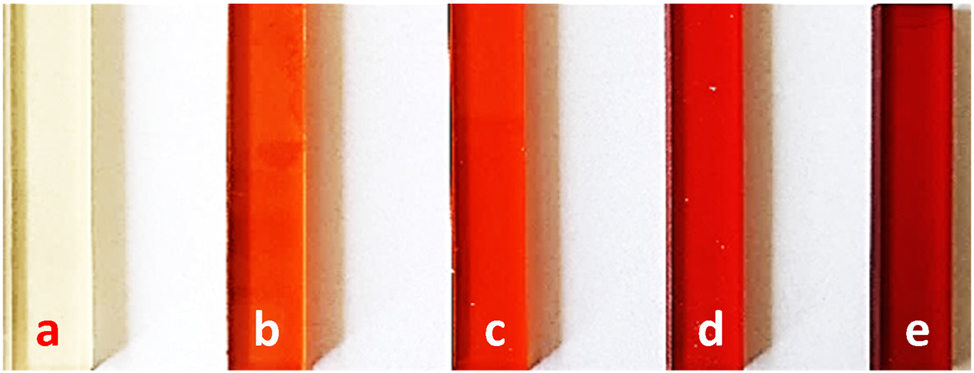
Images of cured, pure unsaturated polyester resin (a) and its materials with different amounts of iron(II) clathrochelate: 0.005 (b), 0.01 (c), 0.03 (d), and 0.05 wt% (e).
The crystallographic study of the iron(II) clathrochelate started with the analysis of the crystal shape observed in the SEM images. In Fig. 4, a view of micro-crystals of FeBd2(Cl(Me2N)Gm)(BF)2 can be observed. The morphology of clathrochelate is mainly styloid, rhomboid, and arrow shaped as confirmed by scanning electron microscopy.

SEM micrographs of micro-crystals of FeBd2(Cl(Me2N)Gm)(BF)2 under various magnifications (a–f).
The styloid morphotype is characterized by the presence of elongated crystals with pointed ends in the front view or truncated on the lateral surface (Fig. 4a–f). Arrow-shaped crystals can be observed in Fig. 4b, where this morphotype, characterized by a sharp end with a V-shaped notch resembling the tip of an arrow, is very noticeable. The parallelogram with unequal adjacent sides in Fig. 4d is an example of a rhomboid crystal morphotype. In addition to large, single monocrystals, smaller ones, so-called crystal sand, can also be observed (Fig. 4a–f).
Based on the above analysis of morphology using SEM, it can be assumed that the iron clathrochelate compound crystallizes in the monoclinic crystal system.
SEM is an appropriate technique to show particle morphology. Determining particle morphology using TEM is no longer straightforward because TEM shows only two-dimensional projected images of three-dimensional particles (Fig. 5).

TEM micrographs of FeBd2(Cl(Me2N)Gm)(BF)2.
Scanning TEM (STEM) elemental mapping showed that iron, oxygen and carbon are uniformly dispersed in FeBd2(Cl(Me2N)Gm)(BF)2 micro-crystals (Fig. 6).

STEM-EDS elemental mapping of FeBd2(Cl(Me2N)Gm)(BF)2.
The crystallographic study was carried out using X-ray diffraction patterns of cured polyester resin and polyester material with 0.05 wt% content of the clathrochelate (Fig. 7). The diffraction patterns for both samples, pure material and that containing the clathrochelate additive, consist of two main crystalline peaks. The values of diffraction angles peaks of about 9° and 20° indicate amorphous and semi-crystalline features of the polymer matrix. The only significant difference observed during the XRD analysis was a decrease of the peak intensity and the shift in the diffraction pattern from 9.35° to 7.51° for the polyester with the iron compound.

X-ray diffraction (XRD) patterns and crystalline structure of the cured polyester resin and polyester material with the 0.05 wt% clathrochelate content.
The transmittance and absorbance data for pure polyester and polyesters modified by clathrochelate in the UV-vis-NIR range are presented in Table 1 and Fig. 8.
Transmittance data from the UV-vis-NIR.
| Sample | Transmittance, % | ||
|---|---|---|---|
| at 460 nm | at 550 nm | at 850 nm | |
| Pure UPR | 63.37 | 70.87 | 84.54 |
| UPR 0.005 % Fe2+ | 6.31 | 39.43 | 85.21 |
| UPR 0.01 % Fe2+ | 3.04 | 34.84 | 88.70 |
| UPR 0.03 % Fe2+ | 0.02 | 10.47 | 89.41 |
| UPR 0.05 % Fe2+ | 0.00 | 2.06 | 89.27 |

UV-vis-NIR spectra: (a) transmittance and (b) absorbance.
Light transmission for the pure polyester with the values of 63.4, 70.9 and 84.5 % appears at the wavelengths of 460, 550 and 850 nm, respectively (Fig. 8 a). For the materials with clathrochelate, a transmittance peak appears at λ range 350–500 nm (visible light) and decreases with the increasing additive concentrations: 0.005 (22.3 %, 407.5 nm), 0.01 (20.6 %, 402 nm), 0.03 (6.0 %, 393.5 nm) and 0.05 (1.2 %, 389 nm). It is worth noting that the clathrochelate-modified polyesters were characterized by higher transmittance above 700 nm (NIR range) compared with the pure polyester.
The comparison of the absorbance of pure polyester and polyester with different amounts of iron(II) clathrochelate is given in Fig. 8b. The spectrum of pure UPR (black line) showed almost zero absorbance in the VIS range (400–700 nm) while for the materials with clathrochelate a peak with the maximum at a wavelength of about 480 nm. There was observed a tendency that with the increasing concentration there was an increase in absorbance in the mentioned region.
The technique of spectrophotometry in both transmission and reflection modes is a versatile tool for studying optical properties of polymeric materials. Characterization of the material appearance using tristimulus values in the CIELAB colour system and related parameters provides useful information for describing physical properties of materials and their interactions with light.
The characteristics of polyesters using the CIELAB scale and coordinates are presented graphically (Fig. 9) and summarized in Table 2. As shown polymer compositions can affect the colour and trichromatic values of the final product.

Variability of the colour parameters L *, a * and b *.
Values of colorimetric parameters for the UPR-based samples.
| Sample name | L * | a * | b * |
|
|---|---|---|---|---|
| Pure UPR (standard) | 79.51 | −1.79 | 15.79 | – |
| UPR 0.005 % Fe2+ | 57.06 | 36.12 | 46.46 | 53.68 |
| UPR 0.01 % Fe2+ | 54.78 | 41.07 | 47.87 | 58.97 |
| UPR 0.03 % Fe2+ | 46.21 | 46.10 | 33.68 | 61.01 |
| UPR 0.05 % Fe2+ | 40.21 | 46.10 | 33.68 | 64.48 |
Our colorimetric measurements have shown that lightness diminishes with the increasing amount of clathrochelate in polyester. For pure UPR and polymer with the largest contents of this additive, the values were 79.5 and 40.2, respectively. This may be due to greater heterogeneity of the composition.
Since the studied polyesters are colourful (Figs. 3 and 9), the values of their chromaticity parameters a* and b* vary towards yellowish and reddish, respectively. Considering the fact that the a* value for all clathrochelate-modified polyesters is positive, materials are in the redness region. The negative value for pure UPR may come from the blue-green colour of the resin.
Large values of the parameter
The effect of the amount of iron(II) clathrochelate on the thermal properties of the studied unsaturated polyesters was summarized and presented in Table 3. In the case of modified materials, there were observed changes in characteristic temperatures compared with pure UPR. The basic difference comparing the thermal research results is 15–20 °C lower than the temperature of 5 % mass loss for the resins cross-linked in the presence of iron(II) clathrochelate compared to the resin cross-linked in the presence of cobalt(II). Significant differences can be also observed for the 50 % mass loss and the maximum decomposition temperatures. These values obtained for the resins cross-linked in the presence of iron(II) are slightly larger than for the reference resin.
Thermogravimetric data for pure UPR and polyesters with iron(II) clathrochelate.
| Sample | T 5 % a, °C | T 10 % b, °C | T 50 % c, °C | T f d, °C | T max e, °C | Mass change, % |
|---|---|---|---|---|---|---|
| Pure UPR | 314.7 | 339.9 | 390.4 | 525.1 | 388.7 519.1 |
−83.37 −14.76 |
| UPR 0.005 % Fe2+ | 299.5 | 336.0 | 392.3 | 535.5 | 390.9 526.1 |
−79.66 −18.35 |
| UPR 0.01 % Fe2+ | 297.9 | 335.6 | 391.9 | 528.5 | 392.2 520.5 |
−79.64 −17.28 |
| UPR 0.03 % Fe2+ | 297.1 | 331.6 | 392.0 | 525.5 | 392.8 516.3 |
−81.16 −15.08 |
| UPR 0.05 % Fe2+ | 300.9 | 333.0 | 393.2 | 514.6 | 393.3 480.9 509.0 |
−79.02 −6.26 −10.25 |
-
aTemperature of 5 % mass loss; bTemperature of 10 % mass loss; cTemperature of 50 % mass loss; dFinal (end) temperature of decomposition; eMaximum decomposition temperature.
Iron compounds in the polyester materials caused a certain slowdown in the initiation of the polyester chains decomposition. Once this process had started, their decomposition was much faster and therefore ended at lower temperatures. The decrease in the decomposition temperature in the presence of Fe2+ ions suggests that the addition of iron compound affects the mechanism of thermal degradation of the polymer and in this case these compounds reduce the thermal stability of polymers by catalyzing their destructive reactions. 40 This may be due to the presence of three decomposition maxima. The main cause of thermal instability is probably the effect of indirect, improved and more efficient heat transfer to the polymer matrix via iron ionic compounds dispersed in it.
The mechanical properties, hardness and impact strength of clathrochelate-resin materials are presented in Table 4.
Thermo-mechanical, mechanical properties, hardness and impact strength of clathrochelate-resin materials.
| Sample | VSTa, °C | E f b, GPa | σ f c, MPa | ε f d, % | HDe, ShD | a cU f, kJ m−2 |
|---|---|---|---|---|---|---|
| Pure UPR | 150.0 | 3.32 | 107.46 | 3.46 | 72.1 | 8.1 |
| UPR 0.005 % Fe2+ | 150.0 | 0.30 | 30.10 | 16.36 | 70.0 | 33.8 |
| UPR 0.01 % Fe2+ | 138.0 | 1.21 | 47.37 | 6.82 | 69.4 | 17.1 |
| UPR 0.03 % Fe2+ | 116.5 | 0.85 | 35.83 | 6.11 | 66.2 | 15.2 |
| UPR 0.05 % Fe2+ | 85.3 | 0.94 | 38.64 | 7.84 | 65.6 | 19.3 |
-
aVicat softening temperature; bFlexural modulus; cFlexural strength; dStrain at break; eShore D hardness; fCharpy impact strength.
Flexural strength refers to the ability of a material to withstand bending under the application of external force, which is determined by the largest stress the material can withstand before failure.
The flexural modulus (bending modulus or modulus of rigidity) represents a tendency of a material to resist bending. Taking into account the values obtained for the resins cured in the presence of iron(II) clathochelate, it should be stated that, regardless of its concentration, they are much lower than for the reference UPR cured in the presence of a divalent cobalt compound. In turn, the strain at break values, also known as elongation at break, are almost twice as large as those of the reference resin.
The results of the resin hardness test are interesting. Namely, the resin cured in the presence of the smallest amount of clathrochelate has a slightly smaller hardness than the reference resin but with the increase in the concentration of iron(II) compound, its decrease of the materials is visible.
Although for the resin with the 0.05 % clathrochelate addition, gelation occurred after 15 min, both this and the other resins cured in the presence of iron(II) clathrochelate are not completely cross-linked. In addition, the change in the iron oxidation state from II to III during curing indicated by the clearly visible brown colour of the cross-linked shapes, leads to the disintegration of the chelate structure and the released ligands can have a plasticizing effect on the resin. 41 , 42
The resins containing iron(II) clathrochelate showed interesting thermomechanical properties. Unfortunately, in our study it was not possible to measure the Vicat softening temperature for all samples. This was due to the temperature limitation resulting from the fact that the apparatus was equipped with an oil bath with a maximum operating temperature of 150 °C. At this temperature the indenter immersed to 0.72 mm in pure UPR and to 0.74 mm in the polyester with the lowest clathrochelate content (0.005 %). In the case of materials containing 0.01 % iron(II) compounds, correct softening temperature measurements were already performed. Compared to the sample without clathrochelate, the polyesters with higher iron addition showed lower Vicat softening temperatures reaching 138.0, 116.5 and 85.3 °C for 0.01, 0.03 and 0.05 %, respectively which is a consequence of the already mentioned plasticizing effect of the ligands.
The impact strength of the pure unsaturated polyester resin was approximately 8.1 kJ m−2, whereas the addition of the iron(II) clathrochelate significant enhanced the values of this parameter in the studied materials.
Increase of the iron content led to a decrease of impact strength, though still remaining above that of the pure UPR. This trend may be linked to the competing effects of network disruption and increased chain mobility. Higher iron concentrations likely promote aggregation or destruction of the metal complexes, which may create stress concentration points and reduce the material’s ability to dissipate energy under impact. Additionally, excessive plasticization can reduce crosslink density, negatively affecting the toughness.
Our studies show that the iron(II) clathrochelate addition offers a tunable approach to enhance the impact resistance of unsaturated polyester resins, with an optimal concentration around 0.005 % balancing increased ductility and network integrity.
The addition of iron(II) clathrochelate to unsaturated polyester resin significantly influences its network structure and thermomechanical properties (Table 5).
Thermomechanical data of UPR-clathrochelate materials obtained by DMA.
| Sample | E ′a | E ″b, MPa | T g c, °C | tanδ d | FWHM e, °C | ν e f, mol m−3 | |
|---|---|---|---|---|---|---|---|
| (20 °C) MPa | (180 °C) MPa | ||||||
| Pure UPR | 3279 | 16.2 | 229.6 | 133.4 | 0.56 | 37 | 1431 |
| UPR 0.005 % Fe2+ | 1815 | 8.9 | 158.9 | 110.4 | 0.70 | 55 | 907 |
| UPR 0.01 % Fe2+ | 2020 | 9.9 | 171.4 | 114.7 | 0.68 | 49 | 1003 |
| UPR 0.03 % Fe2+ | 2322 | 11.5 | 188.5 | 119.8 | 0.65 | 45 | 1154 |
| UPR 0.05 % Fe2+ | 2705 | 13.4 | 209.6 | 125.2 | 0.62 | 42 | 1280 |
-
aStorage modulus, glassy state and rubbery plateau; bLoss modulus; cGlass transition temperature from tan δ curve; dLoss factor; eFull width at half maximum; fCrosslinking density.
A comparative analysis of the thermomechanical data reveals distinct differences between the pure unsaturated polyester resin (UPR) and those modified with various concentrations of Fe2+-clathrochelate. The pure UPR exhibits the highest storage modulus at 20 °C (3279 MPa), glass transition temperature (T g = 133 °C), and crosslinking density (ν e = 1431 mol·m−3), reflecting a highly rigid and densely crosslinked network.
Upon the introduction of 0.005 wt% Fe complex, a notable decrease in stiffness (E ′ = 1815 MPa) and T g (110 °C) is observed, accompanied by the highest tanδ (0.70) and broadest phase transition (FWHM = 55 °C). These features suggest plasticization effect of the network, and increased structural heterogeneity. However, as the Fe2+ content increases from 0.01 % to 0.05 %, a gradual recovery in thermomechanical performance is evident. The storage modulus, T g , and crosslinking density all increase, while tanδ and FWHM progressively decrease.
At 0.05 wt% Fe2+, the modified UPR nearly approaches the properties of the pure system, with E ′ = 2705 MPa, T g = 125 °C, and ν e = 1280 mol·m−3. This suggests that Fe2+ at higher concentrations acts as a crosslinking promoter, reinforcing the polymer network and restoring mechanical integrity.
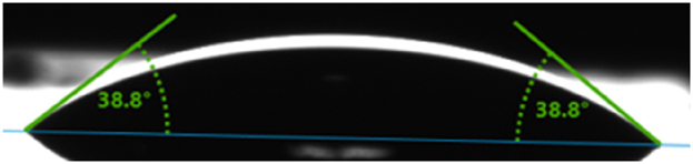
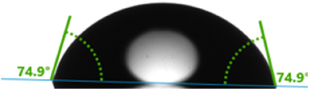
Droplet images and contact angles of water and diiodomethane for the polyesters are shown in Table 6. It was also found that the water contact angles, measured at about 65° for the pure UPR. However, as expected for those with clathrochelate, the water contact angle increased to about 81°, which is a consequence of increased hydrophobicity of polyesters. Diiodomethane contact angles were measured to about 39° for pure polyester. The values for clathrochelate-modified polyesters decreased to about 31°, indicating increased dispersion interactions on the polyester surfaces.
Contact angle images of pure UPR and polyesters with iron(II) clathrochelate.
| Sample | Water droplet | θ w | Diiodomethane droplet | θ d |
|---|---|---|---|---|
| Pure UPR |

|
64.6 |
|
38.8 |
| UPR 0.005 % Fe2+ |

|
68.9 |

|
38.1 |
| UPR 0.01 % Fe2+ |
|
74.9 |

|
37.7 |
| UPR 0.03 % Fe2+ |

|
77.4 |

|
35.8 |
| UPR 0.05 % Fe2+ |

|
80.5 |

|
30.8 |
Surface free energies of the polyesters were calculated from the measured contact angle values and they are shown in Table 7. The decrease in the surface free energy from 49.8 mJ m−2 for the pure polyester to 45.9 mJ m−2 for that polyester with the largest amount of clathrochelate can be attributed to the decrease in polarity caused by the increased clathrochelate content. The polar part decreases from 9.6 to 3.1 mJ m−2 with the increasing clathrochelate content. According to the Table 7, the increase in the dispersive part from 40.2 to 42.8 mJ m−2 confirms the previous suggestion that clathrochelate caused an increase in dispersive interactions.
Calculated surface free energy (γ
S
) and its components
| Sample name | γ S , mJ m−2 |
|
|
|---|---|---|---|
| Pure UPR | 49.80 ± 2.12 | 40.17 ± 0.34 | 9.63 ± 1.78 |
| UPR 0.005 % Fe2+ | 47.62 ± 2.38 | 40.20 ± 1.37 | 7.42 ± 1.01 |
| UPR 0.01 % Fe2+ | 46.72 ± 1.14 | 41.35 ± 0.63 | 5.47 ± 0.51 |
| UPR 0.03 % Fe2+ | 46.21 ± 1.49 | 42.04 ± 0.69 | 4.17 ± 0.80 |
| UPR 0.05 % Fe2+ | 45.87 ± 1.16 | 42.81 ± 0.32 | 3.06 ± 0.84 |
When determining the characteristics of materials for the medical, pharmaceutical, cosmetology or food industries, it is extremely important to determine the resistance of these materials to the adhesion of bacterial biofilm. For this purpose, the biofilm formation test was carried out on the surface of pure UPR and with the addition of Fe2+. The obtained results were compared with the control biofilm growth formed at the bottom of the well of a 24-well polystyrene plate. Bacterial strains responsible for infections in the medical and food industries were used in this test: S. aureus ATCC 25923 or E. faecalis PCM 896, or E. coli ATCC 25992. Planktonic bacterial cells were cultured twice so long to form a mono-species biofilm, i.e. 48 h under appropriate temperature and media.
Bacterial cells present in the environment under the favourable conditions, can form organized biofilm structures from planktonic forms, which pose a health threat. The data (Figs. 10 and 11) indicate that the addition of iron from only 0.03 % and more has a beneficial inhibitory effect on the adhesion of bacterial biofilm. The data show that the presence of 0.01 % iron in the polyester does not change composition, porosity or charge of the material significantly, because in relation to all tested bacterial strains, this material behaves like a pure control material.

Visualization on the CLSM images of biofilm formed by Staphylococcus aureus ATCC 25923, Enterococcus faecalis PCM 896 or Escherichia coli ATCC 25992 on the surface of the tested materials. (Magnification 200×, green fluorescence – viable cells, red and yellowish fluorescence – dead bacteria cells).

Quantification of the bacterial cells adhered to the tested materials compared to the control polystyrene surface (24-wells polystyrene plate).
Such observations can be justified because many factors influence the process of cell adhesion to materials. Namely, the material surface and microstructure can have different properties (smoothness, porosity, roughness, hydrophilicity), which may influence biofilm adhesion to varying degrees. 43 , 44 , 45 As well as electric charge because materials with the opposite charge to bacteria (usually bacteria are negatively charged) could attract biofilm aggregates. According to Renner and Weibel, van der Waals forces and electrostatic interactions are the main forces of bacterial adhesion to material surfaces. 46 The composites mechanical properties, such as hardness and elasticity, also have their consequences because biofilm settles and grows faster on flexible materials than on stiff, inflexible ones. Additionally, the chemical composition is important, i.e. the presence of organic compounds or ions that can be used as an energy source by bacteria if they are available. On one hand, low concentrations of magnesium or iron ions can stimulate the biofilm adhesion to materials. Microorganisms can use iron ions as a source of nutrients, which can increase their metabolic activity and promote surface colonization (Table 8).
Antibiofilm activity as a percentage of inhibition of bacterial biofilm adhesion to the material surface.
| Control biofilm | UPR pure | UPR 0.01 % Fe2+ |
UPR 0.03 % Fe2+ |
UPR 0.05 % Fe2+ |
|
|---|---|---|---|---|---|
| S. aureus | 100 | 37.2 | 34.3 | 78.2 | 94.7 |
| E. faecalis | 100 | 49.5 | 53.2 | 85.4 | 94.2 |
| E. coli | 100 | 72.3 | 71.0 | 75.7 | 68.8 |
On the other hand, high concentrations of such ions can be harmful to microorganisms because they can lead to formation of reactive oxygen species that can damage their cellular structures. High iron concentrations can also inhibit the metabolic activity of microorganisms and hinder formation of a stable biofilm.
Depending on the type of microorganisms and environmental conditions, the optimal concentration of iron ions can vary. Therefore, controlling the concentration of iron ions and other metal ions is important in the context of managing biofilm formation on materials.
Cytotoxicity tests showed that the obtained materials are not-toxic to skin fibroblasts. The WST-8 test proved that both native and enriched variants (pure or iron(II) clathrochelate-modified polyesters) have no negative effect on human normal fibroblasts after 24-h and 48-h incubation of cells with 24-h extracts. Due to the fact that cell viability was not reduced below 70 %, it is noticeable that according to ISO 10993-5 all tested materials are non-toxic (Fig. 12). 36

BJ cell viability after 24 and 48 h of incubation at 37 °C using the WST-8 test.
Cytotoxicity of the obtained polyester-based materials was also determined by the indirect contact method using Live/Dead fluorescent staining of the BJ fibroblasts grown with the 24-h extract of tested materials. The CLSM images presented a healthy monolayer of viable cells with typical spindle-shaped morphology after exposure to all tested extracts, confirming the non-toxic character of the materials (Fig. 13). Importantly, the viability and morphology of the cells exposed to the extracts were comparable to control cells grown in the presence of fresh culture medium.

Live/Dead assay of fibroblast in contact with the PS control, pure UPR and polyesters with different amounts of iron(II) clathrochelate.
A leaching study was used to assess the stability or potential release of iron(II) clathrochelate over time (Fig. 14).

Immersion test of the clathrochelate-resin materials.
Considering the fact that the iron compound is colored, its release from the polymer matrix to the solution should be easily and unambiguously observed. For this purpose, crushed polyester materials were immersed in liquid chemicals and left for 1 month in the dark, determining only the solvent factor.
As expected, the only changes were observed for samples with iron complex immersed in acetone. This aggressive solvent is responsible for the destruction of the polyester matrix, which helps in the release of the chelate or ligand into the liquid. 47 However, it was noted that the colour change of the liquid is insignificant, which also confirms that leaching from the matrix is nevertheless negligible (Fig. 15).

Leaching of clathrochelate from resin materials after 1 month.
Due to the potential use of iron(II) clathrochelate polyesters, these materials will be most exposed to contact with water or aqueous solutions of acids, bases and salts. This is why immersion test in such solvents is most important. No colour changes were observed in these samples.
The use of iron compounds is often limited due to their colour-enhancing properties. 20 However, iron(II) clathrochelates impart unique characteristics to polymeric materials, making them promising candidates for biomedical applications.
Conclusions
The resin curing process was carried out without the presence of a cobalt salt catalyst. It was noticed that its role was taken over by an iron(II) clathrochelate.
Cured resins with different colours were obtained. The addition of clathrochelate did not significantly change the thermal, and mechanical properties of the cross-linked products. However, with the increase in iron content, the water contact angles increased.
Studies have shown that iron(II) clathrochelate acts as both a plasticizer and a crosslinking promoter, depending on its concentration.
Importantly, the performed cytotoxicity tests showed that the obtained materials were not-toxic to skin fibroblasts. It was also observed that the addition of iron to resins from 0.03 % and more has a beneficial inhibitory effect on the adhesion of bacterial biofilm. This suggests that such polyesters may have potential applications in biomedical areas.
Replacing cobalt salts with iron(II) clathrochelate in cross-linking UPR resins derived from recycled PET further reduces the process’s carbon footprint. It reduces dependence on high-impact critical raw materials and lowers embedded CO2 emissions from the curing process.
Acknowledgments
The authors would like to express their appreciation to LERG S.A. for supplying the unsaturated polyester resin based on recycled PET.
-
Research ethics: Not applicable.
-
Informed consent: Not applicable.
-
Author contributions: P. P.: Conceptualization, Methodology, Formal analysis, Investigation, Resources, Writing – Original Draft, Writing – Review & Editing, Visualization, Supervision, S.V.S.: Resources, N.V.S.: Investigation, Writing – Original Draft, A.D.T.: Investigation, K.G.: Formal analysis, Investigation, Visualization, M.W.: Investigation, Visualization, M.M.: Formal analysis, Investigation, Visualization, Writing – Original Draft, M.T.K.: Formal analysis, G.G.: Formal analysis, Writing – Review & Editing, B.G.: Formal analysis, Writing – Review & Editing, All authors have accepted responsibility for the entire content of this manuscript and approved its submission.
-
Use of Large Language Models, AI and Machine Learning Tools: None declared.
-
Conflict of interest: All other authors state no conflict of interest.
-
Research funding: None declared.
-
Data availability: The datasets generated and/or analyzed during the current study are available from the corresponding author on reasonable request.
References
1. Penczek, P.; Czub, P.; Pielichowski, J. Unsaturated Polyester Resins: Chemistry and Technology. In Crosslinking in Materials Science: Technical Applications; Abe, A.; Dušek, K.; Kobayashi, S., Eds.; Springer-Verlag Berlin: Heidelberg, 2005; pp. 1–95.10.1007/b136243Suche in Google Scholar
2. Thomas, S.; Chirayil, C. J. Applications of Unsaturated Polyester Resins: Synthesis, Modifications, and Preparation Methods; Elsevier Inc: Amsterdam, 2023.Suche in Google Scholar
3. Pączkowski, P.; Sigareva, N. V.; Gorelov, B. M.; Terets, M. I.; Sementsov, Y. I.; Kartel, M. T.; Gawdzik, B. The Influence of Carbon Nanotubes on the Physical and Chemical Properties of Nanocomposites Based on Unsaturated Polyester Resin. Nanomaterials 2023, 13 (23), 2981. https://doi.org/10.3390/nano13232981.Suche in Google Scholar
4. Pączkowski, P.; Gawdzik, B. Synthesis, Characterization and Degradation Studies of eco-friendly Composites from Thermoset Resins with Pistachio Shell Waste. J. Therm. Anal. Calorim. 2024, 149 (7), 2789–2804. https://doi.org/10.1007/s10973-023-12872-0.Suche in Google Scholar
5. Malik, M.; Choudhary, V.; Varma, I. K. Current Status of Unsaturated Polyester Resins. J. Macromol. Sci. Part C: Polym. Rev. 2000, 40 (2-3), 139–165. https://doi.org/10.1081/MC-100100582.Suche in Google Scholar
6. Rokicki, G.; Wodzicki, H. Waterborne Unsaturated Polyester Resins. Macromol. Mater. Eng. 2000, 278 (1), 17–22. https://doi.org/10.1002/(SICI)1439-2054(20000501)278:1<17::AID-MAME17>3.0.CO;2-3.10.1002/(SICI)1439-2054(20000501)278:1<17::AID-MAME17>3.0.CO;2-3Suche in Google Scholar
7. Burgueno, R.; Quagliata, M. J.; Mehta, G. M.; Mohanty, A. K.; Misra, M.; Drzal, L. T. Sustainable Cellular Biocomposites from Natural Fibers and Unsaturated Polyester Resin for Housing Panel Applications. J. Polym. Environ. 2005, 13, 139–149. https://doi.org/10.1007/s10924-005-2945-9.Suche in Google Scholar
8. Nourizadeh, H.; Mirzaghorbanali, A.; McDougall, K.; Jeewantha, L. H. J.; Craig, P.; Motallebiyan, A.; Shokri, B. J.; Rastegarmanesh, A.; Aziz, N. Characterization of Mechanical and Bonding Properties of Anchoring Resins Under Elevated Temperature. Int. J. Rock Mech. Min. Sci. 2023, 170, 105506. https://doi.org/10.1016/j.ijrmms.2023.105506.Suche in Google Scholar
9. Anish, M. C.; Pandey, K. K.; Kumar, R. Preparation and Characterization of Unsaturated Polyester Infused Transparent Wood Composites. Eur. J. Wood Wood Prod. 2024, 82 (2), 503–513. https://doi.org/10.1007/s00107-023-02023-5.Suche in Google Scholar
10. Rubeš, D.; Vinklárek, J.; Podzimek, Š.; Honzíček, J. Bio-Based Unsaturated Polyester Resin from Post-consumer PET. RSC Adv. 2024, 14 (12), 8536–8547. https://doi.org/10.1039/D3RA08500G.Suche in Google Scholar
11. Voloshin, Y. Z.; Kostromina, N. A.; Krämer, R. Clathrochelates: Synthesis, Structure and Properties; Elsevier Science BV: Amsterdam, 2002.Suche in Google Scholar
12. Voloshin, Y.; Beltaya, I.; Krämer, R. Cage Metal Complexes: Clathrochelates Revisited; Springer International Publishing AG: Cham, 2017.10.1007/978-3-319-56420-3Suche in Google Scholar
13. Varzatskii, O. A.; Voloshin, Y. Z.; Stuzhin, P. A.; Shulga, S. V.; Volkov, S. V.; Vologzhanina, A. V.; Lebed, E. G.; Bubnov, Y. N. New Cadmium-Promoted Reaction of a C-Nucleophile: Synthesis and X-Ray Structure of the First Dicyanopyrazine Iron(II) Clathrochelate. Inorg. Chem. Commun. 2011, 14 (9), 1504–1507. https://doi.org/10.1016/j.inoche.2011.06.002.Suche in Google Scholar
14. Varzatskii, O. A.; Shulga, S. V.; Belov, A. S.; Novikov, V. V.; Dolganov, A. V.; Vologzhanina, A. V.; Voloshin, Y. Z. Copper(I)-and Copper(0)-Promoted Homocoupling and Homocoupling– Hydrodehalogenation Reactions of Dihalogenoclathrochelate Precursors for C–C Conjugated Iron(II) Bis-Cage Complexes. Dalton Trans. 2014, 43 (48), 17934–17948. https://doi.org/10.1039/C4DT01557F.Suche in Google Scholar PubMed
15. Voloshin, Y. Z.; Novikov, V. V.; Nelyubina, Y. V. Recent Advances in Biological Applications of Cage Metal Complexes. RSC Adv. 2015, 5 (89), 72621–72637. https://doi.org/10.1039/C5RA10949C.Suche in Google Scholar
16. Anxolabéhère-Mallart, E.; Costentin, C.; Fournier, M.; Nowak, S.; Robert, M.; Savéant, J. M. Boron-Capped Tris(Glyoximato) Cobalt Clathrochelate as a Precursor for the Electrodeposition of Nanoparticles Catalyzing H2 Evolution in Water. J. Am. Chem. Soc. 2012, 134 (14), 6104–6107. https://doi.org/10.1021/ja301134e.Suche in Google Scholar PubMed
17. Wise, M. D.; Ruggi, A.; Pascu, M.; Scopelliti, R.; Severin, K. Clathrochelate-Based Bipyridylligands of Nanoscale Dimensions: Easy-To-Access Building Blocks for Supramolecular Chemistry. Chem. Sci. 2013, 4 (4), 1658–1662. https://doi.org/10.1039/C3SC50155H.Suche in Google Scholar
18. Voloshin, Y.; Varzatskii, O.; Shulga, S.; Novikov, V.; Belov, A.; Makarenko, I.; Dubey, I.; Krivorotenko, D.; Negrutska, V.; Zhizhin, K.; Kuznetsov, N.; Bubnov, Y. Clathrochelate Complexes as Promising Molecular Scaffolds for Novel Biomimetic Systems, (Radio)Pharmaceuticals and Topological Drugs. In 10th European Biological Inorganic Chemistry Conference EUROBIC 10; Kessissoglou, D., Salifoglu, T., Eds.; Merimond: Pianoro, Italy, 2010; pp 29–38. https://cassi.cas.org/publication.jsp?P=eCQtRPJo9AQyz133K_ll3zLPXfcr-WXfSgiCpX76Xm5vE7qeDcHlotujkNmJU6b_Ms9d9yv5Zd9E8xUXr5Yowctu6inlelCMMs9d9yv5Zd9e81GLT7-JEWoaltA3PrU_.Suche in Google Scholar
19. Sigareva, N.; Barbash, V.; Yashchenko, O.; Shulga, S. V.; Starokadomsky, D.; Gorelov, B. Influence of Cellulose Particles on Chemical Resistance, Mechanical and Thermal Properties of Epoxy Composites. Biophys. Bull. 2020, 43, 57–70. https://doi.org/10.26565/2075-3810-2020-43-07.Suche in Google Scholar
20. Matušková, E.; Vinklárek, J.; Honzíček, J. Effect of Accelerators on the Curing of Unsaturated Polyester Resins: Kinetic Model for Room Temperature Curing. Ind. Eng. Chem. Res. 2021, 60 (39), 14143–14153. https://doi.org/10.1021/acs.iecr.1c02963.Suche in Google Scholar
21. Simonsen, L. O.; Harbak, H.; Bennekou, P. Cobalt Metabolism and Toxicology—A Brief Update. Sci. Total Environ. 2012, 432, 210–215. https://doi.org/10.1016/j.scitotenv.2012.06.009.Suche in Google Scholar PubMed
22. Lison, D. Cobalt. In Handbook on the Toxicology of Metals, Volume II: Specific Metals; Nordberg, G. F., Costa, M., Eds.; Academic Press: London, England, 2022; pp 221–242.10.1016/B978-0-12-822946-0.00008-8Suche in Google Scholar
23. Burdukov, A. B.; Vershinin, M. A.; Pervukhina, N. V.; Voloshin, Y. Z.; Varzatskii, O. A. Reactivity of Iron(II) Dichloride Clathrochelate: Synthesis and Properties of Mono-and Disubstituted Amino Clathrochelates. Russ. Chem. Bull. Int 2006, 55, 1982–1988. https://doi.org/10.1007/s11172-006-0540-4.Suche in Google Scholar
24. International Organization for Standardization. Plastics – Thermogravimetry (TG) of Polymers – Part 1: General Principles; ISO 11358-1: Geneva, Switzerland, 2014.Suche in Google Scholar
25. International Organization for Standardization. Plastics – Thermoplastic Materials – Determination of Vicat Softening Temperature (VST); ISO 306: Geneva, Switzerland, 2013.Suche in Google Scholar
26. International Organization for Standardization. Plastics – Plastics and Ebonite – Determination of Indentation Hardness by Means of a Durometer (Shore hardness); ISO 868: Geneva, Switzerland, 2003.Suche in Google Scholar
27. International Organization for Standardization. Plastics – Determination of Flexural Properties; ISO 178: Geneva, Switzerland, 2019.Suche in Google Scholar
28. International Organization for Standardization. Plastics – Determination of Charpy Impact Properties-Part 2: Instrumented Impact Test; ISO 179-2: Geneva, Switzerland, 2020.Suche in Google Scholar
29. International Organization for Standardization. Plastics – Determination of Dynamic Mechanical Properties-Part 1: General Principles; ISO 6721-1: Geneva, Switzerland, 2019.Suche in Google Scholar
30. Hashemi, M.; Shojaei, A. Morphology Development and Mechanical Properties of Unsaturated Polyester Resin Containing Nanodiamonds. Polym. Int. 2017, 66 (6), 950–959. https://doi.org/10.1002/pi.5343.Suche in Google Scholar
31. ASTM International. Standard Practice for Computing the Colors of Objects by Using the CIE System; ASTM E308: West Conshohocken, PA, USA, 2018.Suche in Google Scholar
32. Mokrzycki, W. S.; Tatol, M. Colour Difference ΔE: a Survey. Mach. Graphics Vis. 2011, 20 (4), 383–411.Suche in Google Scholar
33. Park, S.-J.; Cho, M.-S.; Lee, J.-R. Studies on the Surface Free Energy of Carbon-Carbon Composites: Effect of Filler Addition on the ILSS of Composites. J. Colloid Interface Sci. 2000, 226 (1), 60–64. https://doi.org/10.1006/jcis.2000.6787.Suche in Google Scholar
34. Pączkowski, P.; Głogowska, K. Preparation and Characterization of Quartz-Reinforced Hybrid Composites Based on Unsaturated Polyester Resin from Post-Consumer PET Recyclate. Materials 2024, 17 (5), 1116. https://doi.org/10.3390/ma17051116.Suche in Google Scholar
35. International Organization for Standardization. Plastics – Methods of Test for the Determination of the Effects of Immersion in Liquid Chemicals; ISO 175: Geneva, Switzerland, 2010.Suche in Google Scholar
36. O’Toole, G. A. Microtiter Dish Biofilm Formation Assay. J. Vis. Exp. 2011, 47, e2437; https://doi.org/10.3791/2437.Suche in Google Scholar
37. International Organization for Standardization. Biological Evaluation of Medical Devices – Part 5: Tests for in Vitro Cytotoxicity; ISO 10993-5: Geneva, Switzerland, 2009.Suche in Google Scholar
38. International Organization for Standardization. Biological Evaluation of Medical Devices – Part 12: Sample Preparation and Reference Materials; ISO 10993-12: Geneva, Switzerland, 2009.Suche in Google Scholar
39. Gawdzik, B.; Pączkowski, P. Sposób Utwardzania Nienasyconych Żywic Poliestrowych, Patent PL 246188 B1, 2024.Suche in Google Scholar
40. Wang, H.; Xu, Z.; Cheng, C.; Wang, T.; Mei, M.; Chen, S.; Liu, J.; Li, J. Unveiling Effect of Iron on Pyrolysis of Bisphenol A Epoxy by Comparisons of Product Characteristics and Reaction Mechanisms. Process Saf. Envirn. Prot. 2023, 171, 365–373. https://doi.org/10.1016/j.psep.2023.01.035.Suche in Google Scholar
41. Voloshin, Y. Z.; Varzatskii, O. A.; Kron, T. E.; Belsky, V. K.; Zavodnik, V. E.; Strizhakova, N. G.; Palchik, A. V. Triribbed-Functionalized Clathrochelate Iron(II) Dioximates as a New and Promising Tool to Obtain Polynucleating and Polynuclear Compounds with Improved Properties. Inorg. Chem. 2000, 39 (9), 1907–1918. https://doi.org/10.1021/ic990476n.Suche in Google Scholar
42. Bakar, M.; Djaider, F. Effect of Plasticizers Content on the Mechanical Properties of Unsaturated Polyester Resin. J. Thermoplast. Compos. Mater. 2007, 20 (1), 53–64. https://doi.org/10.1177/0892705707068820.Suche in Google Scholar
43. An, Y. H.; Friedman, R. J. Concise Review of Mechanisms of Bacterial Adhesion to Biomaterial Surfaces. J. Biomed. Mater. Res. 2002, 43 (3), 338–348. https://doi.org/10.1002/(SICI)1097-4636(199823)43:3<338::AID-JBM16>3.0.CO;2-B.10.1002/(SICI)1097-4636(199823)43:3<338::AID-JBM16>3.0.CO;2-BSuche in Google Scholar
44. Bazaka, K.; Jacob, M. V.; Crawford, R. J.; Ivanova, E. P. Efficient Surface Modification of Biomaterial to Prevent Biofilm Formation and the Attachment of Microorganisms. Appl. Microbiol. Biotechnol. 2012, 95, 299–311. https://doi.org/10.1007/s00253-012-4144-7.Suche in Google Scholar
45. Song, F.; Koo, H.; Ren, D. Effects of Material Properties on Bacterial Adhesion and Biofilm Formation. J. Dent. Res. 2005, 94 (8), 1027–1034. https://doi.org/10.1177/0022034515587690.Suche in Google Scholar
46. Renner, L. D.; Weibel, D. B. Physicochemical Regulation of Biofilm Formation. MRS Bull. 2011, 36 (5), 347–355. https://doi.org/10.1557/mrs.2011.65.Suche in Google Scholar PubMed PubMed Central
47. Pączkowski, P.; Puszka, A.; Gawdzik, B. Effect of Eco-Friendly Peanut Shell Powder on the Chemical Resistance, Physical, Thermal, and Thermomechanical Properties of Unsaturated Polyester Resin Composites. Polymers 2021, 13 (21), 3690. https://doi.org/10.3390/polym13213690.Suche in Google Scholar PubMed PubMed Central
© 2025 the author(s), published by De Gruyter, Berlin/Boston
This work is licensed under the Creative Commons Attribution-NonCommercial-NoDerivatives 4.0 International License.
Artikel in diesem Heft
- Frontmatter
- Special Issue Articles
- Magnesium oxide nanoparticles impregnated pyrolyzed coconut coir as an antifungal agent
- Comparative analysis of physicochemical, nutritional, functional, and sensory properties of rice bran oil from white (Bg 300) and brown rice (At 362)
- Electrochemical synthesis of porous polyaniline for supercapacitor application
- Modelling an amorphous biochar structure using classical molecular dynamics simulations
- Photocatalytic activity of C, N and S doped SnO2: effective band gap engineering to increase the quantum efficiency and exploration of the change in the position of Fermi energy with dopant concentration and its influence on photoreactivity
- Temporal variation of heavy metals in groundwater of North Central Province of Sri Lanka
- Regular Articles
- Synthesis and characterization of thermally stable quinoxaline-based polyamides
- Density-based solvent separation method for recycling mixed low-value plastic waste
- Synthesis, characterization and cytotoxic behavior against HeLa of iridium (III) complexes, half sandwich type
- Assess the sensitivity of gas and liquid chromatography for detecting trace substances in the environment
- Novel hybrid indazole-based 2,4-dihydro-3H-1,2,4-triazole-3-thione derivatives: design, synthesis, spectroscopic characterization, SAR, molecular docking, pharmacokinetics and toxicological activities
- Metal ions complexes with an azo compound derived from 2-amino-5-nitrothiazole: spectrophotometric determination and antioxidant activity in spiked samples
- Synthesis of N-formyl dihydropyrazoles as urease inhibitors
- Moisture resistant K-loaded ZIF-8 catalyst for glycerol carbonate production
- Synthesis, properties and application prospects in biomedical areas of unsaturated polyester resin modified with iron(II) clathrochelate