Biomaterial-based nanoreactors, an alternative for enzyme delivery
-
Rina Koyani
Rina Koyani received her PhD in Botany from The Maharaja Sayajirao University of Baroda (Gujarat, India) in 2012. She is currently a postdoctoral fellow at Centro de Nanociencias y Nanotecnología, Universidad Nacional Autónoma de México, (Ensenada, Baja California, Mexico). Her current research interests focus on bionanotechology and biotransformation.
Javier Pérez-Robles is a Postdoctoral researcher at Centro de Nanociencias y Nanotecnología, UNAM. His research has been related to genetics and biotechnology in aquatic organisms. At present, his work is focused on the encapsulation of catalytic proteins into recombinant virus-like nano-particles (VLPs).
Ruben D. Cadena-Nava is an Associate Professor at the Center of Nanoscience and Nanotechnology (CNyN) of National Autonomous University of Mexico (UNAM). He received his PhD degree in Physics from the Autonomous University of San Luis Potosí, Mexico. In 2006, he was a postdoctoral fellow in the Department of Chemistry and Biochemistry, University of California, Los Angeles, USA. His current research interests focus on bionanotechnology and nanomedicine.
Rafael Vazquez-Duhalt is a full professor at the Center for Nanosciences and Nanotechnology of the National University of Mexico. He is an Industrial Chemical Engineer from the National Polytechnic Institute in Mexico City. He earned his PhD degree in Biological Sciences from the University of Geneva, Switzerland. In addition, Dr. Vazquez-Duhalt carried out a 3-year postdoctoral work in the University of Alberta, Canada, and he has been a visiting professor at the University of Maryland and at the University of California, San Diego, USA. Dr. Vazquez-Duhalt is an Associate Director of the CaliBaja Center for Resilient Materials and Systems at the Jacobs School of Engineering, University of California at San Diego, USA. Prof. Vazquez-Duhalt is the Editor-in-chief of the journal “Biocatalysis” and is a member of the editorial boards of five scientific journals.


Abstract
Application of nanotechnology is making huge progress in the biomedical and environmental fields. The design and production of nanoreactors based on the combination of catalytic properties of enzymes and the unique characteristics of nano-sized materials is, certainly, an opportunity to solve different challenges in biomedical and environmental fields. Most of the research efforts to combine enzymes and nanostructured materials have been made using ceramic, metallic, or carbon-based materials. Nevertheless, biomaterials, or materials from biological origin, have two main advantages for biomedical and environmental applications when compared with non-biological nanomaterials; they are biocompatible and biodegradable materials. In this work, a critical review of the literature information on nanostructured biomaterials for enzyme delivery is shown.
‘The goal of life is to live in agreement with nature’
Zeno (335–264 BC)
1 Introduction
Nanobiotechnology, or Bionanotechnology, is the combination of the sophisticated mechanisms from biological systems with the exceptional properties of nanomaterials. This new scientific and technological field appeared just 15 years ago opening new approaches to solving old technological problems in the medical, environmental, and industrial areas. In less than two decades, nanobiotechnology has obtained spectacular advances, and the number of institutions worldwide carrying out research in this field is rapidly increasing [1].
Nature has developed molecular machines much more accurate and efficient than any of the manufacture processes developed by men. A single bacterial cell of Escherichia coli with 1 μm diameter stores approximately 10 Mbit of information in its genome [2], one ribosome is able to ensemble more than 50 different proteins, eukaryotic cells synthesize DNA molecules with an error rate of less than 10−11 per nucleotide added, and plants convert more energy and synthesize more amount of fine chemicals than all chemical industries [3]. On the other hand, biomolecular analogs to conventional devices such as structural components, tubes, motors, wires, pumps, synthesis processes, sensors, and programmable control systems, always exist in nature without exception. Among the main technological challenges, there is the mimicry of biological systems at nanometric scale to convert and transport energy, synthesize fine chemicals and macromolecules, store information, recognize, sense, move, auto-assembly, and produce smart materials. This is the main task of nanobiotechnology.
Enzymes are widely used in several applications in the food, feed, agriculture, paper, leather, textiles, pharmaceutical, and chemical industries, resulting in significant cost reductions and more energetically efficient processes. Enzymes catalyze chemical reactions with great specificity and very high transformation rate, which are orders of magnitude better than chemical catalysts. Enzymes are remarkable catalysts showing an exquisite selectivity, catalyzing reactions with insuperable chiral (enantio-) and positional (regio-) selectivity. This high selectivity also provides efficient reactions with minimal by-products, making enzymes an environment-friendly and low-energy alternative to conventional chemical catalysts. These reactions are the basis of the metabolism of all living organisms and, thus, provide tremendous opportunities for industries to carry out elegant, efficient, and economic biocatalytic conversions [4], [5], [6]. It is expected that there will be an accelerated growth of biocatalytic processes in industries that already have developed some expertise in this area, and many other industries will incorporate enzymatic transformations in their processes [7], [8], [9].
Humanity faces two important challenges when dealing with energy and the environmental issues that are intimately connected: (1) How can all the goods that society needs, and will need in the future, be produced using energetically efficient and environment-friendly processes? (2) How can the dependence on fossil energy sources be reduced and the environmental impact of human activities be minimized?
Green chemistry gains importance, opening new opportunities for research and development, to improve the efficiency of chemical processes while simultaneously reducing the materials and energy intensity and minimize or eliminate the dispersion of harmful chemicals in the environment. Among the challenges for green chemists, there is the discovery and development of new efficient catalysts, performing under mild reaction conditions with environment-friendly solvents and showing high selectivity for energy minimization, less toxic and inherently safer chemicals.
On the other hand, the field of Nanomedicine, which is the medical use of nanotechnology, is also emerging rapidly. The medical applications of nanotechnology include nanoelectronic biosensors, drug delivery, medical imaging, and molecular nanotechnology to prevent, treat, and cure major diseases [10]. The main goal of nanomedicine is to design, produce, and supply research tools and products clinically useful for the near future [11]. There are several research groups worldwide that are focused on the use of nanoparticles for drug delivery. However, the use of nanoparticles as carriers for enzymatic activity has less attention, even if the potential medical applications are evident [12], [13]. The improvement of chemotherapy by delivering cytochrome P450 activity into tumor cells has been achieved by targeted virus-like nanoparticles loaded with CYP [14], [15]. Most of the drugs used in chemotherapy are administered as pro-drugs, and they have to be activated into the active anticarcinogenic compound by the cytochrome P450 superfamily [16]. Unfortunately, the CYP expression varies significantly in the different tissues [17], and even more, in some cases, the healthy cells near the tumor cells express higher CYP activity [18], preferentially activating the cytotoxic agent, and affecting these healthy cells. Thus, the increase in CYP activity in the tumor tissue will improve the treatment efficiency and will reduce the doses, and importantly, a significant decrease in the side effects are expected [15]. The potential applications for the enzyme delivery are not limited to cancer. There are numerous illnesses originated by the lack of enzymatic activity. Among them, galactosemia and illnesses related to the lack of enzymes are involved in the amino acid metabolism such as albinism, alkaptonury, argininemia, argininosuccinic aciduria, deficit of carbamoyl phosphate synthetase I, homocystinuria, methylmalonic acidemia, phenylketonuria, and finally, the congenital adrenal hyperplasia (CAH), which encompasses a group of autosomal recessive disorders, each of which involves a deficiency 21-hydroxylase, resulting from mutations or deletions of CYP21A involved in the synthesis of cortisol, aldosterone, or both. This disorder affects between 1/10,000 and 1/15,000 of the population.
The use of therapeutic enzymes is not new. Duve [19] in the 1960s described the use of enzymes with the aim of treating disorders originated by enzymatic activity deficiencies. The therapeutic enzymes could be employed independently or in combination with specific drugs for the treatment of different disorders in a safe way [20]. Recently, the use of nanoparticles for transport therapeutic enzymes has been reviewed [21]. Nevertheless, the work is focused in cross-linked enzymatic aggregates (CLEA), metal-, silica-, and carbon-based nanoparticles, including synthetic polymers. In this work, biomaterials (materials from a biological origin) are reviewed, discussed, and proposed as better materials for the design and production of nanoparticle delivery of enzymes with medical and also with environmental interest.
On the other hand, nanoreactors are potential platforms to mimic the special organization of metabolic enzymes. In contrast to the enzymatic reactions with diluted enzymes, cells contain a high concentration of macromolecules resulting in crowding conditions that affect the structure and function of enzymes [22]. The encapsulation of a chain of metabolic enzymes in a nanoreactor could increase the catalytic efficiency. In a multi-enzymatic nanoreactor, enzyme active sites for consecutive reactions are placed in close proximity, which reduces the intermediate diffusion distance and, therefore, increases the probability that a metabolic intermediate undergoes a sequential reaction step before diffusing away [22].
2 Biopolymer nanoparticles
Nanotechnology, soon after its emergence, has been successfully applied to many engineering and technical fields where a number of metals, ceramics, and synthetic organic/inorganic polymers were utilized for the purpose. Among the potential applications, nanomedicine procured the use of biopolymers for nanoparticle manufacturing with enhanced possibilities of their successful uses for diagnostics, medical supplies, therapeutic treatments, and emerging regenerative medicine such as tissue-engineered tissues. Biopolymers, being organic, also offer a versatility that is unmatched by metals and ceramics. The wide spectrum of physical, mechanical, and chemical properties provided by biopolymers has fueled the extensive research, development, and applications of polymeric biomaterials [23]. Moreover, utilizing biodegradable polymers over synthetic non-degradable materials paved the alternative way to overcome degradation issues and environmental concerns. Conversely, the potential toxicological effect of some biomaterials, in addition to unease structuration at nano-size scale, limits their utilization. Nevertheless, extensive research and bioengineering advances certainly would help to solve these challenges.
The current overarching concept of engineering of biopolymeric nanoparticles has established a new vision for their utilization over synthetic materials in a range of medical applications. A major emphasis has been made on the use of biodegradable polymers like liposomes, proteins, polysaccharides, and virus-like particles (VLPs) for nanoparticle preparation. The focus for these biomaterials has turned toward tissue engineering, sophisticated cell, drug and gene delivery systems, and other applications in biotechnology, where the need for specific and direct interactions between biomaterials and tissue components become a priority [24]. With the potential use of biopolymers for medical purposes, a new paradigm for biocompatibility has emerged, and continuous efforts for understanding the biocompatibility mechanisms are being made.
During the last decade, several protocols for the fabrication of biopolymer-based nanoparticles have been well established (Table 1) and have shown notable advantages including easy preparation, biodegradability, and high stability. The increasing interest for biopolymeric nanoparticles (BNPs) is originated by its implication in medical science (drug delivery), with advantages including enhanced bioavailability, high drug loading, reduced side effects, and controlled and targeted drug release. Delivery of a variety of chemicals, nutraceuticals, flavors, antimicrobials, dietary fibers, and minerals through BNP encapsulation is becoming progressively significant [67]. Moreover, applications of BNPs in gene therapy and tissue engineering are another important aspect potential for their medical applications. Bioactive compound encapsulation in biomaterials also allows the modification of their stability, texture, mechanical, and optical properties [68]. The incorporation of BNPs as co-catalysts enhances the photocatalytic water oxidation process when compared with commercially available non-stabilized cobalt nanoparticles [69].
Biopolymers for engineered nanoparticles – recent reports.
| Biopolymers | References |
|---|---|
| Animal-derived proteins | |
| Albumin | [25], [26] |
| Gelatine | [27] |
| Caseins | [28] |
| Fibroin | [29], [30] |
| Sericin | [31], [32] |
| Keratin | [33] |
| Whey protein | [34], [35] |
| Collagen | [36] |
| Elastin | [37], [38] |
| Plant-derived Proteins | |
| Gliadin | [39], [40] |
| Zein | [41], [42] |
| Pea legumin | [43] |
| Soy protein isolate | [44] |
| Polysaccharides | |
| Agar | [45] |
| Alginate | [46], [47] |
| Chitosan | [48], [49], [50] |
| Dextran | [51], [52] |
| Carrageenan | [53], [54] |
| Cellulose | [55], [56] |
| Gellan gum | [57] |
| Inulin | [58] |
| Pectin | [59], [60] |
| Pullulan | [61], [62] |
| Heparin | [63], [64] |
| Starch and its hydrolysates | [65], [66] |
Enzymes, used as catalysts for several biochemical processes, are also known for their extensive applications through nanoencapsulation in biopolymeric materials as a possible solution of some limiting factors like stability and reusability (Figure 1). Immobilization of ligninolytic enzymes such as laccases and peroxidases on BNPs improves their performances for bioremediation processes [48], whereas encapsulated glucose oxidases in BNP can be used to build biosensors. An interesting approach of enzyme encapsulation technology with biopolymeric matrix also provides self-regulated degradation mechanism, which is mandatory for the applications such as hard tissue replacement and some drug delivery systems [70]. Enzyme immobilization on biopolymeric materials has also been proven to prevent bacterial biofilm formation on implanted medical devices such as endotracheal tubes [71]. The most recent-emerged DNA nanotechnology involves the use of DNA hydrogels for enzyme entrapment. This technique was applied to enzymatic biobattery that holds a great promise to improve bioelectrocatalysis-based devices [72], and DNases can degrade it. Converting natural sensitive biocatalysts to stable and environmentally adaptive catalysts through their encapsulation or immobilization on BNPs open novel strategies and uses for industrial processes.
Biosynthesis of macromolecules through enzymatic polymerizations of specific molecular structures constitutes a critical step in the evolution of living organisms. Tailored fabrication of BNPs seems to be the next step and could hold particular promise for their potential applications, but this technology is still in its infancy. BNPs are being investigated for their prospective in many areas at laboratory scale. Thus, it is still important to take up the challenge for economically achievable large-scale commercial production of BNPs with desired and uncompromised properties or functional attributes.
Several methodologies have been developed and optimized for BNP preparation, but still, their sensitivity to the operational conditions needs to be addressed. Associative interaction of proteins and polysaccharides can also be one of the intrinsic sources for BNP fabrication with a variety of appropriate combinations. However, such particles are under threat of dissociation due to changes in environmental conditions that may induce protein unfolding or aggregation and limit the use of multiple methodological combinations [67]. Novel sources of biopolymers, different combinations, innovative and simple new preparation methods with high protein loading efficiency, and ensuring biological safety should also certainly be emphasized. The toolbox offered by BNPs opens research opportunities to all fascinating interdisciplinary fields for innovation, which is truly imperative for the application of nanotechnology in medicine and green technology.
3 Liposomes
Early investigations on bimolecular-based nanoparticles emerged with the formation of “liposomes” defined as the spherical vesicles composed of lipid bilayers. These liposome bilayers can be formed from different phospholipids wherein its constituent characteristics reflect in resultant particles [73]. The unique, and appreciated, nature of liposomes that shows a hydrophilic core and hydrophobic bilayer favors both encapsulation and entrapment of hydrophilic as well as hydrophobic compounds simultaneously. Liposomes have been employed as a “safe platform” for carrying antibodies, antigens, proteins, peptides, enzymes, hormones, vaccines, and photosensitizers to be able to reach the target tissue. Investigations developed toward enzyme-containing liposomes, where lipid vesicles are carrying water-soluble enzymes, had appeared as one of the most practically favorable technique. Enzyme-containing liposomes are mainly known for their two main potential applications, i.e. enzyme-replacement therapy in biomedical sciences and food processing, where liposomes chiefly play the role of carriers [74]. The role of liposome-encapsulated enzymes in accelerating cheese ripening process and flavor development [75], and in the treatment of lactose intolerance [76], provided good examples for their real industrial applications.
Targeting and on-time release of enzymes are essential factors to be controlled in order to obtain effective outcomes and to avoid unwanted catalytic reactions, and they can be certainly achievable by using liposomes. Their application is not restricted to cancer therapy, and their use as carriers for infectious and autoimmune diseases, their application for drug delivery, treatment of myocardial diseases, or any kind of enzyme-replacement therapy could be envisaged. Liposomes containing butyrylcholinesterase as toxin scavengers [77] and phosphotriesterase for the destruction of toxic organophosphates [78] are other examples of biocatalytic liposomes. On the other hand, highly sensitive chemiluminescence immunosensor has also been developed with horseradish peroxidase encapsulation in liposomes for prostate-specific antigen (PSA) detection [79]. Furthermore, utilization for environmental processes has also been considered, and the decontamination of metal-laden industrial wastes through phosphatase containing lipid vesicles has been explored [80].
The demonstration of liposome capacity for drug delivery has been stimulated by their biocompatibility and biodegradability, but has been restricted due to their physical stability and degradation prior to targeted therapeutical application. However, major breakthroughs through intensive research for years began to elucidate the understanding of lipid compositions for producing liposomes with added-value properties, i.e. stability, enhanced drug bioavailability, protection against drug hydrolysis or oxidation, increased drug lifetime, better pharmacokinetic properties, and improvement of therapeutic index [81]. The manipulation of liposomal structures by the addition of different molecules in the liposome bilayer, with both active and passive targeting strategies, achieved a liposomal formulation containing the anthracycline drug for the treatment of cancer and AIDS-related Kaposi sarcoma, and multiple myelomas [73], [82]. Modified liposomes have been proposed as potential vaccine preparations against a number of pathogens such as tuberculosis [83]. An important recent contribution has been reported, in which liposomes display viral proteins (named by the authors as “virosome”). These preparations have been proposed for prophylactic applications of hepatitis B vaccines and to improve the vaccine-based preparation against influenza [83], [84], [85]. These modification strategies certainly could be applied to other biocatalytic liposomes.
Since their invention 50 years ago, liposomes were proposed first as vehicles for drug delivery, and they have significantly achieved much attention due to their fascinating contribution to the medical sciences. However, there is still a huge potential of this technology that has not been entirely exploited. The design of tailored liposomes with multifunctional properties is an important opportunity niche for research. Stability, toxicity, controlled release, immunity-promoted delivery, selective tissue targeting, and many more issues should be explored to establish a robust technology that can be rapidly taken up by the medical and pharmaceutical industries. Regardless of significant research and development in liposomal technology, inadequate commercialization and low-quality control have limited its application; thus, it is imperative to establish regulation rules for the production and use of liposomes.
Like liposomes, “polymersomes” are also composed of lipid bilayers with aggregated amphiphilic macromolecules, but they are less dynamic than liposomes due to lower critical aggregation concentration and larger dimensions of the amphiphilic block copolymers, and moreover, their low permeability to water limits its utilization as nanoreactors [86]. This limitation could be solved by the inclusion of channel proteins [87] or proton pumps [88] in the polymeric bilayer. However, the porous nature of the polymersomes allows the enzyme encapsulation and positioning of different enzymes to specific separate domains and is possible as well, which is demonstrated by Vriezema et al. [89] and van Dongen et al. [90]. Thus, polymersomes have a potential for constructing nanoreactors with multiple enzymes.
4 Virus-like particles (VLPs) as nanovehicles for enzymes
VLPs are nanoparticles similar to the original virus but lacking the viral genome. The VLPs are dynamic structures forming many different shapes and sizes according to the medium pH and ionic strength. They are monodisperse, chemically stable, and they maintain some properties of the original virus. In addition, the basic structure of the VLPs can be programmed in different ways to place inside various types of molecules. VLPs can also be conjugated with other molecules to form more complex nanoparticles [91], [92], [93]. The use of the VLPs as nanocarriers confers several advantages:(i) prevents premature degradation of the cargo and avoid the cargo interaction with the environment, (ii) improves the absorption and/or delivery of its cargo, and (iii) provides better-controlled distribution of the carried substances [92].
VLPs show an enormous potential, especially for medical applications because of their versatility, structure, safety, and capacity to protect their cargo. VLPs are (i) highly ordered and self-assembled structures. (ii) They show high surface area to bind ligands. (iii) They contain a diversity of reactive groups for ligand anchorage. (iv) They contain empty cavities for cargo encapsulation. (v) Importantly for reactors, they are porous structures for substrate intake and product release. Finally, some of the VLPs may perform cell internalization.
The capacity of virus capsids or VLPs as enzyme containers to form nanoreactors and as enzyme platforms has been demonstrated (Table 2) [101], [102], [103], [104], [105], [106]. Enzymatic nanoreactors or enzyme nanocarriers are not unbound to some limitations and necessity of improvements. VLPs could be coated with enzyme molecules [107], but without a doubt, the chemical characteristics of the empty capsids should be considered for the design of different strategies for creating functional enzymatic VLPs (Figure 2). The outer and inner charge, size, nucleation, optimal temperature, and pH for self-assembly and the formation of secondary structures at different ionic strengths, degradation, and stability in non-optimal conditions should be considered [107], [108], [109], [110]. Considering these properties, the encapsulation of different enzymes such as alcohol dehydrogenase [111], glycosidase [100], and cytochrome P450 (CYP) [14], [15], [112] has been achieved (Figure 3). The design of synthetic “metabolomes” or multi-enzyme systems has also been proposed [113].
Enzyme encapsulation in virus-like particles (VLPs).
| Enzyme | VLP | Mconf(mM) | Enzyme per capsid | kcat KM−1 related to free enzyme | Encapsulation method | Ref. |
|---|---|---|---|---|---|---|
| Cytosine deaminase | SV40 | ND | ND | Lower (VNR) | Fusion protein with internal protein of the capsid (in vivo) | [94] |
| Peptidase E | Bacteriophage Qβ | ND | 2–18 | Lower 3× | Fusion protein with RNA (in vivo) | [95] |
| Luciferase | Bacteriophage Qβ | ND | 4–8 | Lower 30× | Fusion protein with RNA (in vivo) | [96] |
| Pseudozyme antartica lipase B | CCMV | 1 | 1.3–4 | Higher (kcat) | Fusion protein with coiled-coil motif (in vitro) | [97] |
| Alkaline phosphatase | Bacteriophage MS2 | 0.5 | 3.2 (monomers) | Same | Electrostatic charges. Fusion protein with a negative peptide (in vitro) | [98] |
| Alcohol dehydrogenase | Bacteriophage P22 | 7.2 | 249 ± 13 | Lower 1.6× | Fusion protein with scaffold protein (in vivo) | [99] |
| CelB glucosidase | Bacteriophage P22 | 2.4 | 87 ± 3.5 (monomers) | Same | Fusion protein with scaffold protein (in vivo) | [100] |
| Phosphotriesterase | Bacteriophage P22 | 1.1 | 40 ± 10 (monomers) | Lower 600× | Fusion protein with scaffold protein (in vivo) | [98] |
| Cytochrome P450 | CCMV | 4.9 | 14 | Lower 10× | Trapping by complementary charges (in vitro) | [14] |
| Cytochrome P450 | Bacteriophage P22 | 3.1 | 110 | Same | Fusion protein with scaffold protein (in vivo) | [15] |
Mconf, molar confinement (enzyme concentration inside the capsid); ND, not determined; NRV, not reported value.
VLPs from different viruses have been synthesized including the non-enveloped icosahedral MVM (Minute virus of mice), PaV (Pariacoto virus), CCMV (Cowpea chlorotic mottle virus), NV (Norwalk virus), and bacteriophages U29, k, HK97, and P22. Likewise, they have also been produced from icosahedral capsids of enveloped viruses such as HBV (hepatitis B virus), HSV-1 (herpes simplex virus type 1), MLV (murine leukemia virus), HIV-1 (human immunodeficiency virus type 1), and influenza virus [114].
In addition to the protection and delivery of enzymes with a specific catalytic activity, VLPs can be directed to specific target tissues or cells, which is a major issue of interest in nanomedicine. Because of its protein nature and the presence of reactive groups on the capsid surface (i.e. free amino and carboxylic groups), they could be easily functionalized with specific ligand molecules to be recognized for receptors in the target cell surface and then internalized [115]. Antibodies have been used to functionalize some types of nanoparticles; however, the size (~160 kDa) could hinder the penetration of nanoparticles into the target tissues. Another drawback of antibodies is that they may have a high affinity for other structures, and they are subjected to degradation [116], [117]. To solve such problems, antibody fragments, or construct from small antibodies, have been used [118]. On the other hand, smaller molecules such as peptides or micropeptides seem to be an attractive and efficient alternative for VLP functionalization [119]. These molecules are small, easily synthesized, chemically stable, able to be conjugated with radionuclides, and they can adhere to viral capsids and different types of micelles. In addition, the use of artificial small peptides from d-amino acids, which are more stable to proteolysis, could be envisaged [116], [120], [121], [122]. Cytosine deamidase, which transforms the prodrug 5-fluorocytosine to the active drug 5-fluorouracyl, has been encapsulated into the SV40 capsid as a carrier to deliver the enzymatic activity to CV-1 cells with the aim to increase the drug sensitivity and to induce apoptosis [94]. Recently, a multienzymatic VLP-based nanoparticle has been produced. Three enzymes: CelB glucosidase, ATP-galactosidase, and ADP-glucokinase, involved in the carbohydrate metabolism, were encapsulated into VLPs from bacteriophage P22 [113]. Unexpectedly, no increase in the efficiency of the cascade reaction was found, concluding that it is important to consider the right balance of kinetic parameters of each of the involved enzymes in the design of efficient catalytic systems. Nevertheless, the production of synthetic metabolome based on the enzyme encapsulation in VLPs could generate complex catalytic systems with various potential applications.
The enzyme concentration inside the virus capsids could reach the mM order. Thus, it is possible to study catalysis in crowded environments, simulating those found at the cellular level, to better understand the enzymatic catalysis inside cells. Despite that the catalytic activity of many encapsulated enzymes is lower than these found with free enzyme, nanoreactors with new properties are generated with biocatalytic VLPs, such as thermostability, proteolysis resistance, protection during lyophilization process [120], [121], [122], reduction of substrate inhibition [111], and improvement of performance under certain operational conditions [101].
Without a doubt, the development of VLPs as enzymatic nano-vehicles is still in their infancy. However, so far, the capacity to carry biocatalytic cargos has been clearly demonstrated, and their functionalization and targeting to specific tissues should also be considered. In addition, the use multi-enzymatic nanoparticles that mimic metabolomes is still scarce. Without a doubt, multi-enzymatic VLPs are an open opportunity to find new and innovative applications.
Finally, functionalized VLPs loaded with active enzymes are an attractive alternative for the treatment of several illnesses that are originated by an enzyme deficiency. The application of VLP-nanoreactors is not limited to medical issues, but they could be envisaged for environmental applications as well. However, further research is needed to improve the stability, functionalization, degradation, and biocompatibility of VLPs, and still a research effort should be focused to optimize the transformation of different substrates, as suggested by Comellas-Aragones et al. [101], Cardinale et al. [99], and Patterson et al. [113].
5 DNA nanostructures as enzyme carriers
The postulate proposed by Seeman in 1982 [123] pointing out the use of DNA as a building biomaterial for the creation of predesigned nanostructures through molecular self-assembly has started the emergent field of DNA nanotechnology. The real impulse in the field was observed with the invention of the DNA origami technique, which allowed DNA strands to be molded according to the nanostructure requirement [124]. The enormous potential of DNA-based enzyme nanoreactors has been recently reviewed [125] in which the diversity of DNA-based nanostructures as single or multi-enzymatic systems and their potential applications are discussed. Moreover, the non-toxic and biocompatible nature of DNA, together with the possibility to design a desired architecture allowed to explore their implementation in many fields, especially in biomedical applications. DNA nanostructures can be site-specifically engineered and can be the base of structures like autonomous walkers [126], nanotweezers [127], and nanocages [128]. So far, numerous applications of DNA-based nanostructures have been demonstrated in both biological and non-biological areas such as plasmonic and nanoelectronics/nanophotonics device fabrication [129], [130], scale rulers for optical imaging [131], DNA computing [132] targeted drug delivery [128], [133], gene therapy [134], and development of biosensors for cancer diagnosis [135].
Furthermore, DNA nanoarchitecture also serves as a powerful platform for appropriate activation of enzyme cascades to improve their biological efficiency (Figure 4). Control of relative distance and orientation of enzymatic components or facilitation of interface between enzymes/cofactors and electrode surfaces enables protein-DNA assemblies to organize cascades of enzymatic reactions [136], [137]. The multi-enzymatic DNA nanoreactors have been studied and demonstrated [138]. However, the regulation of the enzymatic activities in these systems is still a critical issue. The manipulation of key parameters like position, stoichiometry, and inter-enzyme distance for optimal activity as well the use of programmed DNA nanostructures increases the potential for the design of multi-enzymatic nanoreactors [139]. Fu et al. [140] have systematically studied the effect of inter-enzyme distance, in which the activity of a GOx/HRP cascade spatially organized on a DNA nanostructure increased when the inter-enzyme distance is reduced. The DNA tweezer-actuated enzymatic nanoreactor has been successfully developed controlling the inhibition and activation of dehydrogenase and NAD+ cofactor [141]. The introduction of feedback or feed-forward control loops and the concept of responsive enzymatic nanodevices [142] could be applied for diagnostic and production of high-value chemicals. Rational control and regulation of DNA-based nanodevices containing multi-enzyme systems also expand the uses of DNA nanotechnology for enzyme-induced drug delivery and enzyme-triggered nanostructure assembly [143]. Many expected biomedical applications of bicatalytic nanoreactors include the intracellular delivery of enzymes or their efficient cellular uptake. The cell internalization could be enhanced by noncovalent modification of origami structures [144], [145]. A significant enhancement of cell uptake has been obtained by coating the DNA origami nanostructures with virus capsid proteins [146]. This strategy utilizes purified capsid proteins that can bind and self-assemble on the origami surface through electrostatic interactions and further pack the origami nanostructures. Though several approaches have been developed and attempted to reach this goal, it is always hampered with transport-limiting factors like size, charge, and stability of enzymes. However, utilization of designed DNA nanostructures as a carrier for the enzymatic cargo was introduced as an important strategy [125]. Efficiently designed DNA containers developed by Kuzuya and Komiyama [147], Zhao et al. [148], and Kiviaho et al. [149] protect the enzymes from biological degradation, enhance the catalytic activity, and may tune the enzymatic reaction rates. The strategy of delivering β-galactosidase into cells can be improved by coating it with a dense shell of oligonucleotides [150], which is also an innovative attribute of DNA nanotechnology.
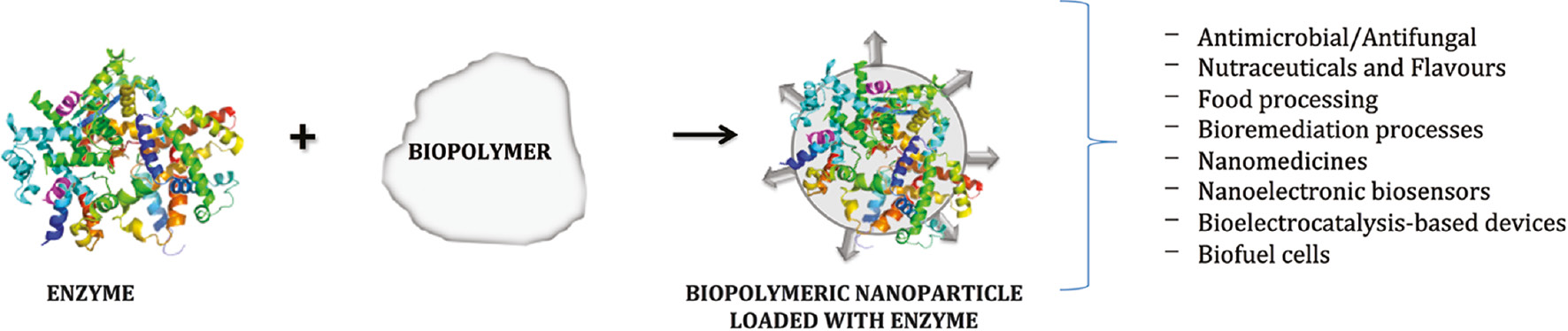
Biopolymer-based biocatalytic nanoparticle and their possible applications.
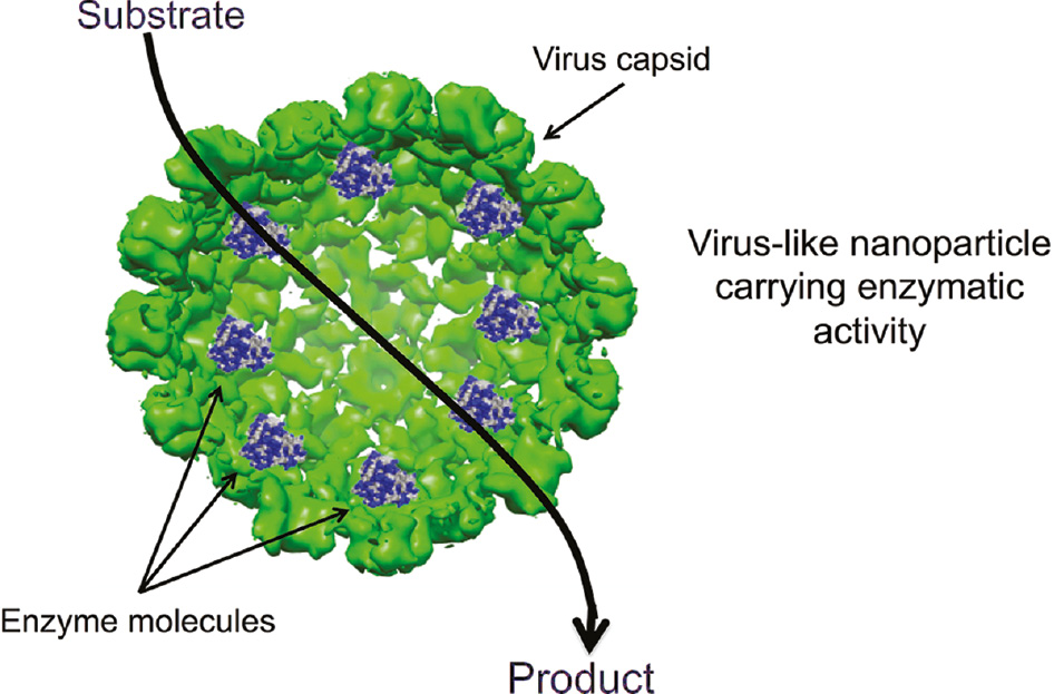
Virus-like particles (VLPs) as nano-vehicles for enzyme delivery. The enzyme molecules are encapsulated inside the virus capsid. The capsid is porous allowing the substrate internalization, and the product can diffuse outside the nanoreactor.
![Figure 3: Transmission electronic microscopy of P22 capsids containing cytochrome P450 [112].](/document/doi/10.1515/ntrev-2016-0071/asset/graphic/j_ntrev-2016-0071_fig_003.jpg)
Transmission electronic microscopy of P22 capsids containing cytochrome P450 [112].
![Figure 4: Scheme of an enzymatic reactor build from DNA and enzyme molecules. Enzyme molecules are attached with precision on the highly modulated DNA nanostructures. Glucose oxidase (GOx, purple) and horseradish peroxidase (HRP, green) have been assembled into two tubular DNA origami nanostructures (orange and yellow cages). The reaction cascade starts with the production of hydrogen peroxide from glucose oxidase, which is the substrate for peroxidase-mediated oxidation. This figure was taken from Linko et al. [125].](/document/doi/10.1515/ntrev-2016-0071/asset/graphic/j_ntrev-2016-0071_fig_004.jpg)
Scheme of an enzymatic reactor build from DNA and enzyme molecules. Enzyme molecules are attached with precision on the highly modulated DNA nanostructures. Glucose oxidase (GOx, purple) and horseradish peroxidase (HRP, green) have been assembled into two tubular DNA origami nanostructures (orange and yellow cages). The reaction cascade starts with the production of hydrogen peroxide from glucose oxidase, which is the substrate for peroxidase-mediated oxidation. This figure was taken from Linko et al. [125].
Without a doubt, the rapid development of DNA nanotechnology offers a huge potential for enzyme-based nanoreactors. Nevertheless, additional research efforts should be performed to reach the goal of establishing an easy, efficient, scalable, and economic strategy for the production of DNA-based nanostructures with desirable characteristics. The number of studies on DNA nanostructure loaded with enzymes is still scarce, but an increasing interest on DNA-nanostructures as the carrier of engineered enzymes with multiple functions and new potential applications is expected.
6 Physical chemistry of nanoreactors
Physicochemical properties of nanoreactors are influenced or modulated by external parameters such as temperature, pH, or ionic strength. Alterations in these environmental parameters allow the control of both nanoparticle stability and catalytic activity, which is possible by controlling the diffusion of substrate and products through the nanoreactor surface, adjusting the optimal concentration of catalytic molecules and reactants, and by the suitable nano-environment inside the nanoreactor. The enzymatic activity of a nanoreactor can be affected by several factors such as mass transfer limitations, the obstruction of the catalytic site of enzymes, and the decrease in the diffusion rate of substrates, due to the high concentration of enzymes. Therefore, in order to produce an efficient nanoreactor, it is very important to consider an optimal number of enzyme molecules and avoid enzyme crowding.
The efficiency of the nanoreactors has been measured through their physicochemical properties where different techniques are being used to qualify their performance. The diffusion-controlled reactions in nanoreactors have been experimentally studied by means of luminescence quenching [151], by a theoretical diffusion model of spherical cavity [152], or different hollow geometries [153]. Another model has been developed considering the diffusion coefficients of the reacting molecules and critical distance for the reaction with the Stokes-Einstein equation, in which the diffusion coefficient of a species is correlated to its radius and the solvent viscosity. From this approach, a Smoluchowski reaction expression is obtained, that consider a steric factor, which characterizes the diffusion rate into the porous cavity by absorption [154]. However, the Smoluchowski reaction rate constant does not consider the nanoreactor geometry and the crowding effects.
On the other hand, the “ζ” potential describes the surface charge of nanoparticles [155] and can be used to study the stability of a nanoreactor in suspension or in the environments where it is being used. The ζ potential of nanoparticles can be determined through dynamic light scattering (DLS) or by measuring their electrophoretic mobility. It is well known that nanoparticles with ζ potential values greater than +25 mV or less than −25 mV typically have high degrees of stability [156]. Moreover, dispersions with a low ζ potential value will eventually aggregate due to van der Waals interparticle attractions [145], [157]. DLS is a versatile characterization technique that can be used for particle size measurements and polydispersity determination together with other structural parameters such as weight molar mass, the radius of gyration, and hydrodynamic radius [155]. The critical aggregation concentration, the particle morphology, and the change in size or shape with the variation in pH, temperature, and ionic strength could also be determined by DLS [158].
The dynamic properties of fluorescent molecules can be studied using the fluorescence correlation spectroscopy (FCS). By monitoring fluctuations of fluorescence intensity in a confocal volume and by performing the correlation analysis of the fluctuations, it is possible to estimate the size, diffusion coefficients, and concentrations of nanoparticles [159], [160]. The number of fluorescent molecules in the nanocontainers was determined by this technique [161]. The FCS is a promising technique that can also be used to study the diffusion through the porous surface, stability, and the enzymatic activity of nanoreactors. To the best our knowledge, the DLS and CFS techniques have not been fully exploited to study the physiochemical properties of nanoreactors.
Electron microscopy techniques, such as transmission electron microscopy (TEM) and cryo-TEM, are useful techniques to characterize and study the shape, size, thickness, and structure of self-assembled viral capsids and nanoreactors [162], [163]. In addition, atomic force microscopy (AFM) is a powerful tool to investigate the mechanical properties of viral capsids [164], [165].
The concept of “smart nanoreactor” could be fulfilled only with the theoretical and experimental studies of all the physicochemical properties of different components at any possible environment in which the nanoreactor could be exposed.
7 Concluding remarks
Without a doubt, biomaterial-based nanoparticles have huge potential for enzyme delivery. There are numerous illnesses originated by the lack of enzymatic activity. Biomaterial nanoparticles seem to be a better alternative for biomedical applications than inorganic preparations due to its biocompatibility, biodegradability, available functional groups, and adjustable structural characteristics. Still, basic research efforts should be performed in order to solve challenges such as immunogenic response, particle stability in physiological fluids, and catalytic performance, among others. Nevertheless, it is clear, from the information available, that enzyme delivery by biomaterial-based nanoparticles is an important alternative in the new field of nanomedicine.
About the authors
Rina Koyani received her PhD in Botany from The Maharaja Sayajirao University of Baroda (Gujarat, India) in 2012. She is currently a postdoctoral fellow at Centro de Nanociencias y Nanotecnología, Universidad Nacional Autónoma de México, (Ensenada, Baja California, Mexico). Her current research interests focus on bionanotechology and biotransformation.
Javier Pérez-Robles is a Postdoctoral researcher at Centro de Nanociencias y Nanotecnología, UNAM. His research has been related to genetics and biotechnology in aquatic organisms. At present, his work is focused on the encapsulation of catalytic proteins into recombinant virus-like nano-particles (VLPs).
Ruben D. Cadena-Nava is an Associate Professor at the Center of Nanoscience and Nanotechnology (CNyN) of National Autonomous University of Mexico (UNAM). He received his PhD degree in Physics from the Autonomous University of San Luis Potosí, Mexico. In 2006, he was a postdoctoral fellow in the Department of Chemistry and Biochemistry, University of California, Los Angeles, USA. His current research interests focus on bionanotechnology and nanomedicine.

Rafael Vazquez-Duhalt is a full professor at the Center for Nanosciences and Nanotechnology of the National University of Mexico. He is an Industrial Chemical Engineer from the National Polytechnic Institute in Mexico City. He earned his PhD degree in Biological Sciences from the University of Geneva, Switzerland. In addition, Dr. Vazquez-Duhalt carried out a 3-year postdoctoral work in the University of Alberta, Canada, and he has been a visiting professor at the University of Maryland and at the University of California, San Diego, USA. Dr. Vazquez-Duhalt is an Associate Director of the CaliBaja Center for Resilient Materials and Systems at the Jacobs School of Engineering, University of California at San Diego, USA. Prof. Vazquez-Duhalt is the Editor-in-chief of the journal “Biocatalysis” and is a member of the editorial boards of five scientific journals.
Acknowledgments
This work has been funded by the National Council of Science and Technology of Mexico (Grants IFC 2015-1 and CB-251241). R. Koyani is postdoctoral fellow of DGAPA-UNAM and J. Perez-Robles is a postdoctoral fellow of CONACyT.
References
[1] Chan WCW. Bionanotechnology progress and advances. Biol. Blood Marrow Transpl. 2006, 12, 87–91.10.1016/j.bbmt.2005.10.004Search in Google Scholar
[2] Zhirnov VV, Cavin RK. Microsystems for Bioelectronics: Scaling and Performance Limits, 2nd ed., Elsevier: Amsterdam, the Netherlands, 2015, pp. 256.Search in Google Scholar
[3] Drexler E. Building molecular machine systems. Trends Biotechnol. 1999, 17, 5–7.10.1016/S0167-7799(98)01278-5Search in Google Scholar
[4] Beilen JBV, Li Z. Enzyme technology: an overview. Curr. Opin. Biotechnol. 2002, 13, 338–344.10.1016/S0958-1669(02)00334-8Search in Google Scholar PubMed
[5] Schmid A, Dordick JS, Hauer B, Kiener A, Wubbolts M, Witholt B. Industrial biocatalysis today and tomorrow. Nature 2001, 409, 258–268.10.1038/35051736Search in Google Scholar PubMed
[6] Küchler A, Yoshimoto M, Luginbühl S, Mavelli F, Walde P. Enzymatic reactions in confined environments. Nat. Nanotechnol. 2016, 11, 409–420.10.1038/nnano.2016.54Search in Google Scholar PubMed
[7] Tao J, Xu JH. Biocatalysis in development of green pharmaceutical processes. Curr. Opin. Chem. Biol. 2009, 13, 43–50.10.1016/j.cbpa.2009.01.018Search in Google Scholar PubMed
[8] Choi JM, Han SS, Kim HS. Industrial applications of enzyme biocatalysis: current status and future aspects. Biotechnol. Adv. 2015, 33, 1443–1454.10.1016/j.biotechadv.2015.02.014Search in Google Scholar PubMed
[9] Porter JL, Rusli RA, Ollis DL. Directed evolution of enzymes for industrial biocatalysis. ChemBioChem. 2016, 17, 197–203.10.1002/cbic.201500280Search in Google Scholar PubMed
[10] Doane TL, Burda C. The unique role of nanoparticles in nanomedicine: imaging, drug delivery and therapy. Chem. Soc. Rev. 2012, 41, 2885–2911.10.1039/c2cs15260fSearch in Google Scholar PubMed
[11] Hare JI, Lammers T, Ashford MB, Puri S, Storm G, Barry ST. Challenges and strategies in anti-cancer nanomedicine development: an industry perspective. Adv. Drug Deliv. Rev. 2017, 108, 25–38.10.1016/j.addr.2016.04.025Search in Google Scholar PubMed
[12] Bosio VE, Islan GA, Martínez YN, Durán N, Castro GR. Nanodevices for the immobilization of therapeutic enzymes. Crit. Rev. Biotechnol. 2016, 36, 447–464.10.3109/07388551.2014.990414Search in Google Scholar PubMed
[13] Rostro-Alanis MJ, Mancera-Andrade EI, Gómez Patiño MB, Arrieta-Baez D, Cardenas B, Martinez-Chapa SO, Parra R. Nanobiocatalysis: nanostructured materials a minireview. Biocatalysis 2016, 2, 1–24.10.1515/boca-2016-0001Search in Google Scholar
[14] Sánchez-Sánchez L, Cadena-Nava RD, Palomares LA, Ruiz-Garcia J, Koay MST, Cornelissen JJLM, Vazquez-Duhalt R. Chemotherapy pro-drug activation by biocatalytic virus-like nanoparticles containing cytochrome P450. Enzyme Microb. Tech. 2014, 60, 24–31.10.1016/j.enzmictec.2014.04.003Search in Google Scholar
[15] Sánchez-Sánchez L, Tapia-Moreno A, Juarez-Moreno K, Patterson DP, Cadena-Nava RD, Douglas T, Vazquez-Duhalt R. Design of a VLP-nanovehicle for CYP450 enzymatic activity delivery. J. Nanobiotechnol. 2015, 13, 66.10.1186/s12951-015-0127-zSearch in Google Scholar
[16] Huttunen KM, Mahonen N, Raunio H, Rautio J. Cytochrome P450-activated prodrugs: targeted drug delivery. Curr. Med. Chem. 2008, 15, 2346–2365.10.2174/092986708785909120Search in Google Scholar PubMed
[17] Choudhary D, Jansson I, Schenkman JB, Sarfarazi M, Stoilov I. Comparative expression profiling of 40 mouse cytochrome P450 genes in embryonic and adult tissues. Arch. Biochem. Biophys. 2003, 414, 91–100.10.1016/S0003-9861(03)00174-7Search in Google Scholar PubMed
[18] Zhao YN, Zhang W, Chen YC, Fang F, Liu X-Q. Relative imbalances in the expression of catechol-O-methyltransferase and cytochrome P450 in breast cancer tissue and their association with breast carcinoma. Maturitas 2012, 72, 139–145.10.1016/j.maturitas.2012.03.003Search in Google Scholar PubMed
[19] Duve CD. The significance of lysosomes in pathology and medicine. Proc. Inst. Med. Chicago. 1966, 26, 73–76.Search in Google Scholar
[20] Kaur R, Sekhon BS. Enzymes as drugs: an overview. J. Pharm. Edu. Res. 2012, 3, 29–41.Search in Google Scholar
[21] Bosio VE, Islan GA, Martínez YN, Durán N, Castro GR. Nanodevices for the immobilization of therapeutic enzymes. Crit. Rev. Biotechnol. 2016, 36, 447–464.10.3109/07388551.2014.990414Search in Google Scholar PubMed
[22] Conrado RJ, Varner JD, DeLisa MP. Engineering the spatial organization of metabolic enzymes: mimicking nature’s synergy. Curr. Op. Biotechnol. 2008, 19, 492–499.10.1016/j.copbio.2008.07.006Search in Google Scholar PubMed
[23] He W, Benson R. Polymeric biomaterials. In Handbook of Polymer Applications in Medicine and Medical Devices, Ebnesajjad, S, Modjarrad, K, Eds., William Andrew Publishing: Oxford, 2014, pp. 55–76.10.1016/B978-0-323-22805-3.00004-9Search in Google Scholar
[24] Williams DF. On the mechanisms of biocompatibility. Biomaterials 2008, 29, 2941–2953.10.1016/j.biomaterials.2008.04.023Search in Google Scholar PubMed
[25] Noorani L, Stenzel M, Liang R, Pourgholami MH, Morris DL. Albumin nanoparticles increase the anticancer efficacy of albendazole in ovarian cancer xenograft model. J. Nanobiotechnol. 2015, 13, 25.10.1186/s12951-015-0082-8Search in Google Scholar PubMed PubMed Central
[26] Yu Z, Yu M, Zhang Z, Hong G, Xiong Q. Bovine serum albumin nanoparticles as controlled release carrier for local drug delivery to the inner ear. Nanoscale Res. Lett. 2014, 9, 343.10.1186/1556-276X-9-343Search in Google Scholar PubMed PubMed Central
[27] Azimi B, Nourpanah P, Rabiee M, Arbab S. Producing gelatin nanoparticles as delivery system for bovine serum albumin. Iran. Biomed. J. 2014, 18, 34–40.Search in Google Scholar PubMed
[28] Shapira A, Davidson I, Avni N, Assaraf YG, Livney YD. β-Casein nanoparticle-based oral drug delivery system for potential treatment of gastric carcinoma: stability, target-activated release and cytotoxicity. Eur. J. Pharma. Biopharma. 2012, 80, 298–305.10.1016/j.ejpb.2011.10.022Search in Google Scholar PubMed
[29] Gupta V, Aseh A, Ríos CN, Aggarwal BB, Mathur AB. Fabrication and characterization of silk fibroin-derived curcumin nanoparticles for cancer therapy. Int. J. Nanomed. 2009, 4, 115–122.10.2147/IJN.S5581Search in Google Scholar PubMed PubMed Central
[30] Numata K, Yamazaki S, Naga N. Biocompatible and biodegradable dual-drug release system based on silk hydrogel containing silk nanoparticles. Biomacromolecules 2012, 13, 1383–1389.10.1021/bm300089aSearch in Google Scholar PubMed
[31] Das SK, Dey T, Kundu SC. Fabrication of sericin nanoparticles for controlled gene delivery. RSC Adv. 2014, 4, 2137–2142.10.1039/C3RA44990DSearch in Google Scholar
[32] Pereda MDCV, Polezel MA, de Campos Dieamant G, Nogueira C, Marcelino AG, Rossan MR, Andrade Santana MH. Sericin cationic nanoparticles for application in products for hair and dyed hair. Google Patents US20120164196 A1, 2012.Search in Google Scholar
[33] Zhi X, Wang Y, Li P, Yuan J, Shen J. Preparation of keratin/chlorhexidine complex nanoparticles for long-term and dual stimuli-responsive release. RSC Adv. 2015, 5, 82334–82341.10.1039/C5RA16253JSearch in Google Scholar
[34] Giroux HJ, Houde J, Britten M. Preparation of nanoparticles from denatured whey protein by pH-cycling treatment. Food Hydrocoll. 2010, 24, 341–346.10.1016/j.foodhyd.2009.10.013Search in Google Scholar
[35] Gülseren İ, Fang Y, Corredig M. Whey protein nanoparticles prepared with desolvation with ethanol: characterization, thermal stability and interfacial behavior. Food Hydrocoll. 2012, 29, 258–264.10.1016/j.foodhyd.2012.03.015Search in Google Scholar
[36] Parenteau-Bareil R, Gauvin R, Berthod F. Collagen-based biomaterials for tissue engineering applications. Materials 2010, 3, 1863.10.3390/ma3031863Search in Google Scholar
[37] Daamen WF, Veerkamp JH, van Hest JCM, van Kuppevelt TH. Elastin as a biomaterial for tissue engineering. Biomaterials 2007, 28, 4378–4398.10.1016/j.biomaterials.2007.06.025Search in Google Scholar PubMed
[38] Nitta S, Numata K. Biopolymer-based nanoparticles for drug/gene delivery and tissue engineering. Int. J. Mol. Sci. 2013, 14, 1629.10.3390/ijms14011629Search in Google Scholar PubMed PubMed Central
[39] Joye IJ, Nelis VA, McClements DJ. Gliadin-based nanoparticles: Fabrication and stability of food-grade colloidal delivery systems. Food Hydrocoll. 2015, 44, 86–93.10.1016/j.foodhyd.2014.09.008Search in Google Scholar
[40] Sharma K, Deevenapalli M, Singh D, Chourasia MK, Bathula SR. Preparation and characterization of paclitaxel-loaded gliadin nanoparticles. J. Biomater. Tissue Eng. 2014, 4, 399–404.10.1166/jbt.2014.1182Search in Google Scholar
[41] Lai LF, Guo HX. Preparation of new 5-fluorouracil-loaded zein nanoparticles for liver targeting. Int. J. Pharma. 2011, 404, 317–323.10.1016/j.ijpharm.2010.11.025Search in Google Scholar PubMed
[42] Girija R, Balasubramanian S, Dhandayudhapani B, Fukuda T, Yoshida Y, Maekawa T, Kumar SD. Biocompatible fluorescent zein nanoparticles for simultaneous bioimaging and drug delivery application. Adv. Natural Sci. Nanosci. Nanotechnol. 2012, 3, 025006.10.1088/2043-6262/3/2/025006Search in Google Scholar
[43] Yin B, Zhang R, Yao P. Influence of pea protein aggregates on the structure and stability of pea protein/soybean polysaccharide complex emulsions. Molecules 2015, 20, 5165.10.3390/molecules20035165Search in Google Scholar PubMed PubMed Central
[44] Liu F, Tang CH. Soy protein nanoparticle aggregates as Pickering stabilizers for oil-in-water emulsions. J. Agri. Food Chem. 2013, 61, 8888–8898.10.1021/jf401859ySearch in Google Scholar PubMed
[45] Lee EJ, Park JK, Khan SA, Lim KH. Preparation of agar nanoparticles by W/O emulsification. J. Chem. Eng. Japan 2011, 44, 502–508.10.1252/jcej.11we016Search in Google Scholar
[46] Paques JP, van der Linden E, van Rijn CJM, Sagis LMC. Preparation methods of alginate nanoparticles. Adv. Colloid Interface Sci. 2014, 209, 163–171.10.1016/j.cis.2014.03.009Search in Google Scholar PubMed
[47] Sarei F, Dounighi NM, Zolfagharian H, Khaki P, Bidhendi SM. Alginate nanoparticles as a promising adjuvant and vaccine delivery system. Ind. J. Pharma. Sci. 2013, 75, 442–449.10.4103/0250-474X.119829Search in Google Scholar PubMed PubMed Central
[48] Koyani R, Vazquez-Duhalt R. Laccase encapsulation in chitosan nanoparticles enhances the protein stability against microbial degradation. Environ. Sci. Pollut. Res. 2016, 23, 18850–18857.10.1007/s11356-016-7072-8Search in Google Scholar PubMed
[49] Piras AM, Maisetta G, Sandreschi S, Gazzarri M, Bartoli C, Grassi L, Esin S, Chiellini F, Batoni G. Chitosan nanoparticles loaded with the antimicrobial peptide temporin B exert a long-term antibacterial activity in vitro against clinical isolates of Staphylococcus epidermidis. Fron. Microbiol. 2015, 6, 372.10.3389/fmicb.2015.00372Search in Google Scholar PubMed PubMed Central
[50] Hosseini SF, Rezaei M, Zandi M, Farahmandghavi F. Preparation and characterization of chitosan nanoparticles-loaded fish gelatin-based edible films. J. Food Process Eng. 2015, 39, 521–529.10.1111/jfpe.12246Search in Google Scholar
[51] Katas H, Hussain Z, Awang SA. Bovine serum albumin-loaded chitosan/dextran nanoparticles: preparation and evaluation of ex vivo colloidal stability in serum. J. Nanomater. 2013, 2013, 64.Search in Google Scholar
[52] Wu F, Zhou Z, Su J, Wei L, Yuan W, Jin T. Development of dextran nanoparticles for stabilizing delicate proteins. Nanoscale Res. Lett. 2013, 8, 197.10.1186/1556-276X-8-197Search in Google Scholar PubMed PubMed Central
[53] Rodrigues S, Costa AMR, Grenha A. Chitosan/carrageenan nanoparticles: effect of cross-linking with tripolyphosphate and charge ratios. Carbohydr. Polym. 2012, 89, 282–289.10.1016/j.carbpol.2012.03.010Search in Google Scholar PubMed
[54] Grenha A, Gomes ME, Rodrigues M, Santo VE, Mano JF, Neves NM, Reis RL. Development of new chitosan/carrageenan nanoparticles for drug delivery applications. J. Biomed. Mater. Res. A 2010, 92A, 1265–1272.10.1002/jbm.a.32466Search in Google Scholar PubMed
[55] Fattahi MT, Dadashian F, Mir Mohamad SG, Ebrahimi ZAH. Spherical cellulose nanoparticles preparation from waste cotton using a green method. Powder Technol. 2014, 261, 232–240.10.1016/j.powtec.2014.04.039Search in Google Scholar
[56] Sharma PR, Varma AJ. Functionalized celluloses and their nanoparticles: morphology, thermal properties, and solubility studies. Carbohydr. Polym. 2014, 104, 135–142.10.1016/j.carbpol.2014.01.015Search in Google Scholar PubMed
[57] Jaya Prakash SSAJS. Optimization of process variables for the preparation of gellan gum-raloxifene nanoparticles using statistical design. Am. J. Pharm. Health Res. 2014, 2, 34–49.Search in Google Scholar
[58] Fares MM, Salem MS. Dissolution enhancement of curcumin via curcumin–prebiotic inulin nanoparticles. Drug Dev. Indus. Pharm. 2015, 41, 1785–1792.10.3109/03639045.2015.1004184Search in Google Scholar PubMed
[59] Jonassen H, Treves A, Kjøniksen AL, Smistad G, Hiorth M. Preparation of ionically cross-linked pectin nanoparticles in the presence of chlorides of divalent and monovalent cations. Biomacromolecules 2013, 14, 3523–3531.10.1021/bm4008474Search in Google Scholar PubMed
[60] Yu CY, Wang YM, Li NM, Liu GS, Yang S, Tang GT, He DX, Tan XW, Wei H. In vitro and in vivoevaluation of pectin-based nanoparticles for hepatocellular carcinoma drug chemotherapy. Mol. Pharma. 2014, 11, 638–644.10.1021/mp400412cSearch in Google Scholar PubMed
[61] Dionísio M, Cordeiro C, Remuñán-López C, Seijo B, Rosa da Costa AM, Grenha A. Pullulan-based nanoparticles as carriers for transmucosal protein delivery. Eur. J. Pharma. Sci. 2013, 50, 102–113.10.1016/j.ejps.2013.04.018Search in Google Scholar PubMed
[62] Wang Y, Liu Y, Liu Y, Wang Y, Wu J, Li R, Yang J, Zhang N. pH-sensitive pullulan-based nanoparticles for intracellular drug delivery. Polym. Chem. 2014, 5, 423–432.10.1039/C3PY00817GSearch in Google Scholar
[63] Nurunnabi M, Khatun Z, Moon WC, Lee G, Lee YK. Heparin based nanoparticles for cancer targeting and noninvasive imaging. Quant. Imaging Med. Surg. 2012, 2, 219–226.Search in Google Scholar PubMed
[64] Kemp MM, Linhardt RJ. Heparin-based nanoparticles. Nanomed. Nanobiotechnol. 2010, 2, 77–87.10.1002/wnan.68Search in Google Scholar PubMed
[65] Le Corre D, Angellier-Coussy H. Preparation and application of starch nanoparticles for nanocomposites: a review. React. Funct. Polym. 2014, 85, 97–120.10.1016/j.reactfunctpolym.2014.09.020Search in Google Scholar
[66] Chin SF, Azman A, Pang SC. Size controlled synthesis of starch nanoparticles by a microemulsion method. J. Nanomater. 2014, 2014, 7.10.1155/2014/763736Search in Google Scholar
[67] Jones OG, Lesmes U, Dubin P, McClements DJ. Effect of polysaccharide charge on formation and properties of biopolymer nanoparticles created by heat treatment of β-lactoglobulin–pectin complexes. Food Hydrocoll. 2010, 24, 374–383.10.1016/j.foodhyd.2009.11.003Search in Google Scholar
[68] Arroyo-Maya IJ, McClements DJ. Biopolymer nanoparticles as potential delivery systems for anthocyanins: fabrication and properties. Food Res. Int. 2015, 69, 1–8.10.1016/j.foodres.2014.12.005Search in Google Scholar
[69] Kim YY, Meldrum FC, Walsh D. Biopolymer stabilized nanoparticles as co-catalysts for photocatalytic water oxidations. Polymer Chem. 2011, 2, 1375–1379.10.1039/c1py00037cSearch in Google Scholar
[70] Azevedo HS, Reis RL. Encapsulation of α-amylase into starch-based biomaterials: an enzymatic approach to tailor their degradation rate. Acta Biomater. 2009, 5, 3021–3030.10.1016/j.actbio.2009.04.039Search in Google Scholar PubMed
[71] Thallinger B, Prasetyo EN, Nyanhongo GS, Guebitz GM, Antimicrobial enzymes: An emerging strategy to fight microbes and microbial biofilms. Biotechnol. J. 2013, 8, 97–109.10.1002/biot.201200313Search in Google Scholar PubMed
[72] Van Nguyen K, Minteer SD. DNA-functionalized Pt nanoparticles as catalysts for chemically powered micromotors: toward signal-on motion-based DNA biosensor. Chem. Commun. 2015, 51, 4782–4784.10.1039/C4CC10250ASearch in Google Scholar
[73] Malam Y, Loizidou M, Seifalian AM. Liposomes and nanoparticles: nanosized vehicles for drug delivery in cancer. Trend. Pharmacol. Sci. 2009, 30, 592–599.10.1016/j.tips.2009.08.004Search in Google Scholar
[74] Walde P, Ichikawa S. Enzymes inside lipid vesicles: preparation, reactivity and applications. Biomol. Eng. 2001, 18, 143–177.10.1016/S1389-0344(01)00088-0Search in Google Scholar PubMed
[75] Piard JC, El Soda M, Alkhalaf W, Rousseau M, Desmazeaud M, Vassal L, Gripon JC. Acceleration of cheese ripening with liposome-entrapped proteinase. Biotechnol. Lett. 1986, 8, 241–246.10.1007/BF01030505Search in Google Scholar
[76] Matsuzaki M, McCafferty F, Karel M. The effect of cholesterol content of phospholipid vesicles on the encapsulation and acid resistance of β-galactosidase from E. coli. Int. J. Food Sci. Technol. 1989, 24, 451–460.10.1111/j.1365-2621.1989.tb00666.xSearch in Google Scholar
[77] Schumacher I, Arad A, Margalit R. Butyrylcholinesterase formulated in liposomes. Biotechnol. Appl. Biochem. 1999, 30, 225–230.Search in Google Scholar PubMed
[78] Allen TM. Long-circulating (sterically stabilized) liposomes for targeted drug delivery. Trends Pharmacol. Sci. 1994, 15, 215–220.10.1016/0165-6147(94)90314-XSearch in Google Scholar PubMed
[79] Zheng Y, Chen H, Liu XP, Jiang JH, Luo Y, Shen GL, Yu RQ. An ultrasensitive chemiluminescence immunosensor for PSA based on the enzyme encapsulated liposome. Talanta 2008, 77, 809–814.10.1016/j.talanta.2008.07.038Search in Google Scholar
[80] Jeong BC, Hawes C, Bonthrone KM, Macaskie LE. Localization of enzymatically enhanced heavy metal accumulation by Citrobacter sp. and metal accumulation in vitro by liposomes containing entrapped enzyme. Microbiology 1997, 143, 2497–2507.10.1099/00221287-143-7-2497Search in Google Scholar PubMed
[81] Kraft JC, Freeling JP, Wang Z, Ho RJY. Emerging research and clinical development trends of liposome and lipid nanoparticle drug delivery systems. J. Pharma. Sci. 2014, 103, 29–52.10.1002/jps.23773Search in Google Scholar PubMed PubMed Central
[82] Ning YM, He K, Dagher R, Sridhara R, Farrell AT, Justice R, Pazdur R. Liposomal doxorubicin in combination with bortezomib for relapsed or refractory multiple myeloma. Oncology 2007, 21, 1503–1508.Search in Google Scholar
[83] Smith JD, Morton LD, Ulery BD. Nanoparticles as synthetic vaccines. Curr. Opin. Biotechnol. 2015, 34, 217–224.10.1016/j.copbio.2015.03.014Search in Google Scholar PubMed
[84] Felnerova D, Viret J-F, Glück R, Moser C. Liposomes and virosomes as delivery systems for antigens, nucleic acids and drugs. Curr. Opin. Biotechnol. 2004, 15, 518–529.10.1016/j.copbio.2004.10.005Search in Google Scholar PubMed
[85] Monto AS, Ansaldi F, Aspinall R, McElhaney JE, Montaño LF, Nichol KL, Puig-Barberà J, Schmitt J, Stephenson I. Influenza control in the 21st century: optimizing protection of older adults. Vaccine 2009, 27, 5043–5053.10.1016/j.vaccine.2009.06.032Search in Google Scholar PubMed
[86] Discher DE, Eisenberg A. Polymer Vesicles. Science 2002, 297, 967–973.10.1126/science.1074972Search in Google Scholar PubMed
[87] Nallani M, Benito S, Onaca O, Graff A, Lindemann M, Winterhalter M, Meier W, Schwaneberg U. A nanocompartment system (Synthosome) designed for biotechnological applications. J. Biotechnol. 2006, 123, 50–59.10.1016/j.jbiotec.2005.10.025Search in Google Scholar PubMed
[88] Choi HJ, Montemagno CD. Artificial organelle: ATP synthesis from cellular mimetic polymersomes. Nano Lett. 2005, 5, 2538–2542.10.1021/nl051896eSearch in Google Scholar PubMed
[89] Vriezema DM, Garcia PML, Oltra NS, Hatzakis NS, Kuiper SM, Nolte RJM, Rowan AE, van Hest JCM. Positional assembly of enzymes in polymersome nanoreactors for cascade reactions. Angew. Chem. Int. Ed. 2007, 46, 7378–7382.10.1002/anie.200701125Search in Google Scholar PubMed
[90] van Dongen SFM, Nallani M, Cornelissen JJLM, Nolte RJM, van Hest JCM. A three-enzyme cascade reaction through positional assembly of enzymes in a polymersome nanoreactor. Chem. Eur. J. 2009, 15, 1107–1114.10.1002/chem.200802114Search in Google Scholar PubMed
[91] Destito G, Schneemann A, Manchester M. Biomedical nanotechnology using virus-based nanoparticles. In Viruses and Nanotechnology, Manchester, M, Steinmetz, NF, eds., Heidelberg: Springer Berlin Heidelberg, Berlin, 2009, pp. 95–122.10.1007/978-3-540-69379-6_5Search in Google Scholar PubMed
[92] Ma Y, Nolte RJM, Cornelissen JJLM. Virus-based nanocarriers for drug delivery. Adv. Drug Delivery Rev. 2012, 64, 811–825.10.1016/j.addr.2012.01.005Search in Google Scholar PubMed
[93] Strable E, Finn MG. Chemical modification of viruses and virus-like particles. In Viruses and Nanotechnology, Manchester, M, Steinmetz, NF, eds., Heidelberg: Springer Berlin Heidelberg, Berlin, 2009, pp. 1–21.10.1007/978-3-540-69379-6_1Search in Google Scholar PubMed
[94] Inoue T, Kawano MA, Takahashi RU, Tsukamoto H, Enomoto T, Imai T, Kataoka K, Handa H. Engineering of SV40-based nano-capsules for delivery of heterologous proteins as fusions with the minor capsid proteins VP2/3. J. Biotechnol. 2008, 134, 181–92.10.1016/j.jbiotec.2007.12.006Search in Google Scholar PubMed
[95] Fiedler JD, Brown SD, Lau JL, Finn MG. RNA-directed packaging of enzymes within virus-like particles. Angew. Chem. Int. Ed. 2010, 49, 9648–9651.10.1002/anie.201005243Search in Google Scholar PubMed PubMed Central
[96] Minten IJ, Claessen VI, Blank K, Rowan AE, Nolte RJM, Cornelissen JJLM. Catalytic capsids: the art of confinement. Chem. Sci. 2011, 2, 358–362.10.1039/C0SC00407CSearch in Google Scholar
[97] Glasgow JE, Capehart SL, Francis MB, Tullman-Ercek D. Osmolyte-mediated encapsulation of proteins inside MS2 viral capsids. ACS Nano 2012, 6, 8658–8664.10.1021/nn302183hSearch in Google Scholar PubMed PubMed Central
[98] O’Neil A, Prevelige PE, Douglas T. Stabilizing viral nano-reactors for nerve-agent degradation. Biomater. Sci. 2013, 1, 881–886.10.1039/c3bm60063gSearch in Google Scholar PubMed
[99] Cardinale D, Carette N, Michon T. Virus scaffolds as enzyme nano-carriers. Trends Biotechnol. 2012, 30, 369–376.10.1016/j.tibtech.2012.04.001Search in Google Scholar PubMed
[100] Patterson DP, Schwarz B, El-Boubbou K, van der Oost J, Prevelige PE, Douglas T. Virus-like particle nanoreactors: programmed encapsulation of the thermostable CelB glycosidase inside the P22 capsid. Soft Matter. 2012, 8, 10158–10166.10.1039/c2sm26485dSearch in Google Scholar
[101] Comellas-Aragones M, Engelkamp H, Claessen VI, Sommerdijk NA, Rowan AE, Christianen PC, Maan JC, Verduin BJ, Cornelissen JJ, Nolte RJ. A virus-based single-enzyme nanoreactor. Nat. Nanotechnol. 2007, 2, 635–639.10.1038/nnano.2007.299Search in Google Scholar PubMed
[102] Douglas T, Strable E, Willits D, Aitouchen A, Libera M, Young M. Protein engineering of a viral cage for constrained nanomaterials synthesis. Adv. Mater. 2002, 14, 415–418.10.1002/1521-4095(20020318)14:6<415::AID-ADMA415>3.0.CO;2-WSearch in Google Scholar
[103] Douglas T, Young M. Host-guest encapsulation of materials by assembled virus protein cages. Nature 1998, 393, 152–155.10.1038/30211Search in Google Scholar
[104] Seebeck FP, Woycechowsky KJ, Zhuang W, Rabe JP, Hilvert D. A simple tagging system for protein encapsulation. J. Am. Chem. Soc. 2006, 128, 4516–4517.10.1021/ja058363sSearch in Google Scholar PubMed
[105] Varpness Z, Peters JW, Young M, Douglas T. Biomimetic synthesis of a H2catalyst using a protein cage architecture. Nano Lett. 2005, 5, 2306–2309.10.1021/nl0517619Search in Google Scholar PubMed
[106] Vriezema DM, Comellas Aragonès M, Elemans JAAW, Cornelissen JJLM, Rowan AE, Nolte RJM. Self-assembled nanoreactors. Chem. Rev. 2005, 105, 1445–1490.10.1021/cr0300688Search in Google Scholar PubMed
[107] Cadena-Nava RD, Hu Y, Garmann RF, Ng B, Zelikin AN, Knobler CM, Gelbart WM. Exploiting fluorescent polymers to probe the self-assembly of virus-like particles. J. Phys. Chem. B. 2011, 115, 2386–2391.10.1021/jp1094118Search in Google Scholar PubMed
[108] Patterson DP, LaFrance B, Douglas T. Rescuing recombinant proteins by sequestration into the P22 VLP. Chem. Commun. 2013, 49, 10412–10414.10.1039/c3cc46517aSearch in Google Scholar
[109] Running WE, Ni P, Kao CC, Reilly JP. Chemical reactivity of brome mosaic virus capsid protein. J. Mol. Biol. 2012, 423, 79–95.10.1016/j.jmb.2012.06.031Search in Google Scholar PubMed
[110] Yildiz I, Shukla S, Steinmetz NF. Applications of viral nanoparticles in medicine. Curr. Opin. Biotechnol. 2011, 22, 901–908.10.1016/j.copbio.2011.04.020Search in Google Scholar PubMed
[111] Patterson DP, Prevelige PE, Douglas T. Nanoreactors by Programmed Enzyme Encapsulation Inside the Capsid of the Bacteriophage P22. ACS Nano 2012, 6, 5000–5009.10.1021/nn300545zSearch in Google Scholar PubMed
[112] Sánchez-Sánchez L, Tapia-Moreno A, Juarez-Moreno K, Patterson DP, Cadena-Nava RD, Douglas T, Vazquez-Duhalt R. Design of a VLP-nanovehicle for CYP450 enzymatic activity delivery. J. Nanobiotechnol. 2015, 13, 1–10.10.1186/s12951-015-0127-zSearch in Google Scholar
[113] Patterson DP, Schwarz B, Waters RS, Gedeon T, Douglas T. Encapsulation of an enzyme cascade within the bacteriophage P22 virus-like particle. ACS Chem. Biol. 2014, 9, 359–365.10.1021/cb4006529Search in Google Scholar PubMed
[114] Mateu MG. Assembly, stability and dynamics of virus capsids. Arch. Biochem. Biophys. 2013, 531, 65–79.10.1016/j.abb.2012.10.015Search in Google Scholar PubMed
[115] Hommersom CA, Matt B, van der Ham A, Cornelissen JJLM, Katsonis N. Versatile post-functionalization of the external shell of cowpea chlorotic mottle virus by using click chemistry. Org. Biomol. Chem. 2014, 12, 4065–4069.10.1039/C4OB00505HSearch in Google Scholar PubMed
[116] Aina OH, Liu R, Sutcliffe JL, Marik J, Pan C-X, Lam KS. From combinatorial chemistry to cancer-targeting peptides. Mol. Pharma. 2007, 4, 631–651.10.1021/mp700073ySearch in Google Scholar
[117] Petrenko VA, Jayanna PK. Phage protein-targeted cancer nanomedicines. FEBS Lett. 2014, 588, 341–349.10.1016/j.febslet.2013.11.011Search in Google Scholar PubMed
[118] Todorovska A, Roovers RC, Dolezal O, Kortt AA, Hoogenboom HR, Hudson PJ. Design and application of diabodies, triabodies and tetrabodies for cancer targeting. J. Immun. Methods. 2001, 248, 47–66.10.1016/S0022-1759(00)00342-2Search in Google Scholar
[119] Huang R, Ke W, Han L, Li J, Liu S, Jiang C. Targeted delivery of chlorotoxin-modified DNA-loaded nanoparticles to glioma via intravenous administration. Biomaterials 2011, 32, 2399–2406.10.1016/j.biomaterials.2010.11.079Search in Google Scholar PubMed
[120] DeRoock IB, Pennington ME, Sroka TC, Lam KS, Bowden GT, Bair EL, Cress AE. Synthetic peptides inhibit adhesion of human tumor cells to extracellular matrix proteins. Cancer Res. 2001, 61, 3308–3313.Search in Google Scholar PubMed
[121] Pennington ME, Lam KS, Cress AE. The use of a combinatorial library method to isolate human tumor cell adhesion peptides. Mol. Div. 1996, 2, 19–28.10.1007/BF01718696Search in Google Scholar PubMed
[122] Zhang X-X, Eden HS, Chen X. Peptides in cancer nanomedicine: drug carriers, targeting ligands and protease substrates. J. Cont. Release. 2012, 159, 2–13.10.1016/j.jconrel.2011.10.023Search in Google Scholar
[123] Seeman NC. Nucleic acid junctions and lattices. J. Theor. Biol. 1982, 99, 237–247.10.1016/0022-5193(82)90002-9Search in Google Scholar PubMed
[124] Rothemund PWK. Folding DNA to create nanoscale shapes and patterns. Nature 2006, 440, 297–302.10.1038/nature04586Search in Google Scholar PubMed
[125] Linko V, Nummelin S, Aarnos L, Tapio K, Toppari JJ, Kostiainen MA. DNA-based enzyme reactors and systems. Nanomaterials 2016, 6, 139.10.3390/nano6080139Search in Google Scholar PubMed PubMed Central
[126] Lund K, Manzo AJ, Dabby N, Michelotti N, Johnson-Buck A, Nangreave J, Taylor S, Pei R, Stojanovic MN, Walter NG, Winfree E, Yan H. Molecular robots guided by prescriptive landscapes. Nature 2010, 465, 206–210.10.1038/nature09012Search in Google Scholar PubMed PubMed Central
[127] Zhou C, Yang Z, Liu D. Reversible regulation of protein binding affinity by a DNA machine. J. Am. Chem. Soc. 2012, 134, 1416–1418.10.1021/ja209590uSearch in Google Scholar PubMed
[128] Douglas SM, Bachelet I, Church GM. A logic-gated nanorobot for targeted transport of molecular payloads. Science 2012, 335, 831–834.10.1126/science.1214081Search in Google Scholar PubMed
[129] Teschome B, Facsko S, Gothelf KV, Keller A. Alignment of gold nanoparticle-decorated DNA origami nanotubes: substrate prepatterning versus molecular combing. Langmuir 2015, 31, 12823−12829.10.1021/acs.langmuir.5b02569Search in Google Scholar PubMed
[130] Gür FN, Schwarz FW, Ye J, Diez S, Schmidt TL. Toward self-assembled plasmonic devices: high-yield arrangement of gold nanoparticles on DNA origami templates. ACS Nano 2016, 10, 5374−5382.10.1021/acsnano.6b01537Search in Google Scholar PubMed
[131] Jungmann R, Avendaño MS, Woehrstein JB, Dai M, Shih WM, Yin P. Multiplexed 3D cellular super-resolution imaging with DNA-PAINT and exchange-PAINT. Nat. Methods 2014, 11, 313–318.10.1038/nmeth.2835Search in Google Scholar PubMed PubMed Central
[132] Adleman LM. Molecular computation of solutions to combinatorial problems. Science 1994, 266, 1021–1024.10.1126/science.7973651Search in Google Scholar PubMed
[133] Perrault SD, Walkey C, Jennings T, Fischer HC, Chan WCW. Mediating tumor targeting efficiency of nanoparticles through design. Nano Lett. 2009, 9, 1909–1915.10.1021/nl900031ySearch in Google Scholar PubMed
[134] Mastorakos P, da Silva AL, Chisholm J, Song E, Choi WK, Boyle MP, Morales MM, Hanes J, Suk JS. Highly compacted biodegradable DNA nanoparticles capable of overcoming the mucus barrier for inhaled lung gene therapy. PNAS 2015, 112, 8720–8725.10.1073/pnas.1502281112Search in Google Scholar PubMed PubMed Central
[135] Bohunicky B, Mousa SA. Biosensors: the new wave in cancer diagnosis. Nanotechnol. Sci. Appl. 2011, 4, 1–10.Search in Google Scholar
[136] Teller C, Willner I. Organizing protein–DNA hybrids as nanostructures with programmed functionalities. Trends Biotechnol. 2010, 28, 619–628.10.1016/j.tibtech.2010.09.005Search in Google Scholar PubMed
[137] Erkelenz M, Kuo CH, Niemeyer CM. DNA-mediated assembly of cytochrome P450 BM3 subdomains. J. Am. Chem. Soc. 2011, 133, 16111–16118.10.1021/ja204993sSearch in Google Scholar PubMed
[138] Linko V, Eerikäinen M, Kostiaine MA. A modular DNA origami-based enzyme cascade nanoreactor. Chem. Commun., 2015, 51, 5351–1354.10.1039/C4CC08472ASearch in Google Scholar PubMed
[139] Fu J, Yang YR, Johnson-Buck A, Liu M, Liu Y, Walter NG, Woodbury NW, Yan H. Multi-enzyme complexes on DNA scaffolds capable of substrate channeling with an artificial swinging arm. Nat. Nanotechnol. 2014, 9, 531.10.1038/nnano.2014.100Search in Google Scholar PubMed
[140] Fu J, Liu M, Liu Y, Woodbury NW, Yan H. Interenzyme substrate diffusion for an enzyme cascade organized on spatially addressable DNA nanostructures. J. Am. Chem. Soc. 2012, 134, 5516−5519.10.1021/ja300897hSearch in Google Scholar PubMed PubMed Central
[141] Liu M, Fu J, Hejesen C, Yang Y, Woodbury NW, Gothelf K, Liu Y, Yan H. A DNA tweezer-actuated enzyme nanoreactor. Nat. Commun. 2013, 4, 2127.10.1038/ncomms3127Search in Google Scholar PubMed
[142] Wang D, Zhang Y, Wang M, Dong Y, Zhou C, Isbell MA, Yang Z, Liu H, Dongsheng L. A switchable DNA origami nanochannel for regulating molecular transport at the nanometer scale. Nanoscale 2016, 8, 3944–3948.10.1039/C5NR08206DSearch in Google Scholar PubMed
[143] Grosso ED, Dallaire AM, Vallée-Bélisle A, Ricci F. Enzyme-operated DNA-based nanodevices. Nano Lett. 2015, 15, 8407−8411.10.1021/acs.nanolett.5b04566Search in Google Scholar PubMed
[144] Brglez J, Nikolov P, Angelin A, Niemeyer CM. Designed intercalators for modification of DNA origami surface properties. Chem. Eur. J. 2015, 21, 9440–9446.10.1002/chem.201500086Search in Google Scholar
[145] Schaffert DH, Okholm AH, Sørensen RS, Nielsen JS, Tørring T, Rosen CB, Kodal ALB, Mortensen MR, Gothelf KV, Kjems J. Intracellular delivery of a planar DNA origami structure by the transferrin-receptor internalization pathway. Small 2016, 12, 2634–2640.10.1002/smll.201503934Search in Google Scholar PubMed
[146] Mikkilä J, Eskelinen A-P, Niemelä EH, Linko V, Frilander MJ, Törmä P, Kostiainen MA. Virus-encapsulated DNA origami nanostructures for cellular delivery. Nano Lett. 2014, 14, 2196–2200.10.1021/nl500677jSearch in Google Scholar PubMed
[147] Kuzuya A, Komiyama M. Design and construction of a box-shaped 3D-DNA origami. Chem. Commun. 2009, 28, 4182–4184.10.1039/b907800bSearch in Google Scholar
[148] Zhao Z, Fu J, Dhakal S, Johnson-Buck A, Liu M, Zhang T, Woodbury NW, Liu Y, Walter NG, Yan H. Nanocaged enzymes with enhanced catalytic activity and increased stability against protease digestion. Nat. Commun. 2016, 7, 60.10.1038/ncomms10619Search in Google Scholar
[149] Kiviaho JK, Linko V, Ora A, Tiainen T, Järvihaavisto E, Mikkilä J, Tenhu H, Nonappa J, Kostiainen MA. Cationic polymers for DNA origami coating Examining the binding efficiency and tuning the enzymatic reaction rates. Nanoscale 2016, 8, 11674–11680.10.1039/C5NR08355ASearch in Google Scholar PubMed
[150] Brodin JD, Sprangers AJ, McMillan JR, Mirkin CA. DNA-mediated cellular delivery of functional enzymes. J. Am. Chem. Soc. 2015, 137, 14838−14841.10.1021/jacs.5b09711Search in Google Scholar PubMed
[151] Tachiya M. Diffusion-controlled reaction in a micelle. Chem. Phys. Lett. 1980, 69, 605–607.10.1016/0009-2614(80)85138-4Search in Google Scholar
[152] Bug ALR, Grossman EL, Morgan DD, Berne BJ. Diffusion-limited reactions in spherical cavities. J. Chem. Phys. 1992, 96, 8840–8852.10.1063/1.462241Search in Google Scholar
[153] Lü Y, Bülow M. Analysis of diffusion in hollow geometries. Adsorption 2000, 6, 125–136.10.1023/A:1008913301145Search in Google Scholar
[154] Piazza F, Traytak SD. Diffusion-influenced reactions in a hollow nano-reactor with a circular hole. Phys. Chem. Chem. Phys. 2015, 17, 10417–10425.10.1039/C4CP05605ASearch in Google Scholar
[155] Zhang Y, Yang M, Portney NG, Cui D, Budak G, Ozbay E, Ozkan M, Ozkan CS. Zeta potential: a surface electrical characteristic to probe the interaction of nanoparticles with normal and cancer human breast epithelial cells. Biomed. Microdev. 2008, 10, 321–328.10.1007/s10544-007-9139-2Search in Google Scholar
[156] Júnior JAA, Baldo JB. The behavior of zeta potential of silica suspensions. New J. Glass Ceramics 2014, 4, 9.Search in Google Scholar
[157] Lebovka NI. Aggregation of charged colloidal particles. In Polyelectrolyte Complexes in the Dispersed and Solid State I: Principles and Theory, Müller M, ed. Heidelberg: Springer Berlin Heidelberg, Berlin, 2014, pp. 57–96.Search in Google Scholar
[158] Owen SC, Doak AK, Wassam P, Shoichet MS, Shoichet BK. Colloidal aggregation affects the efficacy of anticancer drugs in cell culture. ACS Chem. Biol. 2012, 7, 1429–1435.10.1021/cb300189bSearch in Google Scholar PubMed
[159] Borodavka A, Tuma R, Stockley PG. A two-stage mechanism of viral RNA compaction revealed by single molecule fluorescence. RNA Biol. 2013, 10, 481–48.10.4161/rna.23838Search in Google Scholar PubMed
[160] Comas-Garcia M, Garmann RF, Singaram SW, Ben-Shaul A, Knobler CM, Gelbart WM. Characterization of viral capsid protein self-assembly around short single-stranded RNA. J. Phys. Chem. B 2014, 118, 7510–7519.10.1021/jp503050zSearch in Google Scholar
[161] Axthelm F, Casse O, Koppenol WH, Nauser T, Meier W, Palivan CG. Antioxidant nanoreactor based on superoxide dismutase encapsulated in superoxide-permeable vesicles. J. Phys. Chem. B 2008, 112, 8211–8217.10.1021/jp803032wSearch in Google Scholar PubMed
[162] Speir JA, Munshi S, Wang G, Baker TS, Johnson JE. Structures of the native and swollen forms of cowpea chlorotic mottle virus determined by X-ray crystallography and cryo-electron microscopy. Structure 1995, 3, 63–78.10.1016/S0969-2126(01)00135-6Search in Google Scholar PubMed
[163] Aniagyei SE, DuFort C, Kao CC, Dragnea B. Self-assembly approaches to nanomaterial encapsulation in viral protein cages. J. Mater. Chem. 2008, 18, 3763–3774.10.1039/b805874cSearch in Google Scholar PubMed PubMed Central
[164] Cuellar JL, Meinhoevel F, Hoehne M, Donath E. Size and mechanical stability of norovirus capsids depend on pH: a nanoindentation study. J. Gen. Virol. 2010, 91, 2449–2456.10.1099/vir.0.021212-0Search in Google Scholar PubMed
[165] Cieplak M, Robbins MO. Nanoindentation of 35 virus capsids in a molecular model: relating mechanical properties to structure. PLoS One 2013, 8, e63640.10.1371/journal.pone.0063640Search in Google Scholar PubMed PubMed Central
©2017 Walter de Gruyter GmbH, Berlin/Boston
This article is distributed under the terms of the Creative Commons Attribution Non-Commercial License, which permits unrestricted non-commercial use, distribution, and reproduction in any medium, provided the original work is properly cited.
Articles in the same Issue
- Frontmatter
- In this issue
- Reviews
- Recent developments in the use of nanoparticles for treatment of biofilms
- Biomaterial-based nanoreactors, an alternative for enzyme delivery
- The role of defects and dimensionality in influencing the charge, capacitance, and energy storage of graphene and 2D materials
- Hydrogel based 3D carriers in the application of stem cell therapy by direct injection
- Magnetic targeting with superparamagnetic iron oxide nanoparticles for in vivo glioma
- Nanomaterials for cancer therapies