Multimodal Purcell enhancement and optical coherence of Eu3+ ions in a single nanoparticle coupled to a microcavity
-
Timon Eichhorn
 ,
Nicholas Jobbitt
,
Nicholas Jobbitt


Abstract
Europium-doped nanocrystals constitute a promising material for a scalable future quantum computing platform. Long-lived nuclear spin states could serve as qubits addressed via coherent optical transitions. In order to realize an efficient spin-photon interface, we couple the emission from a single nanoparticle to a fiber-based microcavity under cryogenic conditions. The spatial and spectral tunability of the cavity permits us to place individual nanoparticles in the cavity, to measure the inhomogeneous linewidth of the ions, and to show a multi-modal Purcell-enhancement of two transition in Eu3+. A halving of the free-space lifetime to 1.0 ms is observed, corresponding to a 140-fold enhancement of the respective transition. Furthermore, we observe a narrow optical linewidth of 3.3 MHz for a few-ion ensemble in the center of the inhomogeneous line. The results represent an important step towards the efficient readout of single Eu3+ ions, a key requirement for the realization of single-ion-level quantum processing nodes in the solid state.
1 Introduction
Rare-earth ions (REI) doped into inorganic crystals constitute a promising platform for future quantum technologies due to their long optical and spin coherence times. Among the various species of REIs, europium features among the narrowest optical linewidths (sub-kHz) [1] and longest spin coherence lifetimes of over 6 h [2], making it an ideal candidate for storing quantum information. Furthermore, state-selective optical transitions connected to the hyperfine (qubit) states open up the possibility for fast, all-optical single-qubit gate operations [3]. The difference in the permanent electric dipole moments of the ground and optically excited states gives rise to a dipole blockade, similar to the Rydberg blockade [4], which can be used to entangle two qubits [5] at few-nanometer distances. The beneficial ratio between the inhomogeneous and the narrow homogeneous optical linewidth of about 103–105 opens up a way to spectrally address many individual ions. To meet these requirements, a limited number of ions has to be spatially confined, e.g. in a nanoparticle. Such a nanoscale quantum processor, based on europium ions, would then be a scalable alternative to existing quantum processor architectures with the potential for high qubit interconnectivity [6] and high gate fidelities [3], [7].
The dipole-forbidden nature of the coherent 4f-4f transitions in REIs, which are weakly induced in low-symmetry crystals, leads to long lifetimes (∼ms) of the optically excited state. This renders single ion readout difficult due to low fluorescence count rates. However, enhancing the density of states of electromagnetic modes and increasing their vacuum field by an optical cavity gives rise to the Purcell effect [8], which manifests as a lifetime shortening. The Purcell factor F P, quantifying the increase in the spontaneous emission rate, for a Fabry–Pérot type cavity is given as:
Here,
Here, we present important steps towards the detection of single europium ions. We study the optical and coherence properties of small ion ensembles inside individual europium-doped nanoparticles (NPs) at cryogenic temperatures. To reach a high Purcell factor, we incorporate the NPs into a fiber-based Fabry–Pérot microcavity [15], [16], which features a high finesse and a small mode waist. The cavity’s spatial tunability allows us to study six different NPs and to perform cavity-enhanced spectroscopy. The spectral tunability of the cavity is then employed to demonstrate a multimodal Purcell-enhancement of two decay channels of the same excited state. Furthermore, we use transient spectral hole burning to determine an upper bound of the homogeneous linewidth. The measured parameters allow for a realistic estimation of the count rate that we expect from a single europium ion. The system performance demonstrated here should enable the detection and characterization of single ions.
2 Methods
We operate a fully-tunable, open-access, fiber-based Fabry–Pérot microcavity inside a liquid helium flow cryostat. The cavity assembly is mounted on the cold plate of the cryostat and is cooled by helium exchange gas down to a minimum temperature of 3.5 K. The microcavity consists of a concave fiber mirror and a macroscopic planar mirror carrying the nanoparticles as shown in Figure 1(a). We use a single-mode (SM) optical fiber where a concave profile with radius of curvature of 25 μm has been machined onto the end facet by CO2 laser machining [17], [18]. The fiber is then coated with a distributed Bragg reflector (DBR) mirror with 25 ppm transmission. The planar opponent is a DBR mirror with higher transmission of 200 ppm to optimize the out-coupled rate of photons R out (see below) in the presence of mirror loss and nanoparticle scattering. Together with the absorption and scattering losses of the mirrors, this results in a bare cavity finesse of 17,500 at 580 nm and 9,500 at 611 nm. The mode waist at the planar mirror is about 1.4 μm, resulting in a nominal Purcell factor of F P = 580 (330) at 580 nm (611 nm) according to Eq. (1). The fiber position can be fine-tuned with sub-picometer precision in all directions by piezo actuators. Electrical motors achieve a coarse tuning of the cavity length over hundreds of microns and lateral movement of the mirror of up to one mm [19]. The nanopositioning stage is fully operational in the temperature range from room temperature down 3.5 K. The root mean square cavity length jitter, which has a strong influence on the Purcell-effect, can be as low as 2.5 pm under active stabilization [19]. For the measurements presented here, the cavity length jitter was around 8 pm under active stabilization for all measurements except the pulsed lifetime histograms. For those, we only applied a slow drift compensation to stay on resonance resulting in a few picometer worse cavity stability.

Cavity schematic and optical spectrum. (a) Schematic drawing of the fiber-based Fabry–Pérot microcavity. Laser light enters the cavity via a single mode optical fiber (SM-fiber), and the transmission is collected by a photodiode or a single photon counting module (see optical setup in Supplementary Material). (b) Relevant level scheme of europium. (c) Emission spectrum of the 5 D 0 excited state for the transitions depicted in B (blue line), together with a simulation of the cavity spectrum for longitudinal mode orders q = 18–21 of the 580 nm resonance (red lines).
The investigated sample is Eu3+: Y2O3 nanoparticles (NP) of an average diameter of 60 nm, which are doped with europium ions at a concentration of 0.3 %. The NPs were synthesized by homogeneous precipitation followed by an annealing step of 18 h at 800 °C and a high-power oxygen plasma annealing (2 × 3 min at 900 W power using a 2.45 GHz microwave source) [20], [21], [22], [23]. The small particle size was chosen to achieve small scattering losses inside the cavity, which amount to about 13 ppm roundtrip losses for a 60 nm particle.
The relevant level structure of Eu3+ at the C
2 point-symmetry site of yttria is sketched in Figure 1(b). We are exciting the ions via the ground stexttate transition from the 7
F
0 to the 5
D
0 level at 580.8 nm with a frequency doubled diode laser (Toptica DLPro) at cryogenic temperatures. This transition shows a narrow homogenous linewidth of down to 116 kHz as measured by photon echoes in a powder of NPs at 1.4 K (see Figure 6 in Supplementary Material). However, the
To distribute individual NPs homogeneously on the planar mirror, we examined a novel method using an aerosol printer (Aerosol Jet 5X, Optomec Inc.). Therefore, the powder of NPs is dispersed in water together with the surfactant sodium dodecyl sulfate (SDS) to form a colloidal solution. The latter is used as an ink in the aerosol printer, which focuses an aerosol stream with droplet sizes of a few microns onto the mirror surface [24], [25]. By carefully adjusting the different printer parameters, we obtain a homogeneous distribution of single nanoparticles as well as small agglomerations with an average inter-particle distance larger than the cavity mode diameter of about 3 μm. A certain nanoparticle can then be located inside the cavity by imaging the scattering losses using scanning cavity microscopy [26]. A transmission scan of two large particles or agglomerations can be seen in Figure 2(a). Here, one can observe the TEM01 cavity mode profile, yielding the dominant signal contribution in this particular measurement. For comparison, a scanning cavity image of different NPs in Section 2 of the Supplementary Material shows the TEM00 as well as TEM10 modes of the cavity within one scan. Since not every scatterer on the mirror turns out to be an europium-doped NP, we additionally employ a fluorescence cavity scan method. In Figure 2(b) the same area is scanned as in Figure 2(a), and the cavity-enhanced fluorescence of the 611 nm transition is recorded while the 580 nm laser continuously excites the ions via the double resonance. Simultaneously, the laser transmission is used to actively stabilize the cavity to the side-of-fringe. It can be seen that only the right scatterer shows an increased fluorescence count rate of more than 300 counts per second (cps) and thus contains europium ions. The fluorescence distribution reveals the dominantly coupling cavity mode, which is the fundamental TEM00 mode.

Scanning cavity microscopy scans recording the peak transmission through the cavity (a) and the fluorescence count rate (b) of the same region on the planar mirror.
3 Inhomogeneous linewidths
After identifying suitable NPs as described above, we perform cavity-enhanced photoluminescence excitation (PLE) spectroscopy of the inhomogeneous line of the

PLE scans of the inhomogeneous line of europium ions from two different nanoparticles (a) and (b) at 20 K. A Lorentzian line (red) is fit to the data to extract the full width at half maximum (FWHM). A second scan (light blue and light green) is offset vertically to show the reproducibility of the measurement. In total, five different nanoparticles were measured in this way. (c) The FWHMs are plotted against the corresponding diameter of the nanoparticle.
Plot C in Figure 3 summarizes the inhomogeneous linewidths for five different nanoparticles that were investigated. The diameter of each particle can be determined with a high precision from the observed scattering losses,
4 Multimodal Purcell enhancement
Next, we quantify the lifetime shortening of the europium ions due to the Purcell effect in the cavity. The effective Purcell factor can be determined experimentally by comparing the excited state lifetime in the cavity T 1,c with the free-space lifetime T 1, i.e. without cavity enhancement:
To measure the free-space lifetime, we recorded a photon count histogram under pulsed excitation with a confocal microscope at room temperature. Here, we excite all ions within a NP by off-resonant excitation using a 532 nm laser (Cobolt Samba, Hübner Photonics) and collecting the fluorescence of the 611 nm transition. An exemplary histogram of the exponential decay is shown in Figure 4 A together with a fit (grey line). The average determined lifetime of four different nanoparticles residing on the cavity mirror gives a value of T 1 = 2.0 ± 0.1 ms. For some nanoparticles, we also measured the free-space lifetime at cryogenic temperatures inside the cavity under single resonance conditions by detuning the cavity resonance from the laser by six HWHM of the cavity linewidth. This permits us to resonantly excite and detect a sub-ensemble of ions but suppressing the Purcell enhancement to about 3 % of the Purcell factor on resonance. A negligible overlap between the cavity and free-space modes avoids Purcell suppression of the fluorescence. Such measurements yield consistent results with the room temperature confocal measurements. To retrieve the cavity-enhanced lifetime, we make use of the cavity length tunability and selectively enhance only the 580 nm transition, only the 611 nm, or both transitions simultaneously. Since there is no spectral overlap between both transitions, the Purcell factors add up linearly when both transitions are resonant with the cavity (see Section 6 in Supplementary Material):

Purcell enhancement of two transitions. (a) Lifetime histograms of the free-space emission in a confocal microscope (blue) and inside the cavity (orange and green). Tuning the cavity length is used to Purcell-enhance only the 580 nm transition (orange) or both the 580 nm and 611 nm transitions simultaneously (green). (b) and (c) Comparison of the effective Purcell factors measured for six different nanoparticles together with a theoretical estimation of the maximum (blue) and ensemble averaged (green) Purcell factor dependent on the nanoparticle size. In (b) only the 580 nm transition is enhanced by the cavity, whereas (c) shows the multimodal Purcell-enhancement when the cavity is resonant with both transitions.
Therefore, we expect and observe the shortest lifetimes for the multi-modal Purcell enhancement as shown by the green datapoints in Figure 4(a). For this NP, we measure a lifetime of 1.3 (0.1) ms for the 580 nm transition and 1.1 (0.1) ms for the twofold Purcell enhancement.
The resulting effective Purcell factors that we measured for six different nanoparticles are summarized in Figure 4(b) and (c) for enhancing only the 580 nm (both) transition(s). The solid green (blue) line shows the calculation of the ensemble averaged (maximum) effective Purcell factors dependent on the size of the NP together with the error interval of plus and minus one standard deviation (shaded regions). The ensemble averaged Purcell factor takes into account the random dipole orientations of the emitters due to the polycrystalline nature of the nanoparticles. Furthermore, an average of the electric field amplitude at the random positions of the ions inside the nanoparticle is taken into account. The maximum effective Purcell factor assumes a perfect overlap between the dipole and electric field vectors and a position at the field maximum of the standing wave. In both cases the RMS cavity length jitter of 8 pm was taken into account using the formula given in [19]. Details on the derivation of the effective Purcell factor can be found in the Supplementary Material.
We measured a mean, ensemble-averaged, effective Purcell factor of 0.6 (0.3) for enhancing only the 580 nm transition and a value of 0.8 (0.2) for both transitions. In both cases, the maximal observed effective Purcell factor is 1.0 (0.2). This agrees well with the theoretical estimation for the sizes of the particular NPs. Therefore, we can deduce a Purcell-enhancement of up to 2.5 and 3.0 for a perfectly coupling single europium ion inside a 60 nm NP. Although, we only observe a halving of the free-space lifetime inside the cavity, this amounts to a Purcell factor of
The two transitions couple very differently to the cavity: The 580 nm transition has a low branching ratio ζ = 0.7 % and is significantly narrower than the cavity linewidth κ and thus couples in the bad cavity regime. On the other hand, the 611 nm transition has a dominant branching ratio ζ = 36 % and features a broad homogeneous linewidth of Γ
h
= 680 (20) GHz, which leads to the bad emitter regime. In this regime the effective Purcell factor reduces according to
5 Optical coherence
Finally, we determine the homogeneous linewidth of a sub-ensemble of ions in the center of the inhomogeneous line at 4.3 K using transient spectral hole burning [28], [29]. This method requires saturation of the transition, and we therefore study the power dependence of the emission rate. Coherently driving the ions with a high laser power leads to power-broadening of the linewidth

Saturation behaviour in the center of the inhomogeneous line at 20 K. The data is background-corrected and a fit of a power law (red) is shown.
The homogeneous linewidth at a certain power level can now be determined by imprinting a frequency comb of N teeth onto the laser using an electro-optic modulator and scanning the frequency spacing between the comb teeth. At zero detuning, we expect a count rate of
reveals an upper bound of the homogeneous linewidth, which amounts to 3.3 (6) MHz, corresponding to a coherence time of
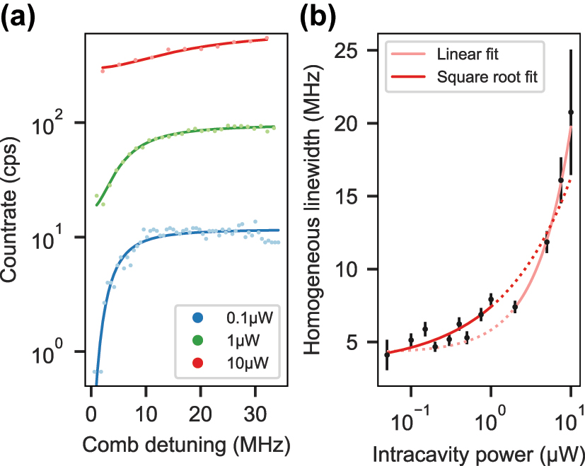
Homogeneous linewidth measurement. (a) Transient spectral holes at three different intracavity power levels. The solid lines display fits of an inverted Lorentzian line. (b) Half-width of the transient spectral hole as a function of the intracavity power. Fitting the square root function of Eq. (2) (dark red) to the data permits to extrapolate an upper bound of the homogeneous linewidth. The four datapoints at highest power were excluded from the fit since a linear function (light red) yields better agreement.
The measured linewidth is about a factor of two higher than the value of 1.6 MHz measured at 4 K by [23] using spectral hole burning spectroscopy in a large ensemble, and much larger than the value obtained from photon echo measurements (116 kHz) as shown in Figure 6 in the Supplementary Information. The comparably broad linewidth can be explained by the high power levels above saturation that are required for this method. In this regime, instantaneous spectral diffusion (ISD) [30] due to dipole–dipole interactions between resonantly but also off-resonantly excited ions broadens the linewidth. Since all data points remain in this regime, an extrapolation to zero power can–not yield a linewidth that is unaffected by ISD as one would expect it for frequency selective single ion addressing.
Using the measured linewidth, Γ h = 2π × 3.3 MHz, we are able to calculate the saturation intensity I sat of a perfectly coupling single ion. Starting from the resonant saturation parameter given in [31] and expressing the Rabi frequency in terms of cavity QED parameters for a Fabry–Pérot type cavity mode, we arrive at the following expression:
This results in a value of I sat = 2 × 10⁴ W/m2 or a intracavity saturation power of P sat = 61 nW for the 580 nm transition at 4.3 K.
We note that with the performed spectroscopy, we already reached the level of the single ion saturation power and probe only a very small number of ions: We simulate the random distribution of the total number of ions (derived from the NP diameter and doping concentration) over the measured inhomogeneous profile taking into account the hyperfine structure to estimate the number of ions that we address within a power-broadened linewidth at the lowest power level of 50 nW intracavity power in Figures 5 and 6. This yields about 15 (2) europium ions.
6 Single ion count rate estimation
The agreement between the measured and estimated ensemble averaged Purcell factors in Figure 4(b) and (c) as well as the confirmation of a homogeneous linewidth much narrower than the cavity linewidth (bad cavity regime) enables us to estimate the count rate that we expect from a perfectly coupling single europium ion. We simulate the count rate for pulsed, resonant excitation on the 580 nm transition with a 1 µs excitation pulse and variable length of the detection time window dependent on the repetition rate R rep = 1/(t ex + t det). A 3-tone excitation pulse matching the ground state hyperfine splitting is assumed, to avoid optical pumping. Since we expect incoherent excitation, we assume an excited state population of p ex = 0.5 for each trial. Further, we take the same cavity parameters as for the measurements presented above, i.e. a bare finesse of 17,500 (9,500) for the 580 nm (611 nm) transition, and a fiber radius of curvature of 25 µm resulting in mode waists of 1.41 µm (1.44 µm) at the double-resonance condition at about 6 µm cavity length, and 1.18 µm for contact mode at 2.5 µm cavity length. The cavity length jitter strongly affects the Purcell factor quantified by eq. (1) in [19] and thus the fluorescence count rate. Here, we set the cavity length stability to the best measured values of 2.5 pm (0.8 pm) for an open cavity (contact mode) as presented in [19]. The detected count rate can then be calculated as,
with the cavity outcoupling efficiency η out ≤ 0.55, which is dependent on the nanoparticle scattering losses. The detector has an efficiency of η det ≈ 0.65 at a dark count rate of 20 Hz and the transmission of the collection optics path amounts to T path = 0.8. Altogether, this results in a probability of up to 30 % to detect a photon which is emitted into the cavity mode. The pulsed emission rate into the cavity mode is given by:
The plot in Figure 7(a) shows that in contact mode, where the cavity fiber touches the planar mirror and the smallest cavity length is achieved, count rates above 300 cps could be reached for a 40 nm NP, which should be within the size distribution of the batch used here. The plot also shows that smaller NPs lead to higher count rates since the Rayleigh-scattering losses are smaller. In this scenario, a maximum Purcell factor of 5.6 could be achieved. In Figure 7(b), a cut for a NP diameter of 70 nm, the smallest measured so far, across the plot in A is shown together with the estimations for an open cavity configuration. Enhancing and collecting both transitions (green) can significantly increase the count rate compared to the case where only the 580 nm transition is collected (light blue). However, for sufficiently high repetition rates, the contact mode provides even higher signals due to an increased cavity length stability and smaller mode waist. Although the estimated count rates are still low compared to other solid state emitters, with the current setup, it should be feasible to detect single europium ions inside a 70 nm NP in contact mode with a signal-to-noise ratio of about

Calculation of the detected count rate for a perfectly coupling, single europium ion in a pulsed, resonant measurement scheme. (a) The cavity is operated in contact mode with an RMS length jitter of 0.8 pm. The blue line marks the nanoparticle size of 70 nm assumed for the calculation in B. (b) Dependence of the count rate on the repetition rate for different operation modes of the cavity. At the double-resonance condition, the count rate can be increased by additionally collecting the 611 nm fluorescence (both, green) compared to only the 580 nm light (light blue).
7 Conclusions
We have performed cavity-enhanced spectroscopy of few-ion ensembles of europium inside individual yttria nanoparticles. Using an aerosol printer, we established a new method to disperse individual nanoparticles on the surface of the planar mirror in a controlled manner. Scanning cavity transmission and fluorescence scans were conducted to locate six different nanoparticles with diameters ranging from 70 nm to 95 nm. The widths of the inhomogeneous linewidths have been measured to be between 30 GHz and 74 GHz, which is broader than in bulk crystals but possibly advantageous for single ion selection. The high degree of control of the cavity length permits us to selectively enhance just the
To summarize our findings, the complete set of parameters describing the cavity-emitter system for the 580 nm as well as the 611 nm transition, respectively, can be found in Table 1. The figure of merit of such a system is the cooperativity C defined as [32]:
which relates the cavity-emitter coupling rate g to the loss rate of the cavity κ and the dephasing rate of the emitter Γ h = 1/(2π T 1) + Γd. Here, Γ d is the pure dephasing rate.
Summary of the measured, best-case parameters describing the cavity-emitter system of a 70 nm nanoparticle and T 1 = 2.0 ms.
| Parameter | 5 D 0 → 7 F 0 | 5 D 0 → 7 F 2 |
|---|---|---|
| λ (nm) | 580.8 | 611 |
| g (MHz) | 2π × 0.4 | 2π × 2.4 |
| κ (GHz) | 2π × 1.8 | 2π × 2.5 |
| Γ h (MHz) | 2π × 3.3 | 2π × 0.7 × 106 |
|
|
3.4 | 0.47 |
| C | 8 × 10⁻⁵ | 5 × 10⁻¹¹ |
For future quantum information applications it would be desirable to reach C ≈ 1, which allows for a near-deterministic entanglement between the photonic and atomic quantum states as e.g. shown by [33]. In order to reach this regime, improved optical coherence is crucial, e.g. Γ h = 2π × 25 kHz as has been observed for slightly larger Eu3+: Y2 O3 nanoparticles [21]. With a small mirror radius of curvature (r c = 8 μm) and a short cavity length in contact mode, one can reach a mode waist of 0.7 μm. Together with a finesse of 55,000, this reaches unity cooperativity. In this regime, efficient single-shot readout of hyperfine qubit states becomes possible, as well as entanglement generation between separate cavity-ion nodes [34], laying the grounds for distributed quantum processing nodes.
Funding source: NEQSIS
Award Identifier / Grant number: 16KISQ029K
Funding source: National Natural Science Foundation of China
Award Identifier / Grant number: 12304454
Funding source: Collaborative Re- search Centre “4f for Future
Award Identifier / Grant number: 471424360
Award Identifier / Grant number: 2021A1515110191
Funding source: SQUARE European Union Quantum Flagship initiative
Award Identifier / Grant number: 820391
Acknowledgments
We thank A. Quintilla and P. Brenner at the Nanostructure Service Laboratory (CFN-NSL) at KIT for carrying out the AFM and SEM measurements, and Julia Benedikter, Bernardo Casabone, and Thomas Hümmer for contributions in the early phase of the experiment. Further, we thank Chetan Deshmukh and Eduardo Beattie for many fruitful discussions. We acknowledge support from Leonhard Neuhaus to adapt the Pyrpl software package to our needs.
-
Research funding: This work has been financially supported by the European Union Quantum Flagship initiative under grant agreement No. 820391 (SQUARE), the Karlsruhe School of Optics and Photonics (KSOP), the BMBF project NEQSIS (contract no. 16KISQ029K), and the Deutsche Forschungsgemeinschaft (DFG) through the Collaborative Research Centre “4f for Future” (CRC 1573 project number 471424360, project C2). SL acknowledges support by the National Natural Science Foundation of China (Grant No. 12304454), and Guangdong Basic and Applied Basic Research Foundation (Grant No. 2021A1515110191).
-
Author contributions: TE: Conceptualization; Data curation; Formal analysis; Investigation; Methodology; Software; Validation; Visualization; Writing – original draft; Writing – review & editing. NJ: Conceptualization; Data curation; Formal analysis; Investigation; Methodology; Writing – review & editing. SL: Sample fabrication and characterization; Writing – review & editing. DS: Sample fabrication and characterization; Writing – review & editing. RH: Aerosolprinting; Writing – review & editing. TK: Cavity fiber fabrication; Writing – review & editing. SB: Cavity fiber fabrication; Laser stabilization setup; Writing – review & editing. UL: Funding acquisition; Methodology; Project administration; Resources; Supervision; Validation; Writing – review & editing. HR: Conceptualization; Funding acquisition; Methodology; Project administration; Resources; Supervision; Validation; Writing – review & editing. PG: Conceptualization; Funding acquisition; Methodology; Project administration; Resources; Supervision; Validation; Writing – review & editing. DH: Conceptualization; Funding acquisition; Methodology; Project administration; Resources; Supervision; Validation; Writing – review & editing. All authors have accepted responsibility for the entire content of this manuscript and approved its submission.
-
Conflict of interest: Authors state no conflict of interest.
-
Informed consent: Informed consent was obtained from all individuals included in this study.
-
Data availability: The datasets generated during and/or analyzed during the current study are available from the corresponding author on reasonable request.
References
[1] R. Macfarlane and R. Shelby, “Sub-kilohertz optical linewidths of the 7F0 – 5D0 transition in Y2O3:Eu3+,” Opt. Commun., vol. 39, no. 3, pp. 169–171, 1981. https://doi.org/10.1016/0030-4018(81)90048-1.Search in Google Scholar
[2] M. Zhong, et al.., “Optically addressable nuclear spins in a solid with a six-hour coherence time,” Nature, vol. 517, pp. 177–180, 2015. https://doi.org/10.1038/nature14025.Search in Google Scholar PubMed
[3] A. Kinos, L. Rippe, S. Kröll, and A. Walther, “Designing gate operations for single-ion quantum computing in rare-earth-ion-doped crystals,” Phys. Rev. A, vol. 104, 2021, Art. no. 052624. https://doi.org/10.1103/physreva.104.052624.Search in Google Scholar
[4] D. Jaksch, J. I. Cirac, P. Zoller, S. L. Rolston, R. Côté, and M. D. Lukin, “Fast quantum gates for neutral atoms,” Phys. Rev. Lett., vol. 85, pp. 2208–2211, 2000. https://doi.org/10.1103/physrevlett.85.2208.Search in Google Scholar
[5] N. Ohlsson, R. Krishna Mohan, and S. Kröll, “Quantum computer hardware based on rare-earth-ion-doped inorganic crystals,” Opt. Commun., vol. 201, nos. 1–3, pp. 71–77, 2002. https://doi.org/10.1016/s0030-4018(01)01666-2.Search in Google Scholar
[6] A. Kinos, L. Rippe, D. Serrano, A. Walther, and S. Kröll, “High-connectivity quantum processor nodes using single-ion qubits in rare-earth-ion-doped crystals,” Phys. Rev. A, vol. 105, 2022, Art. no. 032603. https://doi.org/10.1103/physreva.105.032603.Search in Google Scholar
[7] A. Kinos, et al.., “Roadmap for rare-earth quantum computing,” Preprint arXiv:2103.15743, 2021.Search in Google Scholar
[8] E. M. Purcell, “Spontaneous emission probabilities at radio frequencies,” Phys. Rev., vol. 69, no. 681, 1946.Search in Google Scholar
[9] A. Dibos, M. Raha, C. Phenicie, and J. Thompson, “Atomic source of single photons in the telecom band,” Phys. Rev. Lett., vol. 120, 2018, Art. no. 243601. https://doi.org/10.1103/physrevlett.120.243601.Search in Google Scholar PubMed
[10] J. M. Kindem, A. Ruskuc, J. G. Bartholomew, J. Rochman, Y. Q. Huan, and A. Faraon, “Control and single-shot readout of an ion embedded in a nanophotonic cavity,” Nature, vol. 580, pp. 201–204, 2020. https://doi.org/10.1038/s41586-020-2160-9.Search in Google Scholar PubMed
[11] S. Chen, M. Raha, C. M. Phenicie, S. Ourari, and J. D. Thompson, “Parallel single-shot measurement and coherent control of solid-state spins below the diffraction limit,” Science, vol. 370, pp. 592–595, 2020. https://doi.org/10.1126/science.abc7821.Search in Google Scholar PubMed
[12] K. Xia, et al.., “Tunable microcavities coupled to rare-earth quantum emitters,” Optica, vol. 9, p. 445, 2022. https://doi.org/10.1364/optica.453527.Search in Google Scholar
[13] A. Ulanowski, B. Merkel, and A. Reiserer, “Spectral multiplexing of telecom emitters with stable transition frequency,” Sci. Adv., vol. 8, p. eabo4538, 2022. https://doi.org/10.1126/sciadv.abo4538.Search in Google Scholar PubMed PubMed Central
[14] C. Deshmukh, et al.., “Detection of single ions in a nanoparticle coupled to a fiber cavity,” Optica, vol. 10, p. 1339, 2023. https://doi.org/10.1364/optica.491692.Search in Google Scholar
[15] D. Hunger, T. Steinmetz, Y. Colombe, C. Deutsch, T. W. Hänsch, and J. Reichel, “A fiber Fabry–Perot cavity with high finesse,” New J. Phys., vol. 12, 2010, Art. no. 065038. https://doi.org/10.1088/1367-2630/12/6/065038.Search in Google Scholar
[16] H. Pfeifer, et al.., “Achievements and perspectives of optical fiber Fabry–Perot cavities,” Appl. Phys. B, vol. 128, p. 29, 2022. https://doi.org/10.1007/s00340-022-07752-8.Search in Google Scholar
[17] D. Hunger, C. Deutsch, R. J. Barbour, R. J. Warburton, and J. Reichel, “Laser micro-fabrication of concave, low-roughness features in silica,” AIP Adv., vol. 2, 2012, Art. no. 012119. https://doi.org/10.1063/1.3679721.Search in Google Scholar
[18] A. Muller, E. B. Flagg, J. R. Lawall, and G. S. Solomon, “Ultrahigh-finesse, low-mode-volume fabry–perot microcavity,” Opt. Lett., vol. 35, p. 2293, 2010. https://doi.org/10.1364/ol.35.002293.Search in Google Scholar
[19] M. Pallmann, T. Eichhorn, J. Benedikter, B. Casabone, T. Hümmer, and D. Hunger, “A highly stable and fully tunable open microcavity platform at cryogenic temperatures,” APL Photonics, vol. 8, 2023, Art. no. 046107. https://doi.org/10.1063/5.0139003.Search in Google Scholar
[20] J. G. Bartholomew, K. De Oliveira Lima, A. Ferrier, and P. Goldner, “Optical line width broadening mechanisms at the 10 kHz level in Eu 3+ :Y 2 O 3 nanoparticles,” Nano Lett., vol. 17, pp. 778–787, 2017. https://doi.org/10.1021/acs.nanolett.6b03949.Search in Google Scholar PubMed
[21] S. Liu, D. Serrano, A. Fossati, A. Tallaire, A. Ferrier, and P. Goldner, “Controlled size reduction of rare earth doped nanoparticles for optical quantum technologies,” RSC Adv., vol. 8, no. 65, pp. 37098–37104, 2018. https://doi.org/10.1039/c8ra07246a.Search in Google Scholar PubMed PubMed Central
[22] S. Liu, A. Fossati, D. Serrano, A. Tallaire, A. Ferrier, and P. Goldner, “Defect engineering for quantum grade rare-earth nanocrystals,” ACS Nano, vol. 14, pp. 9953–9962, 2020. https://doi.org/10.1021/acsnano.0c02971.Search in Google Scholar PubMed
[23] A. Fossati, D. Serrano, S. Liu, A. Tallaire, A. Ferrier, and P. Goldner, “Optical line broadening mechanisms in rare-earth doped oxide nanocrystals,” J. Lumin., vol. 263, 2023, Art. no. 120050. https://doi.org/10.1016/j.jlumin.2023.120050.Search in Google Scholar
[24] G. Gramlich, R. Huber, F. Häslich, A. Bhutani, U. Lemmer, and T. Zwick, “Process considerations for Aerosol-Jet printing of ultra fine features,” Flexible Printed Electron., vol. 8, 2023, Art. no. 035002. https://doi.org/10.1088/2058-8585/ace3d8.Search in Google Scholar
[25] R. Huber, Gedruckte Temperatursensor-Arrays mit hoher räumlicher Auflösung, Ph.D. thesis, Karlsruher Institut für Technologie, 2024.Search in Google Scholar
[26] M. Mader, J. Reichel, T. W. Hänsch, and D. Hunger, “A scanning cavity microscope,” Nat. Commun., vol. 6, p. 7249, 2015. https://doi.org/10.1038/ncomms8249.Search in Google Scholar PubMed PubMed Central
[27] B. Casabone, et al.., “Cavity-enhanced spectroscopy of a few-ion ensemble in Eu 3+ :Y 2 O 3,” New J. Phys., vol. 20, 2018, Art. no. 095006. https://doi.org/10.1088/1367-2630/aadf68.Search in Google Scholar
[28] S. Völker, “Hole-burning spectroscopy,” Annu. Rev. Phys. Chem., no. 40, pp. 499–530, 1989. https://doi.org/10.1146/annurev.pc.40.100189.002435.Search in Google Scholar
[29] A. Gritsch, L. Weiss, J. Früh, S. Rinner, and A. Reiserer, “Narrow optical transitions in erbium-implanted silicon waveguides,” Phys. Rev. X, vol. 12, 2022, Art. no. 041009. https://doi.org/10.1103/physrevx.12.041009.Search in Google Scholar
[30] A. Kinos, L. Rippe, A. Walther, and S. Kröll, “Microscopic treatment of instantaneous spectral diffusion and its effect on quantum gate fidelities in rare-earth-ion-doped crystals,” Phys. Rev. A, vol. 105, 2022, Art. no. 032608. https://doi.org/10.1103/physreva.105.032608.Search in Google Scholar
[31] B. Julsgaard, A. Walther, S. Kröll, and L. Rippe, “Understanding laser stabilization using spectralhole burning,” Opt. Express, vol. 15, pp. 11444–11465, 2007.10.1364/OE.15.011444Search in Google Scholar PubMed
[32] A. Auffèves, D. Gerace, J.-M. Gérard, M. F. Santos, L. C. Andreani, and J.-P. Poizat, “Controlling the dynamics of a coupled atom-cavity system by pure dephasing,” Phys. Rev. B, vol. 81, 2010, Art. no. 245419. https://doi.org/10.1103/physrevb.81.245419.Search in Google Scholar
[33] S. Daiss, et al.., “A quantum-logic gate between distant quantum-network modules,” Science, vol. 371, pp. 614–617, 2021. https://doi.org/10.1126/science.abe3150.Search in Google Scholar PubMed
[34] A. Reiserer, “Colloquium : cavity-enhanced quantum network nodes,” Rev. Mod. Phys., vol. 94, 2022, Art. no. 041003. https://doi.org/10.1103/revmodphys.94.041003.Search in Google Scholar
Supplementary Material
This article contains supplementary material (https://doi.org/10.1515/nanoph-2024-0721).
© 2025 the author(s), published by De Gruyter, Berlin/Boston
This work is licensed under the Creative Commons Attribution 4.0 International License.
Articles in the same Issue
- Frontmatter
- Editorial
- Quantum light: creation, integration, and applications
- Reviews
- Low-dimensional solid-state single-photon emitters
- Solid-state single-photon sources operating in the telecom wavelength range
- Quantum super-resolution imaging: a review and perspective
- Perspectives
- New opportunities for creating quantum states of light and matter with intense laser fields
- On-chip frequency-bin quantum photonics
- Building photonic links for microwave quantum processors
- Remote quantum networks based on quantum memories
- Sensing with quantum light: a perspective
- Letter
- Electro-optic frequency shift of single photons from a quantum dot
- Research Articles
- Quantum efficiency of the B-center in hexagonal boron nitride
- Localized exciton emission from monolayer WS2 nanoribbon at cryogenic temperature
- Single-photon emitters in PECVD-grown silicon nitride films: from material growth to photophysical properties
- A fiber-pigtailed quantum dot device generating indistinguishable photons at GHz clock-rates
- Sub-MHz homogeneous linewidth in epitaxial Y2O3: Eu3+ thin film on silicon
- Multimodal Purcell enhancement and optical coherence of Eu3+ ions in a single nanoparticle coupled to a microcavity
- All-optical control of charge-trapping defects in rare-earth doped oxides
- Ultra-broadband and passive stabilization of ultrafast light sources by quantum light injection
- Tunable quantum light by modulated free electrons
- Second-harmonic radiation by on-chip integrable mirror-symmetric nanodimers with sub-nanometric plasmonic gap
- Mie metasurfaces for enhancing photon outcoupling from single embedded quantum emitters
- Design and fabrication of robust hybrid photonic crystal cavities
- Enhanced zero-phonon line emission from an ensemble of W centers in circular and bowtie Bragg grating cavities
- Freeform thin-film lithium niobate mode converter for photon-pair generation
- Luminescence thermometry based on photon emitters in nanophotonic silicon waveguides
- Collective single-photon emission and energy transfer in thin-layer dielectric and plasmonic systems
- Description of ultrastrong light–matter interaction through coupled harmonic oscillator models and their connection with cavity-QED Hamiltonians
- Bound polariton states in the Dicke–Ising model
- Collective multimode strong coupling in plasmonic nanocavities
- Improving quantum metrology protocols with programmable photonic circuits
- Fluorescence enabled phonon counting in an erbium-doped piezo-optomechanical microcavity
- Non-perturbative cathodoluminescence microscopy of beam-sensitive materials