Cryogenic nonlinear microscopy of high-Q metasurfaces coupled with transition metal dichalcogenide monolayers
-
Alena A. Nazarenko
 ,
Alexander I. Musorin
,
Alexander S. Shorokhov
,
Lu Ding
,
Vytautas Valuckas
,
Alexander I. Musorin
,
Alexander S. Shorokhov
,
Lu Ding
,
Vytautas Valuckas


Abstract
Monolayers of transition metal dichalcogenides (TMDCs) demonstrate plenty of unique properties due to the band structure. Symmetry breaking brings second-order susceptibility to meaningful values resulting in the enhancement of corresponding nonlinear effects. Cooling the TMDC films to cryogenic temperatures leads to the emergence of two distinct photoluminescence peaks caused by the exciton and trion formation. These intrinsic excitations are known to enhance second harmonic generation. The nonlinear signal can be greatly increased if these material resonances are boosted by high-quality factor geometric resonance of all-dielectric metasurfaces. Here, we experimentally observe optical second harmonic generation caused by excitons of 2D semiconductor MoSe2 at room and cryogenic temperatures enhanced by spectrally overlapped high-Q resonance of TiO2 nanodisks metasurface. The enhancement reaches two orders of magnitude compared to the case when the resonances are not spectrally overlapped.
1 Introduction
Nonlinear photonics gives a new breath to fundamental science and applications in data storage and information processing. Classical nonlinear optical effects require phase-matching fulfillment between fundamental and harmonic waves and are observed for prolongated anisotropic media or in materials with anomalous dispersion. Plasmonics and all-dielectric nanophotonics suggest a new approach to boost the effects through high local electromagnetic field confinement at a subwavelength area [1], [2]. Surface plasmon resonance in metals is known to enhance harmonic generation processes [3], [4], [5], [6], [7], [8], [9], [10]. However, light is concentrated only at the interface of the material and environment with weak penetration inside, limiting the efficiency of nonlinear conversion. On the contrary, high-index nonmetallic nano objects exhibit Mie scattering resonances and provide light–matter interaction within the entire volume of nonlinear material [11], [12], [13], [14], [15], [16], [17], [18], [19].
The improvement of fundamental radiation conversion to higher harmonics stimulates the exploration of new materials with high nonlinear susceptibility. 2D semiconductors attract more attention owing to the violation of inverse symmetry and the meaningful second-order susceptibility values [20], [21], [22]. Monolayers of transition metal dichalcogenides (TMDCs) demonstrate an absolute value of χ (2) of a few nm/V, which is much higher than that for classical nonlinear crystals [23], [24]. Mechanically exfoliated monolayer of MoS2 possesses a second-order nonlinear susceptibility that even reaches the order of 10−7 m/V [25], while its bulk counterpart exhibits the value of only 10−14 m/V [26]. Meanwhile, monolayers of MoSe2 possess nonlinear susceptibility of about 10−11 m/V [23]. Two-dimensional nature of TMDC usually forces to express a sheet second-order nonlinear susceptibility with the values of 10−20 m2/V [27], [28].
The symbiosis of TMDC with resonant structures proves to be a powerful way to enhance nonlinear conversion efficiency [7], [29], [30], [31]. Numerous papers focus on doubly resonant cavities [32], [33], [34]. The samples were designed to concentrate light outside the nanoresonators in the area of a TMDC monolayer, which is usually located at the top or bottom of the structure. Both strong light localization and the presence of appropriate electric field components oscillating in the plane of the monolayer are needed for harmonic generation. The enhancement up to 7,000 times is experimentally demonstrated in judiciously structured sub-20 nm-wide trenches on a 150 nm-thick gold film with WSe2 monolayer on top [7]. The design allows manipulating SHG intensity by rotating linear polarization of the pump laser. Patterned TMDC by itself can also enhance harmonic generation due to the high refractive index and excitation of Mie resonances [35], [36].
At helium temperatures (4–10 K), substances demonstrate unusual behavior governed by quantum rules. Some semiconductors become superconductive, while others, such as quantum dots or quantum wells, exhibit quantum effects that influence their optical properties – increased absorption and fluorescence, the appearance of higher-order excitons, etc. Coulomb interaction between one electron and one hole results in the generation of a neutral exciton (X 0), whose behavior is similar to a hydrogen atom. These quasiparticles can be transformed into the charged form of a three-particle exciton – trion, by binding to an additional electron (X −) or an additional hole (X +). The electronic and optical properties of such states are also of great importance for fundamental science and optoelectronic applications, ranging from emitting sources [37] to quantum logical devices [38], [39]. Both exciton and trion show a very sensitive dependence on temperature [40]. Measuring the photoluminescence (PL) spectrum is an easy way to track changes in the electronic configuration of MoSe2 films during the cooling process. It possesses one broadband peak at room temperature and two narrow peaks at cryogenic temperatures (15 K) corresponding to bright exciton and trion. These states become more pronounced at low temperatures. The PL spectrum line shape at 15 K differs for X 0 and X −: the peak of the neutral exciton is symmetric and has homogeneous thermal broadening, while X − gives an asymmetric profile with a long low-energy tail consistent with electron-recoil effects. At temperatures above 55 K, the trion peak disappears since electrons leave the quasiparticle, which in turn occurs due to the increased influence of thermal fluctuations. This mechanism makes it possible to observe an increase in the value of nonlinear susceptibility for specific wavelengths, which leads to the enhancement of nonlinear optical signal compared to the room-temperature case. In the case of MoS2, experiments have shown the increasing time-integrated photoluminescence signal with cooling [41]. In contrast, for WSe2 monolayer, temperature-dependent PL decreases at the bright exciton peak and a 30-meV shifted dark state appearances during the cooling process. This different behavior is explained by the spin splitting of the conduction band, the sign of which differs for different TMDC materials.
The spectral overlap of optical resonances of high-index nanocavities with excitonic states of TMDC monolayers leads to significant amplification of optical harmonic generation [30], [34], [42]. However, the interplay between cryogenic cooling and coupling of TMDC exciton with high-quality (high-Q) resonance of all-dielectric metasurfaces has not been studied yet. Here, we explore the nonlinear optical spectra of TiO2 metasurface covered by MoSe2 monolayer to elucidate this effect (see Figure 1).

The idea of the experiment. Image to the left demonstrates second optical harmonic generation (SHG) in a MoSe2 monolayer coupled to the TiO2 metasurface at room temperature. This effect is governed by the high-Q geometric resonance of the nanostructure and a broad excitonic resonance of the 2D material. Image to the right demonstrates SHG boost caused by the interplay between the geometric resonance of the metasurface and narrowed material resonances of the MoSe2 monolayer under cryogenic temperature.
2 Results and discussion
2.1 Linear spectroscopy
As a resonant system, we study a dielectric metasurface that consists of a densely packed 2D array of TiO2 nanodisks on a transparent SiO2 substrate. The square lattice has the periods P of 470 nm. The cylinder diameter d is 450 nm and the height h is 120 nm. The size of the metasurface is 50 μm. The details on the sample fabrication can be found in the Supporting Materials (S1). The transmittance spectra of the sample for normal incidence are shown in Figure 2(a). The red curve is the experimental results and the black one is the numerical modeling. The inset shows a scanning electron microscopy (SEM) image of the metasurface. The spectral sensitivity of the linear experimental setup is limited by 900 nm and the scheme of the setup is presented in the Supporting Materials (S2.1). The details of numerical modeling can be found in the Supporting Materials as well (S3.1). The spectra have two resonant features. The nature of these resonances is the overlapped electrical dipole (ED, red curve) with magnetic quadrupole (MQ, blue curve) for the resonance at the wavelength of 745 nm and overlapped magnetic dipole (MD, green curve) with electric quadrupole (EQ, magenta curve) for the one at the wavelength of 695 nm according to the multipole decomposition shown in the Figure 2(b). The inset shows the geometry of the sample and the experiment. These resonances here and thereafter will be called geometric underlying in that they are not connected with the material resonances and properties of the media but are dictated by the nanostructuring only. The details of the decomposition can be found in the Supporting Materials (S3.2). The quality factor of the left resonance is about 185 and for the right one is approximately 80 extracted from the numerical data. The fitting formula for the resonances is the Fano formula [43]:
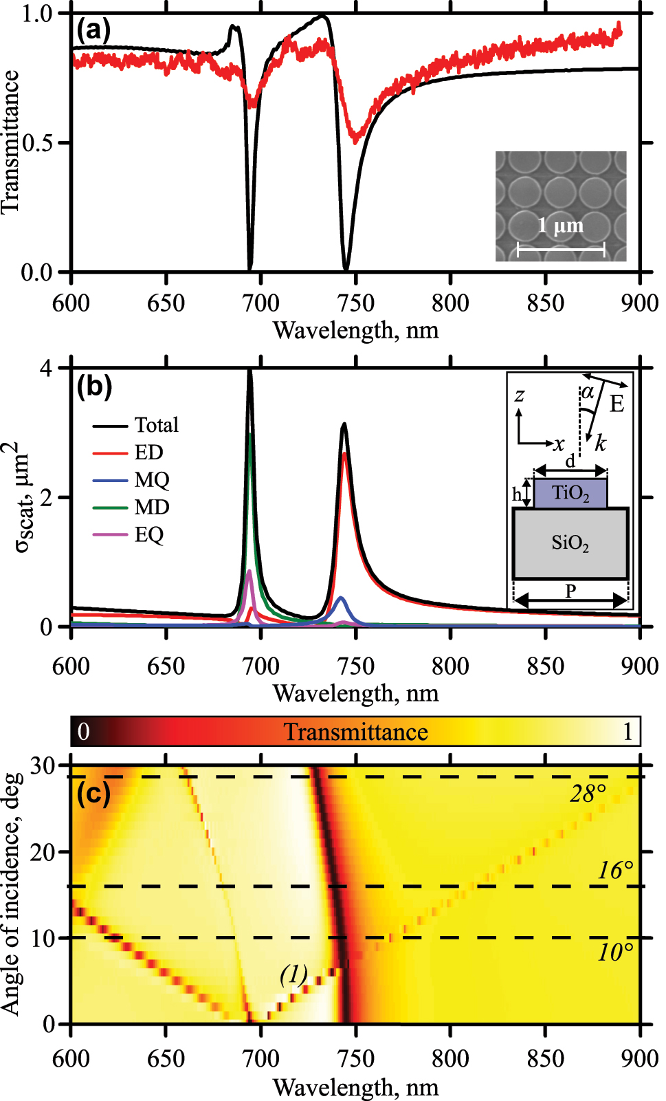
Linear optical characterization. (a) Transmittance spectra of the metasurface for normal incidence. The black curve is numerical calculations, red – experiment. The inset shows an SEM image of the sample. (b) Multipole decomposition. The inset shows the geometry of the sample and the experiment. (c) Angle-of-incidence-resolved numerical transmittance spectra of the sample. The dashed lines are angles chosen for nonlinear experiments.
Here, ω 0 is the central frequency of the resonance, γ is the damping factor, and a 1, a 2, and b are real constants. The spectral positions of both resonances agree well for experimental and numerical data. The difference in depth of the dips is explained by deviations of the disk diameters in the array during the fabrication process averaging this parameter from the exact calculated one (evaluation gives ±10 nm) and finite convergence angle of incoming radiation in the optical experiment averaging the spectral width of the resonance (evaluation gives ±3°). The latter one limits the resolution of the spectra measurements. The numerical calculation also shows that the cooling process does not affect the transmittance spectrum significantly (see the Supporting Materials S3.3).
A comparison of field distribution for two polarizations (S and P) in the case of oblique incident radiation is carried out. The results are presented in the Supporting Materials (S3.4). In this situation, we are interested not only in large values of local fields in the structure but also in overlapping of these fields with nonlinear material. All radiation can be concentrated in the resonator and as a result make a small contribution to the generation of harmonics in the film. Thus, the localization of the field in the monolayer area is of prior importance when choosing the polarization of the incident wave. The strong field localization at the top of the cylinder is observed for P-polarized incoming light. In the case of S-polarized light, the field concentrates between the neighboring disks instead of their top. In addition, local field distributions for P-polarized light, obtained for SHG fields at λ/2, when excited at the fundamental wavelength λ, also exhibit local field concentration in the area of the flake (S3.7). All the above explains the necessity to use P-polarized light.
Figure 2(c) shows transmittance spectra for various angles of incidence of P-polarized incoming light. Variation of the angle of incidence from the normal case leads to shifts of the resonances. We are interested in the one that is marked on the graph as ‘(1).’ It moves toward longer wavelengths. Analyzing the behavior of diffraction maxima on the metasurface, we conclude that the described resonances are hybrid modes of the Mie resonance and the Rayleigh anomaly. The details of the analysis can be found in the Supporting Materials (S3.5). The shift of the resonance toward longer wavelengths allows the metasurface resonance to be spectrally overlapped with the resonances of the TMDC monolayer by tilting the sample. The MoSe2 exciton resonances are observed in vicinity of 800 nm [23]. However, the position of photoluminescence peak strongly depends on the temperature [40] and strain [44]. It is also sensitive to electrostatic potential. When transferring the film on top of different materials, strain value may be the same, but local electrostatic potential may behave differently. That is why, the constituent materials of the structure also play an important role in the position of the exciton resonance. TiO2 is not exactly a standard material for exfoliation. The results obtained for the monolayer exfoliated on TiO2 metasurface may differ from previously reported data for Si or SiO2 substrates. As for the results obtained in this article, three cases will be considered in the nonlinear experiments: 10° when resonances are not overlapped; 16° – overlapping the metasurface resonance with bright exciton of TMDC; 28° – overlapping of the metasurface resonance with the localized exciton of the MoSe2. These angles are marked in Figure 2(c) by the dash lines.
2.2 Nonlinear spectroscopy
The monolayer of MoSe2 is used as a nonlinear material, which has a high value of nonlinear susceptibility and an excitonic state around the wavelength of 800 nm. The flake is deterministically transferred onto the TiO2 metasurface using an aligned transfer technique (described in the Supporting Materials (S1)). Briefly, monocrystalline flakes are mechanically exfoliated onto PDMS film, and subsequently, monolayer flakes are identified using an optical microscope. The flakes are then picked up by a polymer stamp and positioned onto the nanostructure using dual 4-axis stages integrated with an optical microscope. This technique ensures micrometer-level accuracy in the pick-and-place process.
Figure 3 shows both experimental and numerical results on second harmonic generation (SHG) spectroscopy. The nonlinear spectra are calculated only for the room temperature case by the finite element method in COMSOL Multiphysics software. The details of nonlinear calculations can be found in the Supporting Materials (S3.4). The measurements are performed at both room temperature (red dots) and 10 K (blue dots). All nonlinear spectra are normalized to the maximum SHG value observed for the case of overlapped geometric and excitonic resonances at 10 K. This value later will be denoted as
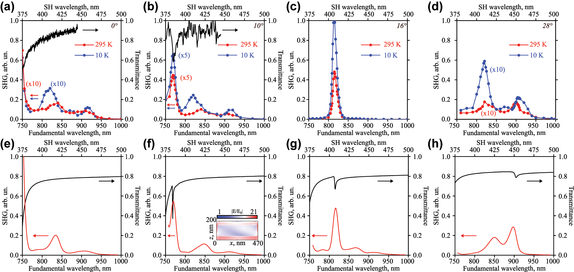
Optical second harmonic spectra for room temperature (red curves) and 10 K (blue curves), and transmittance spectra (black curves) for numerous angles of incidence: (a, e) 0°, (b, f) 10°, (c, g) 16°, (d, h) 28°. (a–d) Experiment, (e–h) numerical results. The inset on panel (f) shows normalized electric field distribution at the wavelength of the resonance of 770 nm in the plane of incidence XOZ in log scale.
The geometric resonances are almost absent for normal incidence in the studied spectral region as discussed above in the linear spectroscopy section (see Figures 2 and 3(a, e)). One of the resonances is centered around 745 nm and its edge appears in Figure 3(a). The nonlinear spectrum at room temperature (see Figure 3(a), red dots) demonstrates enhancement in the vicinity of these wavelengths. Three other emerged resonant features are now associated with the band structure of the nonlinear 2D material. The nature of the left one (λ = 840 nm (1.47 eV)) is the bright exciton in MoSe2 [23]. The value of the SHG signal is only 1.6 % of
The calculated nonlinear spectrum (see Figure 3(e)) at the room temperature case reproduces all main peculiarities: the enhancement of the signal for the geometric resonance near 750 nm, the signal for excitonic (λ = 840 nm) and localized (λ = 905 nm) resonances (compare red curves in the panels (a) and (e) in the Figure 3). The spectral positions are identical with the experiment, and the absolute values differ due to numerous factors that cannot be fully taken into account.
The minimum angle of incidence required to observe the geometric resonance for the studied wavelength range is 10° (see Figure 3(b, f)). The experimental (Figure 3(b)) and numerical (Figure 3(f)) transmittance spectra show the resonance near the wavelength of 770 nm. This high-Q feature arises from nearly equal contributions of the MD and EQ, with minor contributions from the ED and MQ multipoles according to the decomposition analysis (see the Supporting Materials (S3.2)). The SHG spectra demonstrate resonant amplification at both room and cryogenic temperatures. The spectral position does not change during cooling, which is expected, since this is dielectric-metasurface-induced resonance and there is no temperature dependence of TiO2 as mentioned above (Section 2.1 and Supporting Materials (S3.3)). The signal of the second harmonic at room temperature is 9 % of
If the angle of incidence is set to 16°, the geometric resonance is spectrally overlapped with the material one of MoSe2 near the wavelength of 815 nm (see Figure 3(c, g)). The nonlinear signal increases by two orders of magnitude compared to the previously discussed results. The low-temperature signal of SHG is 2 times greater than that at room temperature. The nonlinear signal reaches the highest value of
The next angle of incidence of 28° is chosen to overlap the geometric resonance with the localized excitonic state of MoSe2 near the wavelength of 900 nm (see Figure 3(d, h)). Nonlinear measurements (Figure 3(d)) show signal increase for the wavelengths of the bright (λ = 840 nm) and the localized (λ = 905 nm) excitons at room temperature. They reach 1.7 %
3 Conclusions
We experimentally observed the enhancement of optical second harmonic generation at cryogenic and room temperatures when material resonances of TMDC monolayers are spectrally overlapped with high-Q features of the all-dielectric metasurface. The SHG enhancement reaches two orders of magnitude compared to the case when the resonances are not at the same wavelength due to the interplay between the geometric and material resonances of the sample. The results can be inspiring for novel approaches to boost the development of optoelectronic science and nonlinear photonics.
Award Identifier / Grant number: 22-1-3-5-1
Funding source: Russian Science Foundation
Award Identifier / Grant number: 20-12-00371
Acknowledgments
Authors thank D.F. Yagudin for the assistance with experiment, V.N. Mantsevich and M.R. Shcherbakov for fruitful discussion.
-
Research funding: This work was supported by the Russian Science Foundation (no. 20-12-00371, experiment). The study was carried with the equipment provided by Development Program of MSU. M.A.I. thanks the Foundation for the Development of Theoretical Physics and Mathematics “BASIS” for the support (no. 22-1-3-5-1, calculations).
-
Author contributions: All authors have accepted responsibility for the entire content of this manuscript and approved its submission.
-
Conflict of interest: Authors state no conflict of interest.
-
Data availability: The datasets generated during and/or analyzed during the current study are available from the corresponding author on reasonable request.
References
[1] N. Yu and F. Capasso, “Flat optics with designer metasurfaces,” Nat. Mater., vol. 13, no. 2, pp. 139–150, 2014. https://doi.org/10.1038/nmat3839.Search in Google Scholar PubMed
[2] A. I. Kuznetsov, A. E. Miroshnichenko, M. L. Brongersma, Y. S. Kivshar, and B. Luk’yanchuk, “Optically resonant dielectric nanostructures,” Science, vol. 354, no. 6314, p. aag2472, 2016. https://doi.org/10.1126/science.aag2472.Search in Google Scholar PubMed
[3] W. Cai, A. P. Vasudev, and M. L. Brongersma, “Electrically controlled nonlinear generation of light with plasmonics,” Science, vol. 333, no. 6050, pp. 1720–1723, 2011. https://doi.org/10.1126/science.1207858.Search in Google Scholar PubMed
[4] M. Celebrano, et al.., “Mode matching in multiresonant plasmonic nanoantennas for enhanced second harmonic generation,” Nat. Nanotechnol., vol. 10, no. 5, pp. 412–417, 2015. https://doi.org/10.1038/nnano.2015.69.Search in Google Scholar PubMed
[5] M. Lippitz, M. A. van Dijk, and M. Orrit, “Third-harmonic generation from single gold nanoparticles,” Nano Lett., vol. 5, no. 4, pp. 799–802, 2005. https://doi.org/10.1021/nl0502571.Search in Google Scholar PubMed
[6] A. Minovich, et al.., “Liquid crystal based nonlinear fishnet metamaterials,” Appl. Phys. Lett., vol. 100, no. 12, pp. 121113-1–121113-4, 2012. https://doi.org/10.1063/1.3695165.Search in Google Scholar
[7] Z. Wang, et al.., “Selectively plasmon-enhanced second-harmonic generation from monolayer tungsten diselenide on flexible substrates,” ACS Nano, vol. 12, no. 2, pp. 1859–1867, 2018. https://doi.org/10.1021/acsnano.7b08682.Search in Google Scholar PubMed
[8] B. I. Afinogenov, A. A. Popkova, V. O. Bessonov, B. Lukyanchuk, and A. A. Fedyanin, “Phase matching with Tamm plasmons for enhanced second-and third-harmonic generation,” Phys. Rev. B, vol. 97, no. 11, 2018, Art. no. 115438. https://doi.org/10.1103/PhysRevB.97.115438.Search in Google Scholar
[9] H. Aouani, M. Rahmani, M. Navarro-Cía, and S. A. Maier, “Third-harmonic-upconversion enhancement from a single semiconductor nanoparticle coupled to a plasmonic antenna,” Nat. Nanotechnol., vol. 9, no. 4, pp. 290–294, 2014. https://doi.org/10.1038/nnano.2014.27.Search in Google Scholar PubMed
[10] V. K. Valev, et al.., “Asymmetric optical second-harmonic generation from chiral g-shaped gold nanostructures,” Phys. Rev. Lett., vol. 104, no. 12, 2010, Art. no. 127401. https://doi.org/10.1103/PhysRevLett.104.127401.Search in Google Scholar PubMed
[11] M. R. Shcherbakov, et al.., “Enhanced third-harmonic generation in silicon nanoparticles driven by magnetic response,” Nano Lett., vol. 14, no. 11, pp. 6488–6492, 2014. https://doi.org/10.1021/nl503029j.Search in Google Scholar PubMed
[12] Y. Yang, et al.., “Nonlinear fano-resonant dielectric metasurfaces,” Nano Lett., vol. 15, no. 11, pp. 7388–7393, 2015. https://doi.org/10.1021/acs.nanolett.5b02802.Search in Google Scholar PubMed
[13] S. S. Kruk, et al.., “Nonlinear optical magnetism revealed by second-harmonic generation in nanoantennas,” Nano Lett., vol. 17, no. 6, pp. 3914–3918, 2017. https://doi.org/10.1021/acs.nanolett.7b01488.Search in Google Scholar PubMed
[14] G. Li, S. Zhang, and T. Zentgraf, “Nonlinear photonic metasurfaces,” Nat. Rev. Mater., vol. 2, no. 5, pp. 1–14, 2017. https://doi.org/10.1038/natrevmats.2017.10.Search in Google Scholar
[15] P. P. Vabishchevich, S. Liu, M. B. Sinclair, G. A. Keeler, G. M. Peake, and I. Brener, “Enhanced second-harmonic generation using broken symmetry III–V semiconductor Fano metasurfaces,” ACS Photonics, vol. 5, no. 5, pp. 1685–1690, 2018. https://doi.org/10.1021/acsphotonics.7b01478.Search in Google Scholar
[16] R. Camacho-Morales, et al.., “Nonlinear generation of vector beams from AlGaAs nanoantennas,” Nano Lett., vol. 16, no. 11, pp. 7191–7197, 2016. https://doi.org/10.1021/acs.nanolett.6b03525.Search in Google Scholar PubMed
[17] A. Fedotova, et al.., “Second-harmonic generation in resonant nonlinear metasurfaces based on lithium niobate,” Nano Lett., vol. 20, no. 12, pp. 8608–8614, 2020. https://doi.org/10.1021/acs.nanolett.0c03290.Search in Google Scholar PubMed
[18] M. Semmlinger, et al.., “Generating third harmonic vacuum ultraviolet light with a TiO2 metasurface,” Nano Lett., vol. 19, no. 12, pp. 8972–8978, 2019. https://doi.org/10.1021/acs.nanolett.9b03961.Search in Google Scholar PubMed
[19] L. Carletti, S. S. Kruk, A. A. Bogdanov, C. De Angelis, and Y. Kivshar, “High-harmonic generation at the nanoscale boosted by bound states in the continuum,” Phys. Rev. Res., vol. 1, no. 2, 2019, Art. no. 023016. https://doi.org/10.1103/PhysRevResearch.1.023016.Search in Google Scholar
[20] J. W. You, S. R. Bongu, Q. Bao, and N. C. Panoiu, “Nonlinear optical properties and applications of 2D materials: theoretical and experimental aspects,” Nanophotonics, vol. 8, no. 1, pp. 63–97, 2018. https://doi.org/10.1515/nanoph-2018-0106.Search in Google Scholar
[21] A. A. Popkova, et al.., “Optical third-harmonic generation in hexagonal boron nitride thin films,” ACS Photonics, vol. 8, no. 3, pp. 824–831, 2021. https://doi.org/10.1021/acsphotonics.0c01759.Search in Google Scholar
[22] A. A. Popkova, et al.., “Bloch surface wave-assisted ultrafast all-optical switching in graphene,” Adv. Opt. Mater., vol. 10, no. 4, 2022, Art. no. 2101937. https://doi.org/10.1002/adom.202101937.Search in Google Scholar
[23] C. T. Le, et al.., “Nonlinear optical characteristics of monolayer MoSe2,” Ann. Phys., vol. 528, nos. 7–8, pp. 551–559, 2016. https://doi.org/10.1002/andp.201600006.Search in Google Scholar
[24] M. M. Choy and R. L. Byer, “Accurate second-order susceptibility measurements of visible and infrared nonlinear crystals,” Phys. Rev. B, vol. 14, no. 4, p. 1693, 1976. https://doi.org/10.1103/PhysRevB.14.1693.Search in Google Scholar
[25] N. Kumar, et al.., “Second harmonic microscopy of monolayer MoS2,” Phys. Rev. B, vol. 87, no. 16, 2013, Art. no. 161403. https://doi.org/10.1103/PhysRevB.87.161403.Search in Google Scholar
[26] G. A. Wagoner, P. D. Persans, E. A. Van Wagenen, and G. M. Korenowski, “Second-harmonic generation in molybdenum disulfide,” JOSA B, vol. 15, no. 3, pp. 1017–1021, 1998. https://doi.org/10.1364/JOSAB.15.001017.Search in Google Scholar
[27] Y. R. Shen, “Optical second harmonic generation at interfaces,” Annu. Rev. Phys. Chem., vol. 40, no. 1, pp. 327–350, 1989. https://doi.org/10.1146/annurev.pc.40.100189.001551.Search in Google Scholar
[28] L. Lafeta, et al.., “Second-and third-order optical susceptibilities across excitons states in 2D monolayer transition metal dichalcogenides,” 2D Mater., vol. 8, no. 3, 2021, Art. no. 035010. https://doi.org/10.1088/2053-1583/abeed4.Search in Google Scholar
[29] J. K. Day, M. H. Chung, Y. H. Lee, and V. M. Menon, “Microcavity enhanced second harmonic generation in 2D MoS2,” Opt. Mater. Express, vol. 6, no. 7, pp. 2360–2365, 2016. https://doi.org/10.1364/OME.6.002360.Search in Google Scholar
[30] F. J. F. Löchner, et al.., “Hybrid dielectric metasurfaces for enhancing second-harmonic generation in chemical vapor deposition grown MoS2 monolayers,” ACS Photonics, vol. 8, no. 1, pp. 218–227, 2020. https://doi.org/10.1021/acsphotonics.0c01375.Search in Google Scholar
[31] H. Chen, et al.., “Enhanced second-harmonic generation from two-dimensional MoSe2 on a silicon waveguide,” Light: Sci. Appl., vol. 6, no. 10, p. e17060, 2017. https://doi.org/10.1038/lsa.2017.60.Search in Google Scholar PubMed PubMed Central
[32] F. Yi, et al.., “Optomechanical enhancement of doubly resonant 2D optical nonlinearity,” Nano Lett., vol. 16, no. 3, pp. 1631–1636, 2016. https://doi.org/10.1021/acs.nanolett.5b04448.Search in Google Scholar PubMed
[33] S. Zanotti, M. Minkov, S. Fan, L. C. Andreani, and D. Gerace, “Doubly-resonant photonic crystal cavities for efficient second-harmonic generation in III–V semiconductors,” Nanomaterials, vol. 11, no. 3, p. 605, 2021. https://doi.org/10.3390/nano11030605.Search in Google Scholar PubMed PubMed Central
[34] P. Hong, L. Xu, and M. Rahmani, “Dual bound states in the continuum enhanced second harmonic generation with transition metal dichalcogenides monolayer,” Opto-Electron. Adv., vol. 5, no. 7, pp. 200097-1–200097-8, 2022. https://doi.org/10.29026/oea.2022.200097.Search in Google Scholar
[35] A. A. Popkova, et al.., “Nonlinear exciton-Mie coupling in transition metal dichalcogenide nanoresonators,” Laser Photonics Rev., vol. 16, no. 6, 2022, Art. no. 2100604. https://doi.org/10.1002/lpor.202100604.Search in Google Scholar
[36] S. Busschaert, R. Reimann, M. Cavigelli, R. Khelifa, A. Jain, and L. Novotny, “Transition metal dichalcogenide resonators for second harmonic signal enhancement,” ACS Photonics, vol. 7, no. 9, pp. 2482–2488, 2020. https://doi.org/10.1021/acsphotonics.0c00751.Search in Google Scholar
[37] G. D. Scholes and G. Rumbles, “Excitons in nanoscale systems,” Nat. Mater., vol. 5, no. 9, pp. 683–696, 2006. https://doi.org/10.1038/nmat1710.Search in Google Scholar PubMed
[38] B. Stébé and A. Ainane, “Ground state energy and optical absorption of excitonic trions in two dimensional semiconductors,” Superlattices Microstruct., vol. 5, no. 4, pp. 545–548, 1989. https://doi.org/10.1016/0749-6036(89)90382-0.Search in Google Scholar
[39] M. Kira, S. W. Koch, R. P. Smith, A. E. Hunter, and S. T. Cundiff, “Quantum spectroscopy with Schrödinger-cat states,” Nat. Phys., vol. 7, no. 10, pp. 799–804, 2011. https://doi.org/10.1038/nphys2091.Search in Google Scholar
[40] J. S. Ross, et al.., “Electrical control of neutral and charged excitons in a monolayer semiconductor,” Nat. Commun., vol. 4, no. 1, p. 1474, 2013. https://doi.org/10.1038/ncomms2498.Search in Google Scholar PubMed
[41] X. X. Zhang, Y. You, S. Y. F. Zhao, and T. F. Heinz, “Experimental evidence for dark excitons in monolayer WSe2,” Phys. Rev. Lett., vol. 115, no. 25, 2015, Art. no. 257403. https://doi.org/10.1103/PhysRevLett.115.257403.Search in Google Scholar PubMed
[42] N. Bernhardt, et al.., “Quasi-BIC resonant enhancement of second-harmonic generation in WS2 monolayers,” Nano Lett., vol. 20, no. 7, pp. 5309–5314, 2020. https://doi.org/10.1021/acs.nanolett.0c01603.Search in Google Scholar PubMed
[43] Y. Yang, I. I. Kravchenko, D. P. Briggs, and J. Valentine, “All-dielectric metasurface analogue of electromagnetically induced transparency,” Nat. Commun., vol. 5, no. 1, p. 5753, 2014. https://doi.org/10.1038/ncomms6753.Search in Google Scholar PubMed
[44] J. Liang, et al.., “Monitoring local strain vector in atomic-layered MoSe2 by second-harmonic generation,” Nano Lett., vol. 17, no. 12, pp. 7539–7543, 2017. https://doi.org/10.1021/acs.nanolett.7b03476.Search in Google Scholar PubMed
[45] R. Zhou, A. Krasnok, N. Hussain, S. Yang, and K. Ullah, “Controlling the harmonic generation in transition metal dichalcogenides and their heterostructures,” Nanophotonics, vol. 11, no. 13, pp. 3007–3034, 2022. https://doi.org/10.1515/nanoph-2022-0159.Search in Google Scholar
[46] E. Mostaani, et al.., “Diffusion quantum Monte Carlo study of excitonic complexes in two-dimensional transition-metal dichalcogenides,” Phys. Rev. B, vol. 96, no. 7, 2017, Art. no. 075431. https://doi.org/10.1103/PhysRevB.96.075431.Search in Google Scholar
[47] L. Jian-Jun, L. Yu-xian, K. Xiao-Jun, and L. Shu-Shen, “Binding energy of excitons bound to neutral donors in two-dimensional semiconductors,” Chin. Phys. Lett., vol. 16, no. 7, p. 526, 1999. https://doi.org/10.1088/0256-307X/16/7/021.Search in Google Scholar
[48] H. Fang, et al.., “Localization and interaction of interlayer excitons in MoSe2/WSe2 heterobilayers,” Nat. Commun., vol. 14, no. 1, p. 6910, 2023. https://doi.org/10.1038/s41467-023-42710-8.Search in Google Scholar PubMed PubMed Central
[49] K. De Greve, et al.., “Photon antibunching and magnetospectroscopy of a single fluorine donor in ZnSe,” Appl. Phys. Lett., vol. 97, no. 24, pp. 241913-1–241913-3, 2010. https://doi.org/10.1063/1.3525579.Search in Google Scholar
[50] K. Tran, et al.., “Evidence for moiré excitons in van der Waals heterostructures,” Nature, vol. 567, no. 7746, pp. 71–75, 2019. https://doi.org/10.1038/s41586-019-0975-z.Search in Google Scholar PubMed PubMed Central
[51] K. L. Seyler, et al.., “Signatures of moiré-trapped valley excitons in MoSe2/WSe2 heterobilayers,” Nature, vol. 567, no. 7746, pp. 66–70, 2019. https://doi.org/10.1038/s41586-019-0957-1.Search in Google Scholar PubMed
[52] C. Chakraborty, K. M. Goodfellow, and A. N. Vamivakas, “Localized emission from defects in MoSe2 layers,” Opt. Mater. Express, vol. 6, no. 6, pp. 2081–2087, 2016. https://doi.org/10.1364/OME.6.002081.Search in Google Scholar
[53] J. Jadczak, J. Kutrowska-Girzycka, P. Kapuściński, Y. S. Huang, A. Wójs, and L. Bryja, “Probing of free and localized excitons and trions in atomically thin WSe2, WS2, MoSe2 and MoS2 in photoluminescence and reflectivity experiments,” Nanotechnology, vol. 28, no. 39, 2017, Art. no. 395702. https://doi.org/10.1088/1361-6528/aa87d0.Search in Google Scholar PubMed
[54] J. Joshi, T. Zhou, S. Krylyuk, A. V. Davydov, I. Zutic, and P. M. Vora, “Localized excitons in NbSe2-MoSe2 heterostructures,” ACS Nano, vol. 14, no. 7, pp. 8528–8538, 2020. https://doi.org/10.1021/acsnano.0c02803.Search in Google Scholar PubMed PubMed Central
Supplementary Material
This article contains supplementary material (https://doi.org/10.1515/nanoph-2024-0182).
© 2024 the author(s), published by De Gruyter, Berlin/Boston
This work is licensed under the Creative Commons Attribution 4.0 International License.
Articles in the same Issue
- Frontmatter
- Editorial
- New frontiers in nonlinear nanophotonics
- Reviews
- Tailoring of the polarization-resolved second harmonic generation in two-dimensional semiconductors
- A review of gallium phosphide nanophotonics towards omnipotent nonlinear devices
- Nonlinear photonics on integrated platforms
- Nonlinear optical physics at terahertz frequency
- Research Articles
- Second harmonic generation and broad-band photoluminescence in mesoporous Si/SiO2 nanoparticles
- Second harmonic generation in monolithic gallium phosphide metasurfaces
- Intrinsic nonlinear geometric phase in SHG from zincblende crystal symmetry media
- CMOS-compatible, AlScN-based integrated electro-optic phase shifter
- Symmetry-breaking-induced off-resonance second-harmonic generation enhancement in asymmetric plasmonic nanoparticle dimers
- Nonreciprocal scattering and unidirectional cloaking in nonlinear nanoantennas
- Metallic photoluminescence of plasmonic nanoparticles in both weak and strong excitation regimes
- Inverse design of nonlinear metasurfaces for sum frequency generation
- Tunable third harmonic generation based on high-Q polarization-controlled hybrid phase-change metasurface
- Phase-matched third-harmonic generation in silicon nitride waveguides
- Nonlinear mid-infrared meta-membranes
- Phase-matched five-wave mixing in zinc oxide microwire
- Tunable high-order harmonic generation in GeSbTe nano-films
- Si metasurface supporting multiple quasi-BICs for degenerate four-wave mixing
- Cryogenic nonlinear microscopy of high-Q metasurfaces coupled with transition metal dichalcogenide monolayers
- Giant second-harmonic generation in monolayer MoS2 boosted by dual bound states in the continuum
- Quasi-BICs enhanced second harmonic generation from WSe2 monolayer
- Intense second-harmonic generation in two-dimensional PtSe2
- Efficient generation of octave-separating orbital angular momentum beams via forked grating array in lithium niobite crystal
- High-efficiency nonlinear frequency conversion enabled by optimizing the ferroelectric domain structure in x-cut LNOI ridge waveguide
- Shape unrestricted topological corner state based on Kekulé modulation and enhanced nonlinear harmonic generation
- Vortex solitons in topological disclination lattices
- Dirac exciton–polariton condensates in photonic crystal gratings
- Enhancing cooperativity of molecular J-aggregates by resonantly coupled dielectric metasurfaces
- Symmetry-protected bound states in the continuum on an integrated photonic platform
- Ultrashort pulse biphoton source in lithium niobate nanophotonics at 2 μm
- Entangled photon-pair generation in nonlinear thin-films
- Directionally tunable co- and counterpropagating photon pairs from a nonlinear metasurface
- All-optical modulator with photonic topological insulator made of metallic quantum wells
- Photo-thermo-optical modulation of Raman scattering from Mie-resonant silicon nanostructures
- Plasmonic electro-optic modulators on lead zirconate titanate platform
- Miniature spectrometer based on graded bandgap perovskite filter
- Far-field mapping and efficient beaming of second harmonic by a plasmonic metagrating