Solutions to global challenges and industrial progress are closely linked to the availability of suitable materials and technologies. Materials are not only carriers but often the very drivers of technological innovation. To secure technological leadership, materials must be developed rapidly, and their entire life cycle – from synthesis to recycling – must be understood in an integrated way. This is only achievable if characterization is embedded into a continuous feedback loop with process optimization, ensuring that insights from structural and functional analysis directly inform materials design, processing, and application strategies. Such an approach requires coverage across all length scales: from the macroscopic behavior of devices and components to the micro- and nanostructures that ultimately determine functionality, stability, and failure.
X-ray computed tomography (CT) has undergone remarkable advances in recent years, both in spatial resolution and in automated, AI-assisted data processing. Micro-CT has become an established method for non-destructive three-dimensional analysis of complete parts. Novel optical magnification strategies have largely decoupled resolution from sample size, enabling the study of complex, functional systems with sub-micron precision. Complementary to this, Nano-CT now provides insights into mesoscale and nanoscale structures at resolutions below 50 nm, extending the analytical reach into domains once reserved for electron microscopy or synchrotron-based CT.
The true power of CT emerges when it is embedded into correlative analytical workflows. Beginning with the holistic three-dimensional assessment of entire devices at the macroscale, Micro-CT captures structural integrity, phase distribution, and early signatures of failure. Targeted Nano-CT investigations then resolve microstructures, interfaces, and partitioning processes at the mesoscale. When combined with complementary electron- (SEM) and ion-based microscopy techniques, the analytical scope expands even further: integrated modalities such as Energy-Dispersive-X-ray (EDX) detection for chemical composition, Electron-Backscatter-Diffraction (EBSD) for crystallographic orientation, Atomic-Force-Microscopy (AFM)-in-SEM for electromechanical properties, cathodoluminescence (CL) for opto-electronic characterization, and conductive AFM (cAFM) variants for local electrical response provide powerful multimodal datasets. Together, these methods enable a seamless analytical chain that bridges functional performance and degradation phenomena across orders of magnitude in length scale, up to and including atomistic detail.
The contributions gathered in this volume, originating from an international workshop on tomographic three-dimensional (3D) analytics with X-ray microscopy (XRM) and computed tomography (CT), demonstrate the breadth of CT applications. They span functional materials, energy systems, structural composites, porous membranes, and even biological tissues. A particular strength of CT is its ability to probe 3D volumes in in situ and operando experiments, revealing function, transformation, and failure in longitudinal operational studies under realistic conditions. Crucially, CT occupies the starting point in an “analytical food chain”: it identifies where in a complex system critical phenomena occur, effectively finding the needle in the haystack. Once such events are localized, for example, a crack propagating in a brittle composite or a metallic dendrite initiating a short circuit in a battery during operation, highly specialized analyses can be directed precisely to the regions of interest. In this way, CT not only provides broad, non-destructive overview data, but also serves as the essential navigational tool that enables targeted, high-resolution investigations with complementary methods. This versatility highlights CT not as a discipline-specific instrument, but as a material-agnostic backbone for modern correlative analytics.
Taken together, the advances presented here illustrate how CT forms the central axis of contemporary correlative analysis. By linking structural and functional information across scales and material classes, CT provides a unifying methodology that accelerates materials discovery, supports sustainable innovation, and fosters a deeper understanding of complex systems. We believe this volume will serve as a key reference point for the ongoing evolution and application of X-ray tomography in correlative science.

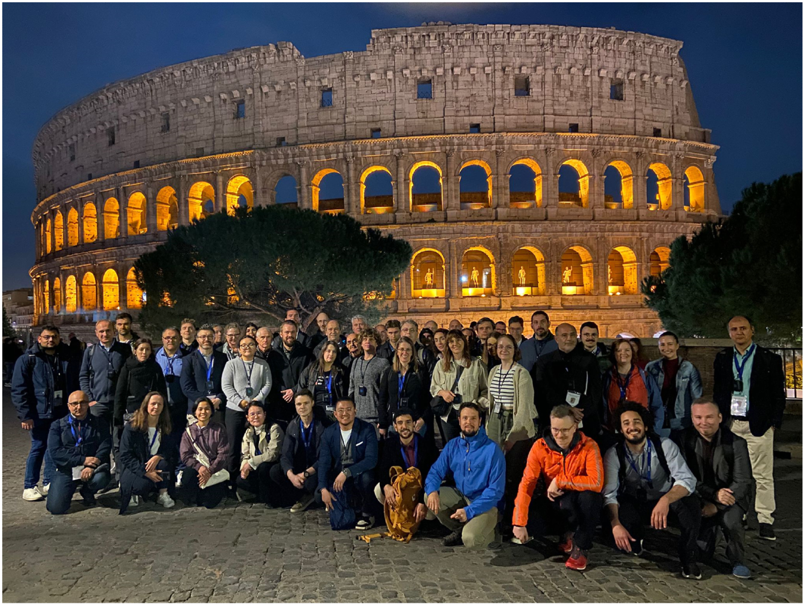
Photograph of the Workshop participants of the Zeiss X-ray Microscopy User Meeting held from 11–13 November 2024 at Rome, at Sapienza University, near the historical monument shown in the image.
-
Research ethics: Not applicable.
-
Informed consent: Informed consent was obtained from all individuals included in this study, or their legal guardians or wards.
-
Author contributions: All authors have accepted responsibility for the entire content of this manuscript and approved its submission.
-
Use of Large Language Models, AI and Machine Learning Tools: None.
-
Conflict of interest: None.
-
Research funding: None.
-
Data availability: Not applicable.
© 2025 the author(s), published by De Gruyter on behalf of Thoss Media
This work is licensed under the Creative Commons Attribution 4.0 International License.

