Effect of Fiber Laser Treating on Magnetic Domains in the Grain-Oriented Silicon Steel: Imaging Domains by Bitter, MFM and Kerr Microscopy
-
V. Puchý
 ,
F. Kováč
,
F. Kováč

Abstract
A magnetic domain laser scribing technique of grain-oriented 3.2% silicon steel has been investigated for the direct influencing on the magnetic domain wall. The magneto-optical Kerr effect was employed to obtain a visible contrast between antiparallel domains. The effects of laser treating on domain wall were observed. The Bitter, MFM and Kerr microscope pictures showed that domain-wall positions did not repeat precisely from cycle to cycle, particularly at high inductions, and that the average domain-wall spacing decreased with increasing density of laser scribing lines. Two phenomena have been discovered that are difficult to explain (1) that the hardness decreases with increasing laser energy and (2) that the coercivity decreases with decreasing laser energy. A semi-quantitative relationship has been found between the domain patterns and used fiber laser treating method. The behavior of patterns in an applied magnetic field is shown, and based on that a two-dimensional laser lines configuration is proposed for one of the less complicated surface patterns.
Introduction
In modern cold-rolled grain-oriented (GO) electrical steels, a perfect cube-on-edge crystallographic texture (110)[001] is usually produced to improve magnetic permeability. Silicon steels known as electrical steels are used for transformer cores. In the power industry electrical voltage is almost always AC and at low frequency, 50/60 Hz. At these frequencies eddy currents are generated in the transformer core. Alloying the Fe with Si has a large marked effect on the electric resistivity and also has the benefit of reducing the magnetostriction and the magnetocrystalline anisotropy. In addition, the material is used in the form of laminations, typically 0.3/0.7 mm thick. The addition of too much silicon makes the material extremely brittle and difficult to produce, giving a practical limitation of 4 wt% to the amount of Si that can be added. Typically most electrical steels will contain between 3 and 4 wt% Si [1]. In this case, in the Fe-3% Si alloy the only tetragonal axis [001] (easy axis) lying in the ribbon plane is close to the rolling direction and makes small angles with it, about 2°–3°. The core loss of grain-oriented electrical steel can be improved by an additional treatment process by laser scribing method. For the industrial utilization, the CO2 or Nd:YAG lasers are most widely used for laser scribing with typical core loss improvements of about 10%. In this work, a fiber laser with power up to 400 W is investigated with the purpose of industrial utilization. It was found [2] that a specific parameter range for laser beam source is necessary to improve the core losses for the applied parameter range. Furthermore, an increase of the core loss improvement of 0.35 mm electrical steel was investigated by using the fiber laser [3]. One of the ways of the reduction of magnetic losses via the decrease in the width of domains in modern highly textured electrical steels is the creation of magnetostructural barriers using local laser irradiation. GO silicon steels exhibit a relative simple domain structure with 180° Bloch walls aligned in the single grain along the [001] direction and supplementary closure secondary domains. With increasing misorientation of the [001] direction with respect to the plane of the sheet the relative proportion of the closure domain increases with corresponding increase of coercivity and decrease of permeability. Laser scribing can be used to reduce the ferromagnetic domain size, and thus core loss, in grain-oriented electrical steels. The localized stress required for domain-size control can be produced either by shock deformation, optimally associated with beam dwell times (pulse lengths), or by thermal stressing occurring with dwell times, optimally longer than 1×10⁻⁴ sec. In this paper, we show that comparable core loss improvements can be obtained at greatly different dwell times, but that the associated changes in the surface condition of coated silicon steel laser scribed for maximum core loss reduction very dramatically as dwell time varies. The short times characterizing shock deformation are achieved using pulse laser radiation. Under optimum dwell times for thermal expansion effects, achieved using continuous-wave mode, there is no significant effect on the coating, and no postscribing treatment is required. At intermediate dwell times, however, significant core loss reduction is associated with extensive disruption of the coating and with melting of the steel. At dwell times of approximately 1×10⁻⁵ sec using pulse laser radiation, there is an unstable transition between the shock (vaporization) and heating modes, with intermittent melting [4].
Experimental materials and techniques
Preparation of Goss-oriented silicon steel materials
The FeSi grain-oriented electrical steel samples (ArcelorMittal Frýdek-Místek a.s., steel grade M165-35S) with the dimensions of 30 mm×10 mm×0.35 mm were used in this study and its chemical composition is listed in Table 1. All of the samples were for microstructural examination prepared by cutting with the electroerosive CNC cutter, then they were suffused into resin molds and grinded with five grades of silicon carbide papers (no. 320, 500, 1,200, 2,400 and 4,000), followed by cleaning in deionized water and degreasing in ethanol by ultrasonic vibration. Subsequently, in order to remove a surface oxidation film formed by the laser radiation, mechano-chemical polishing in an alkaline SiO2 mechano-chemical suspension was first applied. Then, the samples were cut out from the resin and fine polished using a Lectropol-5 electrolytic polishing machine using electrolytic bath containing 500 ml CH3COOH and 25 ml HClO4, in about 50V/20°C so as to effectively mirror-finish the steel sheet surface. In this way, the grain-oriented silicon steel sheet having smooth surface can be obtained for the excellent domain observation. This procedure was used to completely remove the influence of surface grinding and should result in an excellent surface whose properties are undistorted by polishing steps.
Chemical composition of tested materials (in wt%).
| Sample | Si | C | Mn | S | Cu | P | Al | V | N | Cr |
|---|---|---|---|---|---|---|---|---|---|---|
| FeSi3 | 3.2 | 0.04 | 0.18 | 0.003 | 0.54 | 0.003 | 0.004 | 0.046 | 0.003 | 0.008 |
Laser treatment of silicon steel samples
The samples after preparation were mounted in a sample holder and set into the experimental laser workstation TRUMPF 3003 with laser source of TruFiber400. The samples were then laser treated with the programmable focused optic head (PFO) by computerized X–Y stage (CNC table). The samples were treated in air through a fiber CW and pulsed mode laser (1,064 nm). The laser spot diameter on the surface of the sample was approximately 30 µm. The power density of the laser beam was from 12 W to 100 W with the pulsed duration from 50 µs to 500 µs (from 0.6 mJ to 50 mJ energy in one pulse) and the frequency 1 Hz. The schematic diagram of the laser scribing process is shown in Figure 1. All of the samples after the laser treatment were air cooled to avoid deformation due to thermal shocks and differential contractions. The baking thermal treatment was not used after laser treatment.
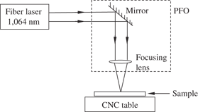
The schematic diagram of the laser scribing process.
Microstructural, textural and magnetic domain characterization
Scanning electron microscopy (SEM) was used to observe laser-treated surfaces in order to determine the grain sizes. The polished surfaces were transferred to the SEM (JEOL JSM-7000F, 10 kV, working distance 9–12 mm) for examination. The surface morphology was characterized by SEM equipped with energy-dispersive spectrum (EDS). The magnetic domains were observed using several visualizing techniques – Bitter microscopy, MFM and Kerr microscopy. The samples for the Bitter observation technique were prepared by mechanical grinding, polishing, and coating with magnetic colloid suspension Ferrofluid and then the magnetic domains were observed with the optical microscope Olympus GX71. For the MFM observation an AFM microscope Dimension Icon (Bruker) with Bruker Co/Cr tip was used. Kerr method for the domain imaging was done with the scanning magneto-optical Kerr microscope model EVICO MAGNETICS. The domain widths were measured with the aid of the software Image J for Windows. A minimum of 50 readings was taken to measure the domain sizes of each material and the results were statistically treated.
Mechanical properties and electromagnetic testing
Nanohardness measurements were performed on each sample using nanohardness tester Agilent G200 on the laser-treated surface layer (along the depth in the longitudinal section) with the applied load 50 mN and the penetration depth was about 800 nm. The samples before and after laser treatment were tested for electromagnetic properties by coercivity meter. After preparation they were set onto coercimeter holder and then they were set into coercimeter coil. Then the samples were saturated with applied magnetic field. And then the magnetization was reduced to zero with the intensity of the applied magnetic field which gave us the measured coercivity.
Results and discussion
Microstructure and texture
The microstructure along the depth in the longitudinal section of the samples is shown at higher magnification in the SEM images (Figure 2). From this it is evident that the microstructure of the laser untreated materials is basically different compared to laser-treated materials due to the laser heating material which expanded and caused residual thermal stresses. The effect of tensile stresses on magnetic losses is related not only to a decrease in the width of the main domains but also to a rearrangement of 90° domains into 180° ones [5]. In addition, their action leads to the removal of closure domains and a reduction in core loss.

(a) The laser pulse with 50 mJ energy (100 W, 500 µs, focus+0 mm) (b) detail of the laser pulse.
Magnetic domains
The refined magnetic domains of the laser-treated samples compared to the untreated material are mainly dependent on the residual stresses that remain in the material after the laser effect, similarly, as it was observed in other investigations [6, 7]. Together with the residual stresses created by the thermal shocks due to laser pulses also other characteristic processing defects were present in our materials, similarly as reported in all the works dealing with similar materials and techniques [8, 9]. This indicates the still present difficulties in the laser treatment of domain scribing of the silicon steels but also the potential for the improvement of their electromagnetic, functional, and mechanical properties. The laser-treated silicon steel with short pulses was less damaged but the silicon-steel exposed to the long pulses contained high amount of surface damage, which was associated with thermal shocking of the laser ray. The transient and non-uniform temperature distributions resulting from the laser heating cause different zones of the material to expand by different amounts and thermal stress to develop. When the stress is smaller it is absorbed by the elasticity of the material which results in refined magnetic domains (Figure 3). According to literature [10] with increasing laser energy the number of refined magnetic domains increases. In our case relatively large numbers of refined domains were observed on the laser scribed area. The magnetic domains were refined from 45 µm thickness to 15 µm thickness (Figures 4 and 5).

(a) Domains made visible by Ferrofluid in the silicon steel with 5 mJ laser treatment, (b) domains in the 50 mJ laser-treated silicon steel.

(a) Domains made visible by AFM without laser treatment (area 90×90 µm) – magnetic domain sub-structure – lancet domains, (b) domains in the 0.6 mJ laser-treated silicon steel (area 50×50 µm).

(a) Primary magnetic domains made visible by KERR microscopy without laser treatment, (b) refined domains in the 0.6 mJ laser-treated silicon steel – labyrinthine structure near the laser spot.
Mechanical and electro-magnetic properties
We can see that the coercivity (Table 2) significantly decreases by laser irradiation and its minimum value was obtained at around 0.6 mJ/pulse. When the laser energy increased to the 50 mJ/pulse, the coercivity slightly increased. That is to say, an optimum laser energy condition for the improvement of the coercivity in Fe-3.2% grain-oriented silicon steel sheet exists at around 0.6 mJ/pulse. A similar result has been already reported by Ref. [7]. The relationship between the laser energy and the average ratio of magnetic domain wall spacing before and after the laser irradiation is summarized in Table 2. It is obvious that the magnetic domains between the lines were remarkably refined when the laser energy decreased. The magnetic domain spacing becomes much narrower by decreasing the laser energy up to E=0.6 mJ/pulse and then saturates with lower laser energy. This means that the improvement of core loss cannot be simply explained by the magnetic domain refining effect. This is thought to be due to the lower material vaporization by the longer laser pulses with high energy. We found that the local tensile residual stresses are induced at the surface of Fe-3.2%Si grain-oriented silicon steel sheet by the laser irradiation and this state causes the refinement of 180° magnetic domains. Next, we investigated the plastic deformation effect by laser irradiation. It is known, in general, that work hardening occurs due to induced plastic strains for a metal. The amount of plastic deformation might be larger with increasing laser energy since the traces become larger with increasing the laser energy. In order to estimate this effect by laser irradiation, we performed Vickers nanohardness testing experiments. Table 2 show the results of Vickers nanohardness distributions in the laser-irradiated traces with the laser energies of 0.6, 5, 50 mJ/pulse. We can see from this table that the hardness decreases with increasing laser energy in the traces. In particular, a marked decrease of the hardness was observed at E=50 mJ/pulse. This means that the silicon was in a solid solution but in lower amount (Figure 6) caused by mechanism of laser-induced sublimation of silicon. It was confirmed that the magnetic domain-refining phenomenon occurs by laser-induced residual stresses. The laser irradiation has two effects on Fe-3.2%Si grain-oriented silicon steel in relation to the reduction of coercivity: one is the residual stress and the other is plastic deformation at the surface of the material. The total core loss is determined by the balance of these two effects of laser irradiation, of which E=0.6 mJ/pulse is the optimum condition in Fe-3.2%Si grain-oriented silicon steel. The domain structure after laser irradiation was refined with 0.6 and 5 mJ pulse energy but the better results of coercivity were achieved with the smaller pulse energy. Because these pulses create internal stresses which produce finer domains with better decreased coercivity. This behavior is similar as Weidenfeller and Kalinscák observed [1, 11]. However in our case, with the pulse energy 5 and 50 mJ coercivity was slightly higher than sample without laser treatment through to generation of the higher residual stresses and number of surface defects. The 0.6 mJ intensity of laser beam caused a higher number of primary 180° domains and this mechanism leads to a decrease of coercivity. This behavior, the reduction of the coercive force by the introduction of crystal defects similarly like Weidenfeller observed [1], is the Brown’s paradox.
Mechanical and electro-magnetic properties of the Fe-3.2% Si silicon steel.
| Sample no. | Interval of lines [mm] | Pulse energy [mJ] | El. coercivity [A/cm] | Magnetic domain width [µm] | HV0.05 [GPa] in the pulse line |
|---|---|---|---|---|---|
| 1 | 0.21 | 45 ± 4.2 | 4.2 ± 0.1 | ||
| 2 | 3.75 | 0.6 | 0.14 | 23 ± 2.9 | 3.99 ± 0.33 |
| 3 | 5 | 0.205 | 38 ± 3.6 | 3.94 ± 0.27 | |
| 4 | 50 | 0.225 | 50 ± 4.1 | 3.83 ± 0.16 | |
| 5 | 5 | 0.6 | 0.11 | 16 ± 2.1 | 3.96 ± 0.24 |
| 6 | 5 | 0.215 | 39 ± 3.8 | 3.91 ± 0.17 | |
| 7 | 50 | 0.23 | 50 ± 5.5 | 3.85 ± 0.14 | |
| 8 | 7.5 | 0.6 | 0.15 | 25 ± 3.4 | 3.97 ± 0.29 |
| 9 | 5 | 0.21 | 43 ± 4.1 | 3.92 ± 0.25 | |
| 10 | 50 | 0.215 | 47 ± 4.4 | 3.87 ± 0.17 |

SEM crater morphology and EDS result for 50 mJ laser-treated silicon steel.
Conclusions
Primary magnetic domains of grain-oriented silicon steel become smaller in consequence of the formation and development of both, magnetic poles and secondary magnetic domains due to laser-induced internal stresses.
With the increasing of laser energy, the laser affected volume is deeper and thicker and the hardness of laser-treated layer decreases.
The magnetic domain thickness reached minimum when the laser scribe lines were 5 mm and the minimum values of coercivity were achieved at laser energy of 0.6 mJ/pulse (0.11 A/cm for 5 mm line spacing).
The higher number of mobile 180° magnetic domains leads to a decrease of coercivity and it can lead to decrease of hysteresis losses.
Funding statement: Funding: This work was carried out within the framework of the project “High strength electrotechnical composite steels” which is supported by the Slovak Research and Development Agency under the contract No. APVV-0147-11. This work was also partially supported by the Slovak Grant Agency VEGA, project No. 2/0083/13 and also within the frame of the project “Research Centre of Advanced Materials and Technologies for Recent and Future Applications ‘PROMATECH’, which is supported by the Operational Program ‘Research and Development’ financed through European Regional Development Fund”.
References
[1] B. Weidenfeller and W. Riehemann, J. Magn. Magn. Mater., 292 (2005) 210–214.10.1016/j.jmmm.2004.08.036Search in Google Scholar
[2] M. Imafuku, H. Suzuki, K. Akita, K. Iwata and M. Fujikura, Acta Mater., 53 (2005) 939–945.10.1016/j.actamat.2004.10.040Search in Google Scholar
[3] P. Rauscher, J. Hauptmann and E. Beyer, Phys. Procedia, 41 (2013) 312–318.10.1016/j.phpro.2013.03.083Search in Google Scholar
[4] B. Weidenfeller and M. Anhalt, J Magn. Magn. Mater., 322 (2010) 69–72.10.1016/j.jmmm.2009.08.030Search in Google Scholar
[5] Y. Ushigami, M. Mizokami, M. Fujikura, T. Kubota, H. Fujii and K. Murakami, J. Magn. Magn. Mater., 254–255 (2003) 307–314.10.1016/S0304-8853(02)00933-2Search in Google Scholar
[6] J. Li, Y. Gu and Z. Guo, J. Mater. Process. Technol., 74 (1998) 292–297.10.1016/S0924-0136(97)00274-4Search in Google Scholar
[7] J. Li, Y. Gu and Z. Guo, J. Mater. Process. Technol., 69 (1997) 180–185.10.1016/S0924-0136(97)00015-0Search in Google Scholar
[8] K. Suzuki, M. Yoshizumi, T. Izumi, Y. Shiohara, M. Iwakuma, A. Ibi, S. Miyata and Y. Yamada, Physica C, 468 (2008) 1579–1582.10.1016/j.physc.2008.05.076Search in Google Scholar
[9] B. Weidenfeller and W. Riehemann, J. Magn. Magn. Mater., 160 (1996) 287–288.10.1016/0304-8853(96)00143-6Search in Google Scholar
[10] S.V. Ponnaluri, R. Cherukuri and P.A. Molian, J. Mater. Process. Technol., 112 (2001) 199–204.10.1016/S0924-0136(01)00573-8Search in Google Scholar
[11] Z. Kalincsák, J. Takács, P. Kollár and Á. Cziráki, J. Magn. Magn. Mater., 304 (2006) e495–e497.10.1016/j.jmmm.2006.02.134Search in Google Scholar
©2017 by De Gruyter
This article is distributed under the terms of the Creative Commons Attribution Non-Commercial License, which permits unrestricted non-commercial use, distribution, and reproduction in any medium, provided the original work is properly cited.
Articles in the same Issue
- Frontmatter
- Research Articles
- A New Method to Produce Ni–Cr Ferroalloy Used for Stainless Steel Production
- Mechanical and Electrochemical Characterization of Super-Solidus Sintered Austenitic Stainless Steel (316L)
- Effect of γ→α Phase Transformation on Refining Austenite Grains of Microalloyed Steel in Continuous Casting by Simulation
- Fatigue Life Improving of Drill Rod by Inclusion Control
- Influence of Basicity and MgO on Fluidity and Desulfurization Ability of High Aluminum Slag
- Effect of Sputtered AlY Coating on High-Temperature Oxidation Behavior of Stainless Steel
- Optimal Design of Nozzle for Supersonic Atmosphere Plasma Spraying
- Oxidation Behaviors of Inconel 740H in Air and Dynamic Steam
- Line-Profile Analysis Combined with Texture Analysis for Characterizing Dislocation Distribution in Texture Components of Cold-Rolled Copper Sheets
- Microstructure Analysis of HPb59-1 Brass Induced by High Current Pulsed Electron Beam
- Thermal Treatment Method for Synthesis and Characterization of the Octahedral Magnetic Nanostructures of Co3O4 from a New Precursor
- Phases Transition and Consolidation Mechanism of High Chromium Vanadium-Titanium Magnetite Pellet by Oxidation Process
- Effect of Fiber Laser Treating on Magnetic Domains in the Grain-Oriented Silicon Steel: Imaging Domains by Bitter, MFM and Kerr Microscopy